Original and Modified Sauvé–Kapandji Procedures: A Two-Axis Framework for Surgical Decision-Making—A Narrative Review


Abstract
1. Introduction
2. Materials and Methods
3. The Sauvé–Kapandji Procedure: Evolution, Technique, Indications, and Outcomes
3.1. Historical Evolution
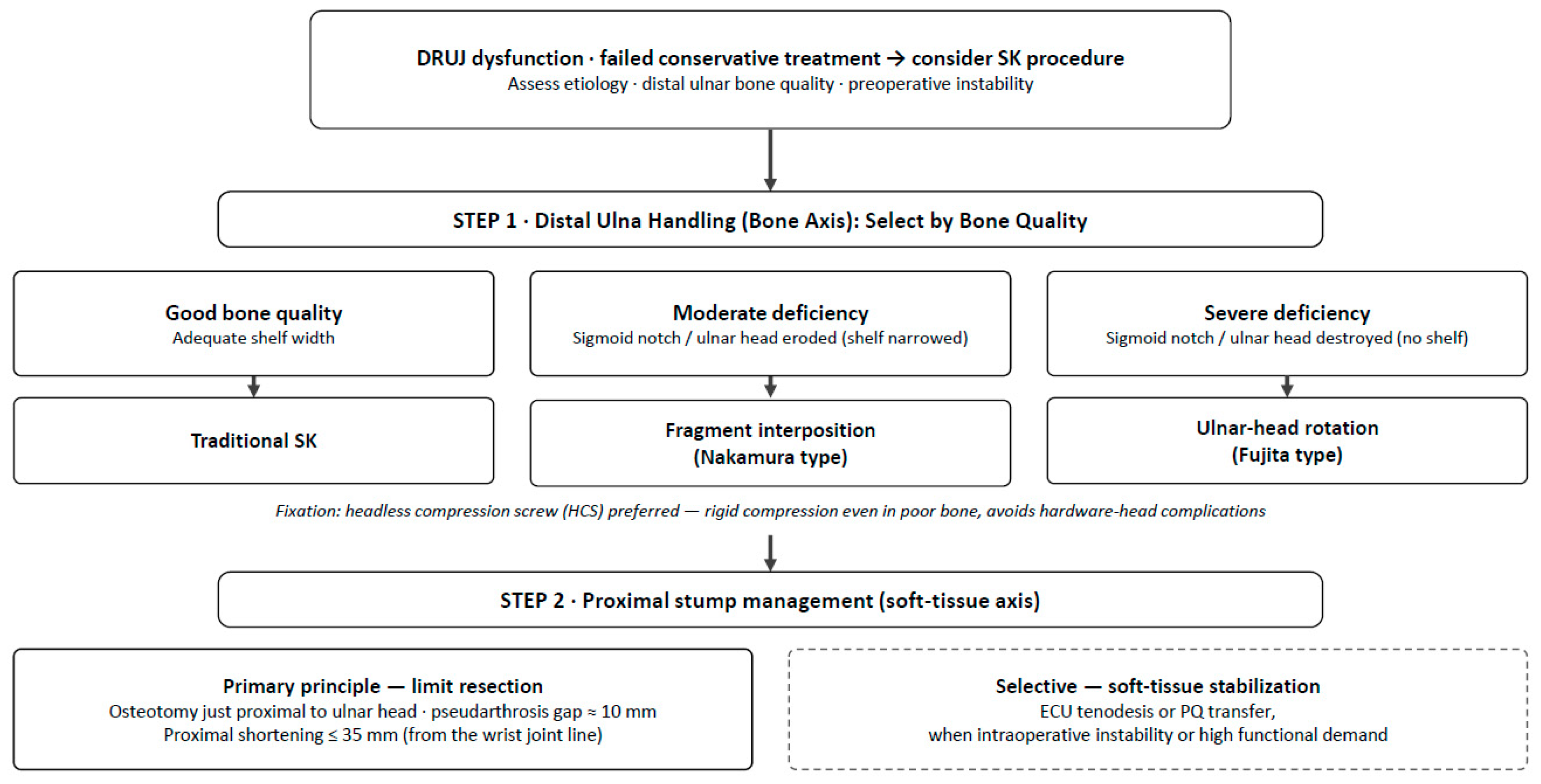
3.2. The Two-Axis Framework
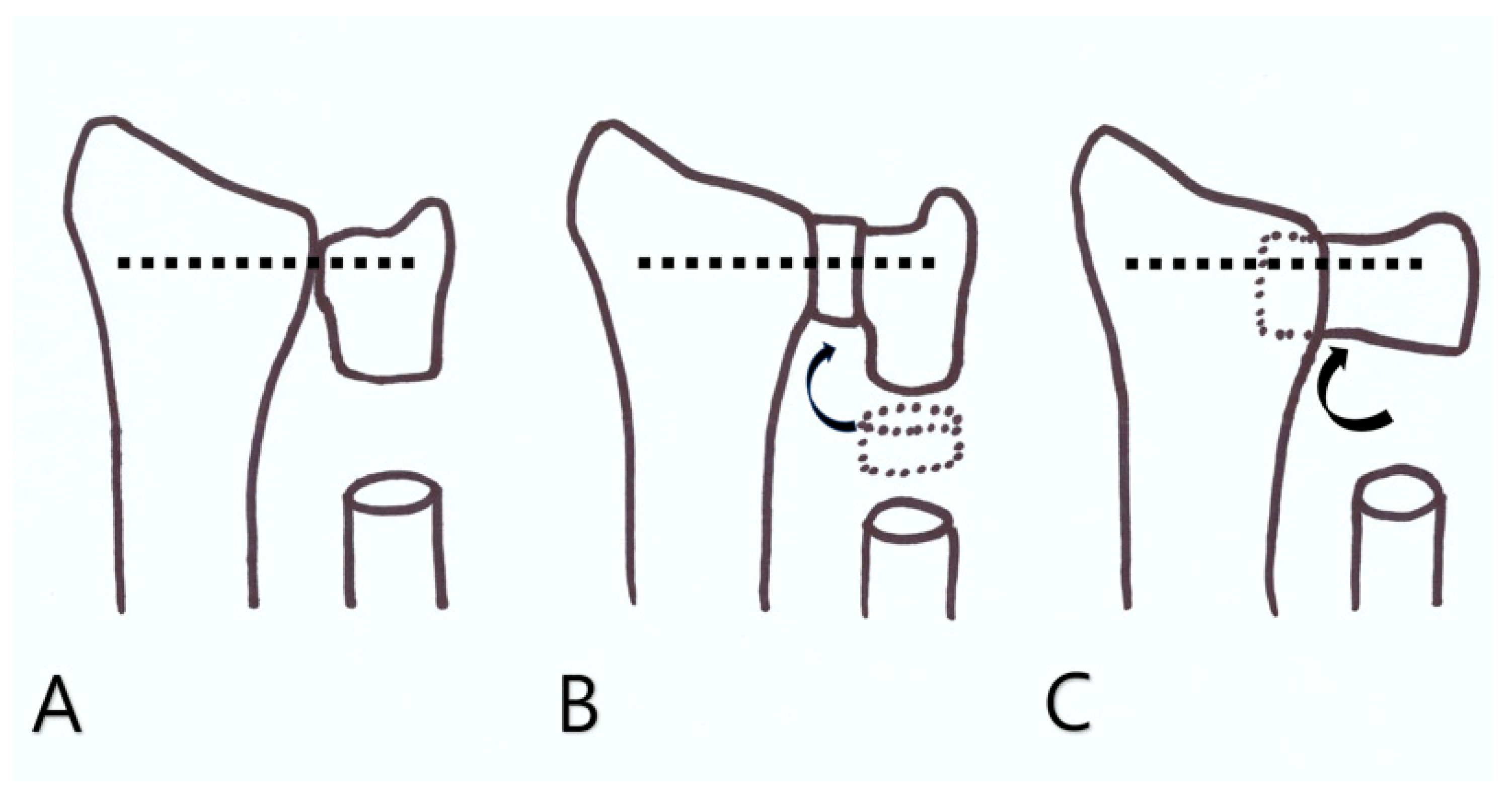
3.2.1. Distal Ulna Handling (Bony Axis)
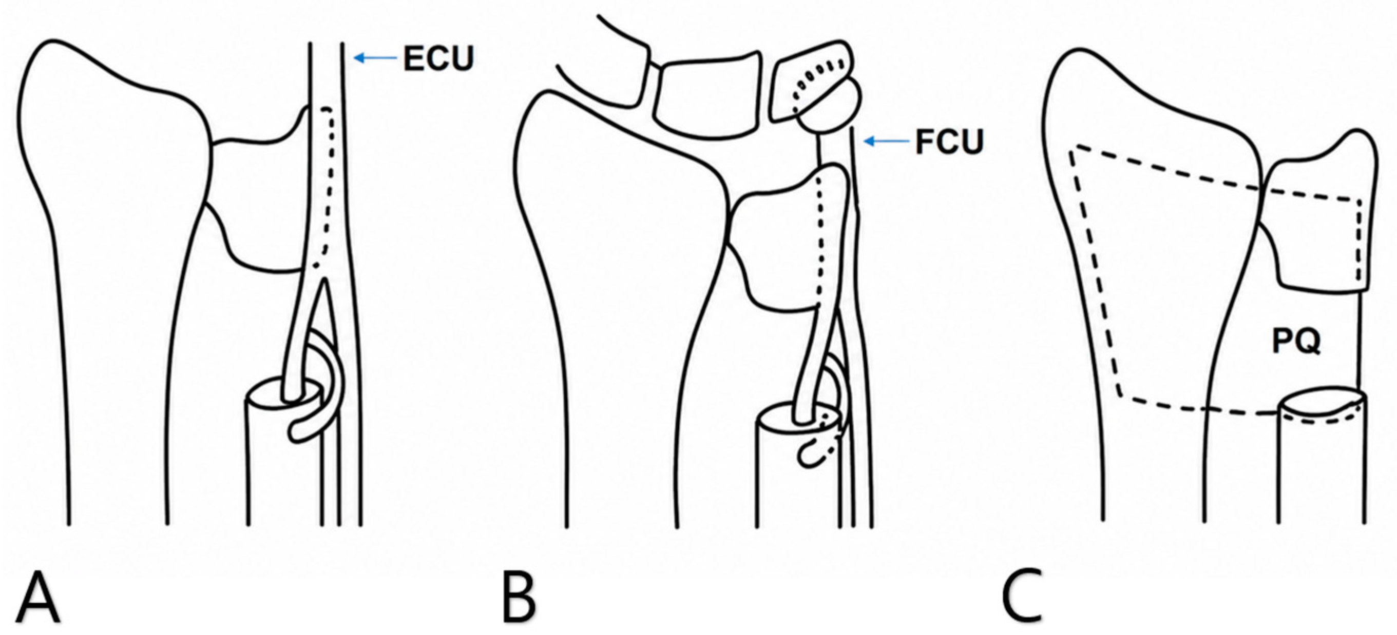
3.2.2. Proximal Stump Stabilization (Soft-Tissue Axis)
3.3. Outcomes and Comparative Evidence
3.4. An Indication-Based Decision Framework and Research Agenda
4. Limitations
5. Conclusions
Author Contributions
Funding
Institutional Review Board Statement
Informed Consent Statement
Data Availability Statement
Acknowledgments
Conflicts of Interest
References
- Breyer, J.M.; Vergara, P. Solutions for the Unstable and Arthritic Distal Radioulnar Joint. Hand Clin. 2020, 36, 523–530. [Google Scholar] [CrossRef] [Scilit] [PubMed]
- Farias-Eisner, G.; Zoller, S.D.; Iannuzzi, N. Treatment Options for Distal Radioulnar Joint Arthritis: Balancing Functional Demand and Bony Resection. Hand Clin. 2021, 37, 575–586. [Google Scholar] [CrossRef] [Scilit] [PubMed]
- Sebastin, S.J.; Larson, B.P.; Chung, K.C. History and Evolution of the Sauvé–Kapandji Procedure. J. Hand Surg. Am. 2012, 37, 1895–1902. [Google Scholar] [CrossRef] [Scilit] [PubMed]
- Vincent, K.A.; Szabo, R.M.; Agee, J.M. The Sauvé–Kapandji Procedure for Reconstruction of the Rheumatoid Distal Radioulnar Joint. J. Hand Surg. Am. 1993, 18, 978–983. [Google Scholar] [CrossRef] [Scilit] [PubMed]
- Kobayashi, A.; Futami, T.; Tadano, I.; Fujita, M.; Watanabe, T.; Moriguchi, T. Radiographic Comparative Evaluation of the Sauvé–Kapandji Procedure and the Darrach Procedure for Rheumatoid Wrist Reconstruction. Mod. Rheumatol. 2005, 15, 187–190. [Google Scholar] [CrossRef] [Scilit] [PubMed]
- Roulet, S.; Williot, A.; Sos, C.; Mazaleyrat, M.; Marteau, E.; Laulan, J.; Bacle, G. Comparison of Subjective Outcomes of Darrach and Sauvé–Kapandji Procedures at a Minimum 2 Years’ Follow-Up. Orthop. Traumatol. Surg. Res. 2021, 107, 102974. [Google Scholar] [CrossRef] [Scilit] [PubMed]
- Reissner, L.; Schweizer, A.; Unterfrauner, I.; Estermann, L.; Nagy, L. Long-Term Results of Sauvé–Kapandji Procedure. J. Hand Surg. Eur. Vol. 2021, 46, 626–631. [Google Scholar] [CrossRef] [Scilit] [PubMed]
- Papp, M.; Papp, L.; Lenkei, B.; Károlyi, Z. Long-Term Results of the Sauvé–Kapandji Procedure in the Rheumatoid Wrist. Acta Orthop. Belg. 2013, 79, 655–659. [Google Scholar] [PubMed]
- Giberson-Chen, C.C.; Leland, H.A.; Benavent, K.A.; Harper, C.M.; Earp, B.E.; Rozental, T.D. Functional Outcomes After Sauvé–Kapandji Arthrodesis. J. Hand Surg. Am. 2020, 45, 408–417. [Google Scholar] [CrossRef] [Scilit] [PubMed]
- Nguyen, M.H.; Lipari, N.; O’Brien, A.L.; Samade, R.; Jain, S.A. Darrach vs. Sauvé–Kapandji: A Comprehensive Meta-Analysis of Surgical Outcomes in Distal Radioulnar Joint (DRUJ) Dysfunction. Indian J. Orthop. 2023, 57, 565–570. [Google Scholar] [CrossRef] [Scilit] [PubMed]
- Lamont, S.; Debkowska, M.; Johnsen, P.; Froehle, A.; Cotterell, I.H.; Isaacs, J. Outcomes of Darrach and Sauvé–Kapandji Procedures: A Systematic Review. Hand 2024, 19, 68–73. [Google Scholar] [CrossRef] [Scilit] [PubMed]
- Nakamura, R.; Tsunoda, K.; Watanabe, K.; Horii, E.; Miura, T. The Sauvé–Kapandji Procedure for Chronic Dislocation of the Distal Radio-Ulnar Joint with Destruction of the Articular Surface. J. Hand Surg. Br. 1992, 17, 127–132. [Google Scholar] [CrossRef] [Scilit] [PubMed]
- Minami, A.; Suzuki, K.; Suenaga, N.; Ishikawa, J. The Sauvé–Kapandji Procedure for Osteoarthritis of the Distal Radioulnar Joint. J. Hand Surg. Am. 1995, 20, 602–608. [Google Scholar] [CrossRef] [Scilit] [PubMed]
- Lamey, D.M.; Fernandez, D.L. Results of the Modified Sauvé–Kapandji Procedure in the Treatment of Chronic Posttraumatic Derangement of the Distal Radioulnar Joint. J. Bone Jt. Surg. Am. 1998, 80, 1758–1769. [Google Scholar] [CrossRef] [Scilit] [PubMed]
- Minami, A.; Kato, H.; Iwasaki, N. Modification of the Sauvé–Kapandji Procedure with Extensor Carpi Ulnaris Tenodesis. J. Hand Surg. Am. 2000, 25, 1080–1084. [Google Scholar] [CrossRef] [Scilit] [PubMed]
- Nakamura, K.; Oda, H.; Tanaka, S.; Kuga, Y.; Yamamoto, M.; Nishikawa, T.; Juji, T.; Shimizu, M. Usefulness of Absorbable Screws in the Sauvé–Kapandji Procedure for Rheumatoid Wrist Reconstruction. Mod. Rheumatol. 2002, 12, 144–147. [Google Scholar] [CrossRef] [Scilit]
- Fujita, S.; Masada, K.; Takeuchi, E.; Yasuda, M.; Komatsubara, Y.; Hashimoto, H. Modified Sauvé–Kapandji Procedure for Disorders of the Distal Radioulnar Joint in Patients with Rheumatoid Arthritis. J. Bone Jt. Surg. Am. 2005, 87, 134–139. [Google Scholar] [CrossRef] [Scilit] [PubMed]
- Daecke, W.; Martini, A.K.; Schneider, S.; Streich, N.A. Amount of Ulnar Resection Is a Predictive Factor for Ulnar Instability Problems After the Sauvé–Kapandji Procedure: A Retrospective Study of 44 Patients Followed for 1–13 Years. Acta Orthop. 2006, 77, 290–297. [Google Scholar] [CrossRef] [Scilit] [PubMed]
- Kawabata, A.; Egi, T.; Hashimoto, H.; Masada, K.; Saito, S. A Comparative Study of the Modified Sauvé–Kapandji Procedure for Rheumatoid Wrist With and Without Stabilization of the Proximal Ulnar Stump. J. Hand Surg. Eur. Vol. 2010, 35, 659–663. [Google Scholar] [CrossRef] [Scilit] [PubMed]
- Toyama, S.; Tamai, K.; Sakamoto, A.; Hirashima, T. The Short-Term Outcome of the Modified Sauvé–Kapandji Procedure Regarding Range of Motion, Carpal Bone Translation and Bony Shelf Size. Mod. Rheumatol. 2011, 21, 37–42. [Google Scholar] [CrossRef] [Scilit] [PubMed]
- Uerpairojkit, C.; Leechavengvongs, S.; Malungpaishorpe, K.; Witoonchart, K.; Buddhavibul, P. Proximal Ulnar Stump Stability After Using the Pronator Quadratus Muscle Transfer Combined with the Sauvé–Kapandji Procedure in Rheumatoid Wrist. Hand Surg. 2014, 19, 25–32. [Google Scholar] [CrossRef] [Scilit] [PubMed]
- Sakuma, Y.; Ochi, K.; Yano, K.; Yoshida, S.; Ikari, K.; Momohara, S. Association Between Position of the Fixed Ulnar Head and Carpal Translocation After the Sauvé–Kapandji Procedure in Patients with Rheumatoid Arthritis. Mod. Rheumatol. 2016, 26, 702–707. [Google Scholar] [CrossRef] [Scilit] [PubMed]
- Maeda, A.; Suzuki, T.; Hasegawa, M.; Kuroiwa, T.; Shizu, K.; Hayakawa, K.; Tsuji, T.; Suzuki, K.; Yamada, H. Sauvé–Kapandji Procedure with Headless Compression Screw in Patients with Rheumatoid Arthritis. Mod. Rheumatol. 2018, 28, 114–118. [Google Scholar] [CrossRef] [Scilit] [PubMed]
- Ikeda, M.; Kawabata, A.; Suzuki, K.; Toyama, M.; Egi, T. Outcome of the Sauvé–Kapandji Procedure for Distal Radioulnar Joint Disorder with Rheumatoid Arthritis or Osteoarthritis: Results of One-Year Follow-Up. Mod. Rheumatol. 2018, 28, 490–494. [Google Scholar] [CrossRef] [Scilit] [PubMed]
- Kato, T.; Suzuki, T.; Kameyama, M.; Okazaki, M.; Morisawa, Y.; Nishiwaki, M.; Nakamura, T.; Sato, K.; Iwamoto, T. Range of Motion After the Sauvé–Kapandji and Darrach Procedures Without Extensor Tendon Rupture. J. Wrist Surg. 2021, 10, 190–195. [Google Scholar] [CrossRef] [Scilit] [PubMed]
- Liu, K.Y.; Wu, C.H.; Ma, C.H.; Jou, I.M.; Tu, Y.K. Modified Sauvé–Kapandji Procedure with Only Extensor Carpi Ulnaris Tenodesis in Advanced-Stage Rheumatoid Wrists: A Retrospective Study of Nine Wrists. Formos. J. Musculoskelet. Disord. 2026, 17, 7–13. [Google Scholar] [CrossRef] [Scilit]
- Sauvé, L.; Kapandji, M. Nouvelle technique de traitement chirurgical des luxations récidivantes isolées de l’extrémité inférieure du cubitus. J. Chir. 1936, 47, 589–594. [Google Scholar]
- Kapandji, I.A. The Kapandji–Sauvé Operation. Its Techniques and Indications in Non-Rheumatoid Diseases. Ann. Chir. Main 1986, 5, 181–193. [Google Scholar] [PubMed]
- Munaretto, N.; Aibinder, W.; Moran, S.; Rizzo, M. Sauvé–Kapandji Remains a Viable Option for Distal Radioulnar Joint Dysfunction. Hand 2022, 17, 963–968. [Google Scholar] [CrossRef] [Scilit] [PubMed]
- Okabe, H.; Maeda, K.; Yamashita, Y.; Zenitani, A.; Nishizawa, T.; Yukawa, M.; Nishimura, R.; Bo, H.; Miyawaki, T.; Saito, M. Biologic Therapy Is Associated with Faster Bone Union After the Sauvé–Kapandji Procedure in Patients with Rheumatoid Arthritis. Rheumatol. Ther. 2026, 13, 855–865. [Google Scholar] [CrossRef] [Scilit] [PubMed]
- Verhiel, S.H.W.L.; Özkan, S.; Ritt, M.J.P.F.; Chen, N.C.; Eberlin, K.R. A Comparative Study Between Darrach and Sauvé–Kapandji Procedures for Post-Traumatic Distal Radioulnar Joint Dysfunction. Hand 2021, 16, 375–384. [Google Scholar] [CrossRef] [Scilit] [PubMed]
- Tomori, Y.; Sawaizumi, T.; Nanno, M.; Takai, S. Functional Outcomes After the Sauvé–Kapandji Procedure for Distal Radio-Ulnar Post-Traumatic Instability: A Case-Control Comparison of Three Different Operative Methods of Stabilization of the Ulnar Stump. Int. Orthop. 2018, 42, 2173–2179. [Google Scholar] [CrossRef] [Scilit] [PubMed]



| Study (Year) [Ref.] | Design n | Etiology | Bone Handling (Axis 1) | Stump Stabilization (Axis 2) | Fixation | Key Finding |
|---|---|---|---|---|---|---|
| Nakamura 1992 [12] | Retro 15 | Chronic dislocation | Fragment interposition | Periosteum | Metal screw | RU convergence/stump instability in all; pain in 4/15 |
| Vincent 1993 [4] | Retro 17 (21 wrists) | RA | Traditional | Soft tissue | Metal screw | Prevented ulnar translation, improved rotation |
| Minami 1995 [13] | Retro 15 patients | Primary OA | Traditional | — | Metal screw | Good pain/function in OA |
| Lamey & Fernandez 1998 [14] | Retro 18 patients | Post-traumatic | Traditional (modified) | FCU | Metal screw | Good results in post-traumatic DRUJ |
| Minami 2000 [15] | Retro 13 wrists | RA/OA | Traditional | ECU tenodesis | Metal screw | Introduced ECU tenodesis modification |
| Nakamura K 2002 [16] | Retro 43 (23 PLLA/20 Metal) | RA | Traditional | — | PLLA absorbable screw | No peri-screw resorption; no removal needed |
| Fujita 2005 [17] | Retro 56 (66 wrists) | RA | Ulnar-head rotation | — | Metal screw | Reconstructed bony shelf in bone deficiency |
| Kobayashi 2005 [5] | Retro comparison (vs Darrach) 20 (26 wrists) | RA | Traditional | — | Metal screw | No difference in carpal collapse/ulnar translation |
| Daecke 2006 [18] | Retro 44 | Mixed | Traditional | Various | Metal screw | More resection → more instability; shortening ≤35 mm |
| Kawabata 2010 [19] | Retro comparison (ECU ±) 41 | RA | Ulnar-head rotation | ECU vs. none | Metal screw | No difference except early pain; 30 mm osteotomy |
| Toyama 2011 [20] | Retro 20 (24 wrists) | RA | Ulnar-head rotation | — | Metal screw | Recommended SWR < 0.77 |
| Papp 2013 [8] | Retro long-term 14 | RA | Traditional | ECU + PQ interposition | Metal screw | Good long-term; no translation/subluxation |
| Uerpairojkit 2014 [21] | Retro 10 | RA | Traditional | PQ transfer | 1 cancellous screw | Sagittal stable; coronal convergence on loading (asymptomatic) |
| Sakuma 2016 [22] | Retro 40 patients | RA | Traditional/interposition/rotation | — | Metal screw | Fixed ulnar-head position associated with carpal translation |
| Maeda 2018 [23] | Retro 41 | RA | Fragment interposition | PQ + periosteum | HCS | High union rate; no hardware complications |
| Ikeda 2018 [24] | Retro 28 (32 wrists) | RA/OA | Traditional | Periosteal coverage | 1 HCS | Stable, painless stump without tenodesis |
| Giberson-Chen 2020 [9] | Retro comparison (ECU vs. PQ) 57 | Mixed | Traditional | ECU vs. PQ | 2 HCS | Two groups similar; 7% reosteotomy for reossification |
| Kato 2021 [25] | Retro comparison (vs. Darrach) 46 | Mixed | Traditional | — | Metal screw or HCS | Flexion 49°→40°; Darrach increases flexion/extension |
| Liu 2026 [26] | Retro 6 (9 wrists) | Advanced RA | Traditional (no graft) | ECU only | 2 HCS | Radiographic convergence/volar migration progress but function improves |
| Complication | Reported Incidence | Major Risk Factors | Preventive Strategies | Management |
|---|---|---|---|---|
| Proximal ulnar stump instability/ulnar impingement | Revision for instability in up to ~18% in one comparative cohort [6] and in 6 of 15 wrists in a 13-year series [7]; ulnar stump pain in 4/15 (26.7%) [12]; radioulnar convergence radiographically common [7,12,21] | Excessive or too-proximal resection (large shortening); inadequate stump stabilization | Limit resection: osteotomy just proximal to the ulnar head; pseudarthrosis gap ~10 mm and proximal shortening ≤35 mm (provisional reference values *) [18]; selective ECU [15], FCU [14], or PQ [21] stabilization | Secondary ECU/FCU tenodesis or PQ transfer; revision |
| Reossification (bony bridging) of the pseudarthrosis | Revision osteotomy in 7.0% (4/57) [9] | Too-small pseudarthrosis gap; incomplete removal of periosteum/interosseous membrane | Adequate pseudarthrosis gap (~10 mm *) [18]; extra-periosteal resection removing periosteum and interosseous membrane [28]; PQ interposition [21] | Revision osteotomy [9] |
| Nonunion/delayed union of the DRUJ arthrodesis | Nonunion in 6 of 19 wrists (31.6%) with conventional screws in an earlier series [17]; high union rates reported with headless compression screws [23], and no nonunions in a contemporary cohort [9] | Poor (osteopenic) RA bone; high-dose methotrexate or NSAIDs [30] | Rigid compression fixation (HCS) [23] with cancellous bone grafting; perioperative optimization (biologic/TNF-inhibitor therapy accelerates union, whereas high-dose MTX and NSAIDs delay it) [30] | Revision arthrodesis with autologous bone grafting |
| Extensor tendon attrition/rupture | Reported (tendon impingement over a prominent or unstable stump) [8,19] | Prominent/unstable proximal stump; osteotomy performed too distally | Osteotomy ~25–30 mm proximal to the joint line; periosteal and soft-tissue coverage of the stump [19] | Tendon transfer (e.g., extensor indicis proprius) or free tendon graft |
| Hardware-related (peri-screw resorption, screw-head irritation) | Peri-screw bone resorption in 5/20 (25%) with metal screws [16]; screw-head discomfort in 2/21 (9.5%) with cannulated cancellous screws [23]; none with PLLA [16] or HCS [23] | Osteopenic RA bone; prominent screw heads | Bioabsorbable (PLLA) screws [16] or headless compression screws [23] | Hardware removal after union if symptomatic |
Disclaimer/Publisher’s Note: The statements, opinions and data contained in all publications are solely those of the individual author(s) and contributor(s) and not of MDPI and/or the editor(s). MDPI and/or the editor(s) disclaim responsibility for any injury to people or property resulting from any ideas, methods, instructions or products referred to in the content. |
© 2026 by the authors. Licensee MDPI, Basel, Switzerland. This article is an open access article distributed under the terms and conditions of the Creative Commons Attribution (CC BY) license.
Share and Cite
Park, I.-J.; Roh, Y.-T. Original and Modified Sauvé–Kapandji Procedures: A Two-Axis Framework for Surgical Decision-Making—A Narrative Review. J. Clin. Med. 2026, 15, 6368. https://doi.org/10.3390/jcm15166368
Park I-J, Roh Y-T. Original and Modified Sauvé–Kapandji Procedures: A Two-Axis Framework for Surgical Decision-Making—A Narrative Review. Journal of Clinical Medicine. 2026; 15(16):6368. https://doi.org/10.3390/jcm15166368
Chicago/Turabian StylePark, Il-Jung, and Youn-Tae Roh. 2026. "Original and Modified Sauvé–Kapandji Procedures: A Two-Axis Framework for Surgical Decision-Making—A Narrative Review" Journal of Clinical Medicine 15, no. 16: 6368. https://doi.org/10.3390/jcm15166368
APA StylePark, I.-J., & Roh, Y.-T. (2026). Original and Modified Sauvé–Kapandji Procedures: A Two-Axis Framework for Surgical Decision-Making—A Narrative Review. Journal of Clinical Medicine, 15(16), 6368. https://doi.org/10.3390/jcm15166368