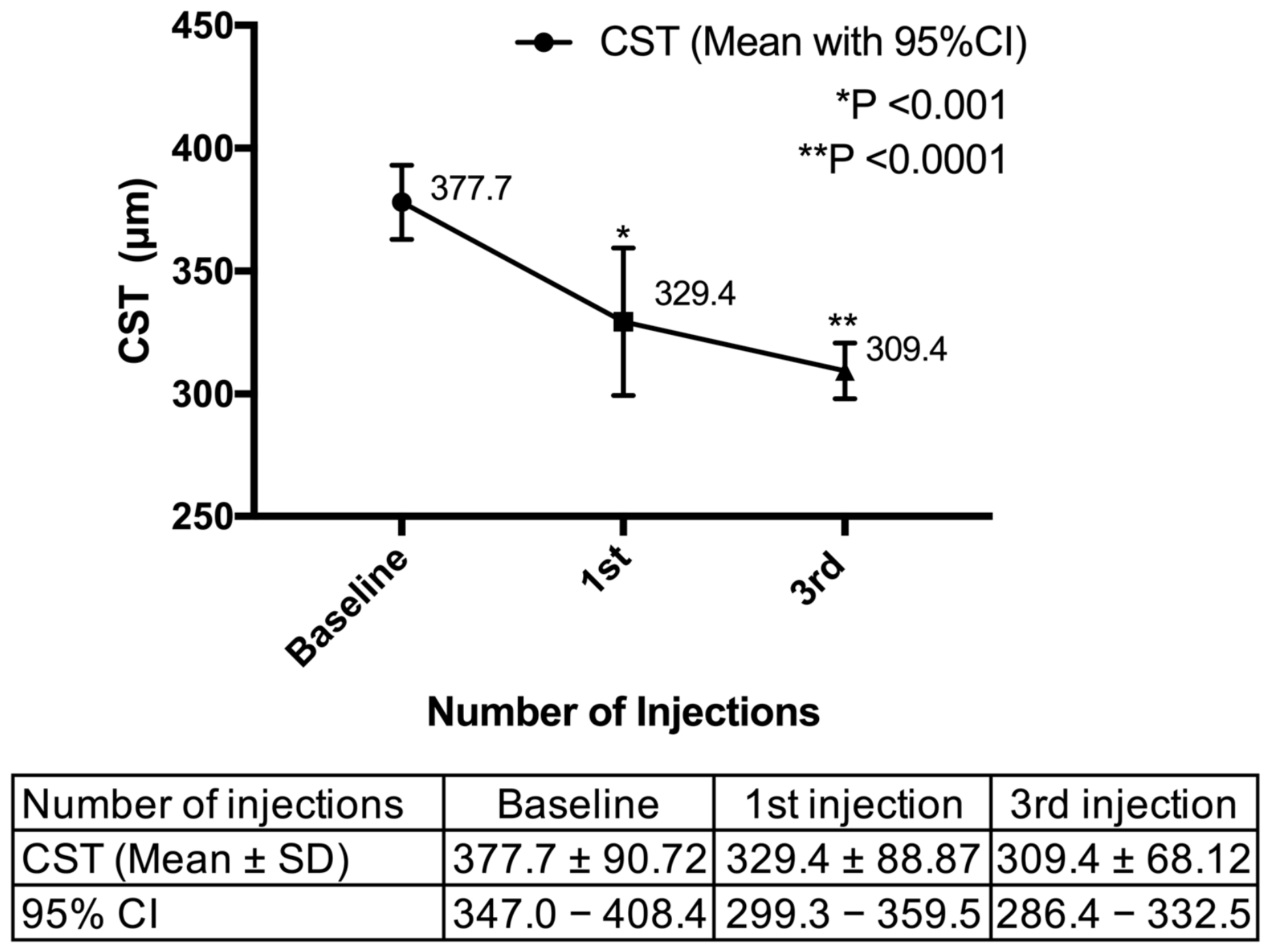
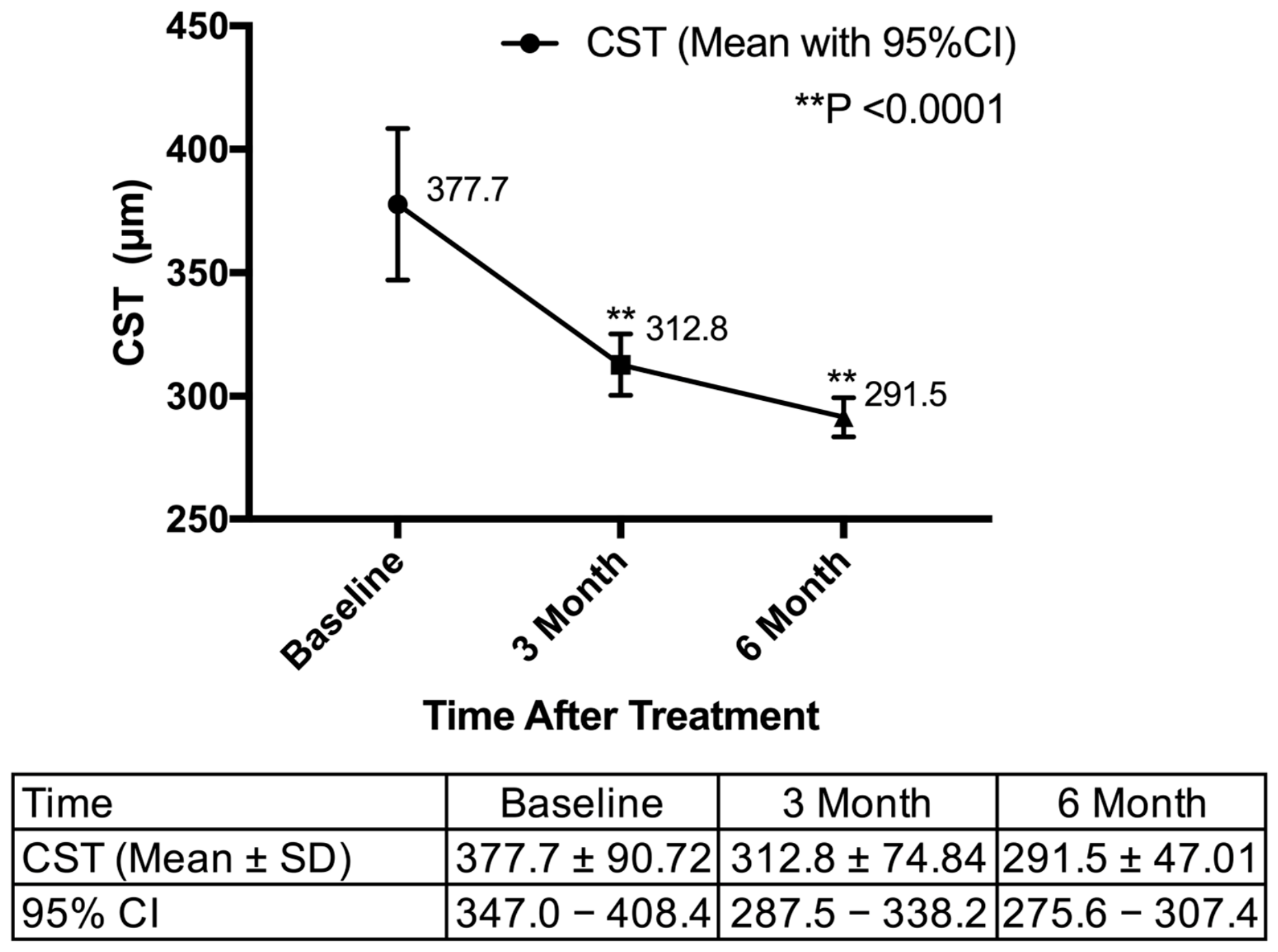
Beyond Central Subfield Thickness: Early Multi-Slice Optical Coherence Tomography Structural Response After Faricimab Injection in Real-World Diabetic Macular Edema
 , , , , , ,
Abstract
1. Introduction
2. Materials and Methods
2.1. Study Design and Patient Selection
2.2. Imaging and Data Acquisition
2.3. Statistical Analysis
3. Results
3.1. Baseline Demographics and Clinical Characteristics
3.2. Anatomical and Functional Outcomes
3.3. Evolution of OCT Biomarkers and Structural Fixation
3.4. Predictive Factors for Treatment Outcomes
4. Discussion
4.1. Early Anatomical Response and the Anatomical–Functional Disconnect
4.2. HEs, a Marker of Leakage Severity, and Observations on ERM
4.3. Structural Fixation and Baseline Outer Retinal Integrity
4.4. Strengths and Limitations
5. Conclusions
Supplementary Materials
Author Contributions
Funding
Institutional Review Board Statement
Informed Consent Statement
Data Availability Statement
Conflicts of Interest
Abbreviations
| Ang-2 | Angiopoietin-2 |
| BCVA | Best-corrected visual acuity |
| CI | Confidence interval |
| CI-DME | Center-involved diabetic macular edema |
| CST | Central subfield thickness |
| ΔCST | Change in central subfield thickness |
| DME | Diabetic macular edema |
| DRIL | Disorganization of the retinal inner layers |
| ERM | Epiretinal membrane |
| EZ | Ellipsoid zone |
| EZD | Ellipsoid zone disruption |
| GLM | General linear model |
| HEs | Hard exudates |
| HRF | Hyperreflective foci |
| IOP | Intraocular pressure |
| IRCs | Intraretinal cysts |
| IVF | Intravitreal faricimab |
| LogMAR | Logarithm of the Minimum Angle of Resolution |
| LONLCs | Large outer-nuclear-layer cavities |
| NPDR | Non-proliferative diabetic retinopathy |
| OCT | Optical coherence tomography |
| PDR | Proliferative diabetic retinopathy |
| PRN | Pro re nata |
| RPE | Retinal pigment epithelium |
| SD | Standard deviation |
| SD-OCT | Spectral-domain optical coherence tomography |
| SRF | Subretinal fluid |
| T&E | Treat-and-extend |
| VEGF | Vascular endothelial growth factor |
| VMI | Vitreomacular interface |
References
- Varma, R.; Bressler, N.M.; Doan, Q.V.; Gleeson, M.; Danese, M.; Bower, J.K.; Selvin, E.; Dolan, C.; Fine, J.; Colman, S.; et al. Prevalence of and risk factors for diabetic macular edema in the United States. JAMA Ophthalmol. 2014, 132, 1334–1340. [Google Scholar] [CrossRef] [Scilit] [PubMed]
- Tan, G.S.; Cheung, N.; Simó, R.; Cheung, G.C.; Wong, T.Y. Diabetic macular oedema. Lancet Diabetes Endocrinol. 2017, 5, 143–155. [Google Scholar] [CrossRef] [Scilit] [PubMed]
- Mesquida, M.; Drawnel, F.; Fauser, S. The role of inflammation in diabetic eye disease. Semin. Immunopathol. 2019, 41, 427–445. [Google Scholar] [CrossRef] [Scilit] [PubMed]
- Chen-Li, G.; Martinez-Archer, R.; Coghi, A.; Roca, J.A.; Rodriguez, F.J.; Acaba-Berrocal, L.; Berrocal, M.H.; Wu, L. Beyond VEGF: Angiopoietin-Tie Signaling Pathway in Diabetic Retinopathy. J. Clin. Med. 2024, 13, 2778. [Google Scholar] [CrossRef] [Scilit] [PubMed]
- Lim, J.I.; Kim, S.J.; Bailey, S.T.; Kovach, J.L.; Vemulakonda, G.A.; Ying, G.S.; Flaxel, C.J. Diabetic Retinopathy Preferred Practice Pattern®. Ophthalmology 2025, 132, P75–P162. [Google Scholar] [CrossRef] [Scilit] [PubMed]
- American Diabetes Association Professional Practice Committee for Diabetes. 12. Retinopathy, Neuropathy, and Foot Care: Standards of Care in Diabetes-2026. Diabetes Care 2026, 49, S261–S276. [Google Scholar] [CrossRef] [Scilit] [PubMed]
- Parmar, M.S. Evaluation and Care of Patients with Diabetic Retinopathy. N. Engl. J. Med. 2020, 383, e31. [Google Scholar] [CrossRef] [Scilit] [PubMed]
- Virgili, G.; Curran, K.; Lucenteforte, E.; Peto, T.; Parravano, M. Anti-vascular endothelial growth factor for diabetic macular oedema: A network meta-analysis. Cochrane Database Syst. Rev. 2023, 2023, Cd007419. [Google Scholar] [CrossRef] [Scilit] [PubMed]
- Sun, J.K.; Beaulieu, W.T.; Melia, M.; Ferris, F.L., 3rd; Maturi, R.K.; Nielsen, J.S.; Solomon, S.D.; Jampol, L.M. Defining “strong” versus “weak” response to antivascular endothelial growth factor treatment for center-involved diabetic macular edema. Retina 2023, 43, 616–623. [Google Scholar] [CrossRef] [Scilit] [PubMed]
- Ashraf, M.; Souka, A.; Adelman, R. Predicting outcomes to anti-vascular endothelial growth factor (VEGF) therapy in diabetic macular oedema: A review of the literature. Br. J. Ophthalmol. 2016, 100, 1596–1604. [Google Scholar] [CrossRef] [Scilit] [PubMed]
- Chang, Y.C.; Huang, Y.T.; Hsu, A.Y.; Meng, P.P.; Lin, C.J.; Lai, C.T.; Hsia, N.Y.; Chen, H.S.; Tien, P.T.; Lin, J.M.; et al. Optical Coherence Tomography Biomarkers in Predicting Treatment Outcomes of Diabetic Macular Edema after Ranibizumab Injections. Medicina 2023, 59, 629. [Google Scholar] [CrossRef] [Scilit] [PubMed]
- Wykoff, C.C.; Abreu, F.; Adamis, A.P.; Basu, K.; Eichenbaum, D.A.; Haskova, Z.; Lin, H.; Loewenstein, A.; Mohan, S.; Pearce, I.A.; et al. Efficacy, durability, and safety of intravitreal faricimab with extended dosing up to every 16 weeks in patients with diabetic macular oedema (YOSEMITE and RHINE): Two randomised, double-masked, phase 3 trials. Lancet 2022, 399, 741–755. [Google Scholar] [CrossRef] [Scilit] [PubMed]
- Nanji, K.; Grad, J.; Hatamnejad, A.; El-Sayes, A.; Mihalache, A.; Gemae, M.; Huang, R.; Phillips, M.; Kaiser, P.K.; Munk, M.R.; et al. Baseline OCT Biomarkers Associated with Visual Acuity in Diabetic Macular Edema: A Systematic Review and Meta-analysis. Ophthalmology 2026, 133, 75–90. [Google Scholar] [CrossRef] [Scilit] [PubMed]
- Li, X.; Yang, L.; Long, H.; Peng, S. Comparative value of retinal structural features versus systemic inflammatory and renal indicators in predicting short-term anti-VEGF response in diabetic macular edema: Focus on OCT biomarkers. Front. Med. 2026, 13, 1761324. [Google Scholar] [CrossRef] [Scilit] [PubMed]
- Nalcaci, S.; Gobeka, H.H.; Sincar, K.; Akkin, C.; Afrashi, F. Intravitreal dexamethasone implant in epiretinal membrane-associated diabetic macular edema refractory to anti-VEGF therapy: A visual performance and retinal ultra-structural analysis. Eur. Rev. Med. Pharmacol. Sci. 2026, 30, 71–82. [Google Scholar] [CrossRef] [Scilit] [PubMed]
- Chou, H.D.; Wu, C.H.; Chiang, W.Y.; Chen, N.N.; Hwang, Y.S.; Chen, K.J.; Lai, C.H.; Wu, P.C.; Chen, Y.H.; Yeung, L.; et al. Optical coherence tomography and imaging biomarkers as outcome predictors in diabetic macular edema treated with dexamethasone implant. Sci. Rep. 2022, 12, 3872. [Google Scholar] [CrossRef] [Scilit] [PubMed]
- Huang, Y.T.; Chang, Y.C.; Meng, P.P.; Lin, C.J.; Lai, C.T.; Hsia, N.Y.; Chen, H.S.; Tien, P.T.; Bair, H.; Lin, J.M.; et al. Optical Coherence Tomography Biomarkers in Predicting Treatment Outcomes of Diabetic Macular Edema After Dexamethasone Implants. Front. Med. 2022, 9, 852022. [Google Scholar] [CrossRef] [Scilit] [PubMed]
- Wells, J.A.; Glassman, A.R.; Ayala, A.R.; Jampol, L.M.; Aiello, L.P.; Antoszyk, A.N.; Arnold-Bush, B.; Baker, C.W.; Bressler, N.M.; Browning, D.J.; et al. Aflibercept, bevacizumab, or ranibizumab for diabetic macular edema. N. Engl. J. Med. 2015, 372, 1193–1203. [Google Scholar] [CrossRef] [Scilit] [PubMed]
- Kusuhara, S.; Kishimoto-Kishi, M.; Matsumiya, W.; Miki, A.; Imai, H.; Nakamura, M. Short-Term Outcomes of Intravitreal Faricimab Injection for Diabetic Macular Edema. Medicina 2023, 59, 665. [Google Scholar] [CrossRef] [Scilit] [PubMed]
- Yu, J.Y.; Du, M.; Elliott, M.H.; Wu, M.; Fu, D.; Yang, S.; Basu, A.; Gu, X.; Ma, J.X.; Aston, C.E.; et al. Extravascular modified lipoproteins: A role in the propagation of diabetic retinopathy in a mouse model of type 1 diabetes. Diabetologia 2016, 59, 2026–2035. [Google Scholar] [CrossRef] [Scilit] [PubMed]
- Shukla, U.V.; Tripathy, K. Diabetic Retinopathy. In StatPearls; StatPearls Publishing LLC.: Treasure Island, FL, USA, 2026. [Google Scholar]
- Heier, J.S.; Singh, R.P.; Wykoff, C.C.; Csaky, K.G.; Lai, T.Y.Y.; Loewenstein, A.; Schlottmann, P.G.; Paris, L.P.; Westenskow, P.D.; Quezada-Ruiz, C. The angiopoietin/tie pathway in retinal vascular diseases: A Review. Retina 2021, 41, 1–19. [Google Scholar] [CrossRef] [Scilit] [PubMed]
- Sahni, J.; Patel, S.S.; Dugel, P.U.; Khanani, A.M.; Jhaveri, C.D.; Wykoff, C.C.; Hershberger, V.S.; Pauly-Evers, M.; Sadikhov, S.; Szczesny, P.; et al. Simultaneous Inhibition of Angiopoietin-2 and Vascular Endothelial Growth Factor-A with Faricimab in Diabetic Macular Edema: BOULEVARD Phase 2 Randomized Trial. Ophthalmology 2019, 126, 1155–1170. [Google Scholar] [CrossRef] [Scilit] [PubMed]
- Ożóg, M.K.; Nowak-Wąs, M.; Rokicki, W. Pathophysiology and clinical aspects of epiretinal membrane—Review. Front. Med. 2023, 10, 1121270. [Google Scholar] [CrossRef] [Scilit] [PubMed]
- Canonica, J.; Foxton, R.; Garrido, M.G.; Lin, C.M.; Uhles, S.; Shanmugam, S.; Antonetti, D.A.; Abcouwer, S.F.; Westenskow, P.D. Delineating effects of angiopoietin-2 inhibition on vascular permeability and inflammation in models of retinal neovascularization and ischemia/reperfusion. Front. Cell. Neurosci. 2023, 17, 1192464. [Google Scholar] [CrossRef] [Scilit] [PubMed]
- Ferro Desideri, L.; Traverso, C.E.; Nicolò, M.; Munk, M.R. Faricimab for the Treatment of Diabetic Macular Edema and Neovascular Age-Related Macular Degeneration. Pharmaceutics 2023, 15, 1413. [Google Scholar] [CrossRef] [Scilit] [PubMed]
- Jeansson, M.; Gawlik, A.; Anderson, G.; Li, C.; Kerjaschki, D.; Henkelman, M.; Quaggin, S.E. Angiopoietin-1 is essential in mouse vasculature during development and in response to injury. J. Clin. Investig. 2011, 121, 2278–2289. [Google Scholar] [CrossRef] [Scilit] [PubMed]
- Kuiper, E.J.; Van Nieuwenhoven, F.A.; de Smet, M.D.; van Meurs, J.C.; Tanck, M.W.; Oliver, N.; Klaassen, I.; Van Noorden, C.J.; Goldschmeding, R.; Schlingemann, R.O. The angio-fibrotic switch of VEGF and CTGF in proliferative diabetic retinopathy. PLoS ONE 2008, 3, e2675. [Google Scholar] [CrossRef] [Scilit] [PubMed]
- Jaffe, G.J.; Deák, G.; Gibson, K.; Khurana, R.N.; Nudleman, E.; Ogura, Y.; Schmidt-Erfurth, U.; Wang, T.; Westenskow, P.D.; Wong, D.; et al. Impact of faricimab versus aflibercept on epiretinal membrane formation over 2 years in patients with diabetic macular edema in the phase 3 Yosemite and Rhine trials. Retina 2025, 45, 2003–2011. [Google Scholar] [CrossRef] [Scilit] [PubMed]
- Saxena, S.; Meyer, C.H.; Akduman, L. External limiting membrane and ellipsoid zone structural integrity in diabetic macular edema. Eur. J. Ophthalmol. 2022, 32, 15–16. [Google Scholar] [CrossRef] [Scilit] [PubMed]
- Kessler, L.J.; Auffarth, G.U.; Bagautdinov, D.; Khoramnia, R. Ellipsoid Zone Integrity and Visual Acuity Changes during Diabetic Macular Edema Therapy: A Longitudinal Study. J. Diabetes Res. 2021, 2021, 8117650. [Google Scholar] [CrossRef] [Scilit] [PubMed]
- Daruich, A.; Matet, A.; Moulin, A.; Kowalczuk, L.; Nicolas, M.; Sellam, A.; Rothschild, P.R.; Omri, S.; Gélizé, E.; Jonet, L.; et al. Mechanisms of macular edema: Beyond the surface. Prog. Retin. Eye Res. 2018, 63, 20–68. [Google Scholar] [CrossRef] [Scilit] [PubMed]
- Spaide, R.F.; Curcio, C.A. Anatomical correlates to the bands seen in the outer retina by optical coherence tomography: Literature review and model. Retina 2011, 31, 1609–1619. [Google Scholar] [CrossRef] [Scilit] [PubMed]
- Simó, R.; Villarroel, M.; Corraliza, L.; Hernández, C.; Garcia-Ramírez, M. The retinal pigment epithelium: Something more than a constituent of the blood-retinal barrier--implications for the pathogenesis of diabetic retinopathy. J. Biomed. Biotechnol. 2010, 2010, 190724. [Google Scholar] [CrossRef] [Scilit] [PubMed]
- Strauss, O. The retinal pigment epithelium in visual function. Physiol. Rev. 2005, 85, 845–881. [Google Scholar] [CrossRef] [Scilit] [PubMed]
- Vecino, E.; Rodriguez, F.D.; Ruzafa, N.; Pereiro, X.; Sharma, S.C. Glia-neuron interactions in the mammalian retina. Prog. Retin. Eye Res. 2016, 51, 1–40. [Google Scholar] [CrossRef] [Scilit] [PubMed]
- Navneet, S.; Wilson, K.; Rohrer, B. Müller Glial Cells in the Macula: Their Activation and Cell-Cell Interactions in Age-Related Macular Degeneration. Investig. Ophthalmol. Vis. Sci. 2024, 65, 42. [Google Scholar] [CrossRef] [Scilit] [PubMed]
- Wu, Q.; Zhang, B.; Hu, Y.; Liu, B.; Cao, D.; Yang, D.; Peng, Q.; Zhong, P.; Zeng, X.; Xiao, Y.; et al. Detection of morphologic patterns of diabetic macular edema using a deep learning approach based on optical coherence tomography images. Retina 2021, 41, 1110–1117. [Google Scholar] [CrossRef] [Scilit] [PubMed]
- Harju, N.; Kauppinen, A.; Loukovaara, S. Fibrotic Changes in Rhegmatogenous Retinal Detachment. Int. J. Mol. Sci. 2025, 26, 1025. [Google Scholar] [CrossRef] [Scilit] [PubMed]
- Sofroniew, M.V.; Vinters, H.V. Astrocytes: Biology and pathology. Acta Neuropathol. 2010, 119, 7–35. [Google Scholar] [CrossRef] [Scilit] [PubMed]



| Variables | All Patients |
|---|---|
| Patients, no. | 26 |
| - Age (years), Mean ± SD | 57.8 ± 13.4 |
| - Sex, female/total (%) | 14/26 (53.8) |
| - HbA1c (%) *, Mean ± SD | 7.9 ± 1.7 |
| Eyes, no. | 36 |
| - Laterality, OD/total (%) | 16/36 (44.4) |
| - Numbers of IVF injection, Mean ± SD | 4.2 ± 1.1 |
| - Lens, pseudophakic/total (%) | 10/36 (27.8) |
| - Initial CST (μm), Mean ± SD | 377.7 ± 90.7 |
| - Initial LogMAR, Mean ± SD | 0.5 ± 0.4 |
| - Initial IOP (mmHg), Mean ± SD | 17.3 ± 2.7 |
| DR severity, n (%) | |
| - Mild NPDR | 5 (13.9) |
| - Moderate NPDR | 8 (22.2) |
| - Severe NPDR | 5 (13.9) |
| - Severe NPDR s/p PRP | 5 (13.9) |
| - PDR | 2 (5.6) |
| - PDR s/p PRP | 11 (30.6) |
| Parameter | Final CST Improved <50 μm (n = 16) | Final CST Improved ≥50 μm (n = 20) | Univariate | Multivariate | ||||
|---|---|---|---|---|---|---|---|---|
| Crude OR | 95% CI | p Value | Adjusted OR * | 95% CI | p Value | |||
| CST, Mean ± SD | 317.1 ± 38.2 | 426.2 ± 20.5 | 1.027 | 1.008, 1.047 | 0.006 | — | — | — |
| SRF, n (%) | 1 (6.25%) | 5 (25.0%) | 5 | 0.520, 48.068 | 0.163 | 3.254 | 0.231, 45.801 | 0.382 |
| IRC, n (%) | 10 (62.5%) | 19 (95.0%) | 11.4 | 1.200, 108.293 | 0.034 | 4.001 | 0.353, 45.353 | 0.263 |
| ERM, n (%) | 7 (43.8%) | 7 (35.0%) | 0.692 | 0.180, 2.668 | 0.593 | 0.349 | 0.052, 2.358 | 00.280 |
| EZD, n (%) | 0 (0%) | 7 (35.0%) | NA | NA | NA | NA | NA | NA |
| DRIL, n (%) | 4 (25.0%) | 16 (80%) | 12 | 2.484, 57.975 | 0.002 | 2.682 | 0.406, 17.709 | 0.306 |
| HE, n (%) | 9 (56.3%) | 17 (85.0%) | 4.407 | 0.912, 21.301 | 0.065 | 22.021 | 0.927, 523.131 | 0.056 |
| HRF, n (%) | 15 (93.8%) | 19 (95.0%) | 1.267 | 0.073, 21.970 | 0.871 | 33.674 | 0.076, 14,942.192 | 0.258 |
| Number of HRF, n (%) | ||||||||
| - ≤20 (ref.) | 8 (50.0%) | 4 (20%) | 1 (Ref.) | — | — | 1 (Ref.) | — | — |
| - >20 | 7 (43.8%) | 15 (75.0%) | 4.286 | 0.958, 19.178 | 0.057 | 8.147 | 0.697, 95.197 | 0.094 |
| LONLCs, n (%) | 9 (56.3%) | 18 (90.0%) | 7 | 1.200, 40.828 | 0.031 | 2.142 | 0.297, 15.462 | 0.450 |
| VMI, n (%) | 7 (43.8%) | 8 (40.0%) | 0.857 | 0.226, 3.249 | 0.821 | 0.363 | 0.053, 2.465 | 0.300 |
| Parameter | Good Responders (n = 12) | Suboptimal Responders (n = 24) | Univariate | Multivariate | ||||
|---|---|---|---|---|---|---|---|---|
| Crude OR | 95% CI | p Value | Adjusted OR * | 95% CI | p Value | |||
| CST, Mean ± SD | 315.5 ± 39.1 | 406.8 ± 95.6 | 1.019 | 1.003, 1.036 | 0.022 | — | — | — |
| SRF, n (%) | 1 (8.3%) | 5 (20.8%) | 2.895 | 0.299, 28.070 | 0.359 | 1.547 | 0.121, 19.713 | 0.737 |
| IRC, n (%) | 7 (58.3%) | 22 (91.7%) | 7.857 | 1.239, 49.834 | 0.029 | 3.516 | 0.491, 25.170 | 0.211 |
| ERM, n (%) | 6 (50.0%) | 8 (33.3%) | 0.500 | 0.122, 2.057 | 0.337 | 0.292 | 0.051, 1.664 | 0.165 |
| EZD, n (%) | 0 (0%) | 7 (29.2%) | NA | NA | NA | NA | NA | NA |
| DRIL, n (%) | 4 (33.3%) | 16 (66.7%) | 4.000 | 0.920, 17.396 | 0.065 | 0.908 | 0.142, 5.789 | 0.918 |
| HE, n (%) | 8 (66.7%) | 18 (75.0%) | 1.500 | 0.330, 6.822 | 0.600 | 1.498 | 0.264, 8.491 | 0.648 |
| HRF, n (%) | 12 (100%) | 22 (91.7%) | NA | NA | NA | NA | NA | NA |
| Number of HRF, n (%) | ||||||||
| - ≤20 (ref.) | 5 (41.7%) | 7 (31.8%) | 1 (Ref.) | — | — | 1 (Ref.) | — | — |
| - >20 | 7 (58.3%) | 15 (68.2%) | 1.531 | 0.357, 6.569 | 0.567 | 1.124 | 0.214, 5.907 | 0.890 |
| LONLCs, n (%) | 6 (50.0%) | 21 (87.5%) | 7.000 | 1.336, 36.686 | 0.021 | 3.041 | 0.510, 18.139 | 0.222 |
| VMI, n (%) | 6 (50.0%) | 9 (37.5%) | 0.600 | 0.148, 2.436 | 0.475 | 4.412 | 0.404, 48.211 | 0.224 |
| Parameter | Unstandardized Coeff. (B) | 95% CI for B | Standardized Coeff. (β) | p Value |
|---|---|---|---|---|
| SRF | 28.927 | −10.010, 67.865 | 0.135 | 0.140 |
| IRC | 9.423 | −30.043, 48.888 | 0.047 | 0.630 |
| ERM | −8.720 | −38.714, 21.274 | −0.053 | 0.558 |
| EZD | 25.916 | −23.962, 75.794 | 0.129 | 0.298 |
| DRIL | 10.463 | −27.298, 48.224 | 0.065 | 0.577 |
| HE | 45.984 | 17.503, 74.465 | 0.259 | 0.002 |
| HRF | −8.206 | −73.160, 56.748 | −0.024 | 0.799 |
| Number of HRF | ||||
| - ≤20 (ref.) | — | — | — | — |
| - >20 | 24.419 | −3.753, 52.591 | 0.160 | 0.087 |
| LONLCs | 3.629 | −33.407, 40.666 | 0.020 | 0.843 |
| VMI | −10.708 | −40.513, 19.096 | −0.066 | 0.470 |
Disclaimer/Publisher’s Note: The statements, opinions and data contained in all publications are solely those of the individual author(s) and contributor(s) and not of MDPI and/or the editor(s). MDPI and/or the editor(s) disclaim responsibility for any injury to people or property resulting from any ideas, methods, instructions or products referred to in the content. |
© 2026 by the authors. Licensee MDPI, Basel, Switzerland. This article is an open access article distributed under the terms and conditions of the Creative Commons Attribution (CC BY) license.
Share and Cite
Liu, D.-Y.; Wu, S.-L.; Hsia, N.-Y.; Tien, P.-T.; Lin, C.-J.; Wang, I.; Lai, C.-T.; Lin, J.-M.; Huang, Y.-T.; Wu, B.-Q.; et al. Beyond Central Subfield Thickness: Early Multi-Slice Optical Coherence Tomography Structural Response After Faricimab Injection in Real-World Diabetic Macular Edema. J. Clin. Med. 2026, 15, 6356. https://doi.org/10.3390/jcm15166356
Liu D-Y, Wu S-L, Hsia N-Y, Tien P-T, Lin C-J, Wang I, Lai C-T, Lin J-M, Huang Y-T, Wu B-Q, et al. Beyond Central Subfield Thickness: Early Multi-Slice Optical Coherence Tomography Structural Response After Faricimab Injection in Real-World Diabetic Macular Edema. Journal of Clinical Medicine. 2026; 15(16):6356. https://doi.org/10.3390/jcm15166356
Chicago/Turabian StyleLiu, De-Yi, Shiao-Ling Wu, Ning-Yi Hsia, Peng-Tai Tien, Chun-Ju Lin, I Wang, Chun-Ting Lai, Jane-Ming Lin, Yu-Te Huang, Bing-Qi Wu, and et al. 2026. "Beyond Central Subfield Thickness: Early Multi-Slice Optical Coherence Tomography Structural Response After Faricimab Injection in Real-World Diabetic Macular Edema" Journal of Clinical Medicine 15, no. 16: 6356. https://doi.org/10.3390/jcm15166356
APA StyleLiu, D.-Y., Wu, S.-L., Hsia, N.-Y., Tien, P.-T., Lin, C.-J., Wang, I., Lai, C.-T., Lin, J.-M., Huang, Y.-T., Wu, B.-Q., Lin, W.-N., Ku, W.-N., Meng, P.-P., Chen, H.-S., & Tsai, Y.-Y. (2026). Beyond Central Subfield Thickness: Early Multi-Slice Optical Coherence Tomography Structural Response After Faricimab Injection in Real-World Diabetic Macular Edema. Journal of Clinical Medicine, 15(16), 6356. https://doi.org/10.3390/jcm15166356