Review

Next-Generation Stroke Biomarkers: Bridging the Gap Between Innovation and Translation

by
Emilia Conti
1,2,
Marzia Baldereschi
3,*,
Antonio Di Carlo
3,
Giulia Barbieri
4,
Francesco Saverio Pavone
2,5,
Anna Maria Gori
4,† and
Betti Giusti
4,†
1
National Institute of Optics, National Research Council, Via Nello Carrara 1, 50019 Sesto Fiorentino, Italy
2
European Laboratory for Non Linear Spectroscopy (LENS), Via Nello Carrara 1, 50019 Sesto Fiorentino, Italy
3
Neuroscience Institute, National Research Council, Via Madonna del Piano 10, 50019 Sesto Fiorentino, Italy
4
Department of Experimental and Clinical Medicine, University of Florence, Largo Brambilla 3, 50134 Firenze, Italy
5
Department of Physics and Astronomy, University of Florence, Via Sansone 1, 50019 Sesto Fiorentino, Italy
*
Author to whom correspondence should be addressed.
These authors contributed equally to this work.
J. Clin. Med. 2026, 15(16), 6249; https://doi.org/10.3390/jcm15166249
Submission received: 29 April 2026 / Revised: 19 July 2026 / Accepted: 8 August 2026 / Published: 12 August 2026
(This article belongs to the Special Issue Current Advances and Future Perspectives of Ischemic Stroke)

Abstract

The identification of valid biomarkers for ischemic stroke would greatly benefit not only diagnostic and prognostic assessment but also the development of new therapies. The development of omics technologies has made it possible to explore various perspectives of the pathophysiology of ischemic stroke, in particular genomics, transcriptomics, proteomics and metabolomics, and well-known clinical variables, such as hypertension, diabetes, heart diseases, and many others. Omics-based research has produced a mass of data evidencing candidate biomarkers at different stages in the disease process. However, further scientific evidence must be presented and affordable, and rapid detection techniques developed before these technologies can be usefully applied to the clinical setting of stroke. Here, we aim to provide stroke clinicians with an updated overview of the contribution of these emerging technologies in the individuation of diagnostic and prognostic biomarkers and the development of specifically targeted therapies. Cross-fertilization (translation and reverse translation) between preclinical and clinical research is of particular relevance in the identification of stroke biomarkers. We believe it is useful to provide, in parallel, an updated review of omics investigations using stroke rodent models. This review presents a detailed discussion of the state of the art, focusing on the strengths and limitations of the individual technologies and an outline of the promising, most recent results of both clinical and preclinical research in this field related to stroke.

1. Introduction

Ischemic stroke (IS) is a leading cause of disability and mortality worldwide. As individuals age, the risk of experiencing a stroke significantly increases, making stroke a major health concern among the elderly population [1]. Clinical care in the acute phase is currently based on neuroimaging and clinical assessment [2]. However, many aspects remain unaddressed or unsolved. There is a need for: (i) a rapid, accurate blood test to identify IS in the pre-hospital phase and to accelerate triage and access to reperfusion treatments in the narrow therapeutic window; (ii) specific biomarkers to predict and possibly counteract futile recanalization that occurs in about half of recanalized patients; and (iii) diagnostic biomarkers for transient ischemic attacks, because the great majority of patients are asymptomatic when examined, and therefore the diagnosis is only anamnestic [3,4]. These challenges reflect the complex and heterogeneous biological nature of IS. Similar clinical features may be caused by distinct pathogenetic mechanisms that circulating biomarkers might identify, thus allowing for rapid clinical decisions. The identification of reliable biomarkers requires approaches capable of individuating the multiple biological processes involved in stroke initiation, progression, and recovery. Omics technologies provide a comprehensive framework for the investigation of molecular alterations at multiple levels and may help overcome some limitations of conventional biomarker discovery strategies. To address the urgent need for reliable and actionable stroke biomarkers a panel of international stroke experts has recently launched a Delphi consensus survey that identified 17 research priorities for stroke biomarker studies [5].
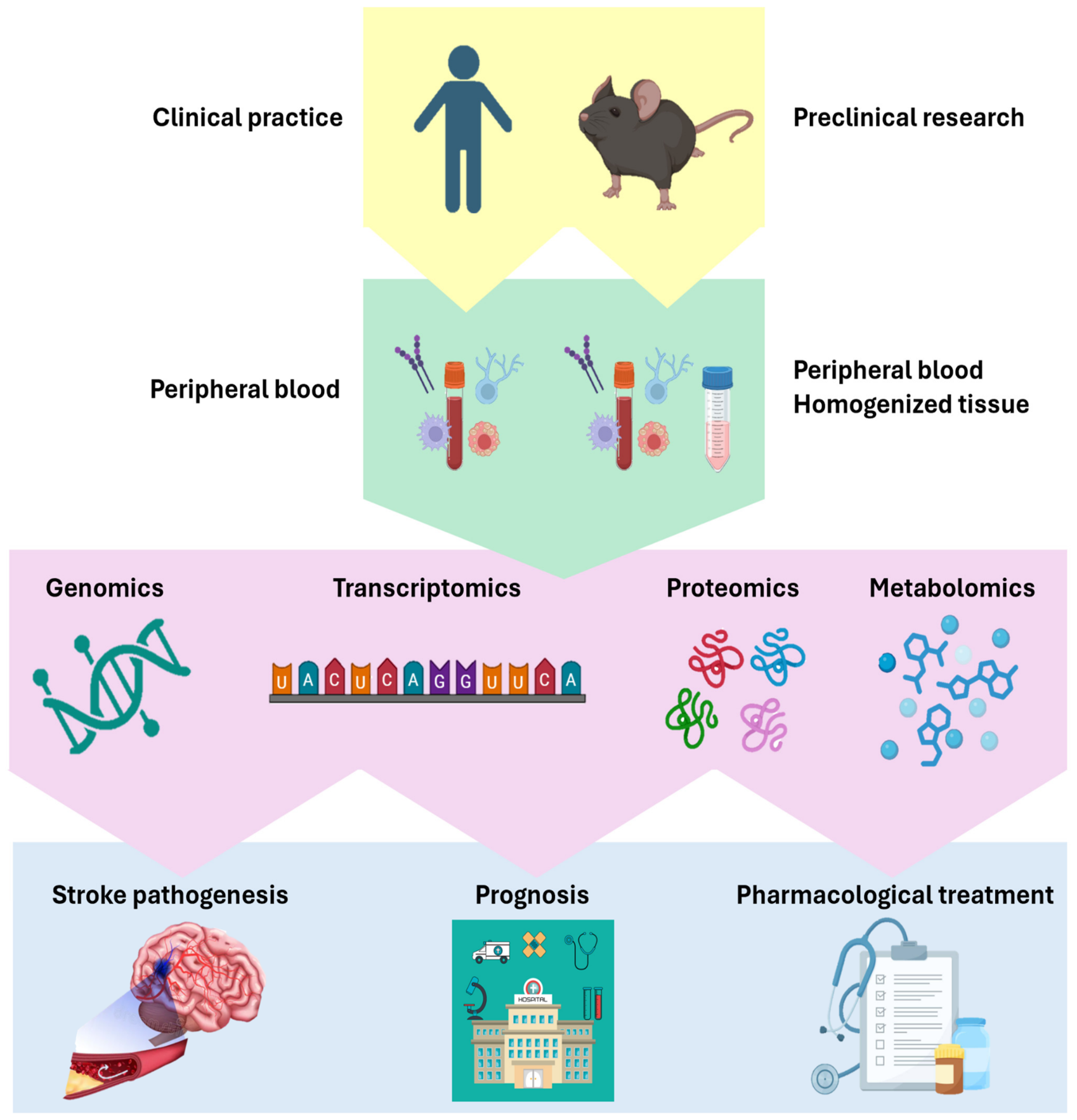
Current knowledge at the molecular, cellular, and network levels is inadequate for a full understanding of the processes underlying stroke progression. An individual patient outcome is influenced by intrinsic differences in the flow capacity of collateral vessels, degree of chronic ischemic disease, ischemic preconditioning, oxidative stress tolerance, microvascular blood flow regulation, and other still unknown factors. Within this framework, preclinical research may help clarify the neuronal and vascular underpinnings of stroke progression and identify potential treatment targets leading to clinical translation. The main advantage of animal models lies in the possibility of selecting various factors, such as genetic background, sex, and age. On the other hand, stroke models allow for dissecting critical features of stroke pathophysiology by employing in vivo and ex vivo strategies, which is not feasible in clinical practice. This feature, together with the high reproducibility of stroke models, allows for the use of fewer biological replicates while minimizing confounding factors. In this framework, preclinical models, combined with high-throughput omics technologies, can provide the otherwise unattainable mechanistic information at the molecular level underlying functional outcome in stroke patients. The successful translation of omics discoveries into clinical practice relies on the continuous mutual interaction between preclinical and clinical research. On the one hand, experimental models provide a controlled environment for the investigation of pathophysiological disease mechanisms, and they can validate already identified candidate biomarkers in patients. On the other hand, clinical studies are essential to confirm their relevance in the heterogeneous human population. In parallel, clinical cohort studies can guide the development of more representative experimental models and generate new mechanistic hypotheses. This bidirectional framework is essential to accelerate the identification of robust biomarkers and therapeutic targets for IS as it is sketched in Figure 1.
Improvements are needed across the whole stroke care pathway, from reperfusion treatments to long-term rehabilitation, to reduce the medical and societal burden of stroke. We consider continuous cross-fertilization between clinical and preclinical research that is necessary to provide novel solutions to unmet clinical needs.
The aim of this review is to illustrate the emerging technologies that could play a strong role in stroke therapy in the near future. Regarding stroke, omics technologies are currently relegated to clinical and preclinical research, but they may soon become an important asset in stroke care. Moreover, the development of advanced detection approaches, such as single-molecule assay (SIMOA) and multi-array systems, in addition to the development of the Point of Care Test (POCT), might make it possible to assess biomarkers at bedside, even in the pre-hospital phase, thus contributing to the fundamental aspect of timing in IS care.
We discuss the application of omics technology in experimental and clinical settings, emphasizing its potential role as a bridge between mechanistic discoveries and the development of clinically relevant biomarkers in stroke. The aim is to update stroke clinicians on the state of the art, as well as the strengths and limitations of each type of technology, giving an overview of the most recent and promising results from both clinical and rodent stroke model omics research. In addition, we highlight possible bottlenecks in the clinical translation of research findings.

2. Integrated Omics (Genomics, Transcriptomics, Proteomics, and Metabolomics)

2.1. Genomics

Genetics and genomics could revolutionize stroke care by shifting the focus from reactive treatment to proactive prevention and personalized medicine. Genetic technologies for stroke have been recently reviewed [6]. The study of genomics in stroke has already given promising results related to prevention (risk reduction), the prediction of response to acute-phase treatments (therefore the outcome), and the identification of the different degrees of response to antiplatelet drugs (pharmacogenomics) [7,8]. Apart from rare Mendelian forms, IS must be considered a polygenic and multifactorial disease. Accordingly, recent studies have increasingly used Genome-Wide Association Studies (GWAS), scanning the whole genome to look for genetic variants associated with an increased risk of stroke [8]. Recent large-scale genomic investigations have substantially expanded the catalogue of IS-associated susceptibility loci and improved the biological interpretation of GWAS findings by integrating genetic association data with functional genomic analyses, including the expression of quantitative trait loci (eQTL) and protein quantitative trait loci (pQTL), Mendelian randomization, and single-cell transcriptomic analyses. These approaches have allowed for the identification of new candidate genes regarding vascular biology, inflammation, lipid metabolism, iron homeostasis, endothelial dysfunction, and neuronal injury. In all likelihood, they will lead to the identification of new therapeutic targets and biomarkers for precision medicine in IS [9,10]. As reported in Table 1, several genetic loci have been identified with IS diagnosis, complications and prognosis. The identification of low metabolizers of the antiplatelet clopidogrel is of particular relevance: CYP2C19, encoding an antiplatelet-metabolizing enzyme, has proven to be of extreme interest for primary and secondary stroke-prevention therapies. CYP2C19’s loss-of-function alleles have been proven to be associated with higher risk of recurrent IS in patients on clopidogrel [11,12]. Poor metabolizers turned out to have lower clopidogrel activation and a nearly twofold risk of recurrent stroke than normal metabolizers [13]. These findings suggest that genotyping stroke patients for CYP2C19 is worthwhile, because poor metabolizers can be treated with other P2Y12 inhibitors, such as ticagrelor, which has been proven to be effective in reducing the risk of stroke recurrence in CYP2C19 LoF carriers [14]. It is well known that the frequency of the polymorphism and the related phenotype is closely linked to ethnicity. Although most of the early studies were conducted on populations of European ancestry, there is now a considerable body of evidence also on populations of different ancestries. In European ancestry populations, the frequency of the loss-of-function allele is about 15%, while it is 30% in South Asians and 60% in native Oceanians [15].

2.1.1. Genetic Risk Score

A genetic risk score (GRS) summarizes the small effects of several Single-Nucleotide Polymorphisms, previously identified by GWAS, to help improve risk prediction and stratification beyond traditional risk factors and to facilitate personalized prevention strategies [16]. It is well known that the stroke phenotype is highly heterogeneous, and this limits its genomic risk prediction [17]. Recent studies have adopted the construction of the increasingly powerful GRS based on summary statistics of multiple sets of GWAS to overcome their single limited predictivity and to achieve added value in combination with established clinical risk factors [18]. In particular, recent promising findings relate to atrial fibrillation (AF) and stroke and are summarized in Table 1. When a large-scale GRS was incorporated into the CHA2DS2-VAS score, there was a net improvement in levels of stroke risk classification for 2.3% of AF patients [19]. The clinical implication is that if several patients are moved to a different stroke risk profile, this refinement might be particularly useful for “gray area” patients (i.e., those with low or intermediate risk of stroke) who may hide a significant stroke genetic susceptibility [19]. Moreover, the specific GRS performed better than the traditional clinical score (HAS-BLED) [20] in stratifying the risk of intracerebral hemorrhage in patients on anticoagulants [21]. We note that the highest level of predictivity is achieved when GRS data are added to clinical factors. Finally, a reliable estimate of stroke risk in AF patients remains challenging [19]. Recent evidence further supports the role of polygenic risk scores as complementary tools for identifying individuals at increased genetic susceptibility, refining risk stratification and facilitating future implementation of precision prevention strategies in ischemic stroke [9,10].

2.1.2. Preclinical Studies

Regarding preclinical research, in a recent study on mice, Wang and colleagues explored the biological significance of candidate IS-associated genes [9]. Existing mouse knockout models from the Mouse Genome Informatics (MGI) database were leveraged to establish functional correlations [22]. Of the seven prioritized genes, only HSD17B12, ERAP2, and SFXN4 showed strong experimental support. Loss of HSD17B12 led to disruptions in fatty acid metabolism, highlighting a mechanistic link between lipid metabolic imbalance and cerebrovascular pathology. Similarly, ERAP2 and SFXN4 were implicated in immune modulation and iron homeostasis, two processes known to contribute to stroke onset and progression. Although knockout models are not yet available for other genes, in particular C15orf40, CPNE1, LYRM9, and SNX32, existing annotations suggest potential involvement in neuronal development, cell-cycle control, and mitochondrial activity, indicating their possible roles in neuronal vulnerability during ischemic injury. Notably, drug-potential analyses indicate that three of these genes (CPNE1, HSD17B12, and ERAP2) encode proteins already targeted by approved or clinically tested compounds, underscoring their translational relevance and potential as therapeutic entry points in stroke precision medicine [23,24,25].

2.2. Transcriptomics

Gene activity can be investigated through transcriptomic analyses, which encompass bulk RNA sequencing, single-cell RNA sequencing, spatial transcriptomics and extracellular vesicle RNA profiling, providing complementary information on gene regulation in tissue and cells.
Transcriptomic studies on peripheral blood have been extensively and recently reviewed [26] and so have the studies on clot transcriptomics—obtained from endovascular disobstruction interventions [27].
In addition to messenger RNA (mRNA), the transcriptome includes non-coding RNAs (ncRNAs), which do not encode proteins, but they function as regulatory molecules modulating gene expression at the post-transcriptional level [28]. These ncRNAs include microRNAs (miRNAs), lncRNAs, and circular RNAs (circRNAs). miRNAs are small (~21 nucleotides) tissue- and time-specific regulators expressed across human tissues that control gene expression by inducing silencing or degradation of target mRNAs.
Notably, miRNAs released from ischemic brain tissue can cross the blood–brain barrier (BBB) within extracellular vesicles or exosomes and remain stable in biofluids such as plasma and serum, making them promising biomarkers of disease status and prognosis, enabling early identification of patients at risk for poor recovery and possibly informing rehabilitation strategies [29,30]. A specific miRNA signature might possibly support the differential diagnosis. Promising findings may arise from clot transcriptome analysis, which was found to be effective in differentiating the occlusion of cardioembolic origin, rich in red blood cells and fibrin, from that caused by large-vessel atherosclerosis, where platelet and immune activations are predominant, thus assisting in stroke subtyping [31]. In a recent metanalysis of 44 studies (4302 IS patients and 3725 healthy controls) a specific panel of lncRNA showed moderate accuracy (79% sensitivity and 88% specificity) in identifying IS [32]. Cell-free nucleic acids (both DNA and RNA) may lead to the identification of upstream biomarkers for stroke diagnosis, even in pre-hospital settings: testing is minimally invasive and potentially possible by point-of-care testing. A recent systematic review and metanalysis [33] did not provide any conclusive data. Recent single-cell RNA sequencing (scRNA-seq) and spatial transcriptomics have identified cell-type-specific transcriptional responses after ischemic stroke, evidencing distinct activation states of the different cell types involved in IS. These novel approaches have furnished several interesting findings on previously unidentified inflammatory and neurovascular pathways involved in ischemic injury and recovery while preserving tissue spatial organization.
The integration of transcriptomic datasets with GWAS findings and other omics layers has improved the prioritization of causal genes and molecular pathways, supporting the development of precision medicine strategies and individualized therapeutic targets [34,35]. Table 1 summarizes the most relevant findings in transcriptomic human IS research.

Preclinical Studies

Preclinical studies show a substantial overlap between differentially expressed genes in peripheral blood and brain [36,37,38]. Androvic et al. recently reported that in aged mice after stroke, genes involved in axonal and synaptic maintenance were downregulated, while type I interferon (IFN-I) signaling was upregulated [39]. Recent studies have investigated the neuroprotective role of miRNA expression in the hyper-acute phase after injury. Song et al. [40] investigated miR-140-5p in a mouse model of Middle Cerebral Artery Occlusion (MCAO), demonstrating reduced expression after stroke compared to controls. Using qRT-PCR, they showed that miR-140-5p modulates the TLR4/NF-κB pathway and that its upregulation inhibits neuronal apoptosis, suggesting a potential role in limiting ischemic damage. Similarly, Kolosowska et al. [41] examined miR-669c, a member of the chromosome 2 miRNA cluster induced by harmful stimuli, and its involvement in stroke-related neuroinflammation. Intracerebral overexpression of miR-669c following stroke modulated microglial/macrophage activation by directly targeting MyD88, a key adaptor of TLR and IL-1 signaling. Lentiviral-mediated overexpression of miR-669c in a transient MCAO mouse model suppressed pro-inflammatory responses while promoting alternative microglial/macrophage activation, supporting its neuroprotective role. In another recent study [42] Metha et al. reported that the brain-specific lncRNA Fos downstream transcript (FosDT) is markedly upregulated following focal ischemia in rodents. FosDT deficiency reduces brain injury by limiting inflammation, apoptosis, mitochondrial dysfunction, and oxidative stress. Moreover, siRNA-mediated FosDT inhibition administered during the hyperacute or acute phase after transient MCAO significantly improved motor recovery and reduced infarct volume. Similarly, Li et al. [43] identified the lncRNA MEG3 as being overexpressed after stroke in MCAO mice; its inhibition decreased lesion volume and cerebral edema while improving neurobehavioral outcomes.
Spatial transcriptomics is emerging as a technology capable of evaluating gene expression in situ and preserving brain architecture, the infarct area in particular, thus bridging the gap between anatomical area and single-cell genomics. The ischemic lesion is not a uniform area of damage but a highly structured tissue environment, with different cellular populations set up into niches defined by unique transcriptional signatures [44]. This technology has the potential for an in-depth study of the ischemic penumbra, i.e., the salvageable tissue, and possibly to identify molecular targets that are able to predict reperfusion injury [45].

2.3. Proteomics

During an IS, many proteins (expression of neuroinflammation, BBB leakage, and excitotoxicity) change immediately and then over time in quantity, structure, and function. Proteomics allows for the simultaneous identification and quantification of thousands of proteins and is expected to be considered essential for the knowledge of pathophysiological mechanisms of IS because it allows for an investigation of the huge numbers of protein changes that occur in blood, cerebrospinal fluid, and brain tissue, which hopefully will lead to the identification of clinically relevant biomarkers [46].
Both mass spectrometry (MS)-based proteomics and highly multiplexed targeted proteomics have become powerful approaches for quantifying stroke-related changes in proteins, thanks to advances in instrumentation and bioinformatics. There are currently two different approaches in discovery proteomics: one uses untargeted MS to identify thousands of proteins without a priori hypothesis, because it scans the entire proteome; the other involves targeted multiplex platforms, including Olink® Explore (part of Thermo Fisher Scientific, Waltham, MA, USA) and SomaScan® (SomaLogic Operating Co., Inc., Boulder, CO, USA), to quantify preselected proteins with high sensitivity and excellent reproducibility, even for small quantities, complementing untargeted MS-based discovery proteomics [47].
Studies employing high-resolution MS have identified different expressions of hundreds of proteins after symptom onset that may differentiate ischemic from hemorrhagic insult when neuroimaging is unavailable or delayed [48]. Using the same technology, two-protein panels proved accurate in distinguishing the onset of symptoms within the 4.5 h therapeutic window from those beyond this limit [49].
Parallel efforts yielded a four-protein panel that is strongly related to large-vessel occlusion (LVO) IS, which could possibly assist in diagnosis and treatment decisions. Furthermore, a three-protein panel turned out to be significantly associated with collateral status and proved to be an independent predictor of good outcome [50]. Moreover, a panel of 17 proteins proved to be associated with hemorrhagic transformation [51]. In a population-based large-cohort study, distinct pre-stroke protein profiles identified by the Somascan technique proved capable of distinguishing between embolic and atherosclerotic stroke, suggesting the possible development of personalized stroke-prevention strategies [52]. One recent study demonstrated the accuracy and the feasibility of analyzing the proteome in extracellular vesicles (EVs) isolated from small amounts of human plasma/serum. Because EVs preserve the molecular cargo of their cells of origin, they may provide higher tissue specificity than unfractionated plasma proteomics [53]. Specific proteomic signatures of the EV cargo proved accurate in differentiating healthy controls from LVO IS and from hemorrhagic stroke [54].
Although blood-based proteomics remains the most immediately translatable approach, emerging evidence indicates that extracellular vesicle- and thrombus-derived proteomics may provide complementary biological information, enabling improved stroke subtyping, the prediction of treatment response and the identification of novel therapeutic targets [55,56].

Preclinical Studies

Proteomic studies in animal models are crucial for mapping the molecular events underlying acute IS pathophysiological processes and for elucidating possible mechanisms of action of therapeutic interventions. By applying Isobaric Tags for Relative and Absolute Quantitation (iTRAQ)-based proteomics, Datta et al. found an increased expression of many brain-specific proteins (Glial Fibrillar Acidic Protein, GFAP; Ubiquitin C-terminal Hydrolase—L1, UCH-L1; and S100 calcium-binding protein B, S100B) in rat serum [57]. This finding indicates that the hyper-acute opening of the BBB may allow for a leakage of these proteins into the circulation, suggesting a potential role of prognostic biomarkers for predicting therapeutic outcome or disease progression. In other studies, proteomics has been applied to explore molecular mechanisms linked with the development of tissue damage. Zheng’s analysis of rat brain tissue in the acute phase after IS identified 282 proteins (73 upregulated, 209 downregulated) involved mainly in energy liberation, intracellular protein transport, and synaptic plasticity regulation, but they were also identified in neurodegenerative disease-related pathways, including Alzheimer’s Disease and Amyotrophic Lateral Sclerosis, in rat studies [58].
In addition, proteomics has been applied in the assessment of alterations in the chronic phase after stroke. Cortical tissue analysis of MCAO rats from the subacute to chronic phase after injury highlights an altered expression of 1305 proteins within the first 14 days after damage. Moreover, cytoskeleton and synaptic structures, energy metabolism, and inflammatory response were found to be significantly disrupted in the subacute phase after stroke. However, in the long-term phase, there was recovery of the cytoskeleton and the activation of inflammatory pathways, which differed from those activated during the subacute phase [59].

2.4. Metabolomics and Lipoproteomics

Metabolomics is uniquely suited to investigate the earliest biochemical changes after stroke onset and to identify clinically relevant biomarkers [46,60,61]. Metabolites are low-molecular-weight molecules derived from lipids, amino acids, carbohydrates and nucleotides; they reflect the downstream products of gene expression, protein activity and environmental influences [46,62,63]. More than 25,000 have been counted in peripheral blood [60]. Owing to their small size, metabolites are able to pass the BBB, so circulating metabolites rapidly mirror cerebral metabolic alterations occurring during acute ischemic stroke. The metabolomic and lipoproteomic approach allows us to investigate the acute phase of stroke, because the natural history of acute IS is linked to metabolic changes that occur in the first minutes and hours. Thanks to the BBB disruption in the early phase of IS [61], metabolomics/lipoproteomics can systematically detect the biochemical profile and its changes over time in peripheral blood samples, providing insights into stroke research [64,65]. The use of this approach is useful for stroke clinicians in the identification of biomarkers for stroke subtyping [65], as well as for predicting reperfusion injury [66]. A recent study [67] suggests the possibility of a “metabolic clock” to identify the onset of stroke and therefore the access to time-dependent treatments: biliverdin and nicotinamide N-oxide (NAMO) combined have been shown able to distinguish time from onset, ≤4.5 h vs. >4.5 h, with substantial accuracy. The advent of mechanical thrombectomy has also enabled direct metabolomic analysis of cerebral thrombi. Comparative metabolomic profiling of thrombi and paired serum samples demonstrated distinct metabolic signatures and lipid enrichment within thrombi, suggesting that thrombus metabolomics may improve stroke etiological classification while providing insights into thrombus biology and the mechanisms underlying treatment resistance [68]. Finally, recent multi-omics reviews suggest that integrating metabolomics with proteomics, transcriptomics and genomics is likely to improve biomarker discovery, patient stratification and precision medicine approaches in IS [46,62,63].
Although no metabolomic biomarker has yet entered routine clinical practice, accumulating evidence indicates that integrated metabolomic and lipidomic profiling may substantially improve stroke diagnosis, etiological classification, treatment selection and outcome prediction, particularly when combined with proteomic and transcriptomic data [62].
Major findings are reported in Table 1.

2.4.1. Preclinical Studies

Animal metabolomic studies outline the progression of stroke, from excitotoxicity to metabolic failure. Although the brain represents only 2% of body mass, it accounts for about 20% of resting metabolism [69]; therefore, arterial occlusion rapidly disrupts energy metabolism, with notable changes in metabolites such as glucose, lactate, creatine/phosphocreatine, and TCA cycle intermediates [70,71,72]. The accumulation of glucose and glycolytic intermediates is a hallmark of ischemia-induced metabolic dysregulation in rodents [73]. Luo et al. [74] applied 5-(diisopropylamino)amylamine derivatization–UHPLC-Q-TOF/MS to cerebrospinal fluid from permanent MCAO rats to investigate these metabolic changes. In addition, post-stroke energy depletion triggers the rapid release and reuptake of glutamate, the main excitatory neurotransmitter in the CNS, thus activating NMDA receptors and causing massive calcium influx from extracellular and intracellular stores. This cascade elevates free radicals and nitric oxide, activates lipases and proteases, and disrupts mitochondrial function, leading to energy imbalance. Kagiyama et al. showed that I-phenylalanine selectively and reversibly suppressed glutamate receptor activity at excitatory synapses in rats and mice, producing a compensatory response to neurotoxic glutamate levels [75]. Post-ischemic inflammation disrupts phospholipid metabolism, altering lipid composition and making brain cells, rich in lipids, particularly vulnerable [76]. Long-term studies in MCAO mice showed persistent changes in lipid metabolism, including increased plasma triglycerides, free fatty acids, and adipokine release [77].
Table 1. Summary of the major possible contributions of omics technologies to the study of human stroke by stage of the disease, including application scenario and candidate biomarker.
Ischemic Stroke StagePossible ContributionPromising Application ScenarioCandidate Biomarkers [References]
GENOMICS
Primary and secondary preventionIdentification of novel causal genes and biological pathways through integrative multi-omics analysesPrecision medicine, therapeutic target discovery and personalized risk predictionNovel genes identified by integrative GWAS/eQTL/pQTL/TWAS/PWAS analyses (e.g., genes involved in inflammation, endothelial dysfunction, lipid metabolism and iron homeostasis) [10,35]
Primary and secondary preventionRisk stratification for Large-Artery Atherosclerotic StrokeGuidance on correct therapy and close monitoring of adherence and its effectivenessCDKN2A/CDKN2B genes [78,79,80,81],
HDAC9 [81] and LPA MMP12 [10]
Primary and secondary preventionRisk stratification for small vessel diseaseGuidance on correct therapy and close monitoring of adherence and its effectivenessCOL4A1/COL4A2 [82] FOXF2, HTRA1, PRDM16, and PMF1 [10]
Primary and secondary preventionRisk stratification for ischemic strokeGuidance on correct therapy and close monitoring of adherence and its effectivenessSORT1 [83] HDAC9, HTRA1, COL4A2, and ABO [10]
Primary and secondary preventionScreening for atrial fibrillationStrict monitoring of anticoagulant therapyPITX2 [84], ZFHX3 [85] and PRRX1 [10]
Primary and secondary preventionImproving stroke risk stratification in patients with and without atrial fibrillation by genetic risk scoreMedication guidance and monitoring8 genetic loci [19,86,87]
Antiplatelet therapyIdentifying low metabolizer of clopidogrelMedication guidanceCYP2C19 alleles [11,12,13,14]
Outcome/PrognosisPrediction of early neurological instabilitySupporting treatment decisionsADAM23 [88]
Outcome/PrognosisPrediction of 3-month functional outcomePossible future tool to support prognosticationPPP1R21 [89,90]
TRANSCRIPTOMICS
Pathophysiology/Diagnosis Cell-specific molecular mechanismsIdentification of cellular mechanisms and therapeutic targetsSingle-cell RNA sequencing (scRNA-seq) signatures [34,35]
Pathophysiology/DiagnosisSpatial organization of ischemic injuryIdentification of neurovascular and inflammatory pathwaysSpatial transcriptomic signatures [34,35]
DiagnosisEarly diagnosisPoint-of-care panelsCell-free DNA and RNA [33]
DiagnosisDifferential diagnosis stroke vs. transient ischemic attackGuidance for clinical work-up and treatment decisionsMicro RNA23b-3p, 29b-3p and 21-5p [91,92]
DiagnosisDiagnosis of ischemic strokeSupporting diagnosis when and where CT is not availableLong non-coding RNAs H19, GAS5, PVT1, TUG1, and MALAT1 [32]
Stroke subtypingDifferential diagnosis of cardioembolic vs. atherosclerotic (clot transcriptomics)Etiological classification and personalized secondary preventionPPBP/CXCL7, ITGA2B, GP9, VWF [27,31], and cell-type-specific transcriptomic signatures [34]
PrognosisPrediction of hemorrhagic transformation before t-PA administrationCustomizing t-PA procedure and introducing neuroprotectionA 6-gene profile (SMAD4, INPP5D, VEGI, AREG, MARCH7, and MCFD2) [93]
PrognosisPrediction of 30-day functional outcomeSupporting prognostication and druggable targetsATP2B, GRK5, SH3PXD2A, CENPQ, HOXC4, HDAC9, BNC2, PTPN11, PIK3CG, CDK6, and PDE4DIP; TLR2 and TLR4 expression [94,95]
PrognosisIntegrated transcriptomic prediction modelsMachine-learning-based prognostic signaturesIntegrated mRNA/lncRNA/miRNA signatures [34,35]
PROTEOMICS
Diagnosis“Molecular clock” to identify therapeutic window: to distinguish onset before 4.5 h from that after 4.5 hEstimation of symptom onset when unknownProtein 4.2 (EPB42) and Phosphatidylethanolamine-binding protein (PEBP1) [49]
DiagnosisDifferentiating ischemic from hemorrhagic insultSupporting diagnosis when and where CT is not availableSpecific proteomic signature [96,97]
DiagnosisDistinguishing ischemic stroke from stroke mimicsSupporting challenging differential diagnosis30 proteins [98] and
17 peptides [99]
DiagnosisDistinguishing minor ischemic stroke and transient ischemic attack from non-vascular conditionsSupporting physicians in challenging differential diagnosis in time-sensitive situationsIGFBP3 (insulin-like growth factor-binding protein-3) and PON3 (serum paraoxonase/lactonase-3) [100,101]
DiagnosisIdentifying large-vessel occlusion ischemic stroke and hemorrhagic stroke versus healthy controlsSupporting diagnosis when and where CT is not availableProteomic patterns of extracellular vesicles [54,96]
Stroke subtypingIdentifying stroke patients with large-vessel occlusion versus non-stroke controlsSupporting stroke subtyping when imaging is not clearPPBP, THBS1, LYVE1, and IGF2 [50]
Stroke subtyping/PrognosisDistinguishing atherothrombotic and cardioembolic strokes, favorable reperfusion and outcome (clot proteomics)Supporting stroke subtyping and therefore related secondary prevention treatments, as well as prognosticationThrombus proteomic signatures associated with stroke etiology, thrombolysis responsiveness and clinical outcome [102,103,104]
PrognosisIdentifying good collateral brain blood circulation and predicting good 3-month functional outcomeSupporting evaluation of collateral vessels when imaging is not clear and related prognosticationIGF2, LYVE1, and THBS1 [50]
PrognosisPrediction of both favorable and unfavorable outcomesImproving prediction of final outcome beyond traditional clinical features9 proteins [105]
PrognosisBiological response to thrombolysisPrediction of thrombolysis responsiveness and identification of therapeutic targets
METABOLOMICS
Diagnosis“Metabolic clock” to identify therapeutic window: to distinguish onset before 4.5 h from that after 4.5 hHelping physicians estimate time from stroke onset when it is not clear or not reportedCombined biliverdin and nicotinamide N-oxide [67]
DiagnosisTo diagnose acute ischemic stroke vs. controlSupporting diagnosis when and where CT is not available30 metabolites [106]
Treatment response/Stroke biologyThrombus metabolic characterizationUnderstanding thrombus composition and mechanisms of treatment resistanceThrombus-specific lipid metabolites [68]
PrognosisPrediction of reperfusion injuryCustomizing endovascular procedure and introducing neuroprotectionMethionine, acetate, GlyA, MMP-2, CXCL-10, IL-12 and LDL-5 [66]
PrognosisPredicting unfavorable outcomes (worst 3-month functional outcome) and symptomatic hemorrhagic transformation with non-response to rt-PACustomizing recanalization treatments, introducing neuroprotection. Improving prediction of final outcome beyond traditional clinical features3-hydroxybutyrate, acetone, triglycerides high-density lipoprotein (HDL) and triglycerides, low-density lipoprotein (LDL), cholesterol and phospholipid levels [64]
The investigations are blood-based, unless otherwise specified.

2.4.2. Integromics

Although most of the clinical and preclinical studies discussed above have focused on single-omics approaches, increasing evidence indicates that the integration of multiple omics datasets may better highlight the multifaceted pathophysiology of stroke.
The integration of individual omics investigations combines complementary information from different biological levels, such as genomic variation, gene expression, protein abundance, and metabolic alterations, allowing for the identification of molecular networks rather than only isolated biomarkers [107].
A recent integrative study [9] combining multiple large-scale genomic datasets with transcriptomic and proteomic data has helped to identify candidate genes and possible biological pathways involved in IS.
In another work, Jung et al. have demonstrated the potential of integrative-approach profiles to move beyond single-layer analyses and identify molecular mechanisms underlying ischemic injury [108]. As an example of system-level integration, Jung et al. applied a multi-omics framework combining genomic, transcriptomic, epigenomic regulatory, and proteomic information to investigate IS susceptibility. By integrating GWAS findings with molecular quantitative trait loci data, the study provided a more comprehensive view of how genetic variation may influence molecular regulation and disease risk, illustrating the added value of integromics compared with isolated omics approaches.
These recent studies provide preliminary information regarding the use of integrated omics to develop future preventive or therapeutic strategies. The combinations of genomics and transcriptomics [9] or genomics with transcriptomics and proteomics [108], epigenomics, single-cell sequencing, and advanced machine-learning approaches have led to advancements in the development of stroke precision medicine.
Despite these promising advances, integrative multi-omics approaches in stroke research are still in their early stages. Nevertheless, recent studies have already demonstrated their ability to identify robust biomarker panels, molecular subtypes, regulatory networks, and candidate therapeutic targets, highlighting the potential of systems biology to advance precision stroke medicine [10,63,109]. Future studies integrating larger and more diverse datasets, together with clinical, imaging, and functional information, will be essential to fully exploit the potential of integromics for biomarker discovery, patient stratification, and personalized therapeutic strategies.

3. Emerging Advanced Detection Technologies/Advanced Sensing Technologies

3.1. Multiarray System

The advent of multiarray systems (also referred to as multiplex or multi-analyte arrays) is proving to be a significant advancement in the research of IS biomarkers and mechanistic pathways. These platforms allow for the simultaneous measurement of hundreds of analytes (proteins, lipids, cytokines, and metabolites) from a small volume of a biological sample, which is especially important in acute stroke, where sample volume is limited and rapid profiling is a necessity.
Despite promising findings, no molecular panel investigated by means of multiarray systems has provided sufficient accuracy either in differentiating IS from mimics and controls or in stroke subtyping and outcome prediction.
Multiarray systems may also contribute to stroke prognostication.
A large multicenter study measuring 14 plasma biomarkers demonstrated that multi-analyte panels significantly improved the prediction of functional outcomes and recurrent vascular events when combined with clinical information, outperforming individual biomarkers alone [110].
A 92-protein panel was strongly associated with long-term cognitive decline after IS, emphasizing the need to incorporate diverse mechanisms (neuroinflammation, synaptic dysfunction, and neuronal loss) into prognostic models [111].
A comprehensive review of immune–inflammatory biomarkers concluded that composite and multiplex approaches, integrating cytokines and endothelial activation markers, have better prognostic accuracy for infarct expansion, early neurological worsening, poor functional outcome and mortality than any single marker [112].
Multiplexing is expanding beyond plasma: panels of extracellular-vesicle-associated molecules (e.g., neurovascular and inflammatory markers) can stratify patients regarding rehabilitation responsiveness and recovery potential, opening a new avenue for subacute-phase prognostication where conventional biomarkers plateau [113].
There are some important limitations to the clinical use of multiplex arrays. They are non-transportable laboratory-based technologies that are time consuming (sample processing and analysis) and that are not adequate when time is brain.

Preclinical Studies

In preclinical stroke research multiarray systems have emerged as powerful tools for the comprehensive profiling of biomarkers in animal models. Martinez-Sanchez et al. found significant differences in biomarker profiles between two models (embolic stroke and permanent MCAO) of brain ischemia in rats and between them and stroke patients [114]. By investigating the expression of IL-6, TNF-α, and glutamate, they found that the embolic stroke model exhibited larger infarct volumes, an IL-6 temporal profile more closely resembling the one observed in humans, and stronger correlations among the three biomarkers, cell death, and infarct size, compared with the permanent MCAO model. More recently, Conti et al. [115] employed a multiplex system for monitoring circulating biomarker levels of cytokines/chemokines at three different time points (pre-stroke, basal, and 24 h after stroke) in stroke mice with and without recanalization. The analysis performed showed that MCA photothrombotic occlusion triggered a strong inflammatory response that is dramatically reduced after recanalization of the vessel.

3.2. Single-Molecule Assays

Single-molecule array (SIMOA), combining single-molecule immunocapture with fluorescence detection, can detect extremely low concentrations of biomarkers (at the femtomolar/attomolar level) that were previously not detectable. Moreover, some SIMOA technologies have been recently adapted for high-throughput and multiplex detection [116]. SIMOA represents a breakthrough in biomarker detection. It has been used to detect specific markers of neuronal injury (i.e., GFAP, Neurofilament Light chain [NfL], and S100B) that are released immediately on brain ischemic insult and are therefore possible candidates as diagnostic biomarkers (IS versus hemorrhagic stroke [117] or mimics, in addition to the Prehospital Stroke Scale [118]). SIMOA may become a crucial technology in the identification of stroke prognosis markers. Simoa assays for NfL predict: the final infarct volume [119,120,121]; IS in AF patients [122,123]; early neurological deterioration in minor-stroke patients [124]; short- and long-term poor functional and neurological outcomes (even after adjustment for age, stroke severity, and timing of sampling [119]); as well as white matter hyperintensity progression at 7-year follow-up [119]. These findings must be considered with caution because Nfl is not stroke-specific, and its levels are elevated in any condition involving axonal injury [125].
The clinical question of whether Nfl can significantly contribute to prognostic assessment by increasing the predictivity of imaging has not yet been definitively answered. In any case, given the very low levels of Nfl in peripheral blood (subfemtomolar), SIMOA remains the only appropriate detection technology currently available [126].
SIMOA also has the same limitations as multiarray systems when considering its introduction into acute stroke clinical practice (see above).

Preclinical Research

In a recent study, Becktel et al. [127] exploited SIMOA to address the expression of NfL over time in mice after stroke (24 h and 1w–7w after injury), finding that NfL expression was significantly elevated compared to healthy animals throughout the study. In addition, they found a positive correlation between NfL plasma levels and infarct volumes 24 h after injury.
Recently, advances in biosensor technology have enabled rapid and accurate quantification of potential stroke biomarkers. Chen et al. developed and characterized a novel biosensor for quantifying the potential stroke biomarker Neuron-Specific Enolase (NSE) in a mouse model of IS [128]. The proposed biosensor compared to the standard method of analysis has revealed good accuracy. Interestingly, the high sensitivity of the biosensor requires a very small quantity of whole blood (20 μL), and detection is completed within 5 min, which is an advantage for point-of-care (POC) applicability.

3.3. Nanoparticle Biosensors

Although the above-mentioned methods for detecting stroke blood biomarkers play a pivotal role in advancing our understanding of stroke pathophysiology and improving patient care, their limitations highlight the need for more rapid, sensitive and accessible diagnostic tools. This gap could be bridged thanks to the emerging field of biosensing. In fact, the advent of nanotechnology-enabled POC biosensors is poised to significantly transform IS diagnosis using rapid, ultra-sensitive, bedside detection of stroke-relevant biomarkers [129,130]. The development of POC biosensors could anticipate stroke diagnosis to pre-hospital settings, guaranteeing faster appropriate treatments, which ultimately lead to better outcomes.
Biosensing technologies are based on the recognition of biological molecules using physical sensors that transform molecules into detectable and quantifiable signals (e.g., optical fluorescence and colorimetric analysis) [123]. Recent reviews highlight that nanomaterial-based sensors can achieve ultra-sensitive detection of stroke-relevant biomarkers at bedside, including low-abundance proteins, microRNAs, and oxidative markers that correlate with brain injury severity and recovery potential. In particular, novel biosensor systems employing nanomaterials can detect biomarkers in whole blood or serum with minimal sample volumes and within minutes [131,132]. For example, an optical biosensor demonstrated sensitive detection of GFAP in human serum, achieving a linear photoluminescence response correlated with biomarker concentration, thus illustrating a potential ambulance-compatible assay for early IS detection [131]. Moreover, research into surface-enhanced Raman spectroscopy (SERS) using silver nanostar conjugates combined with machine learning demonstrated a promising proof of concept for differentiating ischemic versus hemorrhagic stroke in plasma in about 15 min [133]. In addition, the integration of nanotechnology and POC biosensor systems creates a new frontier for IS prognostication, which goes beyond prompt diagnosis with real-time monitoring and outcome prediction. In particular, nanosensor platforms designed for biomarkers such as NSE, S100B, GFAP, and NfL have shown promise in detecting subtle changes that relate to infarct volume, penumbral salvage, and functional outcome trajectories [134].

Preclinical Research

Several preclinical studies have applied this technology to investigate the role of different actors of the inflammatory response after IS. In particular, in the last few decades, the role of matrix metalloproteinases (in particular MMP-2 and MMP-9) has been investigated in particular regarding how they exhibit elevated plasma concentrations in patients with IS, suggesting their potential as biomarkers for clinical stroke diagnosis [135,136]. To promote the detection of these enzymes, Gong et al. developed a series of optical interference-free SERS nanotags (CO-nanotags) that allow for multiplexing sensing of different MMPs. This experimental tool has been applied to multiplex detection in a rat model of ischemia, enabling monitoring of IS progression [137]. Given the low invasiveness of the procedure for collecting blood samples from patients, many functionalized nanoparticles and biosensor assays for stroke biomarker detection are directly tested in humans. Preclinical research is strongly focused on testing these particles in other fields, such as diagnostic imaging [138] and treatments [139,140].

4. Limitations of Current Preclinical Stroke Models and Translational Challenges

Although preclinical stroke models have been fundamental for elucidating pathophysiological mechanisms and identifying potential therapeutic targets, several limitations should be kept in mind when applying experimental findings to human clinical settings [141,142]. Rodent models, particularly mice, have provided invaluable insights into ischemic injury mechanisms; however, they do not fully represent the complexity of human stroke [142,143]. Biological variables, such as age and sex, and comorbidities, such as hypertension, diabetes, and dyslipidemia, are major determinants of stroke pathophysiology and recovery, but they are frequently underrepresented in animal studies [141,144,145]. In addition, while clinical trial enrollment concerns heterogeneous populations, preclinical investigations are commonly performed in young adult rodents with a homogeneous genetic background and without pre-existing pathological conditions [144,146]. This experimental approach is advantageous because it allows for the investigation of fundamental mechanisms of ischemic injury with minimal potential confounding factors. However, it may limit the translational relevance of findings obtained in these models [141,143].
Translational challenges go beyond demographic and clinical variables: they include additional species-specific differences in vascular anatomy, immune regulation, metabolism, and brain organization [147,148]. In particular, humans and rodents exhibit differences in immune system regulation, metabolism, brain architecture, and regenerative capacity, all of which potentially affect the expression of molecular signatures identified through omics approaches [147]. These limitations are particularly relevant for omics-based studies, since age, sex, metabolic status, and inflammatory state can profoundly shape gene expression, epigenetic regulation, protein abundance, and metabolomic profiles [46,62]. Moreover, variations in experimental protocols, including stroke models (transient vs. permanent occlusion and embolic vs. mechanical models), sample timing, and the type of tissue analyzed, may influence molecular profiles and limit direct comparisons [46,149]. Translational research should integrate findings from animal models, human-derived samples, and large clinical cohorts, ideally combining multi-omics datasets with clinical and imaging parameters to improve the identification of clinically relevant biomarkers [62,141,149].

5. Discussion

This review presents the current achievements of the most important emerging technologies related to the field of stroke research that could enhance the background knowledge of stroke physicians. The findings of research using these new technologies will hopefully provide valid contributions for better stroke prevention, diagnosis, treatments, and prognosis. To our knowledge, this is the first comprehensive report of the current status, including possible future scenarios, of clinical and preclinical research in the field of IS.
Heterogeneous pathophysiologies are evidenced, even among patients with the same IS subtype, which highlights the need for personalized approaches based on precision medicine, which in turn is based on biomarker profiling.
Unfortunately, no single-omics technique provides decisive answers. Genomics, in particular GWAS, has identified dozens of possible loci, most of which identify non-coding regions. The role is context-specific, and only an integrated approach between the various omics technologies will be able to provide clinically relevant answers [108,150]. Transcriptomics has provided numerous possible biomarkers, but few studies have been reported, and there are many technical–organizational problems of great relevance. In fact, RNA evaluation has become common practice in some diseases (e.g., cervical cancer, HIV, and breast cancer), but the time it takes to obtain the results varies from 4 to 24 h from blood sample acquisition [46]. This timing is inconceivable in the clinical workflow of acute IS.
Proteomic and metabolomic studies provide promising and potentially useful findings in stroke clinical practice, but they involve significant methodological limitations which lead to inconsistent results. Small sample sizes lead to insufficient statistical power to capture true differences among thousands of measured proteins and metabolites; different patient selection criteria and timings of blood sample acquisition, together with platform heterogeneity, introduce important confounders.
Regarding clinical research, the presence of numerous confounding variables, including age, comorbidities, concomitant therapies, timing of stroke onset, and symptoms, make it extremely difficult to enroll truly homogeneous case series. These difficulties lead to important methodological issues which limit comparison with and reproducibility of omics investigations. One major obstacle, in particular, is the batch effect, which can introduce non-biological variability into omics datasets, thus compromising the reproducibility and comparability of findings across studies. Therefore, standardization of both study samples and procedures, together with rigorous quality control strategies are essential to improve data reliability and allow for metanalyses, facilitating independent validation [151]. Differences in patient selection, stroke subtype, disease severity, timing of sampling, treatment received, and analytical workflows may contribute to inconsistent results across studies. Large multicenter studies using pre-determined candidate biomarkers are more valid and clinically significant than cohort-specific associations.
Furthermore, most omics-based discoveries, such as those reported in this review, currently identify correlations rather than causal relationships. A molecular alteration may reflect a downstream consequence of ischemic injury, an adaptive response, or a true driver of disease progression. Therefore, functional validation approaches are necessary to establish biological relevance and causality. Candidate biomarkers should be validated through complementary strategies, including in vitro experiments using relevant cellular models, genetic manipulation approaches (e.g., knockdown or overexpression studies), animal models, and integration with functional datasets such as single-cell transcriptomics, spatial omics, or proteomic analyses. Combining discovery-based omics approaches with mechanistic validation will be essential to move from biomarker identification toward clinically actionable targets in IS.
Table 2 presents the limitations and possible improvements of the individual omics technologies.
For example, genomics rely on natural genetic variations across populations: most GWAS have been carried out on individuals of European ancestry, thus precluding large applicability to other populations. Nevertheless, a recent study on both European and Chinese populations showed that an integrative GRS may be useful for stroke prognostication [153]. Most limiting factors highlight the need for more standardized protocols, larger and more diverse cohorts, and multicenter validation strategies that render biomarkers reliable tools that can be effectively integrated into the diagnostic workup of IS patients.
Preclinical research provides a crucial contribution: it relies on precise scientific rigor to synchronize biological factors (sex and age) and test time points and comorbidities, making great efforts to develop animal models that better resemble the multifaceted scenario of stroke patients [154,155], using aging animals [156,157,158] and genetically modified animal lines [159,160,161,162]. The development of a murine stroke model that is capable of reproducing human large-vessel occlusion is of fundamental importance in translational research. It allows for the generation of preclinical datasets, which are useful for both studying the mechanisms underlying BBB structural modifications [163] and analyzing post-stroke alterations in functional connectivity [164,165,166,167]. Unfortunately, it is not always possible to translate the same protocols and procedures from preclinical settings to clinical trials. It is often difficult to find the same kits used in the clinical setting, and it is not always possible to repeat mouse serum tests due to the limited amount of available blood from these animals. Conti et al. [115] recently published a paper regarding the investigation of stroke inflammatory biomarkers in mice and humans. The same laboratory was used for both mouse and patient samples, ensuring high reliability of the findings.
It is commonplace in preclinical stroke research to analyze ex vivo brain tissue. Recent rodent studies focus on biomarkers in the penumbral tissue, the battleground where prognosis and final outcome of stroke are decided [45,168]. Brain biopsy tissue analysis is not done in stroke clinical practice or research. Rapid diagnosis is always essential and is based on neuroimaging rather than invasive tissue sampling. This means that peripheral blood and other clinically accessible body fluids are the only viable sources for biomarker assessment in human studies. Use of post-mortem samples is often useless and, in most cases, illegal. In any case the findings can provide definitive information only regarding the type of stroke and its extension and cannot be employed in biomarker studies, given the variability in technical timing of analyses in which it can be performed.
Post-mortem examination, according to the literature, is only useful in cases of uncommon types of stroke because in vivo neuroimaging is almost always reliable [169].
The possibility of analyzing infarcted brain tissue is fundamental for precise identification of the numerous and still undefined aspects of stroke progression. Currently, only preclinical research can furnish information on this topic, because it can use both blood and brain tissue in rapid and experimentally defined times, thus ensuring reproducibility.
Returning to human studies, high-throughput molecular technologies have emerged as a powerful strategy for capturing the systemic signatures of stroke-related biological processes and generating biomarker candidates that may overcome the limitations of single-target approaches. The omics approach is data-driven and therefore unbiased because it yields results that are not based on predetermined hypotheses. Omics generate large-scale molecular datasets that can be integrated with each other (integrated omics, integromics), thereby enabling the identification of complex biomarker signatures as well as individual candidate molecules. Nevertheless, despite years of omics-based research, to date, no biomarker has been applied in stroke clinical practice, due to methodological problems related to the technologies and study designs (see Table 2). The study-design problems depend mostly on the high variability of biological fluids, measurement timing and method, etc. [46]. The different assessment techniques introduce significant inhomogeneities: NMR is commonly used for untargeted metabolomic profiling, whereas LC–MS is mainly used to analyze and quantify specific molecules. As a result, meta-analysis across studies is often difficult, limiting the strength of the available evidence and the possibility of subgroup analyses.
Current technologies, e.g., ELISA, qPCR, and LC–MS/MS, are widely used in research but carry applicative problems regarding complexity, lengthy processing times, and complicated equipment requirements, which do not permit clinical use in the IS time-sensitive condition. In recent years ultra-sensitive detection technologies, such as Simoa and biosensors, have achieved significant breakthroughs, with progressively lower detection limits [123].
The advantages of these nanotechnology-driven platforms in terms of miniaturization, rapid detection, multiplexing, and high sensitivity are promising features for their use in IS biomarker identification. The development of portable devices for using these technologies will allow for their clinical application in the pre-hospital phase and stroke emergency department. Moreover, use of artificial intelligence (AI) and machine-learning (ML) algorithms may boost (i the identification of accurate and specific IS biomarkers that span molecular levels (e.g., transcript → protein → metabolite) and their integration into predictive models; (ii the development of diagnostic and prognostic models that incorporate molecular signatures with clinical and imaging data, potentially enhancing outcome prediction and treatment stratification; and (iii mechanistic network discovery that reveals how molecular interactions contribute to infarct expansion, neuroinflammation and repair, guiding therapeutic target identification. Omics datasets are large, complex, and without a linear relationship. Specific ML modalities are required to reduce dimensionality (e.g., Principal Component Analysis and Uniform Manifold Approximation and Projection) and to screen for the most impactful biomarkers using distinctive algorithms in an attempt to obtain reliable stroke subtyping for use in clinical practice. Computational mapping is also available to translate omics data and investigate the interplay across multiple omics layers (e.g., Graph Convolutional Networks and Multi-Omics Transformation). Among many others, Random Forest, LASSO (Least Absolute Shrinkage and Selection Operator) and XGBoost can create predictive models by selecting the most relevant biomarkers avoiding overfitting. In a recent paper the authors used ML to select the most promising biomarkers related to neurotrophic factors in IS from two large publicly available datasets. The LASSO regression algorithm identified seven genes as potential diagnostic biomarkers for IS [170]. GSE16561 and GSE58294 were analyzed in this study and were downloaded from the GEO database (https://www.ncbi.nlm.nih.gov/geo accessed on 15 April 2025). A total of 2040 neurotrophic factor-related genes (NFRGs) were collected from the GeneCards database.
A recent review on this topic underscored that in spite of their apparent discriminative capabilities, current ML stroke models are inadequate. Larger and better designed studies are necessary to develop robust tools for earlier diagnosis and tailored treatments [109]. Moving forward, the synergy of AI and multi-omics holds strong potential for the enhancement of precision medicine in IS by enabling molecularly informed risk stratification, individualized therapy, and improved prognostic accuracy across diverse patient populations.
Omics sciences have significantly expanded our knowledge of disease biology, yet they have important limitations in identifying reliable prognostic biomarkers. The large magnitude of omics data, where thousands of variables are measured in relatively small patient cohorts, increases the risk of overfitting (when variables outnumber observations) and false-positive findings. Biological variability, technical heterogeneity, batch effects (possible measurement bias), and differences in sample processing further complicate reproducibility across studies and populations. Moreover, many identified molecular signatures lack functional validation and may reflect correlation rather than causation, because most of the studies are cross-sectional.
At present the omics sciences cannot be directly applied in the clinical management of IS patients, because the complex methods used require time, expertise, and advanced technical skills. Omics sciences can be used to identify molecular signatures that assist in the stratification of diagnoses and outcomes, adding information to the standard clinical variables. The potential biomarkers could be determined using simpler technologies, such as POCTs, once they are preclinically and clinically validated.
The aim of this comprehensive review is to highlight the value of the emerging assessment technologies rather than emphasize the identification of the molecules involved in IS.

6. Conclusions and Future Directions

In conclusion, all the emerging omics discussed in this work will provide findings that could be translated into clinically operating tools for stroke management in the near future. Obviously, they must not and cannot work alone but in close collaboration with clinics and neuroimaging, supporting clinical decision-making and helping to optimize treatments. By merging clinical and molecular data, accurate predictive models can be built [66], paving the way to personalized treatments and precision stroke medicine.

Author Contributions

Conceptualization, methodology, data collection, writing—original draft preparation, E.C., M.B. and A.M.G.; visualization, G.B.; writing—review and editing, E.C., M.B., A.M.G., A.D.C., G.B., F.S.P. and B.G.; project administration, F.S.P. and B.G.; supervision A.D.C.; funding acquisition M.B. All authors have read and agreed to the published version of the manuscript.

Funding

This research was funded by Fondazione Cassa di Risparmio di Firenze, Grant number codice SIME 2018/1179 id#24055 for the project “Ictus ischemico acuto: dal laboratorio al letto del malato. Studio di biomarcatori ematici e di neuroimaging come predittori di edema cerebrale, estensione della lesione ischemica e dell’outcome funzionale”.

Data Availability Statement

No new data were created or analyzed in this study.

Acknowledgments

The authors thank Diana Sears for her English editing of the manuscript; ChatGPT-4o (https://chatgpt.com/ accessed on 15 April 2025) which was used for proofreading and correcting typographical errors; and the Translational REsEarch on Stroke (TREES) Working Group (in alphabetical order): Allegra Mascaro, A.L.; Baldereschi, M.; Conti, E.; Di Carlo, A.S.; Fainardi, B.; Giusti, A.M.; Gori, E.; Kennedy, J.; Lombardo, I.; Nencini, P.; Palumbo, V.; Piccardi, B.; Sarti, C.; Sodero, A.; and Tudisco, L.

Conflicts of Interest

The authors declare no conflicts of interest.

References

  1. GBD 2019 Stroke Collaborators. Global, regional, and national burden of stroke and its risk factors, 1990–2019: A systematic analysis for the Global Burden of Disease Study 2019. Lancet Neurol 2021, 20, 795–820. [Google Scholar] [CrossRef] [Scilit] [PubMed]
  2. Psychogios, M.; Brehm, A.; López-Cancio, E.; De Marchis, G.M.; Meseguer, E.; Katsanos, A.H.; Kremer, C.; Sporns, P.; Zedde, M.; Kobayashi, A.; et al. European Stroke Organisation guidelines on treatment of patients with intracranial atherosclerotic disease. Eur. Stroke J. 2022, 7. [Google Scholar] [CrossRef] [Scilit] [PubMed]
  3. Nie, X.; Leng, X.; Miao, Z.; Fisher, M.; Liu, L. Clinically Ineffective Reperfusion After Endovascular Therapy in Acute Ischemic Stroke. Stroke 2023, 54, 873–881. [Google Scholar] [CrossRef] [Scilit]
  4. Wang, L.; Xiong, Y. Advances in Futile Reperfusion following Endovascular Treatment in Acute Ischemic Stroke due to Large Vessel Occlusion. Eur. Neurol. 2023, 86, 95–106. [Google Scholar] [CrossRef] [Scilit] [PubMed]
  5. Tiedt, S.; Bustamante, A.; Cameron, A.; Camps-Renom, P.; Grosse, G.M.; Ospel, J.; Volbers, B.; Audebert, H.J.; Aulin, J.; Cheng, B.; et al. Biomarkers for advancing diagnosis and prognosis in stroke. Lancet Neurol. 2026, 25, 406–420. [Google Scholar] [CrossRef] [Scilit] [PubMed]
  6. Xiao, R.; Zang, M.; Liu, X.; Zhu, Q.; Fu, X. Decoding Stroke Etiology: Multi-Omics Advancements in Genetic Mechanisms and Clinical Implication. Brain Behav. 2025, 15, e70792. [Google Scholar] [CrossRef] [Scilit] [PubMed]
  7. Shorbaji, A.; Pushparaj, P.N.; Al-Ghafari, A.B.; Mira, L.S.; Basabrain, M.A.; Naseer, M.I.; Ahmed, F.; Abu-Elmagd, M.; Rasool, M.; Bakhashab, S. A narrative review of research advancements in pharmacogenetics of cardiovascular disease and impact on clinical implications. NPJ Genom. Med. 2025, 10, 54. [Google Scholar] [CrossRef] [Scilit] [PubMed]
  8. Oyovwi, M.O.; Ben-Azu, B.; Jeroh, E.; Friday, F.B. The Role of Genetics in Stroke Risk and Outcome: A Review of Current Evidence. Brain Behav. 2025, 15, e70820. [Google Scholar] [CrossRef] [Scilit] [PubMed]
  9. Wang, M.; Xu, C.; Du, X.; Zhu, T.; Yang, X.; Duan, F.; Wang, G.; Zuo, Y.; Chen, H.; Wang, G. Multi-omics integrative analysis reveals novel genetic loci and candidate genes for ischemic stroke. Mol. Ther. Nucleic Acids 2025, 36, 102633. [Google Scholar] [CrossRef] [Scilit] [PubMed]
  10. Zhao, S.; Liu, L.; Wu, J.; Long, C.; Xu, J.; Huang, F.; He, B.; Wu, D.; Liang, Y.; Yan, C.; et al. Advances in genetics and multi-omics for ischemic stroke: From pathogenesis to clinical translation. Front. Genet. 2026, 17, 1868950. [Google Scholar] [CrossRef] [Scilit] [PubMed]
  11. McDermott, J.H.; Leach, M.; Sen, D.; Smith, C.J.; Newman, W.G.; Bath, P.M. The role of genotyping to guide antiplatelet therapy following ischemic stroke or transient ischemic attack. Expert. Rev. Clin. Pharmacol. 2022, 15, 811–825. [Google Scholar] [CrossRef] [Scilit] [PubMed]
  12. Cargnin, S.; Ferrari, F.; Terrazzino, S. Impact of CYP2C19 Genotype on Efficacy and Safety of Clopidogrel-based Antiplatelet Therapy in Stroke or Transient Ischemic Attack Patients: An Updated Systematic Review and Meta-analysis of Non-East Asian Studies. Cardiovasc. Drugs Ther. 2024, 38, 1397–1407. [Google Scholar] [CrossRef] [Scilit] [PubMed]
  13. Maas, D.P.M.S.M.; Willems, L.H.; Kranendonk, J.; Kramers, C.; Warlé, M.C. Impact of CYP2C19 Genotype Status on Clinical Outcomes in Patients with Symptomatic Coronary Artery Disease, Stroke, and Peripheral Arterial Disease: A Systematic Review and Meta-Analysis. Drugs 2024, 84, 1275–1297. [Google Scholar] [CrossRef] [Scilit] [PubMed]
  14. Wang, Y.; Meng, X.; Wang, A.; Xie, X.; Pan, Y.; Johnston, S.C.; Li, H.; Bath, P.M.; Dong, Q.; Xu, A.; et al. Ticagrelor versus Clopidogrel in Loss-of-Function Carriers with Stroke or TIA. N. Engl. J. Med. 2021, 385, 2520–2530. [Google Scholar] [CrossRef] [Scilit] [PubMed]
  15. Dello Russo, C.; Frater, I.; Kuruvilla, R.; Lip, S.; O’NEill, H.; Burke, K.; Chaplin, V.; Doney, A.S.F.; Elyas, S.; Greaves, N.; et al. CYP2C19 genotype testing for clopidogrel: A guideline developed by the UK Centre of Excellence in Regulatory Science and Innovation in Pharmacogenomics (CERSI-PGx). Br. J. Clin. Pharmacol. 2026, 92, 329–347. [Google Scholar] [CrossRef] [Scilit] [PubMed]
  16. Dichgans, M.; Beaufort, N.; Debette, S.; Anderson, C.D. Stroke Genetics: Turning Discoveries into Clinical Applications. Stroke 2021, 52, 2974–2982. [Google Scholar] [CrossRef] [Scilit] [PubMed]
  17. Malik, R.; Rannikmäe, K.; Traylor, M.; Georgakis, M.K.; Sargurupremraj, M.; Markus, H.S.; Hopewell, J.C.; Debette, S.; Sudlow, C.L.M.; Dichgans, M.; et al. Genome-wide meta-analysis identifies 3 novel loci associated with stroke. Ann. Neurol. 2018, 84, 934–939. [Google Scholar] [CrossRef] [Scilit] [PubMed]
  18. Abraham, G.; Malik, R.; Yonova-Doing, E.; Salim, A.; Wang, T.; Danesh, J.; Butterworth, A.S.; Howson, J.M.M.; Inouye, M.; Dichgans, M. Genomic risk score offers predictive performance comparable to clinical risk factors for ischaemic stroke. Nat. Commun. 2019, 10, 5819. [Google Scholar] [CrossRef] [Scilit] [PubMed]
  19. O’Sullivan, J.W.; Shcherbina, A.; Justesen, J.M.; Turakhia, M.; Perez, M.; Wand, H.; Tcheandjieu, C.; Clarke, S.L.; Rivas, M.A.; Ashley, E.A. Combining Clinical and Polygenic Risk Improves Stroke Prediction Among Individuals with Atrial Fibrillation. Circ. Genom. Precis. Med. 2021, 14, e003168. [Google Scholar] [CrossRef] [Scilit] [PubMed]
  20. Pisters, R.; Lane, D.A.; Nieuwlaat, R.; de Vos, C.B.; Crijns, H.J.; Lip, G.Y. A novel user-friendly score (HAS-BLED) to assess 1-year risk of major bleeding in patients with atrial fibrillation: The Euro Heart Survey. Chest 2010, 138, 1093–1100. [Google Scholar] [PubMed]
  21. Bragazzi, N.L.; Zhang, L.; Omarov, M.; Georgakis, M.K. Genetic Risk Scores in Stroke Research and Care. Stroke 2025, 56, 2327–2336. [Google Scholar] [CrossRef] [Scilit] [PubMed]
  22. Eppig, J.T. Mouse Genome Informatics (MGI) Resource: Genetic, Genomic, and Biological Knowledgebase for the Laboratory Mouse. ILAR J. 2017, 58, 17–41. [Google Scholar] [CrossRef] [Scilit] [PubMed]
  23. Ren, L.; Ning, L.; Yang, Y.; Yang, T.; Li, X.; Tan, S.; Ge, P.; Li, S.; Luo, N.; Tao, P.; et al. MetaboliteCOVID: A manually curated database of metabolite markers for COVID-19. Comput. Biol. Med. 2023, 167, 107661. [Google Scholar] [CrossRef] [Scilit] [PubMed]
  24. Liu, T.; Huang, J.; Luo, D.; Ren, L.; Ning, L.; Huang, J.; Lin, H.; Zhang, Y. Cm-siRPred: Predicting chemically modified siRNA efficiency based on multi-view learning strategy. Int. J. Biol. Macromol. 2024, 264, 130638. [Google Scholar] [CrossRef] [Scilit] [PubMed]
  25. Zhang, Y.; Yang, Y.; Ren, L.; Zhan, M.; Sun, T.; Zou, Q.; Zhang, Y. Predicting intercellular communication based on metabolite-related ligand-receptor interactions with MRCLinkdb. BMC Biol. 2024, 22, 152. [Google Scholar] [CrossRef] [Scilit] [PubMed]
  26. Stamova, B.; Knepp, B.; Rodriguez, F. Molecular heterogeneity in human stroke—What can we learn from the peripheral blood transcriptome? J. Cereb. Blood Flow Metab. 2024, 46, 1424–1446. [Google Scholar] [CrossRef] [Scilit] [PubMed]
  27. Costamagna, G.; Bonato, S.; Corti, S.; Meneri, M. Advancing Stroke Research on Cerebral Thrombi with Omic Technologies. Int. J. Mol. Sci. 2023, 24, 3419. [Google Scholar] [CrossRef] [Scilit] [PubMed]
  28. Dykstra-Aiello, C.; Jickling, G.C.; Ander, B.P.; Shroff, N.; Zhan, X.; Liu, D.; Hull, H.; Orantia, M.; Stamova, B.S.; Sharp, F.R. Altered Expression of Long Noncoding RNAs in Blood After Ischemic Stroke and Proximity to Putative Stroke Risk Loci. Stroke 2016, 47, 2896–2903. [Google Scholar] [CrossRef] [Scilit] [PubMed]
  29. Khoshnam, S.E.; Winlow, W.; Farbood, Y.; Moghaddam, H.F.; Farzaneh, M. Emerging Roles of microRNAs in Ischemic Stroke: As Possible Therapeutic Agents. J. Stroke 2017, 19, 166–187. [Google Scholar] [CrossRef] [Scilit] [PubMed]
  30. Khoshnam, S.E.; Winlow, W.; Farzaneh, M. The Interplay of MicroRNAs in the Inflammatory Mechanisms Following Ischemic Stroke. J. Neuropathol. Exp. Neurol. 2017, 76, 548–561. [Google Scholar] [CrossRef] [Scilit] [PubMed]
  31. Tutino, V.M.; Fricano, S.; Chien, A.; Patel, T.R.; Monteiro, A.; Rai, H.H.; A Dmytriw, A.; Chaves, L.D.; Waqas, M.; I Levy, E.; et al. Gene expression profiles of ischemic stroke clots retrieved by mechanical thrombectomy are associated with disease etiology. J. Neurointerv. Surg. 2023, 15, e33–e40. [Google Scholar] [CrossRef] [Scilit] [PubMed]
  32. Pan, J.; Fan, W.; Gu, C.; Xi, Y.; Wang, Y.; Wang, P. Long Non-Coding RNAs as Diagnostic Biomarkers for Ischemic Stroke: A Systematic Review and Meta-Analysis. Genes 2024, 15, 1620. [Google Scholar] [CrossRef] [Scilit] [PubMed]
  33. Zhang, X.; Cai, Y.; Sit, B.H.M.; Jian, R.X.; Malki, Y.; Zhang, Y.; Ong, C.C.Y.; Li, Q.; Lam, R.P.K.; Rainer, T.H. Cell-Free Nucleic Acids for Early Diagnosis of Acute Ischemic Stroke: A Systematic Review and Meta-Analysis. Int. J. Mol. Sci. 2025, 26, 1530. [Google Scholar] [CrossRef] [Scilit] [PubMed]
  34. Liu, J.; Qing, T.; He, M.; Xu, L.; Wu, Z.; Huang, M.; Liu, Z.; Zhang, Y.; Li, Z.; Yang, W.; et al. Transcriptomics, single-cell sequencing and spatial sequencing-based studies of cerebral ischemia. Eur. J. Med. Res. 2025, 30, 326. [Google Scholar] [CrossRef] [Scilit] [PubMed]
  35. Wang, Z.; Hao, Z.; Xu, S.; Wu, H.; Wu, J.; Zhao, M.; Wang, R.; Xing, L.; Wang, J. Opportunities and challenges for the application of single-cell RNA sequencing and spatial transcriptomics in ischemic stroke. Exp. Neurol. 2026, 404, 115901. [Google Scholar] [CrossRef] [Scilit] [PubMed]
  36. Durocher, M.; Ander, B.P.; Jickling, G.; Hamade, F.; Hull, H.; Knepp, B.; Liu, D.Z.; Zhan, X.; Tran, A.; Cheng, X.; et al. Inflammatory, regulatory, and autophagy co-expression modules and hub genes underlie the peripheral immune response to human intracerebral hemorrhage. J. Neuroinflamm. 2019, 16, 56. [Google Scholar] [CrossRef] [Scilit] [PubMed]
  37. Ramsay, L.; Quillé, M.-L.; Orset, C.; de la Grange, P.; Rousselet, E.; Férec, C.; Le Gac, G.; Génin, E.; Timsit, S. Blood transcriptomic biomarker as a surrogate of ischemic brain gene expression. Ann. Clin. Transl. Neurol. 2019, 6, 1681–1695. [Google Scholar] [CrossRef] [Scilit] [PubMed]
  38. Carmichael, S.T.; Vespa, P.M.; Saver, J.L.; Coppola, G.; Geschwind, D.H.; Starkman, S.; Miller, C.M.; Kidwell, C.S.; Liebeskind, D.S.; Martin, N.A. Genomic profiles of damage and protection in human intracerebral hemorrhage. J. Cereb. Blood Flow. Metab. 2008, 28, 1860–1875. [Google Scholar] [CrossRef] [Scilit] [PubMed]
  39. Androvic, P.; Kirdajova, D.; Tureckova, J.; Zucha, D.; Rohlova, E.; Abaffy, P.; Kriska, J.; Valny, M.; Anderova, M.; Kubista, M.; et al. Decoding the Transcriptional Response to Ischemic Stroke in Young and Aged Mouse Brain. Cell Rep. 2020, 31, 107777. [Google Scholar] [CrossRef] [Scilit] [PubMed]
  40. Song, W.; Wang, T.; Shi, B.; Wu, Z.; Wang, W.; Yang, Y. Neuroprotective effects of microRNA-140-5p on ischemic stroke in mice via regulation of the TLR4/NF-κB axis. Brain Res. Bull. 2021, 168, 8–16. [Google Scholar] [CrossRef] [Scilit] [PubMed]
  41. Kolosowska, N.; Gotkiewicz, M.; Dhungana, H.; Giudice, L.; Giugno, R.; Box, D.; Huuskonen, M.T.; Korhonen, P.; Scoyni, F.; Kanninen, K.M.; et al. Intracerebral overexpression of miR-669c is protective in mouse ischemic stroke model by targeting MyD88 and inducing alternative microglial/macrophage activation. J. Neuroinflamm. 2020, 17, 194. [Google Scholar] [CrossRef] [Scilit] [PubMed]
  42. Mehta, S.L.; Chokkalla, A.K.; Kim, T.; Bathula, S.; Chelluboina, B.; Morris-Blanco, K.C.; Holmes, A.; Banerjee, A.; Chauhan, A.; Lee, J.; et al. Long Noncoding RNA Fos Downstream Transcript Is Developmentally Dispensable but Vital for Shaping the Poststroke Functional Outcome. Stroke 2021, 52, 2381–2392. [Google Scholar] [CrossRef] [Scilit] [PubMed]
  43. Li, L.; Zha, H.; Miao, W.; Li, C.; Wang, A.; Qin, S.; Gao, S.; Sheng, L.; Wang, Y. LncRNA MEG3 promotes pyroptosis via miR-145-5p/TLR4/NLRP3 axis and aggravates cerebral ischemia-reperfusion injury. Metab. Brain Dis. 2025, 40, 201. [Google Scholar] [CrossRef] [Scilit] [PubMed]
  44. Stacho, R.; Zucha, D.; Kirdajova, D.; Valihrach, L. Applications of Spatial Transcriptomics in Ischemic Stroke Research. Am. J. Pathol. 2026, 196, 1447–1457. [Google Scholar] [CrossRef] [Scilit] [PubMed]
  45. Hansen, L.M.B.; Dam, V.S.; Guldbrandsen, H.Ø.; Staehr, C.; Pedersen, T.M.; Kalucka, J.M.; Beck, H.C.; Postnov, D.D.; Lin, L.; Matchkov, V.V. Spatial Transcriptomics and Proteomics Profiling After Ischemic Stroke Reperfusion: Insights into Vascular Alterations. Stroke 2025, 56, 1036–1047. [Google Scholar] [CrossRef] [Scilit] [PubMed]
  46. Montaner, J.; Ramiro, L.; Simats, A.; Tiedt, S.; Makris, K.; Jickling, G.C.; Debette, S.; Sanchez, J.-C.; Bustamante, A. Multilevel omics for the discovery of biomarkers and therapeutic targets for stroke. Nat. Rev. Neurol. 2020, 16, 247–264. [Google Scholar] [CrossRef] [Scilit] [PubMed]
  47. Sissala, N.; Babačić, H.; Leo, I.R.; Cao, X.; Forshed, J.; Eriksson, L.E.; Lehtiö, J.; Fredolini, C.; Åberg, M.; Pernemalm, M. Comparative evaluation of Olink Explore 3072 and mass spectrometry with peptide fractionation for plasma proteomics. Commun. Chem. 2025, 8, 327. [Google Scholar] [CrossRef] [Scilit] [PubMed]
  48. Núñez-Jurado, D.; Fernández-Vega, A.; Del Río, C.; Penalba, A.; Llucià-Carol, L.; Muiño-Acuña, E.; Ezcurra-Díaz, G.; Guasch-Jiménez, M.; Cullell, N.; Serrano-Heras, G.; et al. Plasma proteomics uncovers divergent molecular signatures in ischemic stroke and intracerebral hemorrhage. Biomark. Res. 2025, 13, 136. [Google Scholar] [CrossRef] [Scilit] [PubMed]
  49. Li, Q.; Zhang, X.; Zhang, Y.; Lam, R.P.K.; Fan, W.; Jin, Y.; Ji, C.; Johnson, J.W.; Rainer, T.H. Using proteomic biomarkers to estimate acute ischaemic stroke onset time. Commun. Med. 2025, 5, 183. [Google Scholar] [CrossRef] [Scilit] [PubMed]
  50. Qin, C.; Zhao, X.-L.; Ma, X.-T.; Zhou, L.-Q.; Wu, L.-J.; Shang, K.; Wang, W.; Tian, D.-S. Proteomic profiling of plasma biomarkers in acute ischemic stroke due to large vessel occlusion. J. Transl. Med. 2019, 17, 214. [Google Scholar] [CrossRef] [Scilit] [PubMed]
  51. Qi, Z.; Yuan, S.; Zhou, X.; Ji, X.; Liu, K.J. Isobaric Tags for Relative and Absolute Quantitation-Based Quantitative Serum Proteomics Analysis in Ischemic Stroke Patients with Hemorrhagic Transformation. Front. Cell Neurosci. 2021, 15, 710129. [Google Scholar] [CrossRef] [Scilit] [PubMed]
  52. Johansen, M.C.; Chen, J.; Walker, K.A.; Wang, Z.; Wang, W.; Chen, L.Y.; Kalani, R.; Floyd, J.; Fornage, M.; Pike, J.R.; et al. Proteomics and the Risk of Incident Embolic and Thrombotic Stroke. Ann. Neurol. 2025, 98, 1125–1135. [Google Scholar] [CrossRef] [Scilit] [PubMed]
  53. Couch, Y.; Buzàs, E.I.; Di Vizio, D.; Gho, Y.S.; Harrison, P.; Hill, A.F.; Lötvall, J.; Raposo, G.; Stahl, P.D.; Théry, C.; et al. A brief history of nearly Everything—The rise and rise of extracellular vesicles. J. Extracell. Vesicles 2021, 10, e12144. [Google Scholar] [CrossRef] [Scilit] [PubMed]
  54. Reymond, S.; Gruaz, L.; Schvartz, D.; Penalba, A.; Montaner, J.; Sanchez, J.-C. Proteomic analysis of plasma-derived extracellular vesicles: Insights into acute stroke pathophysiology. J. Proteom. 2025, 319, 105468. [Google Scholar] [CrossRef] [Scilit] [PubMed]
  55. Li, H.; You, W.; Li, X.; Shen, H.; Chen, G. Proteomic-Based Approaches for the Study of Ischemic Stroke. Transl. Stroke Res. 2019, 10, 601–606. [Google Scholar] [CrossRef] [Scilit] [PubMed]
  56. Dayon, L.; Turck, N.; Garcí-Berrocoso, T.; Walter, N.; Burkhard, P.R.; Vilalta, A.; Sahuquillo, J.; Montaner, J.; Sanchez, J.-C. Brain extracellular fluid protein changes in acute stroke patients. J. Proteome Res. 2011, 10, 1043–1051. [Google Scholar] [CrossRef] [Scilit] [PubMed]
  57. Datta, A.; Jingru, Q.; Khor, T.H.; Teo, M.T.; Heese, K.; Sze, S.K. Quantitative neuroproteomics of an in vivo rodent model of focal cerebral ischemia/reperfusion injury reveals a temporal regulation of novel pathophysiological molecular markers. J. Proteome Res. 2011, 10, 5199–5213. [Google Scholar] [CrossRef] [Scilit] [PubMed]
  58. Zheng, F.; Zhou, Y.-T.; Zeng, Y.-F.; Liu, T.; Yang, Z.-Y.; Tang, T.; Luo, J.-K.; Wang, Y. Proteomics Analysis of Brain Tissue in a Rat Model of Ischemic Stroke in the Acute Phase. Front. Mol. Neurosci. 2020, 13, 27. [Google Scholar] [CrossRef] [Scilit] [PubMed]
  59. Wen, M.; Jin, Y.; Zhang, H.; Sun, X.; Kuai, Y.; Tan, W. Proteomic Analysis of Rat Cerebral Cortex in the Subacute to Long-Term Phases of Focal Cerebral Ischemia-Reperfusion Injury. J. Proteome Res. 2019, 18, 3099–3118. [Google Scholar] [CrossRef] [Scilit] [PubMed]
  60. Wishart, D.S.; Guo, A.; Oler, E.; Wang, F.; Anjum, A.; Peters, H.; Dizon, R.; Sayeeda, Z.; Tian, S.; Lee, B.L.; et al. HMDB 5.0: The Human Metabolome Database for 2022. Nucleic. Acids Res. 2022, 50, D622–D631. [Google Scholar] [CrossRef] [Scilit] [PubMed]
  61. Daneman, R. The blood-brain barrier in health and disease. Ann. Neurol. 2012, 72, 648–672. [Google Scholar] [CrossRef] [Scilit] [PubMed]
  62. Li, W.; Shao, C.; Zhou, H.; Du, H.; Chen, H.; Wan, H.; He, Y. Multi-omics research strategies in ischemic stroke: A multidimensional perspective. Ageing Res. Rev. 2022, 81, 101730. [Google Scholar] [CrossRef] [Scilit] [PubMed]
  63. Ardic, N.; Dinc, R. Multi-Omics Integration in Stroke: Neuroinflammatory Endotypes, Immune Cell Crosstalk, and Precision Biomarker Discovery. Int. J. Mol. Sci. 2026, 27, 5984. [Google Scholar] [CrossRef] [Scilit] [PubMed]
  64. Licari, C.; Tenori, L.; Di Cesare, F.; Luchinat, C.; Giusti, B.; Kura, A.; De Cario, R.; Inzitari, D.; Piccardi, B.; Nesi, M.; et al. Nuclear Magnetic Resonance-Based Metabolomics to Predict Early and Late Adverse Outcomes in Ischemic Stroke Treated with Intravenous Thrombolysis. J. Proteome Res. 2023, 22, 16–25. [Google Scholar] [CrossRef] [Scilit] [PubMed]
  65. Au, A. Metabolomics and Lipidomics of Ischemic Stroke. Adv. Clin. Chem. 2018, 85, 31–69. [Google Scholar] [CrossRef] [Scilit] [PubMed]
  66. Vignoli, A.; Sticchi, E.; Piccardi, B.; Palumbo, V.; Sarti, C.; Sodero, A.; Arba, F.; Fainardi, E.; Gori, A.M.; Giusti, B.; et al. Predicting reperfusion injury and functional status after stroke using blood biomarkers: The STROKELABED study. J. Transl. Med. 2025, 23, 491. [Google Scholar] [CrossRef] [Scilit] [PubMed]
  67. Li, Q.; Zhang, X.; Zhang, Y.; Lam, R.P.K.; Jin, Y.; Ji, C.; Fan, W.; Rainer, T.H. Metabolomic biomarkers could be molecular clocks in timing stroke onset. Sci. Rep. 2025, 15, 21645. [Google Scholar] [CrossRef] [Scilit] [PubMed]
  68. Karmelić, I.; Rubić, I.; Starčević, K.; Ozretić, D.; Poljaković, Z.; Sajko, M.J.; Kalousek, V.; Kalanj, R.; Maslov, D.R.; Kuleš, J.; et al. Comparative Targeted Metabolomics of Ischemic Stroke: Thrombi and Serum Profiling for the Identification of Stroke-Related Metabolites. Biomedicines 2024, 12, 1731. [Google Scholar] [CrossRef] [Scilit] [PubMed]
  69. Mink, J.W.; Blumenschine, R.J.; Adams, D.B. Ratio of central nervous system to body metabolism in vertebrates: Its constancy and functional basis. Am. J. Physiol. 1981, 241, R203–R212. [Google Scholar] [CrossRef] [Scilit] [PubMed]
  70. Diaz, F.; Raval, A.P. Simultaneous nicotine and oral contraceptive exposure alters brain energy metabolism and exacerbates ischemic stroke injury in female rats. J. Cereb. Blood Flow. Metab. 2021, 41, 793–804. [Google Scholar] [CrossRef] [Scilit] [PubMed]
  71. Jia, J.; Zhang, H.; Liang, X.; Dai, Y.; Liu, L.; Tan, K.; Ma, R.; Luo, J.; Ding, Y.; Ke, C. Application of Metabolomics to the Discovery of Biomarkers for Ischemic Stroke in the Murine Model: A Comparison with the Clinical Results. Mol. Neurobiol. 2021, 58, 6415–6426. [Google Scholar] [CrossRef] [Scilit] [PubMed]
  72. Sifat, A.E.; Nozohouri, S.; Archie, S.R.; Chowdhury, E.A.; Abbruscato, T.J. Brain Energy Metabolism in Ischemic Stroke: Effects of Smoking and Diabetes. Int. J. Mol. Sci. 2022, 23, 8512. [Google Scholar] [CrossRef] [Scilit] [PubMed]
  73. Zhang, T.; Wang, W.; Huang, J.; Liu, X.; Zhang, H.; Zhang, N. Metabolomic investigation of regional brain tissue dysfunctions induced by global cerebral ischemia. BMC Neurosci. 2016, 17, 25. [Google Scholar] [CrossRef] [Scilit] [PubMed]
  74. Luo, C.; Bian, X.; Zhang, Q.; Xia, Z.; Liu, B.; Chen, Q.; Ke, C.; Wu, J.-L.; Zhao, Y. Shengui Sansheng San Ameliorates Cerebral Energy Deficiency via Citrate Cycle After Ischemic Stroke. Front. Pharmacol. 2019, 10, 386. [Google Scholar] [CrossRef] [Scilit] [PubMed]
  75. Kagiyama, T.; Glushakov, A.V.; Sumners, C.; Roose, B.; Dennis, D.M.; Phillips, M.I.; Ozcan, M.S.; Seubert, C.N.; Martynyuk, A.E. Neuroprotective action of halogenated derivatives of L-phenylalanine. Stroke 2004, 35, 1192–1196. [Google Scholar] [CrossRef] [Scilit] [PubMed]
  76. Nakamura, A.; Otani, K.; Shichita, T. Lipid mediators and sterile inflammation in ischemic stroke. Int. Immunol. 2020, 32, 719–725. [Google Scholar] [CrossRef] [Scilit] [PubMed]
  77. Haley, M.J.; Mullard, G.; Hollywood, K.A.; Cooper, G.J.; Dunn, W.B.; Lawrence, C.B. Adipose tissue and metabolic and inflammatory responses to stroke are altered in obese mice. Dis. Model. Mech. 2017, 10, 1229–1243. [Google Scholar] [CrossRef] [Scilit] [PubMed]
  78. Zhang, W.; Chen, Y.; Liu, P.; Chen, J.; Song, L.; Tang, Y.; Wang, Y.; Liu, J.; Hu, F.B.; Hui, R. Variants on chromosome 9p21.3 correlated with ANRIL expression contribute to stroke risk and recurrence in a large prospective stroke population. Stroke 2012, 43, 14–21. [Google Scholar] [CrossRef] [Scilit] [PubMed]
  79. Meschia, J.F.; Worrall, B.B.; Rich, S.S. Genetic susceptibility to ischemic stroke. Nat. Rev. Neurol. 2011, 7, 369–378. [Google Scholar] [CrossRef] [Scilit] [PubMed]
  80. Anderson, C.D.; Biffi, A.; Rost, N.S.; Cortellini, L.; Furie, K.L.; Rosand, J. Chromosome 9p21 in ischemic stroke: Population structure and meta-analysis. Stroke 2010, 41, 1123–1131. [Google Scholar] [PubMed]
  81. Bellenguez, C.; Bevan, S.; Gschwendtner, A.; A Spencer, C.C.; I Burgess, A.; Pirinen, M.; A Jackson, C.; Traylor, M.; Strange, A.; Su, Z.; et al. Genome-wide association study identifies a variant in HDAC9 associated with large vessel ischemic stroke. Nat. Genet. 2012, 44, 328–333. [Google Scholar] [CrossRef] [Scilit] [PubMed]
  82. Traylor, M.; Persyn, E.; Tomppo, L.; Klasson, S.; Abedi, V.; Bakker, M.K.; Torres, N.; Li, L.; Bell, S.; Rutten-Jacobs, L.; et al. Genetic basis of lacunar stroke: A pooled analysis of individual patient data and genome-wide association studies. Lancet Neurol. 2021, 20, 351–361. [Google Scholar] [CrossRef] [Scilit] [PubMed]
  83. Jagodic, A.; Zivalj, D.; Krsek, A.; Baticic, L. Genetic Architecture of Ischemic Stroke: Insights from Genome-Wide Association Studies and Beyond. J. Cardiovasc. Dev. Dis. 2025, 12, 281. [Google Scholar] [CrossRef] [Scilit] [PubMed]
  84. Lemmens, R.; Buysschaert, I.; Geelen, V.; Fernandez-Cadenas, I.; Montaner, J.; Schmidt, H.; Schmidt, R.; Attia, J.; Maguire, J.; Levi, C.; et al. The association of the 4q25 susceptibility variant for atrial fibrillation with stroke is limited to stroke of cardioembolic etiology. Stroke 2010, 41, 1850–1857. [Google Scholar] [CrossRef] [Scilit] [PubMed]
  85. Gudbjartsson, D.F.; Holm, H.; Gretarsdottir, S.; Thorleifsson, G.; Walters, G.B.; Thorgeirsson, G.; Gulcher, J.; Mathiesen, E.B.; Njølstad, I.; Nyrnes, A.; et al. A sequence variant in ZFHX3 on 16q22 associates with atrial fibrillation and ischemic stroke. Nat. Genet. 2009, 41, 876–878. [Google Scholar] [CrossRef] [Scilit] [PubMed]
  86. Tada, H.; Shiffman, D.; Smith, J.G.; Sjögren, M.; Lubitz, S.A.; Ellinor, P.T.; Louie, J.Z.; Catanese, J.J.; Engström, G.; Devlin, J.J.; et al. Twelve-single nucleotide polymorphism genetic risk score identifies individuals at increased risk for future atrial fibrillation and stroke. Stroke 2014, 45, 2856–2862. [Google Scholar] [CrossRef] [Scilit] [PubMed]
  87. Marston, N.A.; Patel, P.N.; Kamanu, F.K.; Nordio, F.; Melloni, G.M.; Roselli, C.; Gurmu, Y.; Weng, L.-C.; Bonaca, M.P.; Giugliano, R.P.; et al. Clinical Application of a Novel Genetic Risk Score for Ischemic Stroke in Patients with Cardiometabolic Disease. Circulation 2021, 143, 470–478. [Google Scholar] [CrossRef] [Scilit] [PubMed]
  88. Ibanez, L.; Heitsch, L.; Carrera, C.; Farias, F.H.G.; Del Aguila, J.L.; Dhar, R.; Budde, J.; Bergmann, K.; Bradley, J.; Harari, O.; et al. Multi-ancestry GWAS reveals excitotoxicity associated with outcome after ischaemic stroke. Brain 2022, 145, 2394–2406. [Google Scholar] [CrossRef] [Scilit] [PubMed]
  89. Söderholm, M.; Pedersen, A.; Lorentzen, E.; Stanne, T.M.; Bevan, S.; Olsson, M.; Cole, J.W.; Fernandez-Cadenas, I.; Hankey, G.J.; Jimenez-Conde, J.; et al. Genome-wide association meta-analysis of functional outcome after ischemic stroke. Neurology 2019, 92, e1271–e1283. [Google Scholar] [CrossRef] [Scilit] [PubMed]
  90. Carnwath, T.P.; Demel, S.L.; Prestigiacomo, C.J. Genetics of ischemic stroke functional outcome. J. Neurol. 2024, 271, 2345–2369. [Google Scholar] [CrossRef] [Scilit] [PubMed]
  91. Toor, S.M.; Aldous, E.K.; Parray, A.; Akhtar, N.; Al-Sarraj, Y.; Abdelalim, E.M.; Arredouani, A.; El-Agnaf, O.; Thornalley, P.J.; Pananchikkal, S.V.; et al. Circulating MicroRNA Profiling Identifies Distinct MicroRNA Signatures in Acute Ischemic Stroke and Transient Ischemic Attack Patients. Int. J. Mol. Sci. 2023, 24, 108. [Google Scholar] [CrossRef] [Scilit] [PubMed]
  92. Al-Jehani, H.M.; Mousa, A.H.; Alhamid, M.A.; Al-Mufti, F. Role of microRNA in the risk stratification of ischemic strokes. Front. Neurol. 2025, 16, 1499493. [Google Scholar] [CrossRef] [Scilit] [PubMed]
  93. Jickling, G.C.; Liu, D.; Stamova, B.; Ander, B.P.; Zhan, X.; Lu, A.; Sharp, F.R. Hemorrhagic transformation after ischemic stroke in animals and humans. J. Cereb. Blood Flow. Metab. 2014, 34, 185–199. [Google Scholar] [CrossRef] [Scilit] [PubMed]
  94. Amini, H.; Knepp, B.; Rodriguez, F.; Jickling, G.C.; Hull, H.; Carmona-Mora, P.; Bushnell, C.; Ander, B.P.; Sharp, F.R.; Stamova, B. Early peripheral blood gene expression associated with good and poor 90-day ischemic stroke outcomes. J. Neuroinflam. 2023, 20, 13. [Google Scholar] [CrossRef] [Scilit] [PubMed]
  95. Barr, T.L.; VanGilder, R.; Rellick, S.; Brooks, S.D.; Doll, D.N.; Lucke-Wold, A.N.; Chen, D.; Denvir, J.; Warach, S.; Singleton, A.; et al. A genomic profile of the immune response to stroke with implications for stroke recovery. Biol. Res. Nurs. 2015, 17, 248–256. [Google Scholar] [CrossRef] [Scilit] [PubMed]
  96. Kim, T.Y.; Park, J.Y.; Cho, Y.S.; Youn, D.H.; Han, S.W.; Jung, H.; Cho, Y.-J.; Jeong, I.C.; Jeon, J.P.; Ko, K.; et al. Identification of stroke biomarkers using proteomic profiling of extracellular vesicles derived from human blood: A preliminary study. Front. Neurol. 2025, 16, 1587389. [Google Scholar] [CrossRef] [Scilit] [PubMed]
  97. Malicek, D.; Wittig, I.; Luger, S.; Foerch, C. Proteomics-Based Approach to Identify Novel Blood Biomarker Candidates for Differentiating Intracerebral Hemorrhage from Ischemic Stroke—A Pilot Study. Front. Neurol. 2021, 12, 713124. [Google Scholar] [CrossRef] [Scilit] [PubMed]
  98. Penn, A.M.; Saly, V.; Trivedi, A.; Lesperance, M.L.; Votova, K.; Jackson, A.M.; Croteau, N.; Balshaw, R.F.; Bibok, M.B.; Smith, D.S.; et al. Differential Proteomics for Distinguishing Ischemic Stroke from Controls: A Pilot Study of the SpecTRA Project. Transl. Stroke Res. 2018, 9, 590–599. [Google Scholar] [CrossRef] [Scilit] [PubMed]
  99. O’Connell, G.C.; Stafford, P.; Walsh, K.B.; Adeoye, O.; Barr, T.L. High-Throughput Profiling of Circulating Antibody Signatures for Stroke Diagnosis Using Small Volumes of Whole Blood. Neurotherapeutics 2019, 16, 868–877. [Google Scholar] [CrossRef] [Scilit] [PubMed]
  100. Hochrainer, K.; Yang, W. Stroke Proteomics: From Discovery to Diagnostic and Therapeutic Applications. Circ. Res. 2022, 130, 1145–1166. [Google Scholar] [CrossRef] [Scilit] [PubMed]
  101. Penn, A.M.; Bibok, M.B.; Saly, V.K.; Coutts, S.B.; Lesperance, M.L.; Balshaw, R.F.; Votova, K.; Croteau, N.S.; Trivedi, A.; Jackson, A.M.; et al. Validation of a proteomic biomarker panel to diagnose minor-stroke and transient ischaemic attack: Phase 2 of SpecTRA, a large scale translational study. Biomarkers 2018, 23, 793–803. [Google Scholar] [CrossRef] [Scilit] [PubMed]
  102. Jiang, R.-H.; Liu, X.-L.; Xu, X.-Q.; Shi, H.-B.; Liu, S. Proteomic Composition of Acute Ischemic Stroke Thrombi Retrieved via Endovascular Thrombectomy Is Associated with Stroke Etiology. Transl. Stroke Res. 2025, 16, 1452–1460. [Google Scholar] [CrossRef] [Scilit] [PubMed]
  103. Lopez-Pedrera, C.; Oteros, R.; Ibáñez-Costa, A.; Luque-Tévar, M.; Muñoz-Barrera, L.; Barbarroja, N.; Chicano-Gálvez, E.; Marta-Enguita, J.; Orbe, J.; Velasco, F.; et al. The thrombus proteome in stroke reveals a key role of the innate immune system and new insights associated with its etiology, severity, and prognosis. J. Thromb. Haemost. 2023, 21, 2894–2907. [Google Scholar] [CrossRef] [Scilit] [PubMed]
  104. Reda, A.; Ramarajan, M.G.; Ghozy, S.; Alsereidi, F.R.; Mereuta, O.M.; Dai, D.; El Hajj, G.; Ranatunga, W.; Baheti, S.; Kandasamy, R.K.; et al. A multi-omics approach investigating thrombolysis resistance in acute ischemic stroke thrombi. J. Thromb. Thrombolysis 2026, 7, 1–14. [Google Scholar] [CrossRef] [Scilit] [PubMed]
  105. Angerfors, A.; Brännmark, C.; Lagging, C.; Tai, K.; Månsby Svedberg, R.; Andersson, B.; Jern, C.; Stanne, T.M. Proteomic profiling identifies novel inflammation-related plasma proteins associated with ischemic stroke outcome. J. Neuroinflam. 2023, 20, 224. [Google Scholar] [CrossRef] [Scilit] [PubMed]
  106. Liu, P.; Li, R.; Antonov, A.A.; Wang, L.; Li, W.; Hua, Y.; Guo, H.; Wang, L.; Liu, P.; Chen, L.; et al. Discovery of Metabolite Biomarkers for Acute Ischemic Stroke Progression. J. Proteome Res. 2017, 16, 773–779. [Google Scholar] [CrossRef] [Scilit] [PubMed]
  107. Karczewski, K.J.; Snyder, M.P. Integrative omics for health and disease. Nat. Rev. Genet. 2018, 19, 299–310. [Google Scholar] [CrossRef] [Scilit] [PubMed]
  108. Jung, J.; Lu, Z.; de Smith, A.; Mancuso, N. Novel insight into the etiology of ischemic stroke gained by integrative multiome-wide association study. Hum. Mol. Genet. 2024, 33, 170–181. [Google Scholar] [CrossRef] [Scilit] [PubMed]
  109. Yoo, H.Y.; Shin, H.; Kim, E.-J.; Son, Y.-J. Machine Learning for Predicting Stroke Risk Stratification Using Multiomics Data: Systematic Review. J. Med. Internet Res. 2026, 28, e85654. [Google Scholar] [CrossRef] [Scilit] [PubMed]
  110. Faura, J.; Bustamante, A.; Reverté, S.; García-Berrocoso, T.; Millán, M.; Castellanos, M.; Lara-Rodríguez, B.; Zaragoza, J.; Ventura, O.; Hernández-Pérez, M.; et al. Blood Biomarker Panels for the Early Prediction of Stroke-Associated Complications. J. Am. Heart Assoc. 2021, 10, e018946. [Google Scholar] [CrossRef] [Scilit] [PubMed]
  111. Lagging, C.; Pedersen, A.; Petzold, M.; Furutjäll, S.; Samuelsson, H.; Jood, K.; Stanne, T.M.; Jern, C. Profiling 92 circulating neurobiological proteins identifies novel candidate biomarkers of long-term cognitive outcome after ischemic stroke. Sci. Rep. 2025, 15, 15328. [Google Scholar] [CrossRef] [Scilit] [PubMed]
  112. Montellano, F.A.; Ungethüm, K.; Ramiro, L.; Nacu, A.; Hellwig, S.; Fluri, F.; Whiteley, W.N.; Bustamante, A.; Montaner, J.; Heuschmann, P.U. Role of Blood-Based Biomarkers in Ischemic Stroke Prognosis: A Systematic Review. Stroke 2021, 52, 543–551. [Google Scholar] [CrossRef] [Scilit] [PubMed]
  113. Picciolini, S.; Mangolini, V.; Rodà, F.; Montesano, A.; Arnaboldi, F.; Liuzzi, P.; Mannini, A.; Bedoni, M.; Gualerzi, A. Multiplexing Biosensor for the Detection of Extracellular Vesicles as Biomarkers of Tissue Damage and Recovery after Ischemic Stroke. Int. J. Mol. Sci. 2023, 24, 7937. [Google Scholar] [CrossRef] [Scilit] [PubMed]
  114. Martínez-Sánchez, P.; Gutiérrez-Fernández, M.; Fuentes, B.; Masjuán, J.; Cases, M.A.d.L.; Novillo-López, M.E.; Díez-Tejedor, E.; Stroke Project of the Cerebrovascular Diseases Study Group of the Spanish Society of Neurology. Biochemical and inflammatory biomarkers in ischemic stroke: Translational study between humans and two experimental rat models. J. Transl. Med. 2014, 12, 220. [Google Scholar] [PubMed]
  115. Conti, E.; Minetti, A.; Turrini, L.; Carlini, N.; Sarti, C.; Gori, A.M.; Sticchi, E.; Giusti, B.; Spalletti, C.; Baldereschi, M.; et al. A novel model of light-induced middle cerebral artery occlusion and recanalization in mice. Commun. Biol. 2025, 8, 1117. [Google Scholar] [CrossRef] [Scilit] [PubMed]
  116. Dong, R.; Yi, N.; Jiang, D. Advances in single molecule arrays (SIMOA) for ultra-sensitive detection of biomolecules. Talanta 2024, 270, 125529. [Google Scholar] [CrossRef] [Scilit] [PubMed]
  117. Perry, L.A.; Lucarelli, T.; Penny-Dimri, J.C.; McInnes, M.D.; Mondello, S.; Bustamante, A.; Montaner, J.; Foerch, C.; Kwan, P.; Davis, S.; et al. Glial fibrillary acidic protein for the early diagnosis of intracerebral hemorrhage: Systematic review and meta-analysis of diagnostic test accuracy. Int. J. Stroke 2019, 14, 390–399. [Google Scholar] [CrossRef] [Scilit] [PubMed]
  118. Jæger, H.S.; Tranberg, D.; Larsen, K.; Valentin, J.B.; Blauenfeldt, R.A.; Luger, S.; Bache, K.G.; Gude, M.F. Diagnostic performance of Glial Fibrillary Acidic Protein and Prehospital Stroke Scale for identification of stroke and stroke subtypes in an unselected patient cohort with symptom onset < 4.5 h. Scand. J. Trauma. Resusc. Emerg. Med. 2023, 31, 1. [Google Scholar] [CrossRef] [Scilit] [PubMed]
  119. Holmegaard, L.; Jensen, C.; Pedersen, A.; Blomstrand, C.; Blennow, K.; Zetterberg, H.; Jood, K.; Jern, C. Circulating levels of neurofilament light chain as a biomarker of infarct and white matter hyperintensity volumes after ischemic stroke. Sci. Rep. 2024, 14, 16180. [Google Scholar] [CrossRef] [Scilit] [PubMed]
  120. Onatsu, J.; Vanninen, R.; Jäkälä, P.; Mustonen, P.; Pulkki, K.; Korhonen, M.; Hedman, M.; Zetterberg, H.; Blennow, K.; Höglund, K.; et al. Serum Neurofilament Light Chain Concentration Correlates with Infarct Volume but Not Prognosis in Acute Ischemic Stroke. J. Stroke Cerebrovasc. Dis. 2019, 28, 2242–2249. [Google Scholar] [CrossRef] [Scilit] [PubMed]
  121. Ahn, J.W.; Hwang, J.; Lee, M.; Kim, J.H.; Cho, H.-J.; Lee, H.-W.; Eun, M.-Y. Serum neurofilament light chain levels are correlated with the infarct volume in patients with acute ischemic stroke. Medicine 2022, 101, e30849. [Google Scholar] [CrossRef] [Scilit] [PubMed]
  122. Aulin, J.; Sjölin, K.; Lindbäck, J.; Benz, A.P.; Eikelboom, J.W.; Kultima, K.; Oldgren, J.; Wallentin, L.; Burman, J. Neuroglial Biomarkers for Risk Assessment of Ischemic Stroke and Other Cardiovascular Events in Patients with Atrial Fibrillation Not Receiving Oral Anticoagulation. J. Am. Heart Assoc. 2025, 14, e038860. [Google Scholar] [CrossRef] [Scilit] [PubMed]
  123. Liang, Y.; Chen, J.; Chen, Y.; Tong, Y.; Li, L.; Xu, Y.; Wu, S. Advances in the detection of biomarkers for ischemic stroke. Front. Neurol. 2025, 16, 1488726. [Google Scholar] [CrossRef] [Scilit] [PubMed]
  124. Li, J.; Zhang, P.; Zhu, Y.; Duan, Y.; Liu, S.; Fan, J.; Chen, H.; Wang, C.; Yi, X. Serum neurofilament light chain levels are associated with early neurological deterioration in minor ischemic stroke. Front. Neurol. 2023, 14, 1096358. [Google Scholar] [CrossRef] [Scilit] [PubMed]
  125. Kotaich, F.; Caillol, D.; Bomont, P. Neurofilaments in health and Charcot-Marie-Tooth disease. Front. Cell Dev. Biol. 2023, 11, 1275155. [Google Scholar] [CrossRef] [Scilit] [PubMed]
  126. Truffi, M.; Garofalo, M.; Ricciardi, A.; Ramusino, M.C.; Perini, G.; Scaranzin, S.; Gastaldi, M.; Albasini, S.; Costa, A.; Chiavetta, V.; et al. Neurofilament-light chain quantification by Simoa and Ella in plasma from patients with dementia: A comparative study. Sci. Rep. 2023, 13, 4041. [Google Scholar] [CrossRef] [Scilit] [PubMed]
  127. Becktel, D.A.; Frye, J.B.; Le, E.H.; Whitman, S.A.; Schnellmann, R.G.; Morrison, H.W.; Doyle, K.P. Discovering novel plasma biomarkers for ischemic stroke: Lipidomic and metabolomic analyses in an aged mouse model. J. Lipid Res. 2024, 65, 100614. [Google Scholar] [CrossRef] [Scilit] [PubMed]
  128. Hsu Chen, C.; Wang, E.; Lee, T.-H.; Huang, C.-C.; Tai, C.-S.; Lin, Y.-R.; Chen, W.-L. Point-of-Care NSE Biosensor for Objective Assessment of Stroke Risk. Biosensors 2025, 15, 264. [Google Scholar] [CrossRef] [Scilit] [PubMed]
  129. Komane, P.P.; Kumar, P.; Choonara, Y.E.; Pillay, V. Functionalized, Vertically Super-Aligned Multiwalled Carbon Nanotubes for Potential Biomedical Applications. Int. J. Mol. Sci. 2020, 21, 2276. [Google Scholar] [CrossRef] [Scilit] [PubMed]
  130. Lei, J.; Ju, H. Signal amplification using functional nanomaterials for biosensing. Chem. Soc. Rev. 2012, 41, 2122–2134. [Google Scholar] [CrossRef] [Scilit] [PubMed]
  131. Kakkar, P.; Kakkar, T.; Nampi, P.P.; Jose, G.; Saha, S. Upconversion nanoparticle-based optical biosensor for early diagnosis of stroke. Biosens. Bioelectron. 2025, 275, 117227. [Google Scholar] [CrossRef] [Scilit] [PubMed]
  132. Koohkansaadi, G.; Mohagheghi, A.; Mobed, A.; Charsouei, S. Emerging Biosensor Technologies for Stroke Biomarker Detection: A Comprehensive Overview. Anal. Sci. Adv. 2025, 6, e70035. [Google Scholar] [CrossRef] [Scilit] [PubMed]
  133. Freitas, C.; Eleutério, J.; Soares, G.; Enea, M.; Nunes, D.; Fortunato, E.; Martins, R.; Águas, H.; Pereira, E.; Vieira, H.L.A.; et al. Towards Rapid and Low-Cost Stroke Detection Using SERS and Machine Learning. Biosensors 2025, 15, 136. [Google Scholar] [CrossRef] [Scilit] [PubMed]
  134. Yadav, V.K.; Gupta, R.; Assiri, A.A.; Uddin, J.; Ishaqui, A.A.; Kumar, P.; Orayj, K.M.; Tahira, S.; Patel, A.; Choudhary, N. Role of Nanotechnology in Ischemic Stroke: Advancements in Targeted Therapies and Diagnostics for Enhanced Clinical Outcomes. J. Funct. Biomater. 2025, 16, 8. [Google Scholar] [CrossRef] [Scilit] [PubMed]
  135. Wang, C.-Y.; Zhang, C.-P.; Li, B.-J.; Jiang, S.-S.; He, W.-H.; Long, S.-Y.; Tian, Y. MMP-12 as a potential biomarker to forecast ischemic stroke in obese patients. Med. Hypotheses 2020, 136, 109524. [Google Scholar] [CrossRef] [Scilit] [PubMed]
  136. Zhong, C.; Yang, J.; Xu, T.; Xu, T.; Peng, Y.; Wang, A.; Wang, J.; Peng, H.; Li, Q.; Ju, Z.; et al. Serum matrix metalloproteinase-9 levels and prognosis of acute ischemic stroke. Neurology 2017, 89, 805–812. [Google Scholar] [CrossRef] [Scilit] [PubMed]
  137. Gong, T.; Hong, Z.-Y.; Chen, C.-H.; Tsai, C.-Y.; Liao, L.-D.; Kong, K.V. Optical Interference-Free Surface-Enhanced Raman Scattering CO-Nanotags for Logical Multiplex Detection of Vascular Disease-Related Biomarkers. ACS Nano 2017, 11, 3365–3375. [Google Scholar] [CrossRef] [Scilit] [PubMed]
  138. Li, J.; Zhang, Y.; Lou, Z.; Li, M.; Cui, L.; Yang, Z.; Zhang, L.; Zhang, Y.; Gu, N.; Yang, F. Magnetic Nanobubble Mechanical Stress Induces the Piezo1-Ca -BMP2/Smad Pathway to Modulate Neural Stem Cell Fate and MRI/Ultrasound Dual Imaging Surveillance for Ischemic Stroke. Small 2022, 18, e2201123. [Google Scholar] [CrossRef] [Scilit] [PubMed]
  139. Li, F.; Xu, Y.; Li, X.; Wang, X.; Yang, Z.; Li, W.; Cheng, W.; Yan, G. Triblock Copolymer Nanomicelles Loaded with Curcumin Attenuates Inflammation via Inhibiting the NF-κB Pathway in the Rat Model of Cerebral Ischemia. Int. J. Nanomed. 2021, 16, 3173–3183. [Google Scholar] [CrossRef] [Scilit] [PubMed]
  140. Zhang, S.; Peng, B.; Chen, Z.; Yu, J.; Deng, G.; Bao, Y.; Ma, C.; Du, F.; Sheu, W.C.; Kimberly, W.T.; et al. Brain-targeting, acid-responsive antioxidant nanoparticles for stroke treatment and drug delivery. Bioact. Mater. 2022, 16, 57–65. [Google Scholar] [CrossRef] [Scilit] [PubMed]
  141. Candelario-Jalil, E.; Paul, S. Impact of aging and comorbidities on ischemic stroke outcomes in preclinical animal models: A translational perspective. Exp. Neurol. 2021, 335, 113494. [Google Scholar] [CrossRef] [Scilit] [PubMed]
  142. Fluri, F.; Schuhmann, M.K.; Kleinschnitz, C. Animal models of ischemic stroke and their application in clinical research. Drug Des. Devel. Ther. 2015, 9, 3445–3454. [Google Scholar] [CrossRef] [Scilit] [PubMed]
  143. Macrae, I.M. Preclinical stroke research--advantages and disadvantages of the most common rodent models of focal ischaemia. Br. J. Pharmacol. 2011, 164, 1062–1078. [Google Scholar] [CrossRef] [Scilit] [PubMed]
  144. McCann, S.K.; Lawrence, C.B. Comorbidity and age in the modelling of stroke: Are we still failing to consider the characteristics of stroke patients? BMJ Open Sci. 2020, 4, e100013. [Google Scholar] [CrossRef] [Scilit] [PubMed]
  145. Thaysen, M.; Westi, E.; Clarkson, A.N.; Wellendorph, P.; Kristensen, M. Rodent ischemic stroke models and their relevance in preclinical research. Neuroprotection 2024, 2, 296–309. [Google Scholar] [CrossRef] [Scilit] [PubMed]
  146. Conti, E.; Piccardi, B.; Sodero, A.; Tudisco, L.; Lombardo, I.; Fainardi, E.; Nencini, P.; Sarti, C.; Allegra Mascaro, A.L.; Baldereschi, M. Translational Stroke Research Review: Using the Mouse to Model Human Futile Recanalization and Reperfusion Injury in Ischemic Brain Tissue. Cells 2021, 10, 3308. [Google Scholar] [CrossRef] [Scilit] [PubMed]
  147. Sharp, F.R.; Jickling, G.C. Modeling immunity and inflammation in stroke: Differences between rodents and humans? Stroke 2014, 45, e179–80. [Google Scholar] [PubMed]
  148. Becker, K.J. Strain-Related Differences in the Immune Response: Relevance to Human Stroke. Transl. Stroke Res. 2016, 7, 303–312. [Google Scholar] [CrossRef] [Scilit] [PubMed]
  149. Grigorean, V.T.; Pantu, C.; Breazu, A.; Oprea, S.; Munteanu, O.; Radoi, M.P.; Giuglea, C.; Marin, A. Mapping the Ischemic Continuum: Dynamic Multi-Omic Biomarker and AI for Personalized Stroke Care. Int. J. Mol. Sci. 2026, 27, 502. [Google Scholar] [CrossRef] [Scilit] [PubMed]
  150. Labarga, A.; Martínez-Gonzalez, J.; Barajas, M. Integrative Multi-Omics Analysis for Etiology Classification and Biomarker Discovery in Stroke: Advancing towards Precision Medicine. Biology 2024, 13, 338. [Google Scholar] [CrossRef] [Scilit] [PubMed]
  151. Yu, Y.; Mai, Y.; Zheng, Y.; Shi, L. Assessing and mitigating batch effects in large-scale omics studies. Genome Biol. 2024, 25, 254. [Google Scholar] [CrossRef] [Scilit] [PubMed]
  152. Ding, L.; Zhang, M.; Fan, B.; Deng, F.; Li, Z.; Han, Y.; Wu, Y.; Zeng, J.; Lu, L. Metabolomics reveals key biomarkers for ischemic stroke: A systematic review of emerging evidence. Front. Neurol. 2025, 16, 1630390. [Google Scholar] [CrossRef] [Scilit] [PubMed]
  153. Han, Y.; Shi, H.; Yu, C.; Pei, P.; Yang, L.; Millwood, I.Y.; Walters, R.G.; Chen, Y.; Du, H.; Guan, M.; et al. Genetic Risk and Prognosis of the First Incident Stroke Survivors: Findings from China Kadoorie Biobank and UK Biobank. Neurology 2025, 105, e213832. [Google Scholar] [PubMed]
  154. Conti, E.; Carlini, N.; Piccardi, B.; Allegra Mascaro, A.L.; Pavone, F.S. Photothrombotic Middle Cerebral Artery Occlusion in Mice: A Novel Model of Ischemic Stroke. eNeuro 2023, 10, ENEURO-0244. [Google Scholar] [CrossRef] [Scilit] [PubMed]
  155. Sodero, A.; Conti, E.; Piccardi, B.; Sarti, C.; Palumbo, V.; Kennedy, J.; Gori, A.M.; Giusti, B.; Fainardi, E.; Nencini, P.; et al. Acute ischemic STROKE—From laboratory to the Patient’s BED (STROKELABED): A translational approach to reperfusion injury. Study Protocol. Transl. Neurosci. 2024, 15, 20220344. [Google Scholar] [CrossRef] [Scilit] [PubMed]
  156. Albertson, A.J.; Landsness, E.C.; Tang, M.J.; Yan, P.; Miao, H.; Rosenthal, Z.P.; Kim, B.; Culver, J.C.; Bauer, A.Q.; Lee, J.-M. Normal aging in mice is associated with a global reduction in cortical spectral power and network-specific declines in functional connectivity. Neuroimage 2022, 257, 119287. [Google Scholar] [CrossRef] [Scilit] [PubMed]
  157. Mostany, R.; Anstey, J.E.; Crump, K.L.; Maco, B.; Knott, G.; Portera-Cailliau, C. Altered synaptic dynamics during normal brain aging. J. Neurosci. 2013, 33, 4094–4104. [Google Scholar] [CrossRef] [Scilit] [PubMed]
  158. Dickstein, D.L.; Weaver, C.M.; Luebke, J.I.; Hof, P.R. Dendritic spine changes associated with normal aging. Neuroscience 2013, 251, 21–32. [Google Scholar] [CrossRef] [Scilit] [PubMed]
  159. Hermann, D.M.; Doeppner, T.R.; Popa-Wagner, A. Opportunities and Limitations of Vascular Risk Factor Models in Studying Plasticity-Promoting and Restorative Ischemic Stroke Therapies. Neural Plast. 2019, 2019, 9785476. [Google Scholar] [CrossRef] [Scilit] [PubMed]
  160. Hermann, D.M.; Kleinschnitz, C. Modeling Vascular Risk Factors for the Development of Ischemic Stroke Therapies. Stroke 2019, 50, 1310–1317. [Google Scholar] [CrossRef] [Scilit] [PubMed]
  161. Hermann, D.M.; Popa-Wagner, A.; Kleinschnitz, C.; Doeppner, T.R. Animal models of ischemic stroke and their impact on drug discovery. Expert. Opin. Drug Discov. 2019, 14, 315–326. [Google Scholar] [CrossRef] [Scilit] [PubMed]
  162. Dhande, I.S.; Kneedler, S.C.; Zhu, Y.; Joshi, A.S.; Hicks, M.J.; Wenderfer, S.E.; Braun, M.C.; Doris, P.A. Natural genetic variation in Stim1 creates stroke in the spontaneously hypertensive rat. Genes Immun. 2020, 21, 182–192. [Google Scholar] [CrossRef] [Scilit] [PubMed]
  163. Allegra Mascaro, A.L.; Conti, E.; Lai, S.; Di Giovanna, A.P.; Spalletti, C.; Alia, C.; Panarese, A.; Scaglione, A.; Sacconi, L.; Micera, S.; et al. Combined Rehabilitation Promotes the Recovery of Structural and Functional Features of Healthy Neuronal Networks after Stroke. Cell Rep. 2019, 28, 3474–3485.e6. [Google Scholar] [CrossRef] [Scilit] [PubMed]
  164. Kreuz, T.; Senocrate, F.; Cecchini, G.; Checcucci, C.; Mascaro, A.L.A.; Conti, E.; Scaglione, A.; Pavone, F.S. Latency correction in sparse neuronal spike trains. J. Neurosci. Methods 2022, 381, 109703. [Google Scholar] [CrossRef] [Scilit] [PubMed]
  165. Adam, I.; Cecchini, G.; Fanelli, D.; Kreuz, T.; Livi, R.; di Volo, M.; Mascaro, A.L.A.; Conti, E.; Scaglione, A.; Silvestri, L.; et al. Inferring network structure and local dynamics from neuronal patterns with quenched disorder. Chaos Solitons Fractals 2020, 140, 110235. [Google Scholar] [CrossRef] [Scilit]
  166. Cecchini, G.; Scaglione, A.; Allegra Mascaro, A.L.; Checcucci, C.; Conti, E.; Adam, I.; Fanelli, D.; Livi, R.; Pavone, F.S.; Kreuz, T. Cortical propagation tracks functional recovery after stroke. PLoS Comput. Biol. 2021, 17, e1008963. [Google Scholar] [CrossRef] [Scilit] [PubMed]
  167. Scaglione, A.; Conti, E.; Allegra Mascaro, A.L.; Pavone, F.S. Tracking the Effect of Therapy with Single-Trial Based Classification After Stroke. Front. Syst. Neurosci. 2022, 16, 840922. [Google Scholar] [CrossRef] [Scilit] [PubMed]
  168. Choi, I.-A.; Yun, J.H.; Kim, J.-H.; Kim, H.Y.; Choi, D.-H.; Lee, J. Sequential Transcriptome Changes in the Penumbra after Ischemic Stroke. Int. J. Mol. Sci. 2019, 20, 6349. [Google Scholar] [CrossRef] [Scilit] [PubMed]
  169. Hudák, L.; Nagy, A.C.; Molnár, S.; Méhes, G.; Nagy, K.E.; Oláh, L.; Csiba, L. Discrepancies between clinical and autopsy findings in patients who had an acute stroke. Stroke Vasc. Neurol. 2022, 7, 215–221. [Google Scholar] [CrossRef] [Scilit] [PubMed]
  170. Xu, L.; Wang, P.; Yang, L.; Liu, Y.; Li, X.; Yin, Y.; Lan, C. Neurotrophic factor biomarkers for ischemic stroke diagnosis and mechanistic insights. Sci. Rep. 2025, 15, 11906. [Google Scholar] [CrossRef] [Scilit] [PubMed]
Figure 1. Multi-omics approaches for stroke biomarker identification: from preclinical to clinical research and backwards. Created in BioRender. Pavone, F. (https://BioRender.com/h2saxtv) is licensed under CC BY 4.0.
Jcm 15 06249 g001
Table 2. Limitations and possible improvement strategies by technology breakdown.
TechnologyLimitationsPossible Improvements
GenomicsLimited geographical and ethnic representativeness of the samples analyzed in most studies, which further increases the variability of results and reduces their generalizability.Larger and diverse cohorts studied by means of standardized protocols.
Parallel preclinical studies.
TranscriptomicsOnly a limited number of ncRNAs * have been explored.Expand the number of ncRNAs * to be studied, by using single-cell and spatial transcriptomics and integrating data with corresponding proteomics [34].
ProteomicsStudies found downregulation of synaptic protein and upregulation of inflammatory and coagulation proteins, but with inconsistent results [39,114], possibly due to nonuniformity in study design, biological fluids investigated, timing and detection techniques. Furthermore, large datasets can be difficult to interpret.Preclinical studies both on fluids and brain tissue may suggest and reinforce the actual biological relevance of specific proteins.
Artificial intelligence may support the management and the interpretation of large datasets.
Metabolomics/LipoproteomicsInconsistent results, possibly due to small sample sizes that preclude stratification for relevant clinical variables, as well as for different timings of blood sampling and detection techniques across studies [152]. Furthermore, large datasets can be difficult to interpret.Large studies with standardized procedures, possibly including parallel preclinical investigation.
Preclinical studies, both on fluids and brain tissue, may suggest and reinforce the actual biological relevance of specific molecules.
Artificial intelligence may support the management and the interpretation of large datasets.
SIMOA **High cost and need for specialized equipment and variability in assay timing, heterogeneity in cohorts and stroke subtypes, and lack of standardized cut-off values.Large multicenter studies using standardized procedures and inclusion/exclusion criteria to validate cut-off values.
Multiarray systemsStill limited by heterogeneity in biomarker selection, variability in sample timing, and the need for large-scale validation complexity, lengthy processing times, and high equipment requirements.Previous identification of accurate ischemic stroke biomarkers to develop specific multi-analyte arrays.
Nanoparticle biosensorsStill confined in research investigations. Sensitive to environmental interference, and complex reading technology.Previous identification of accurate ischemic stroke biomarkers to develop specific biosensor platforms and standardized procedures.
* Non-coding RNA; ** Single-Molecule Assays.
Disclaimer/Publisher’s Note: The statements, opinions and data contained in all publications are solely those of the individual author(s) and contributor(s) and not of MDPI and/or the editor(s). MDPI and/or the editor(s) disclaim responsibility for any injury to people or property resulting from any ideas, methods, instructions or products referred to in the content.