Liberation from Mechanical Ventilation in Acute Hypoxemic Respiratory Failure or Adult Respiratory Distress Syndrome: A Review
 ,
,
Abstract
1. Introduction
2. Identifying Candidates for Ventilator Liberation
2.1. Assessing Readiness for Weaning
2.2. Weaning Prediction
2.3. Daily Assessments
3. Ventilator Modes and Reducing Ventilator Support
4. Conduct of Spontaneous Breathing Trials
5. Extubation
6. Post-Extubation Use of Noninvasive Respiratory Support Strategies
Author Contributions
Funding
Institutional Review Board Statement
Informed Consent Statement
Data Availability Statement
Conflicts of Interest
References
- Esteban, A.; Alia, I.; Ibanez, J.; Benito, S.; Tobin, M.J.; The Spanish Lung Failure Collaborative Group. Modes of mechanical ventilation and weaning: A national survey of Spanish hospitals. Chest 1994, 106, 1188–1193. [Google Scholar] [CrossRef] [Scilit] [PubMed]
- Pingleton, S.K. Complications of acute respiratory failure. Am. Rev. Respir. Dis. 1988, 137, 1463–1493. [Google Scholar] [CrossRef] [Scilit] [PubMed]
- Niederman, M.S.; Ferranti, R.D.; Ziegler, A.; Merrill, W.; Reynolds, H.Y. Respiratory infection complicating long-term tracheostomy: The implication of persistent gram-negative tracheobronchial colonization. Chest 1984, 85, 39–44. [Google Scholar] [CrossRef] [Scilit] [PubMed]
- Heyland, D.K.; Cook, D.J.; Griffith, L.; Keenan, S.P.; Brun-Buisson, C.; The Canadian Critical Care Trials Group. The attributable morbidity and mortality of ventilator associated pneumonia in the critically ill patient. Am. J. Respir. Crit. Care Med. 1999, 159, 1249–1256. [Google Scholar] [CrossRef] [Scilit] [PubMed]
- MacIntyre, N.R.; Cook, D.J.; Ely, E.W., Jr.; Epstein, S.K.; Fink, J.B.; Heffner, J.E.; Hess, D.; Hubmayer, R.D.; Scheinhorn, D.J.; American College of Chest Physicians; et al. Evidence-based guidelines for weaning and discontinuing ventilatory support. Chest 2001, 120, 375S–395S. [Google Scholar] [CrossRef] [Scilit] [PubMed]
- Kacmarek, R.M. Noninvasive Respiratory Support for Postextubation Respiratory Failure. Respir. Care 2019, 64, 658–678. [Google Scholar] [CrossRef] [Scilit] [PubMed]
- Brault, C.; Mancebo, J.; Suarez Montero, J.-C. The PROMIZING trial enrollment algorithm for early identification of patients ready for unassisted breathing. Crit. Care 2022, 26, 188. [Google Scholar] [CrossRef] [Scilit] [PubMed]
- Zarrabian, B.; Wunsch, H.; Stelfox, H.T.; Iwashyna, T.J.; Gershengorn, H.B. Liberation from invasive mechanical ventilation with continued receipt of vasopressor infusions. Am. J. Crit. Care Med. 2022, 205, 1053–1063. [Google Scholar] [CrossRef] [Scilit]
- Pham, T.; Heunks, L.; Bellani, G.; Madotto, F.; Aragao, I.; Beduneau, G.; Goligher, E.C.; Grasselli, G.; Laake, J.H.; Mancebo, J.; et al. Weaning from mechanical ventilation in intensive care units across 50 countries (WEAN SAFE): A multicentre, prospective, observational cohort study. Lancet Respir. Med. 2023, 11, 465–476. [Google Scholar] [CrossRef] [Scilit] [PubMed]
- Sharshar, T.; Citerio, G.; Andrews, P.J.D.; Chieregato, A.; Latronico, N.; Menon, D.K.; Puybasset, L.; Sandroni, C.; Stevens, R.D. Neurological examination of critically ill patients: A pragmatic approach. Report of an ESICM panel. Intensive Care Med. 2014, 40, 484–495. [Google Scholar] [CrossRef] [Scilit] [PubMed]
- Duan, J.; Zhang, X.; Song, J. Predictive power of extubation failure diagnosed by cough strength: A systematic review and meta-analysis. Crit. Care 2021, 25, 357. [Google Scholar] [CrossRef] [Scilit] [PubMed]
- Yang, K.L.; Tobin, M.J. A prospective study of indexes predicting the outcome of trials of weaning from mechanical ventilation. N. Engl. J. Med. 1991, 324, 1445–1450. [Google Scholar] [CrossRef] [Scilit] [PubMed]
- Esteban, A.; Frutos, F.; Tobin, M.J.; Alía, I.; Solsona, J.F.; Valverdu, V.; Fernández, R.; de la Cal, M.A.; Benito, S.; Tomás, R.; et al. A comparison of four methods of weaning patients from mechanical ventilation. N. Engl. J. Med. 1995, 332, 345–350. [Google Scholar] [CrossRef] [Scilit] [PubMed]
- Vassilakopoulos, T.; Zakynthinos, S.; Roussos, C. The time-tension index and the frequency/tidal volume ratio are major pathophysiologic determinants of weaning failure and success. Am. J. Respir. Crit. Care Med. 1998, 158, 378–385. [Google Scholar] [CrossRef] [Scilit] [PubMed]
- Trivedi, V.; Chaudhuri, D.; Jinah, R.; Piticaru, J.; Agarwal, A.; Liu, K.; McArthur, E.; Sklar, M.C.; Friedrich, J.O.; Rochwerg, B.; et al. The usefulness of the Rapid Shallow Breathing Index in predicting successful extubation: A systematic review and meta-analysis. Chest 2022, 161, 97–111. [Google Scholar] [CrossRef] [Scilit] [PubMed]
- Ely, E.W.; Baker, A.M.; Dunagan, D.P.; Burke, H.L.; Smith, A.C.; Kelly, P.T.; Johnson, M.M.; Browder, R.W.; Bowton, D.L.; Haponik, E.F. Effect of the duration of mechanical ventilation of identifying patients capable of breathing spontaneously. N. Engl. J. Med. 1996, 335, 1864–1869. [Google Scholar] [CrossRef] [Scilit] [PubMed]
- Kollef, M.H.; Shapiro, S.D.; Silver, P.; John, R.E.S.; Prentice, D.; Sauer, S.; Ahrens, T.S.; Shannon, W.; Baker-Clinkscale, D. A randomized, controlled trial of protocol-directed versus physician-directed weaning from mechanical ventilation. Crit. Care Med. 1997, 25, 567–574. [Google Scholar] [CrossRef] [Scilit] [PubMed]
- Hernández Martínez, G.; Rodriguez, P.; Soto, J.; Caritg, O.; Castellví-Font, A.; Mariblanca, B.; García, A.M.; Colinas, L.; Añon, J.M.; Parrilla-Gomez, F.J.; et al. Effect of aggressive vs conservative screening and confirmatory test on time to extubation among patients at low or intermediate risk: A randomized clinical trial. Intensive Care Med. 2024, 50, 258–267. [Google Scholar] [CrossRef] [Scilit] [PubMed]
- Burns, K.E.A.; Wong, J.; Rizvi, L.; Lafreniere-Roula, M.; Thorpe, K.; Devlin, J.W.; Cook, D.J.; Seely, A.; Dodek, P.M.; Tanios, M.; et al. Frequency of Screening and Spontaneous Breathing Trial Techniques: A Randomized Clinical Trial. JAMA 2024, 332, 1808–1821. [Google Scholar] [CrossRef] [Scilit] [PubMed]
- Ouellette, D.R.; Patel, S.; Girard, T.; Morris, P.E.; Schmidt, G.A.; Truwit, J.D.; Alhazzani, W.; Burns, S.M.; Epstein, S.K.; Esteban, A.; et al. Liberation from mechanical ventilation in critically ill adults: An official American College of Chest Physicians/American Thoracic Society clinical practice guideline. Chest 2017, 151, 166–180. [Google Scholar] [CrossRef] [Scilit] [PubMed]
- Devlin, J.W.; Skrobik, Y.; Gelinas, C.; Needham, D.M.; Slooter, A.J.C.; Pandharipande, P.P.; Watson, P.L.; Weinhouse, G.L.; Nunnally, M.E.; Rochwerg, B.; et al. Executive summary: Clinical practice guidelines for the prevention and management of pain, agitation/sedation, delirium, immobility, and sleep disruption in adult patients in the ICU. Crit. Care Med. 2018, 46, 1532–1548. [Google Scholar] [CrossRef] [Scilit] [PubMed]
- Boncyk, C.; Rolfsen, M.L.; Richards, D.; Stollings, J.L.; Mart, M.F.; Hughes, C.G.; Ely, E.W. Management of pain and sedation in the intensive care unit. BMJ 2024, 387, e079789. [Google Scholar] [CrossRef] [Scilit] [PubMed]
- Kress, J.P.; Pohlman, A.S.; O’Connor, M.F.; Hall, J.B. Daily interruption of sedative infusions in critically ill patients undergoing mechanical ventilation. N. Engl. J. Med. 2000, 342, 1471–1477. [Google Scholar] [CrossRef] [Scilit] [PubMed]
- Brook, A.; Ahrens, T.; Schaiff, R.; Prentice, D.; Sherman, G.; Shannon, W.; Kollef, M.H. Effect of a nursing-implemented protocol on the duration of mechanical ventilation. Crit. Care Med. 1999, 27, 2609–2615. [Google Scholar] [CrossRef] [Scilit] [PubMed]
- Mansouri, P.; Javadpour, S.; Zand, F.; Ghodsbin, F.; Sabetian, G.; Masjedi, M.; Tabatabaee, H.R. Implementation of a protocol for integrated management of pain, agitation, and delirium can improve clinical outcomes in the intensive care unit: A randomized clinical trial. J. Crit. Care 2013, 28, 918–922. [Google Scholar] [CrossRef] [Scilit] [PubMed]
- Girard, T.D.; Kress, J.P.; Fuchs, B.D.; Thomason, J.W.; Schweickert, W.D.; Pun, B.T.; Taichman, D.B.; Dunn, J.G.; Pohlman, A.S.; A Kinniry, P.; et al. Efficacy and safety of a paired sedation and ventilator weaning protocol for mechanically ventilated patients in intensive care (Awake and Breathing Controlled trial): A randomized controlled trial. Lancet 2008, 371, 126–134. [Google Scholar] [CrossRef] [Scilit] [PubMed]
- Girard, T.D.; Alhazzani, W.; Kress, J.P.; Morris, P.E.; Ouellette, D.R.; Alhazzani, W.; Burns, S.M.; Epstein, S.K.; Esteban, A.; Fan, E.; et al. An official American Thoracis Society/American College of Chest Physicians clinical practice guideline: Liberation from mechanical ventilation in critically ill adults. Am. J. Respir. Crit. Care Med. 2017, 195, 120–133. [Google Scholar] [CrossRef] [Scilit] [PubMed]
- Blackwood, B.; Burns, K.E.; Cardwell, C.R.; O’HAlloran, P. Protocolized versus non-protocolized weaning for reducing the duration of mechanical ventilation in critically ill adult patients. Cochrane Database Syst. Rev. 2014, 11. [Google Scholar] [CrossRef] [Scilit] [PubMed]
- Esteban, A.; Frutos-Vivar, F.; Muriel, A.; Ferguson, N.D.; Peñuelas, O.; Abraira, V.; Raymondos, K.; Rios, F.; Nin, N.; Apezteguía, C.; et al. Evolution of mortality over time in patients receiving mechanical ventilation. Am. J. Respir. Crit. Care Med. 2013, 188, 220–230. [Google Scholar] [CrossRef] [Scilit] [PubMed]
- Goligher, E.C.; Dres, M.; Fan, E.; Rubenfeld, G.D.; Scales, D.C.; Herridge, M.S.; Vorona, S.; Sklar, M.C.; Rittayamai, N.; Lanys, A.; et al. Mechanical ventilation-induced diaphragm atrophy strongly impacts clinical outcomes. Am. J. Respir. Crit. Care Med. 2018, 197, 204–213. [Google Scholar] [CrossRef] [Scilit] [PubMed]
- Reep, C.A.T.; Wils, E.J.; Fleuren, L.M.; Breskin, A.; Bellani, G.; Laffey, J.G.; Brochard, L.J.; Pham, T.; Heunks, L.; WEAN SAFE Investigators. Early versus Delayed Switching from Controlled to Assisted Ventilation: A Target Trial Emulation. Am. J. Respir. Crit. Care Med. 2025, 211, 975–983. [Google Scholar] [CrossRef] [Scilit] [PubMed]
- Bosma, K.J.; Martin, C.M.; Burns, K.E.A.; Cortes, J.M.; Montero, J.C.S.; Skrobik, Y.; Thorpe, K.E.; Amaral, A.C.K.-B.; Arabi, Y.; Basmaji, J.; et al. Study protocol for a randomized controlled trial of Proportional Assist Ventilation for Minimizing the Duration of Mechanical Ventilation: The PROMIZING study. Trials 2023, 24, 232. [Google Scholar] [CrossRef] [Scilit] [PubMed]
- Kacmarek, R.M.; Villar, J.; Parrilla, D.; Alba, F.; Solano, R.; Liu, S.; Montiel, R.; Rico-Feijoo, J.; Vidal, A.; Ferrando, C.; et al. Neurally adjusted ventilatory assist in acute respiratory failure: A randomized controlled trial. Intensive Care Med. 2020, 46, 2327–2337. [Google Scholar] [CrossRef] [Scilit] [PubMed]
- Bosma, K.; Burns, K.E.A.; Martin, C.M.; Skrobik, Y.; Cortés, J.M.; Mulligan, S.; Lafreniere-Roula, M.; Thorpe, K.E.; Montero, J.C.S.; Chorro, I.M.; et al. Proportional-Assist Ventilation for Minimizing the Duration of Mechanical Ventilation. N. Engl. J. Med. 2025, 393, 1088–1103. [Google Scholar] [CrossRef] [Scilit] [PubMed]
- Burns, K.E.A.; Rizvi, L.; Cook, D.J.; Lebovic, G.; Dodek, P.; Villar, J.; Slutsky, A.S.; Jones, A.; Kapadia, F.N.; Gattas, D.J.; et al. Ventilator Weaning and Discontinuation Practices for Critically Ill Patients. JAMA 2021, 325, 1173–1184. [Google Scholar] [CrossRef] [Scilit] [PubMed]
- Jubran, A.; Grant, B.J.; Duffner, L.A.; Collins, E.G.; Lanuza, D.M.; Hoffman, L.A.; Tobin, M.J. Effect of pressure support vs unassisted breathing through a tracheostomy collar on weaning duration in patients requiring prolonged mechanical ventilation: A randomized trial. JAMA 2013, 309, 671–677. [Google Scholar] [CrossRef] [Scilit] [PubMed]
- Tonelli, R.; Protti, A.; Spinelli, E.; Grieco, D.L.; Yoshida, T.; Jonkman, A.H.; Akoumianaki, E.; Telias, I.; Docci, M.; Rodrigues, A.; et al. Assessing inspiratory drive and effort in critically ill patients at the bedside. Crit. Care 2025, 29, 339. [Google Scholar] [CrossRef] [Scilit] [PubMed]
- Bertoni, M.; Telias, I.; Urner, M.; Long, M.; Del Sorbo, L.; Fan, E.; Sinderby, C.; Beck, J.; Liu, L.; Qiu, H.; et al. A novel non-invasive method to detect excessively high respiratory effort and dynamic transpulmonary driving pressure during mechanical ventilation. Crit. Care 2019, 23, 346. [Google Scholar] [CrossRef] [Scilit] [PubMed]
- Burns, K.E.A.; Rochwerg, B.; Seely, A. Ventilator Weaning and Extubation. Crit. Care Clin. 2024, 40, 391–408. [Google Scholar] [CrossRef] [Scilit] [PubMed]
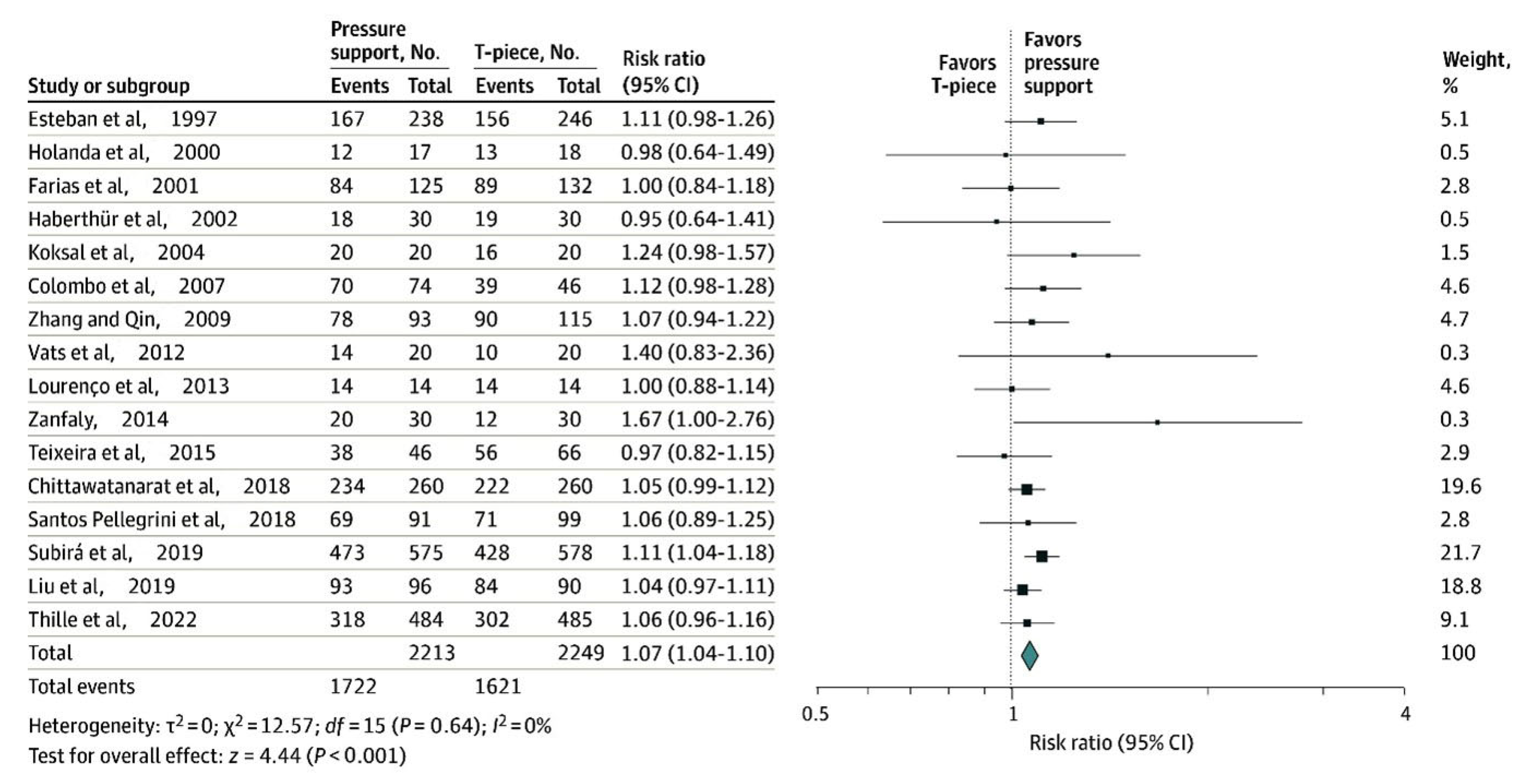
- Subirà, C.; Hernández, G.; Vázquez, A.; Rodríguez-García, R.; González-Castro, A.; García, C.; Rubio, O.; Ventura, L.; López, A.; de la Torre, M.C.; et al. Effect of Pressure Support vs. T-Piece Ventilation Strategies During Spontaneous Breathing Trials on Successful Extubation Among Patients Receiving Mechanical Ventilation: A Randomized Clinical Trial. JAMA 2019, 321, 2175–2182. [Google Scholar] [CrossRef] [Scilit] [PubMed]
- Thille, A.W.; Gacouin, A.; Coudroy, R.; Ehrmann, S.; Quenot, J.-P.; Nay, M.A.; Guitton, C.; Contou, D.; Labro, G.; Reignier, J.; et al. Spontaneous-Breathing Trials with Pressure-Support Ventilation or a T-Piece. N. Engl. J. Med. 2022, 387, 1843–1854. [Google Scholar] [CrossRef] [Scilit] [PubMed]
- Sklar, M.C.; Burns, K.; Rittayamai, N.; Lanys, A.; Rauseo, M.; Chen, L.; Dres, M.; Chen, G.-Q.; Goligher, E.C.; Adhikari, N.K.J.; et al. Effort to Breathe with Various Spontaneous Breathing Trial Techniques. A Physiologic Meta-analysis. Am. J. Respir. Crit. Care Med. 2017, 195, 1477–1485. [Google Scholar] [CrossRef] [Scilit] [PubMed]
- Burns, K.E.A.; Khan, J.; Phoophiboon, V.; Trivedi, V.; Gomez-Builes, J.C.; Giammarioli, B.; Lewis, K.; Chaudhuri, D.; Desai, K.; Friedrich, J.O. Spontaneous Breathing Trial Techniques for Extubating Adults and Children Who Are Critically Ill: A Systematic Review and Meta-Analysis. JAMA Netw. Open 2024, 7, e2356794. [Google Scholar] [CrossRef] [Scilit] [PubMed]
- Burns, K.E.A.; Sadeghirad, B.; Ghadimi, M.; Khan, J.; Phoophiboon, V.; Trivedi, V.; Builes, C.G.; Giammarioli, B.; Lewis, K.; Chaudhuri, D.; et al. Comparative effectiveness of alternative spontaneous breathing trial techniques: A systematic review and network meta-analysis of randomized trials. Crit. Care 2024, 28, 194. [Google Scholar] [CrossRef] [Scilit] [PubMed]
- Esteban, A.; Alia, I.; Gordo, F.; Fernández, R.; Solsona, J.F.; Vallverdú, I.; Macías, S.; Allegue, J.M.; Blanco, J.; Carriedo, D.; et al. Extubation outcome after spontaneous breathing trials with T-tube or pressure support ventilation. Am. J. Respir. Crit. Care Med. 1997, 156, 459–465. [Google Scholar] [CrossRef] [Scilit] [PubMed]
- Holanda, M.A.; Rocha, E.M.; Bandeira, R.M. Pressure support ventilation (PSV) versus T-tube as pre-extubation spontaneous breathing trials (SBT). Am. J. Respir. Crit. Care Med. 2000, 161, A559. [Google Scholar]
- Farias, J.A.; Retta, A.; Alia, I.; Olazarri, F.; Esteban, A.; Golubicki, A.; Allende, D.; Maliarchuk, O.; Peltzer, C.; Ratto, M.; et al. A comparison of twomethods to perform a breathing trial before extubation in pediatric intensive care patients. Intensive Care Med. 2001, 27, 1649–1654. [Google Scholar] [CrossRef] [Scilit] [PubMed]
- Haberthur, C.; Mols, G.; Elsasser, S.; Bingisser, R.; Stocker, R.; Guttmann, J. Extubation after breathing trials with automatic tube compensation, T-tube, or pressure support ventilation. Acta Anaesthesiol. Scand. 2002, 46, 973–979. [Google Scholar] [CrossRef] [Scilit] [PubMed]
- Koksal, G.M.; Sayilgan, C.; Sen, O.; Oz, H. The effects of different weaning modes on the endocrine stress response. Crit. Care 2004, 8, R31–R34. [Google Scholar] [CrossRef] [Scilit] [PubMed]
- Colombo, T.; Boldrini, A.F.; Juliano, S.R.R.; Juliano, M.C.R.; Houly, J.G.S.; Gebara, O.C.E.; Cividanes, G.V.L.; Catão, E.C. Implementation, assessment and comparison of the T-tube and pressure-support weaning protocols applied to the intensive care unit patients who had received mechanical ventilation for more than 48 hours. Rev. Bras. Ter. Intensiva 2007, 19, 31–37. (In Portuguese) [Google Scholar]
- Zhang, B.; Qin, Y.Z. A clinical study of rapid-shallow-breathing index in spontaneous breathing trial with pressure support ventilation and T-piece. Zhongguo Wei Zhong Bing Ji Jiu Yi Xue 2009, 21, 397–401. (In Chinese) [Google Scholar] [PubMed]
- Vats, N.; Singh, J.; Kaira, S. Extubation outcome after spontaneous breathing trials with T-tube or pressure support ventilation. Indian J. Physiother. Occup. Ther. 2012, 6, 86–89. [Google Scholar]
- Lourenco, I.S.; Franco, A.M.; Bassetto, S.; Rodrigues, A.J. Pressure support-ventilation versus spontaneous breathing with “T-tube” for interrupting the ventilation after cardiac operations. Braz. J. Cardiovasc. Surg. 2013, 28, 455–461. [Google Scholar] [CrossRef] [Scilit] [PubMed]
- Zanfaly, H.E. Automatic tube compensation versus pressure support ventilation, continuous positive airway pressure and T-tube during spontaneous breathing trial. Alex. J. Anaesth. Intensive Care 2014, 17, 1–9. [Google Scholar]
- Teixeira, S.N.; Osaku, E.F.; Costa, C.R.L.M.; Toccolini, B.F.; Costa, N.L.; Cândia, M.F.; Leite, M.A.; Jorge, A.C.; Duarte, P.A.D. Comparison of proportional assist ventilation plus, T-tube ventilation, and pressure support ventilation as spontaneous breathing trials for extubation: A randomized study. Respir. Care 2015, 60, 1527–1535. [Google Scholar] [CrossRef] [Scilit] [PubMed]
- Chittawatanarat, K.; Orrapin, S.; Jitkaroon, K.; Mueakwan, S.; Sroison, U. An open label randomized controlled trial to compare low level of pressure support and T-piece strategies for discontinuation of mechanical ventilation in a general surgical intensive care unit. Med. Arch. 2018, 72, 51–57. [Google Scholar] [CrossRef] [Scilit] [PubMed]
- Santos Pellegrini, J.A.; Boniatti, M.M.; Boniatti, V.C.; Zigiotto, C.; Viana, M.V.; Nedel, W.L.; Marques, L.D.S.; Dos Santos, M.C.; De Almeida, C.B.; Pizzol, C.P.D.; et al. Pressure-support ventilation or T-piece spontaneous breathing trials for patients with chronic obstructive pulmonary disease—A randomized controlled trial. PLoS ONE 2018, 13, e0202404. [Google Scholar] [CrossRef] [Scilit] [PubMed]
- Liu, F.; Shao, Q.; Jiang, R.; Zeng, Z.; Liu, Y.; Li, Y.; Liu, Q.; Ding, C.; Zhao, N.; Peng, Z.; et al. High-flow oxygen therapy to speed weaning from mechanical ventilation: A prospective randomized study. Am. J. Crit. Care 2019, 28, 370–376. [Google Scholar] [CrossRef] [Scilit] [PubMed]
- Esteban, A.; Alia, I.; Tobin, M.J.; GIL, A.; Gordo, F.; Vallverdú, I.; Blanch, L.; Bonet, A.; Vázquez, A.; de Pablo, R.; et al. Effect of spontaneous breathing trial duration on outcome of attempts to discontinue mechanical ventilation. Spanish Lung Failure Collaborative Group. Am. J. Respir. Crit. Care Med. 1999, 159, 512–518. [Google Scholar] [CrossRef] [Scilit]
- Chawla, K.; Kupfer, Y.; Goldman, I. The spontaneous breathing trial: How long? Am. J. Respir. Crit. Care Med. 2001, 163, A892. [Google Scholar]
- Perren, A.; Domenighetti, G.; Mauri, S.; Genini, F.; Vizzardi, N. Protocol-directed weaning from mechanical ventilation: Clinical outcome in patients randomized for a 30-min or 120-min trial with pressure support ventilation. Intensive Care Med. 2002, 28, 1058–1063. [Google Scholar] [CrossRef] [Scilit] [PubMed]
- Coudroy, E.; Lejars, A.; Rodriguez, M.; Frat, J.P.; Rault, C.; Arrive, F.; Le Pape, S.; Thille, A.W. Physiologic Effects of Reconnection to the Ventilator for 1 Hour Following a Successful Spontaneous Breathing Trial. Chest 2024, 165, 1406–1414. [Google Scholar] [CrossRef] [Scilit] [PubMed]
- Dadam, M.M.; Goncalves, A.R.R.; Mortari, G.; Klamt, A.P.; Hippler, A.; Lago, J.U.; Ponikieski, C.; Catelano, B.A.; Delvan, D.; Westphal, G.A. The Effect of Reconnection to Mechanical Ventilation for 1 Hour After Spontaneous Breathing Trial on Reintubation Among Patients Ventilated for More Than 12 Hours: A Randomized Clinical Trial. Chest 2021, 160, 148–156. [Google Scholar] [CrossRef] [Scilit] [PubMed]
- Fernandez, M.M.; González-Castro, A.; Magret, M.; Bouza, M.T.; Ibañez, M.; Carolina García, C.; Balerdi, B.; Mas, A.; Arauzo, V.; Añón, J.M.; et al. Reconnection to mechanical ventilation for 1 h after a successful spontaneous breathing trial reduces reintubation in critically ill patients: A multicenter randomized controlled trial. Intensive Care Med. 2017, 43, 1660–1667. [Google Scholar] [CrossRef] [Scilit] [PubMed]
- Baptistella, A.R.; Sarmento, F.J.; da Silva, K.R.; Baptistella, S.F.; Taglietti, M.; Zuquello, R.A.; Nunes Filho, J.R. Predictive factors of weaning from mechanical ventilation and extubation outcome: A systematic review. Crit. Care 2018, 48, 56–62. [Google Scholar] [CrossRef] [Scilit]
- Torrini, F.; Gendreau, S.; Morel, J.; Carteaux, G.; Thille, A.W.; Antonelli, M.; Dessap, A.M. Prediction of extubation outcome in critically ill patients: A systematic review and meta-analysis. Crit. Care 2021, 25, 391. [Google Scholar] [CrossRef] [Scilit] [PubMed]
- GCS Teasdale, G.; Jennett, B. Assessment of coma and impaired consciousness. A practical scale. Lancet 1974, 2, 81–84. [Google Scholar] [CrossRef] [Scilit] [PubMed]
- Sterr, F.; Reintke, M.; Bauernfeind, L.; Senyol, V.; Rester, C.; Metzing, S.; Palm, R. Predictors of weaning failure in ventilated intensive care patients: A systematic evidence map. Crit. Care 2024, 28, 366. [Google Scholar] [CrossRef] [Scilit] [PubMed]
- Grillo Ardila, C.F.; Triana-Moreno, L.C.; Laverde-Sabogal, C.E.; Mora-Arteaga, J.A.; Aguilar-Schotborgh, M.A.; Ramierez-Mosquera, J.J. Diagnostic accuracy of tests for assessing readiness for liberation from mechanical ventilation in adults: An overview of reviews. J. Intensive Care 2026, 14, 14. [Google Scholar] [CrossRef] [Scilit] [PubMed]
- Delisle, S.; Francoeur, M.; Albert, M.; Ouellet, P.; Bellemare, P.; Arsenault, P. Preliminary evaluation of a new index to predict the outcome of a spontaneous breathing trial. Respir. Care. 2011, 56, 1500–1505. [Google Scholar] [CrossRef] [Scilit] [PubMed]
- de Souza, L.C.; Guimarães, F.S.; Lugon, J.R. Evaluation of a new index of mechanical ventilation weaning: The timed inspiratory effort. J. Intensive Care Med. 2015, 30, 37–43. [Google Scholar] [CrossRef] [Scilit] [PubMed]
- Seely, A.J.; Bravi, A.; Herry, C.; Green, G.; Longtin, A.; Ramsay, T.; Fergusson, D.; McIntyre, L.; Kubelik, D.; E Maziak, D.; et al. Do heart and respiratory rate variability improve prediction of extubation outcomes in critically ill patients? Crit. Care 2014, 18, R65. [Google Scholar] [CrossRef] [Scilit] [PubMed]
- Spadaro, S.; Grasso, S.; Mauri, T.; Dalla Corte, F.; Alvisi, V.; Ragazzi, R.; Cricca, V.; Viondi, G.; Di Mussi, R.; Marangoni, E.; et al. Can diaphragmatic ultrasonography performed during the T-tube trial predict weaning failure? The role of diaphragmatic rapid shallow breathing index. Crit. Care 2016, 20, 305. [Google Scholar] [CrossRef] [Scilit] [PubMed]
- Burns, S.M.; Ryan, B.; Burns, J.E. The weaning continuum use of Acute Physiology and Chronic Health Evaluation III, Burns Wean Assessment Program, Therapeutic Intervention Scoring System, and Wean Index scores to establish stages of weaning. Crit. Care Med. 2000, 28, 2259–2267. [Google Scholar] [CrossRef] [Scilit] [PubMed]
- Jiang, J.R.; Yen, S.Y.; Chien, J.Y.; Liu, H.C.; Wu, Y.L.; Chen, C.H. Predicting weaning and extubation outcomes in long-term mechanically ventilated patients using the modified Burns Wean Assessment Program scores. Respirology 2014, 19, 576–582. [Google Scholar] [CrossRef] [Scilit] [PubMed]
- Huaringa, A.J.; Wang, A.; Haro, M.H.; Leyva, F. The weaning index as predictor of weaning success. J. Intensive Care Med. 2013, 28, 369–374. [Google Scholar] [CrossRef] [Scilit] [PubMed]
- Nemer, S.N.; Barbas, C.S.; Caldeira, J.B.; Cárias, T.C.; Santos, R.G.; Almeida, L.C.; Azeredo, L.M.; Noé, R.A.; Guimarães, B.S.; Souza, P.C. A new integrative weaning index of discontinuation from mechanical ventilation. Crit. Care 2009, 13, R152. [Google Scholar] [CrossRef] [Scilit] [PubMed]
- Boniatti, V.M.; Boniatti, M.M.; Andrade, C.F.; Zigiotto, C.C.; Kaminski, P.; Gomes, S.P.; Lippert, R.; Miguel, D.C.; Felix, E.A. The modified integrative weaning index as a predictor of extubation failure. Respir. Care 2014, 59, 1042–1047. [Google Scholar] [CrossRef] [Scilit] [PubMed]
- Kaur, R.; Vines, D.L.; Liu, L.; Balk, R.A. Role of integrated pulmonary index in identifying extubation failure. Respir. Care 2017, 62, 1550–1556. [Google Scholar] [CrossRef] [Scilit] [PubMed]
- Baptistella, A.R.; Mantelli, L.M.; Matte, L.; Medru, C.; Fortunatti, J.A.; Costa, I.Z.; Haro, F.G.; de Turkot, V.L.O.; Baptistella, S.F.; de Carvalho, D.; et al. Prediction of extubation outcome in mechanically ventilated patients: Development and validation of the Extubation Predictive Score (ExPreS). PLoS ONE 2021, 16, e0248868. [Google Scholar] [CrossRef] [Scilit] [PubMed]
- Chaudhuri, S.; Gupta, N.; Adhikari, S.D.; Todur, P.; Maddani, S.S.; Rao, S. Utility of the one-time HACOR score as a predictor of weaning failure from mechanical ventilation: A prospective observational study. Indian J. Crit. Care Med. 2022, 26, 900–905. [Google Scholar] [CrossRef] [Scilit] [PubMed]
- Varón-Vega, F.; Giraldo-Cadavid, L.F.; Uribe, A.M.; Rincón, A.; Palacios, J.; Crevoisier, S.; Tuta-Quintero, E.; Ordoñez, L.; Boada, N.; Rincón, P.; et al. Utilization of spontaneous breathing trial, objective cough test, and diaphragmatic ultrasound results to predict extubation success: COBRE-US trial. Crit. Care 2023, 27, 414. [Google Scholar] [CrossRef] [Scilit] [PubMed]
- Kuo, H.J.; Chiu, H.W.; Lee, C.N.; Chen, T.T.; Chang, C.C.; Bien, M.Y. Improvement in the Prediction of Ventilator Weaning Outcomes by an Artificial Neural Network in a Medical ICU. Respir. Care 2015, 60, 1560–1569. [Google Scholar] [CrossRef] [Scilit] [PubMed]
- Hsieh, M.H.; Hsieh, M.J.; Chen, C.M.; Hsieh, C.C.; Chao, C.M.; Lai, C.C. An Artificial Neural Network Model for Predicting Successful Extubation in Intensive Care Units. J. Clin. Med. 2018, 7, 240. [Google Scholar] [CrossRef] [Scilit] [PubMed]
- Gottschalk, A.; Hyzer, M.C.; Geer, R.T. A comparison of human and machine-based predictions of successful weaning from mechanical ventilation. Med. Decis. Mak. 2000, 20, 160–169. [Google Scholar] [CrossRef] [Scilit] [PubMed]
- Jia, Y.; Kaul, C.; Lawton, T.; Murray-Smith, R.; Habli, I. Prediction of weaning from mechanical ventilation using Convolutional Neural Networks. Artif. Intell. Med. 2021, 117, 102087. [Google Scholar] [CrossRef] [Scilit] [PubMed]
- Yang, H.C.; Hao, A.T.; Liu, S.C.; Chang, Y.C.; Tsai, Y.T.; Weng, S.J.; Chan, M.C.; Wang, C.Y.; Xu, Y.Y. Prediction of Spontaneous Breathing Trial Outcome in Critically Ill-Ventilated Patients Using Deep Learning: Development and Verification Study. JMIR Med. Inform. 2025, 13, e64592. [Google Scholar] [CrossRef] [Scilit] [PubMed]
- Fenske, S.W.; Peltekian, A.; Kang, M.; Markov, N.S.; Zhu, M.; Grudzinski, K.; Bak, M.J.; Pawlowski, A.; Gupta, V.; Mao, Y.; et al. Developing and validating machine learning models to predict next-day extubation. Sci. Rep. 2025, 15, 27552. [Google Scholar] [CrossRef] [Scilit] [PubMed]
- Zhao, Q.Y.; Wang, H.; Luo, J.C.; Luo, M.H.; Liu, L.P.; Yu, S.J.; Liu, K.; Zhang, Y.J.; Sun, P.; Tu, G.W.; et al. Development and Validation of a Machine-Learning Model for Prediction of Extubation Failure in Intensive Care Units. Front. Med. 2021, 8, 676343. [Google Scholar] [CrossRef] [Scilit] [PubMed]
- Park, J.E.; Kim, D.Y.; Park, J.W.; Jung, Y.J.; Lee, K.S.; Park, J.H.; Sheen, S.S.; Park, K.J.; Sunwoo, M.H.; Chung, W.Y. Development of a Machine Learning Model for Predicting Weaning Outcomes Based Solely on Continuous Ventilator Parameters during Spontaneous Breathing Trials. Bioengineering 2023, 10, 1163. [Google Scholar] [CrossRef] [Scilit] [PubMed]
- Burns, K.E.A.; Allan, J.; Herry, C.; Lee, E.; Santos-Taylor, M.; Scales, N.; Kay, P.; Gouskos, A.; Greco, P.; Every, H.; et al. Liberation from mechanical ventilation using Extubation Advisor Decision Support (LEADS): Protocol for a Multicentre Pilot Trial. BMJ Open 2025, 15, e093853. [Google Scholar] [CrossRef] [Scilit] [PubMed]
- Tanios, M.A.; Nevins, M.L.; Hendra, K.P.; Cardinal, P.; Allan, J.E.; Naumova, E.N.; Epstein, S.K. A randomized, controlled trial of the role of weaning predictors in clinical decision making. Crit. Care Med. 2006, 34, 2530–2535. [Google Scholar] [CrossRef] [Scilit] [PubMed]
- Oliveira, J.A.; Klein, F.; Guzatti, N.G.; Kojoroski, A.P.F.; de Vargas Ciello, H.; da Silva, D.J.; Locatelli, J.; da Silva, L.F.; Parise, M.; Spörr, A.V.; et al. Effect of the extubation predictive score (ExPreS) on reintubation in the ICU: A randomized controlled trial. Crit. Care 2026, 30, 343. [Google Scholar] [CrossRef] [Scilit] [PubMed]
- Varón-Vega, F.; Rincón, A.; Giraldo-Cadavid, L.F.; Tuta-Quintero, E.; Palacios, J.; Crevoisier, S.; Duarte, D.C.; Poveda, M.; Cucunubo, L.; Monedero, P. Assessing the reproducibility and predictive value of objective cough measurement for successful withdrawal of invasive ventilatory support in adult patients. BMC Pulm. Med. 2024, 24, 218. [Google Scholar] [CrossRef] [Scilit] [PubMed]
- Kriner, E.J.; Shafazand, S.; Colice, G.L. The endotracheal tube cuff-leak test as a predictor for postextubation stridor. Respir. Care 2005, 50, 1632–1638. [Google Scholar] [CrossRef] [Scilit] [PubMed]
- Salam, A.; Tilluckdharry, L.; Amoateng-Adjepong, Y.; Manthous, C.A. Neurologic status, cough, secretions and extubation outcomes. Intensive Care Med. 2004, 30, 1334–1339. [Google Scholar] [CrossRef] [Scilit] [PubMed]
- Koenig, S.; Mayo, P.; Volpicelli, G.; Millington, S.J. Lung Ultrasound Scanning for Respiratory Failure in Acutely Ill Patients: A Review. Chest 2020, 158, 2511–2516. [Google Scholar] [CrossRef] [Scilit] [PubMed]
- Hermans, G.; Demoule, A.; Heunks, L. How I perform diaphragmatic ultrasound in the intensive care unit. Intensive Care Med. 2024, 50, 2175–2178. [Google Scholar] [CrossRef] [Scilit] [PubMed]
- Kaur, A.; Sharma, S.; Singh, V.P.; Krishna, M.R.; Gautam, P.L.; Singh, G. Sonographic assessment of diaphragmatic thickening and excursion as predictors of weaning success in the intensive care unit: A prospective observational study. Indian J. Anaesth. 2022, 66, 776–782. [Google Scholar] [CrossRef] [Scilit] [PubMed]
- Rabuske, W.C.; Westphal, G.; Rático, G.; do Nascimento, S.A.; Baptistella, A.R. Diaphragmatic ultrasound for extubation readiness: The role of thickening fraction. J. Crit. Care 2026, 91, 155253. [Google Scholar] [CrossRef] [Scilit] [PubMed]
- Parada- Gereda, H.M.; Tibaduiza, A.L.; Rico-Mendoza, A.; Molano-Franco, D.; Nieto, V.H.; Arias-Ortiz, W.A.; Perez-Terán, P.; Masclans, J.R. Effectiveness of diaphragmatic ultrasound as a predictor of successful weaning from mechanical ventilation: A systematic review and meta-analysis. Crit. Care 2023, 27, 174. [Google Scholar] [CrossRef] [Scilit] [PubMed]
- Franchineau, G.; Jonkman, A.H.; Piquilloud, L.; Yoshida, T.; Costa, E.; Rozé, H.; Camporota, L.; Piraino, T.; Spinelli, E.; Combes, A.; et al. Electrical Impedance Tomography to Monitor Hypoxemic Respiratory Failure. Am. J. Respir. Crit. Care Med. 2024, 209, 670–682. [Google Scholar] [CrossRef] [Scilit] [PubMed]
- Wang, G.; Zhang, L.; Li, B.; Niu, B.; Jiang, J.; Li, D.; Yue, Z.; Weng, Y. The Application of Electrical Impedance Tomography During the Ventilator Weaning Process. Int. J. Gen. Med. 2021, 14, 6875–6883. [Google Scholar] [CrossRef] [Scilit] [PubMed]
- Phoophiboon, V.; Antenor Rodrigues, A.; Fernando Vieira, F.; Ko, M.; Madotta, F.; Schreiber, A.; Sun, N.; Sousa, M.L.A.; Docci, M.; Brault, C.; et al. Ventilation distribution during spontaneous breathing trials predicts liberation from mechanical ventilation: The VISION study. Crit. Care 2025, 29, 11. [Google Scholar] [CrossRef] [Scilit] [PubMed]
- Schreiber, A.F.; Bertoni, M.; Coiffard, B.; Fard, S.; Wong, J.; Reid, W.D.; Brochard, L.J.; Piva, S.; Goligher, E.C. Abdominal Muscle Use During Spontaneous Breathing and Cough in Patients Who Are Mechanically Ventilated: A Bi-center Ultrasound Study. Chest 2021, 160, 1316–1325. [Google Scholar] [CrossRef] [Scilit] [PubMed]
- Fernando, S.M.; Tran, A.; Sadeghirad, B.; Burns, K.E.A.; Fan, E.; Brodie, D.; Munshi, L.; Goligher, E.C.; Cook, D.J.; Fowler, R.A.; et al. Noninvasive respiratory support following extubation in critically ill adults: A systematic review and network meta-analysis. Intensive Care Med. 2021, 48, 137–147. [Google Scholar] [CrossRef] [Scilit] [PubMed]
- Pensier, J.; Naudet-Lasserre, A.; Monet, C.; Capdevila, M.; Aarab, Y.; Lakbar, I.; Chanques, G.; Molinari, N.; De Jong, A.; Jaber, S. Noninvasive respiratory support following extubation in critically ill adults with obesity: A systematic review and network meta-analysis. eClinicalMedicine 2025, 79, 103002. [Google Scholar] [CrossRef] [Scilit] [PubMed]
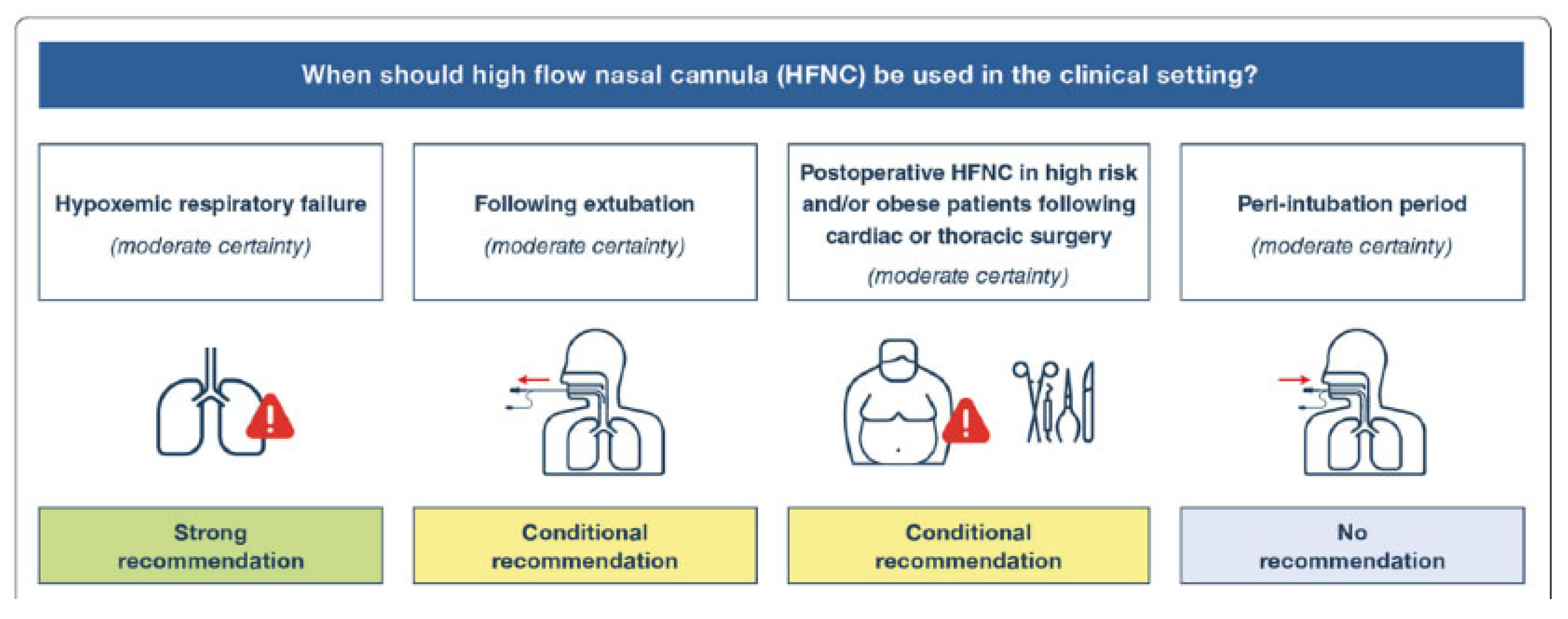
- Rochwerg, B.; Einav, S.; Chaudhuri, D.; Mancebo, J.; Mauri, T.; Helviz, Y.; Goligher, E.C.; Jaber, S.; Ricard, J.-D.; Rittayamai, N.; et al. The role for high flow nasal cannula as a respiratory support strategy in adults: A clinical practice guideline. Intensive Care Med. 2020, 46, 2226–2237. [Google Scholar] [CrossRef] [Scilit] [PubMed]
- Oczkowski, S.; Ergan, B.; Bos, L.; Chatwin, M.; Ferrer, M.; Gregoretti, C.; Heunks, L.; Frat, J.-P.; Longhini, F.; Nava, S.; et al. ERS clinical practice guidelines: High-flow nasal cannula in acute respiratory failure. Eur. Respir. J. 2022, 59, 2101574. [Google Scholar] [PubMed]



| Newer Modes | Main Feature |
|---|---|
| Proportional Assist Ventilation | Airway pressure is amplified according to respiratory mechanics and the set level of assistance to be proportional to the instantaneous effort of the patient. |
| Neurally Adjusted Ventilatory Assist | Measures the electrical activity of the diaphragm (EAdi) and provides positive pressure synchronously and in proportion to the amplitude of the EAdi signal. |
| Adaptive Support Ventilation | Targets a desired minute ventilation by providing the optimal combination of tidal volume and respiratory rate according to Otis’ equation (calculates an ideal respiratory rate that is associated with the least energy expenditure by considering minute ventilation, dead space and the expiratory time constant of the respiratory system). Algorithm calculates the expiratory time constant and adjusts the I:E ratio and inspiratory pressure to reduce work of breathing. |
| SmartCare | Algorithm adjusts the level of pressure support provided according to 8 respiratory diagnoses that aim to achieve a respiratory comfort zone based on respiratory rate, tidal volume and end-tidal carbon dioxide. It automatically conducts spontaneous breathing trials once predetermined settings are achieved. |
| Comparison | Network Odds Ratio (95% CI) | Absolute Risk Difference (95% CI) | Number Needed to Treat | GRADE |
|---|---|---|---|---|
| NIPPV vs. conventional oxygen | 0.65 (0.52–0.82) | –5.18 (–8.09 to –2.26) | 20 (13 to 45) | Moderate a |
| HFNC vs. conventional oxygen | 0.63 (0.45–0.87) | –3.84 (–6.7 to –0.98) | 26 (15 to 102) | Moderate a |
| NIPPV vs. HFNC | 1.04 (0.78–1.38) | –1.34 (–4.4 to 1.72) | N/A | Low a,b |
| HFNC + NIPPV vs. conventional oxygen | 0.38 (0.19–0.74) | –10.25 (–18.49 to –2.01) | 10 (6 to 50) | Moderate a |
| HFNC + NIPPV vs. NIPPV | 0.58 (0.3–1.11) | –5.07 (–13.38 to 3.24) | N/A | Low a,b |
| HFNC + NIPPV vs. HFNC | 0.6 (0.33–1.08) | –6.41 (–14.13 to 1.31) | N/A | Low a,b |
Disclaimer/Publisher’s Note: The statements, opinions and data contained in all publications are solely those of the individual author(s) and contributor(s) and not of MDPI and/or the editor(s). MDPI and/or the editor(s) disclaim responsibility for any injury to people or property resulting from any ideas, methods, instructions or products referred to in the content. |
© 2026 by the authors. Licensee MDPI, Basel, Switzerland. This article is an open access article distributed under the terms and conditions of the Creative Commons Attribution (CC BY) license.
Share and Cite
Burns, K.E.A.; Bosma, K.J.; Ferreyro, B.L.; Chaudhuri, D.; Seely, A.J.E.; Ouellette, D.R. Liberation from Mechanical Ventilation in Acute Hypoxemic Respiratory Failure or Adult Respiratory Distress Syndrome: A Review. J. Clin. Med. 2026, 15, 6019. https://doi.org/10.3390/jcm15156019
Burns KEA, Bosma KJ, Ferreyro BL, Chaudhuri D, Seely AJE, Ouellette DR. Liberation from Mechanical Ventilation in Acute Hypoxemic Respiratory Failure or Adult Respiratory Distress Syndrome: A Review. Journal of Clinical Medicine. 2026; 15(15):6019. https://doi.org/10.3390/jcm15156019
Chicago/Turabian StyleBurns, Karen E. A., Karen J. Bosma, Bruno L. Ferreyro, Dipayan Chaudhuri, Andrew J. E. Seely, and Daniel R. Ouellette. 2026. "Liberation from Mechanical Ventilation in Acute Hypoxemic Respiratory Failure or Adult Respiratory Distress Syndrome: A Review" Journal of Clinical Medicine 15, no. 15: 6019. https://doi.org/10.3390/jcm15156019
APA StyleBurns, K. E. A., Bosma, K. J., Ferreyro, B. L., Chaudhuri, D., Seely, A. J. E., & Ouellette, D. R. (2026). Liberation from Mechanical Ventilation in Acute Hypoxemic Respiratory Failure or Adult Respiratory Distress Syndrome: A Review. Journal of Clinical Medicine, 15(15), 6019. https://doi.org/10.3390/jcm15156019