3. Results
A total of 200 consecutive patients underwent augmentation rhinoplasty with the pyramidal costal cartilage columellar strut graft technique during the study period. Baseline demographic characteristics and operative parameters for the study cohort are summarized in
Table 1.
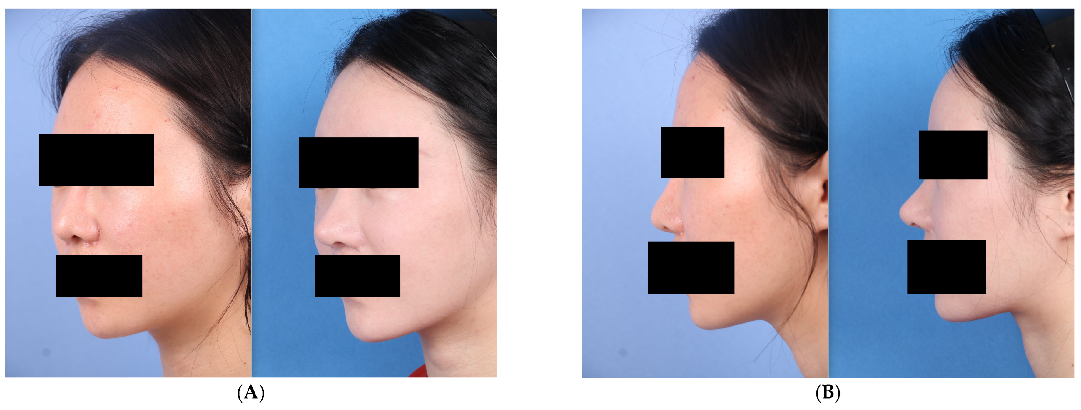
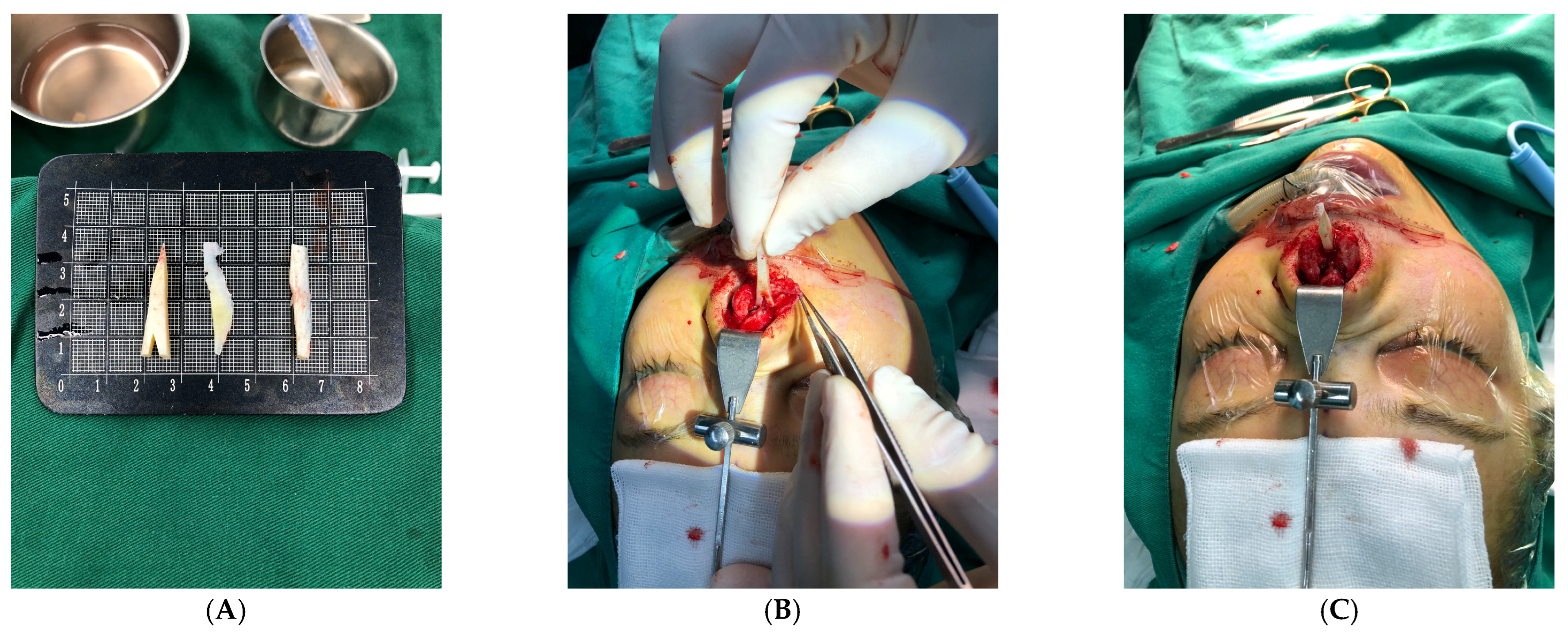
Satisfactory nasal tip projection was achieved in the vast majority of patients and was maintained without significant change throughout the follow-up period. Standardized clinical assessment of postoperative tip mobility suggested maintenance of clinically appreciable tip flexibility on manual examination, supporting favorable clinical observations consistent with the intended floating-tip design of the floating-tip fixation strategy. Patients consistently described the nasal tip as exhibiting a natural tactile quality on palpation and during animated facial expression, in notable contrast to the rigid, immobile character commonly reported following septum-anchored fixation techniques. Intraoperative demonstration of unrestricted columellar mobility following pyramidal strut placement is provided in
Video S1, Supplemental Digital Content (see
Supplemental Digital Content S1, available online with this article, which demonstrates free multidirectional mobility of the pyramidal columellar strut at the anterior nasal spine prior to soft tissue closure). Long-term preservation of physiologic nasal tip mobility at 1 year postoperatively is demonstrated in
Video S2, Supplemental Digital Content (see
Supplemental Digital Content S2, available online with this article, which shows free nasal tip rotation and natural elastic recoil upon manual upturn displacement, confirming sustained tip compliance at 12 months following surgery).
Among the operative cohort, 5 patients (2.5%) presented with mild postoperative nasal tip deviation on follow-up examination. Of these, 4 cases (2%) were determined to fall within the clinically acceptable range of symmetry on blinded standardized photographic analysis and required no further intervention. The remaining 1 cases (0.5%) demonstrated clinically significant tip asymmetry and were managed with revision surgery.
Donor-site outcomes were uniformly favorable throughout the series. No cases of pneumothorax, intraoperative rib fracture, or wound dehiscence were recorded. The 2-cm incision achieved an inconspicuous, well-healed scar appearance by 6 months postoperatively in the majority of patients. Postoperative chest wall pain was characteristically mild and self-limiting, with resolution observed within the first postoperative week in most cases. No patient reported persistent restriction of trunk mobility or inability to resume activities of daily living beyond the immediate perioperative period.
No cases of clinically significant graft resorption, columellar skin necrosis, or delayed surgical site infection were identified at any follow-up interval. No patient required intraoperative or postoperative conversion to an alternative structural fixation technique.
4. Discussion
The present study introduces a novel technical approach to augmentation rhinoplasty that was developed to address two commonly recognized limitations of conventional costal cartilage techniques: donor-site morbidity and iatrogenic restriction of postoperative nasal tip mobility [
1]. The pyramidal columellar strut graft with half-harvest technique represents a methodical integration of established anatomical principles and biomechanical reasoning within the operative framework of costal cartilage rhinoplasty.
Costal cartilage remains the preferred graft material when substantial structural support is required, particularly in revision rhinoplasty and in patients with insufficient septal cartilage [
2,
4]. Previous studies have demonstrated the versatility and long-term durability of costal cartilage in dorsal augmentation, tip support, and complex reconstructive procedures [
3,
11]. Nevertheless, concerns regarding postoperative tip rigidity and donor-site morbidity continue to represent important limitations of conventional costal cartilage techniques. The present technique was developed in response to these commonly encountered clinical concerns while maintaining the structural advantages associated with autologous rib cartilage grafting.
The floating-tip columellar strut concept—wherein the inferior end of the strut is seated at the anterior nasal spine without rigid suture attachment to the caudal septum—has been advocated as a method of preserving a greater degree of postoperative tip flexibility while maintaining structural support [
15]. Compared with previously described septum-anchored constructs, the present technique was designed to permit a greater degree of dynamic tip movement; however, no direct comparative analysis was performed [
6,
7]—such as the septal extension graft, which effectively abolishes the rotational freedom of the medial crura—the floating-tip configuration permits physiologic tip depression and elastic recoil under dynamic functional loads [
16,
17]. The pyramidal strut design introduced in this study advances this principle further by incorporating a graft geometry that anatomically mirrors the native anterior taper of the columellar column, thereby serving as a practical surgical design intended to approximate the native columellar taper while providing basal support and a narrower superior segment suitable for placement between the medial crura.
Septal extension grafts remain among the most reliable techniques for controlling tip projection and rotation and continue to represent a cornerstone of modern structural rhinoplasty [
6,
8]. However, the degree of rigidity associated with long-term fixation may not be desirable in all patients. Accordingly, the present technique was developed as an alternative option for selected patients in whom preservation of a softer and more natural tip feel was considered clinically relevant. Importantly, no direct comparison with septal extension grafts was performed, and therefore no conclusions regarding relative superiority can be drawn from the present study.
An additional consideration in the design of the pyramidal strut was surgical reproducibility. Because the geometry of the graft follows the natural tapering morphology of the columellar complex, the carving process can be standardized and reproduced with relative consistency. Although objective comparisons with alternative graft designs were not performed, the authors found that the pyramidal configuration facilitated stable positioning within the columellar pocket while maintaining a favorable contour profile.
The mortise-and-tenon engagement between the triangular basal notch of the strut and the bony profile of the anterior nasal spine was intended to enhance positional stability at the graft-spine interface. This interlocking configuration was intended to improve positional stability at the graft-spine interface. Although favorable clinical observations were obtained in this series, objective biomechanical validation was not performed Analogous interlocking base geometries have been described in the context of columellar strut fixation to the anterior nasal spine in septum-anchored rhinoplasty frameworks [
18]. By applying this geometric principle within a floating, non-septum-fixed construct, the present technique may provide a balance between basal stability and distal tip flexibility that is without requiring rigid septal fixation.
Therefore, the proposed stabilizing effect of this configuration should be interpreted as a surgical rationale rather than a demonstrated biomechanical advantage. Future cadaveric investigations, finite-element modeling, and mechanical testing studies may further clarify the contribution of this design feature to graft stability.
The half-harvest technique may represent a useful alternative approach for reducing donor-site morbidity of this report. Standard full-thickness costal cartilage harvest requires complete transection of the cartilaginous rib, generating two independently mobile segments whose relative micro-motion during routine trunk movement—particularly rotational and flexural loading—a plausible contributor to postoperative donor-site discomfort, although this mechanism was not directly tested in the present study [
19,
20].
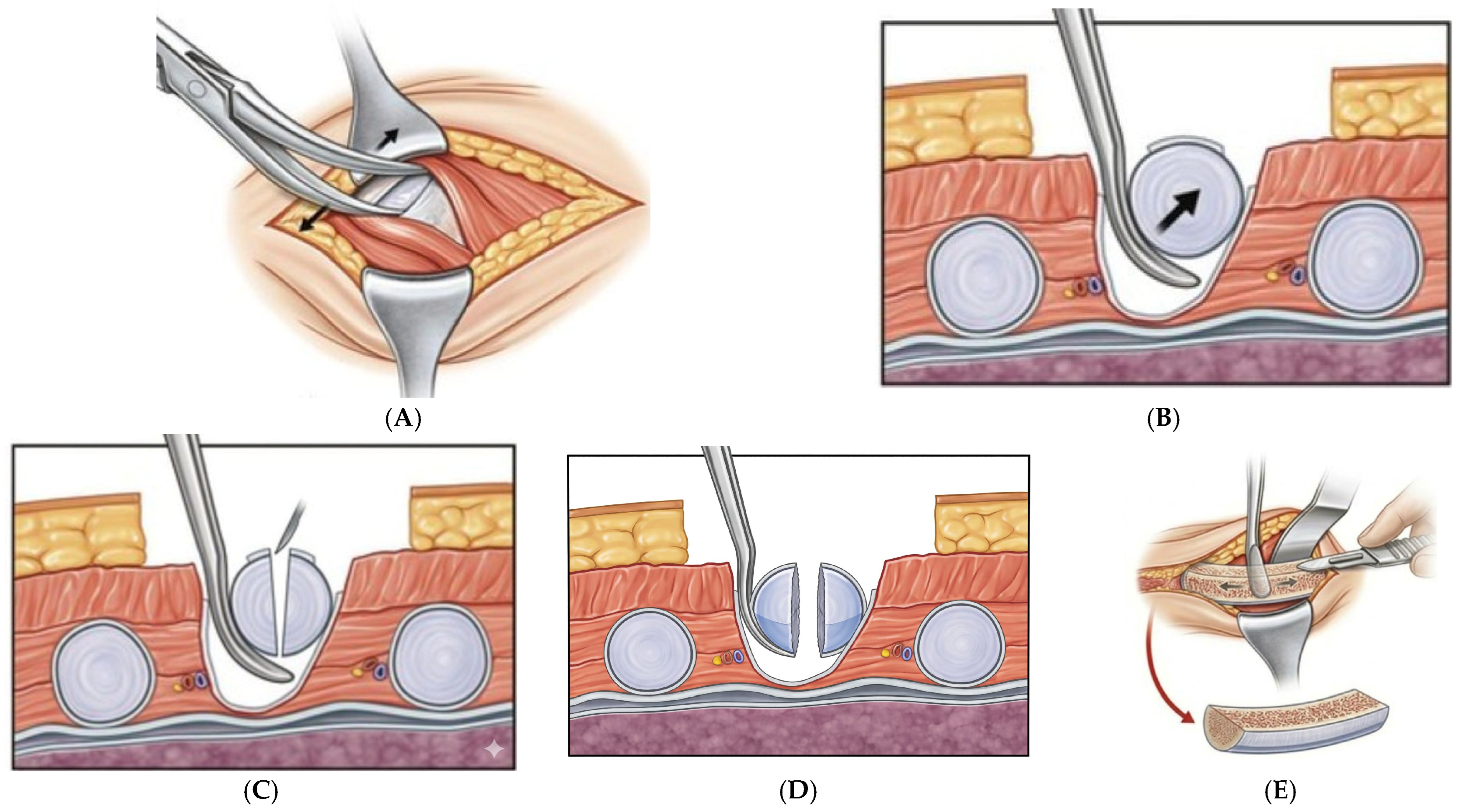
The half-harvest technique constitutes a central innovation of this procedure. In contradistinction to conventional full-thickness rib harvest, only the medial half of the costal cartilage column was resected; the lateral half cortex and the lateral perichondrial layer were intentionally preserved. This unicortical resection strategy confers three distinct biomechanical and biological advantages.
First, retention of the perichondrium on the residual cartilage surface is known to has been reported to possess chondrogenic potential in experimental studies, with the potential to restore donor-site volume during the remodeling phase. Second, preservation of lateral cortical continuity maintains structural continuity of the rib segment during routine daily activities involving trunk rotation, lateral bending, and sit-to-stand transitions. Trunk rotation refers to rotational movements of the torso around the spinal axis, such as turning the upper body from side to side while the pelvis remains relatively stationary, a maneuver commonly used in clinical assessment of postoperative donor-site discomfort and functional recovery. By avoiding complete transection of the costal cartilage, the technique may theoretically reduce stress concentration at the harvest site; however, this proposed mechanism was not directly evaluated in the present study. Third, the absence of complete intercartilaginous discontinuity enables immediate postoperative trunk mobility, permitting patients to resume activities of daily living, including moderate physical exertion, without the prolonged convalescence characteristic of standard full-thickness harvest.
Conventionally, harvesting costal cartilage grafts frequently induces significant donor-site pain, often compromising upper body mobility and severely restricting the patient’s trunk movement during the first postoperative week. In contrast, our refined technique focuses on minimizing surgical trauma to the surrounding intercostal muscles and preserving the structural continuity of the adjacent cartilage framework. Consequently, this approach significantly mitigates donor-site morbidity, allowing patients to tolerate mechanical stress early on. To clinically verify this immediate functional stability, the senior surgeon routinely performed a manual Trunk-Rotation Test prior to discharge, gently twisting the patient’s upper body left and right while holding their hands. The vast majority of patients demonstrated an absence of sharp donor-site pain and successfully returned to normal activities of daily living within 24 h. Notably, even professional fitness trainers within this cohort were able to safely return to their gym routines within one week post-surgery, highlighting the clinical advantage of this technique in preserving immediate trunk mobility.
Interest in minimizing donor-site morbidity has increased substantially as the use of autologous costal cartilage has expanded. Previous investigations have emphasized postoperative pain, contour deformity, and patient concerns regarding chest wall scarring as important considerations during graft selection. Accordingly, techniques that preserve native rib architecture while providing adequate graft volume may offer practical advantages in selected patients. Whether partial-thickness harvest techniques confer measurable clinical benefits over conventional full-thickness harvest methods remains an important topic for future investigation.
Perichondrial preservation may theoretically contribute to cartilage regeneration, although this was not directly evaluated in the present study. Experimental studies have reported chondrogenic potential within preserved perichondrium; however, donor-site regeneration was not directly evaluated in the present study and therefore should be regarded as a theoretical consideration rather than a demonstrated clinical outcome [
21,
22]. Although the clinical magnitude of this regenerative phenomenon in the specific context of partial costal cartilage harvest has not yet been prospectively quantified in humans, its theoretical contribution to donor-site volumetric restoration over the medium and long term represents a meaningful additional argument in favor of the half-harvest approach.
The biological significance of perichondrial preservation remains an area of ongoing investigation. Experimental studies have demonstrated the presence of chondrogenic progenitor cells within the perichondrium and have suggested a potential role in cartilage repair and remodeling [
23]. However, the extent to which these observations translate into clinically meaningful regeneration following costal cartilage harvest remains uncertain. Consequently, any regenerative benefit associated with the present technique should be interpreted cautiously until validated by dedicated clinical studies.
The multilayer soft tissue coverage protocol employed in this series warrants specific discussion. The characteristically thin and relatively inelastic skin of the nasal tip and columella renders any underlying cartilaginous construct susceptible to visible surface irregularities, edge telegraphing, and progressive contour deformity as the overlying soft tissue undergoes atrophy over time. The application of a perichondrial enveloping layer—supplemented by autologous dermal fat when deemed necessary—provides a compliant biological buffer that attenuates graft surface irregularities, facilitates early fibrovascular integration between the construct and its surrounding tissue bed, and may mitigate the long-term risk of skin thinning and graft prominence.
The postoperative immobilization protocol is a critical component of this technique and demands particular emphasis in preoperative patient counseling. The long-term positional stability of the floating-tip columellar strut is contingent upon progressive fibrovascular integration and the development of a stable soft tissue adherence interface—neither of which is instantaneous. Premature mechanical loading of the nasal tip prior to the establishment of adequate graft-tissue adhesion during the early healing phase poses a substantive and specific risk of graft displacement, axial malrotation, and consequent tip asymmetry. Accordingly, structured postoperative protection of the nasal tip was emphasized during the early recovery period and should be clearly discussed with patients as part of preoperative counseling.
From a clinical perspective, the present technique may be particularly applicable in patients requiring substantial structural augmentation while simultaneously expressing concern regarding postoperative tip rigidity or donor-site morbidity. Because the technique utilizes conventional autologous costal cartilage grafting principles and does not require specialized implants or proprietary devices, it can be incorporated into existing rhinoplasty workflows without substantial modification. Furthermore, the technique should be viewed as an additional option within the spectrum of structural rhinoplasty strategies rather than a replacement for established methods such as septal extension grafting.
Several limitations inherent to the present study merit transparent acknowledgment. The retrospective study design and the absence of a prospective concurrent control cohort preclude definitive comparative inferences regarding nasal tip mobility or donor-site outcomes relative to conventional full-thickness harvest or septum-fixed fixation techniques. Objective quantification of nasal tip mobility using validated dynamometric instruments or three-dimensional motion capture analysis was not incorporated, and functional outcome assessment relied upon clinical examination and standardized photographic documentation—methodologies that, while clinically pragmatic, are subject to inter-observer variability. The single-surgeon, single-center design introduces potential performance bias and constrains the external generalizability of the findings. Prospective randomized controlled trials incorporating validated, objective biomechanical outcome instruments are necessary to rigorously substantiate the clinical observations reported in this series.
Additional limitations should be acknowledged. First, the retrospective design and absence of a control group prevent definitive conclusions regarding superiority over conventional septal extension grafts or full-thickness costal cartilage harvest techniques. Second, postoperative tip flexibility was assessed clinically by the operating surgeon without validated objective instruments, dynamic motion analysis, or patient-reported outcome measures such as FACE-Q [
24]. Third, the mean follow-up duration of 8 months limits assessment of long-term graft stability, warping, resorption, and maintenance of tip flexibility. Finally, the private-practice nature of a substantial portion of the cohort may have contributed to loss to follow-up and underestimation of the true long-term revision rate. In addition, because the pyramidal strut configuration, floating fixation strategy, and half-harvest donor technique were applied simultaneously, the independent contribution of each component to the observed outcomes could not be determined. Furthermore, the proposed biomechanical advantages of the pyramidal design and the potential regenerative effects of perichondrial preservation were not directly evaluated and therefore remain theoretical.
Future comparative studies incorporating objective motion analysis, validated patient-reported outcome measures, and longer follow-up durations will be necessary to further define the role of this technique within contemporary costal cartilage rhinoplasty.