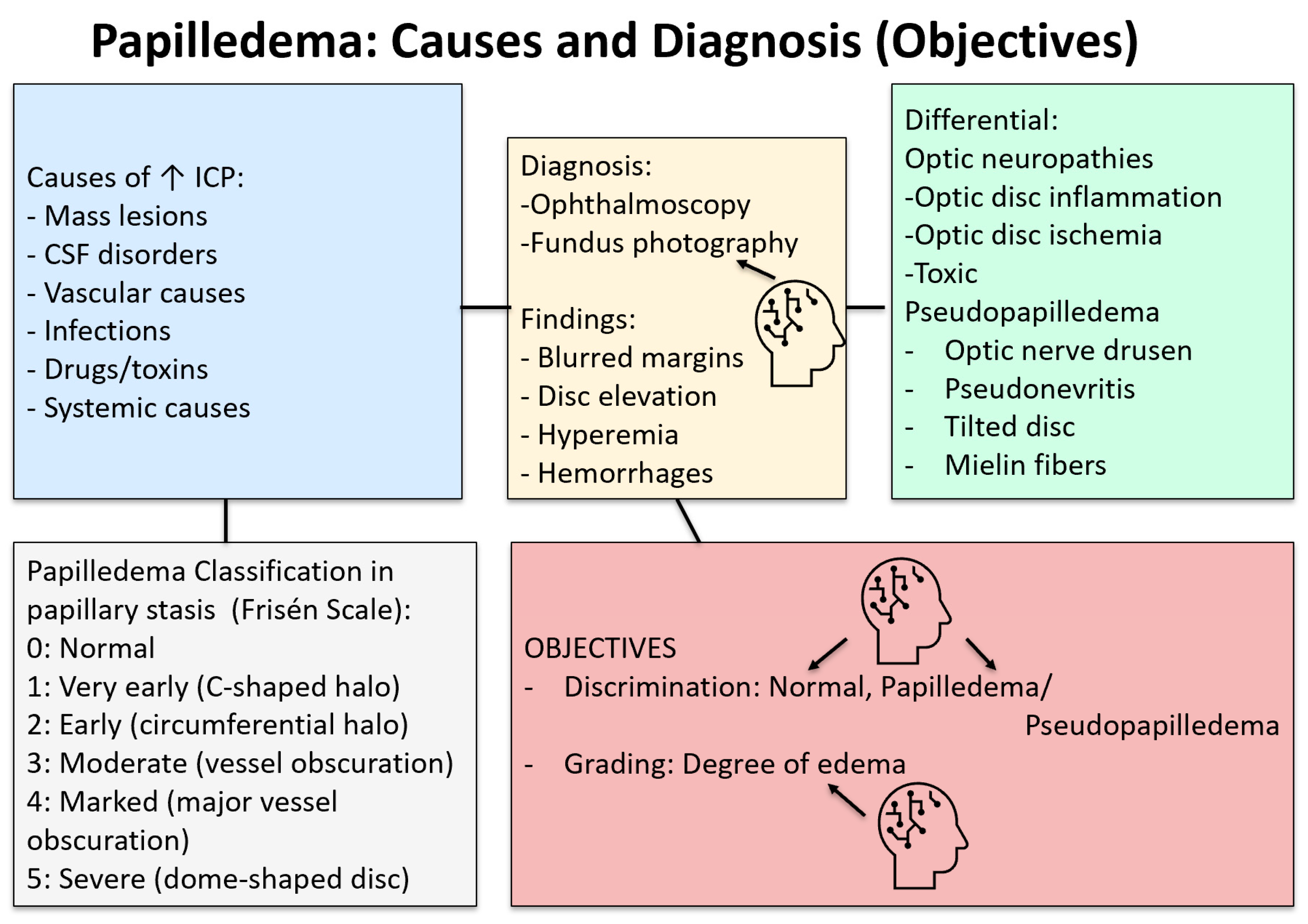
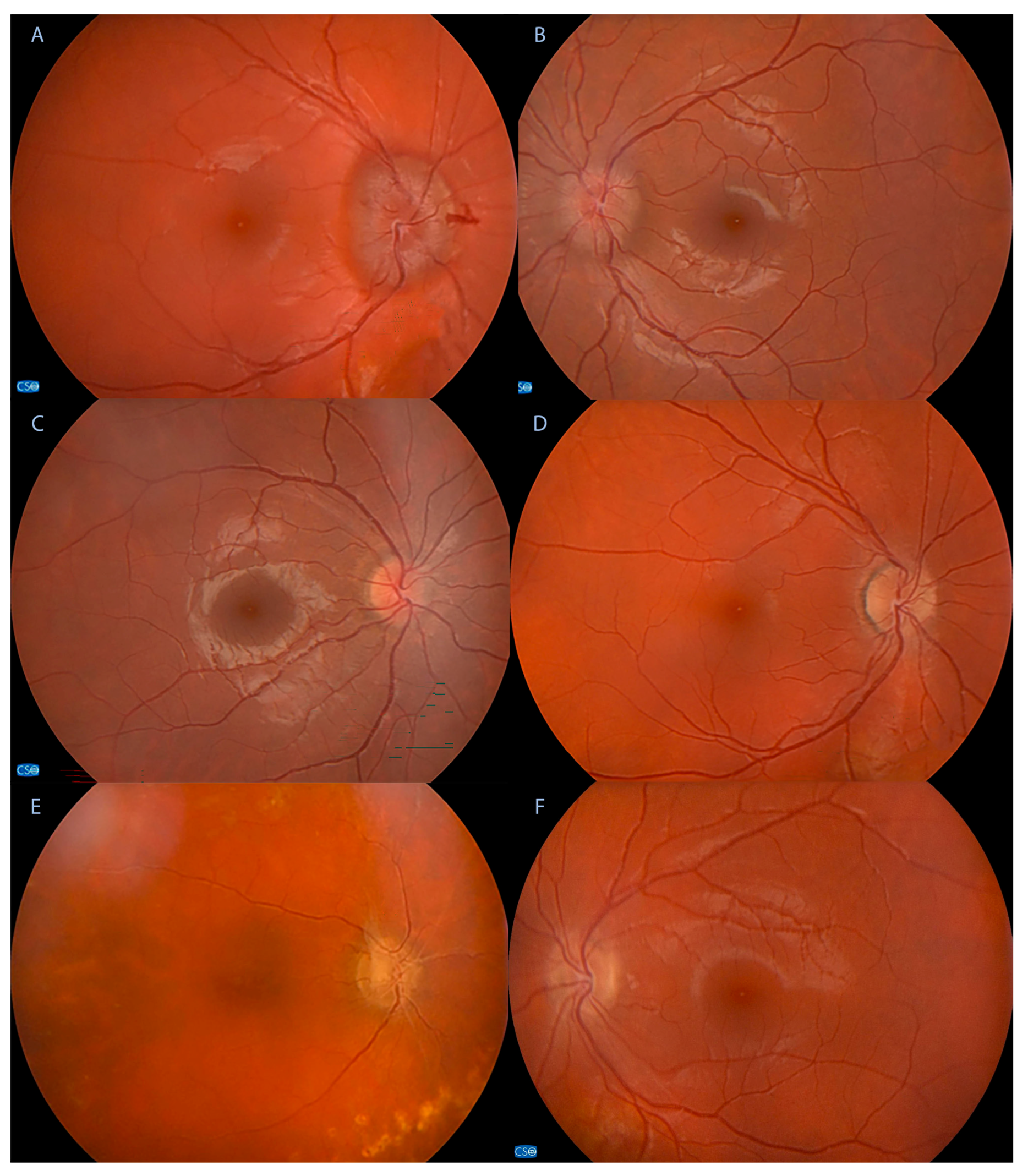
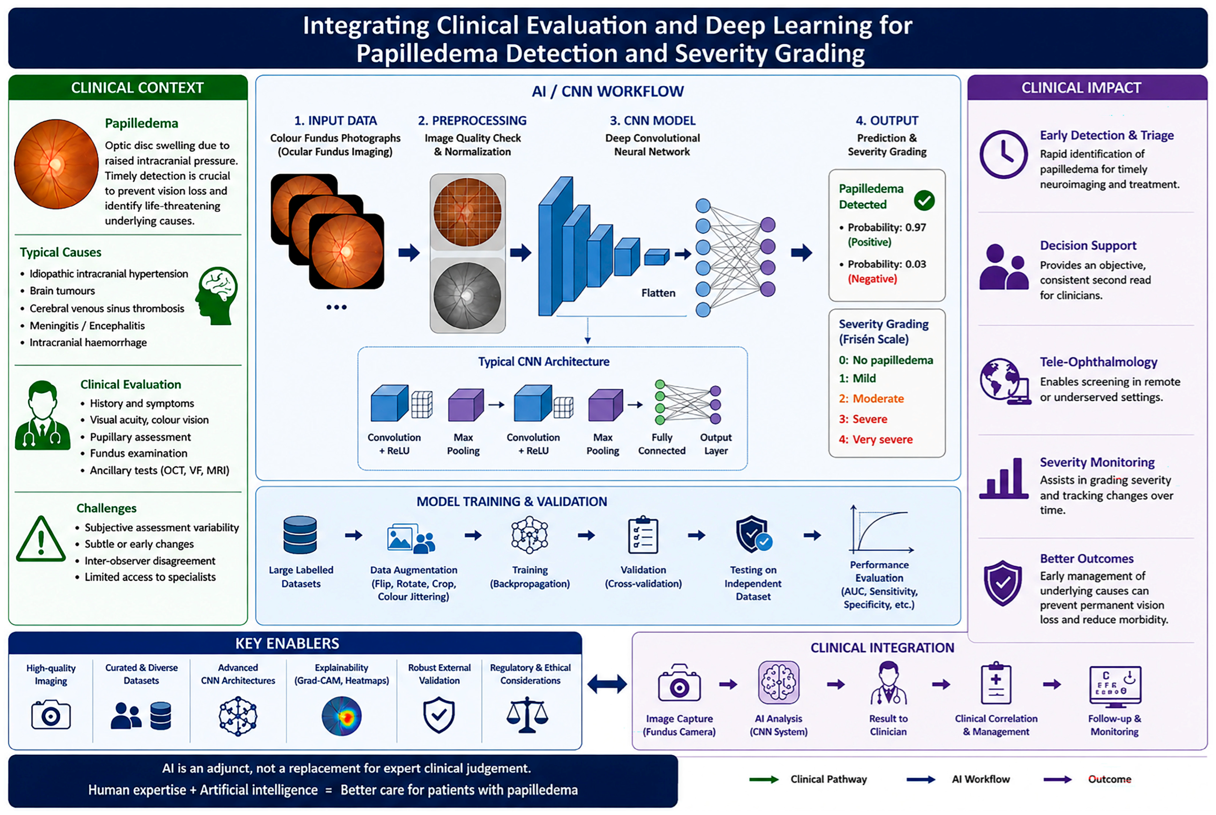
Artificial Intelligence in the Detection of Papilledema: A Systematic Review

Abstract
1. Introduction
1.1. Historical Background
1.2. Fundamentals of AI, ML, and DL
1.3. AI Applications in Ophthalmology
2. Materials and Methods
2.1. Study Design
2.2. Outcomes
2.3. Statistic Analysis and Bias
3. Results
3.1. Literature Research
3.2. Study Analysis
3.3. Quality of Evidence
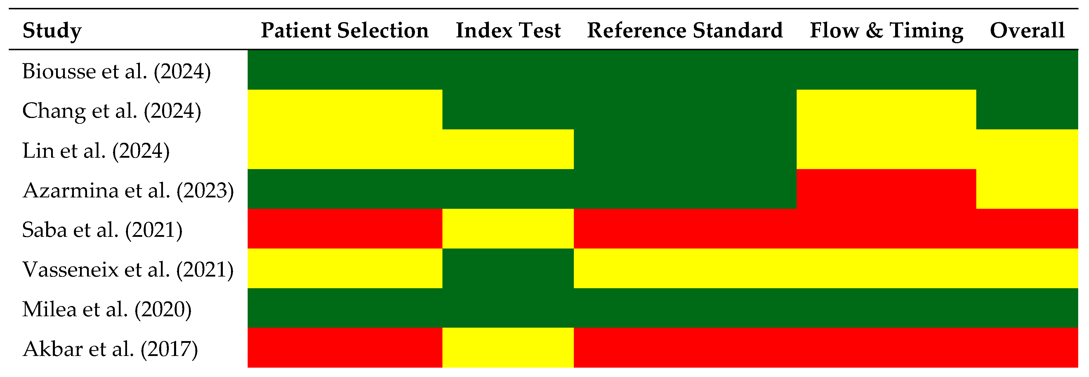
- Patient Selection Bias (evaluation of the study populations, concerning its appropriate selection and representation). Studies by Biousse et al. [55], Azarmina et al. [58], and Milea et al. [61] exhibited low risk of bias in patient selection. The datasets used were appropriately designed for AI-based analysis, ensuring that they included a diverse range of patients and fundus image sources and thus contributing to the broad applicability of the AI models assessed. On the other hand, Saba et al. [59] and Akbar et al. [62] demonstrated a high risk of bias in the patient selection domain due to the use of relatively small and potentially non-representative datasets, as well as limited reporting regarding patient recruitment methodology. In both studies, image selection appeared to rely on retrospective or convenience sampling approaches rather than consecutive or randomized inclusion, increasing the likelihood of selection bias. Furthermore, the datasets lacked broad demographic and clinical heterogeneity, reducing confidence in the generalizability of the models to real-world populations. These limitations are particularly relevant in papilledema research, where optic disc appearance may vary substantially depending on disease severity, patient age, ethnicity, image acquisition conditions, and the presence of confounding optic nerve abnormalities.
- Index Test Bias (for bias in the AI model’s application and interpretation). Most studies showed a low risk of bias in the application of AI algorithms. However, Akbar et al.’s [62] study resulted in a moderate risk due to the use of an SVM model without extensive external validation. In contrast, the studies by Milea et al. [61], Chang et al. [56], and Lin et al. [57] demonstrated strong methodological precision, focusing on external multi-centre validation to minimize bias.
- Reference Standard Bias (examines the accuracy and consistency of the reference group of experts). The reference standard in most studies was expert neuro-ophthalmologists, ensuring a high level of reliability. However, Saba et al. [59] and Akbar et al. [62] were assessed to have a high risk of bias in this section because of the inconsistencies in human grading methods, leading to probable variability in the reference standard.
- Flow and Timing Bias. While Biousse et al. [55] and Milea et al. [61] showed low risk, Azarmina et al. [58], Saba et al. [59], and Akbar et al. [62] were considered as high risk studies due to potential discrepancies in timing between AI–human comparisons. Differences in imaging time points and delays in manual assessment could introduce variations that affect the validation models.
- Low-risk studies: The studies by Milea et al. [61], Chang et al. [56], and Biousse et al. [55] had the lowest risk in all domains evaluated, which strengthens the confidence in their results for future applications. The study by Chang et al. [56] demonstrated overall low risk of bias across most QUADAS-2 domains, with unclear risks in patient selection and flow due to its retrospective design (very common in AI studies), while maintaining low applicability concerns. However, the study is methodologically strong because of multi-centre applications and external validation. Biousse et al. [55] is one of the strongest studies, as a real-world design and use of non-mydriatic fundus photos in ED setting.
4. Discussion
4.1. Principal Findings
4.2. Clinical Implications
4.3. Methodological Quality and Risk of Bias
4.4. Comparison with the Existing Literature
4.5. Limitations and Challenges
4.5.1. Generalizability and Dataset Variability
4.5.2. Real-World Applicability
4.5.3. Interpretability and Trust
4.5.4. Integration and Medico-Legal Considerations
4.5.5. Regulatory and Ethical Considerations
4.6. Future Directions
5. Conclusions
Supplementary Materials
Author Contributions
Funding
Data Availability Statement
Acknowledgments
Conflicts of Interest
Abbreviations
| AI | Artificial Intelligence |
| AMD | Age-Related Macular Degeneration |
| ANN | Artificial Neural Network |
| AUC | Area Under the Curve |
| BONSAI | Brain and Optic Nerve Study with Artificial Intelligence |
| CAD | Computer-Aided Diagnosis |
| CNN | Convolutional neural network |
| CSF | Cerebrospinal Fluid |
| DFN | Deep Feedforward Network |
| DL | Deep Learning |
| ED | Emergency Department |
| EHR | Electronic Health Record |
| EMA | European Medicines Agency |
| FDA | U.S. Food and Drug Administration |
| iDx-DR | Named FDA-Cleared Diabetic Retinopathy System |
| ICP | Intracranial Pressure |
| IRD | Inherited Retinal Disorder |
| KNN | K-Nearest Neighbours |
| MGD | Meibomian Gland Dysfunction |
| ML | Machine Learning |
| MLP | Multilayer Perceptron |
| MRI | Magnetic Resonance Imaging |
| NN | Neural Network |
| OCT | Optical Coherence Tomography |
| PICOS | Population, Intervention, Comparator, Outcomes, Study Design |
| PRISMA | Preferred Reporting Items for Systematic Reviews and Meta-Analyses |
| PUN | Pre-Trained Unsupervised Network |
| QUADAS-2 | Quality Assessment of Diagnostic Accuracy Studies, Version 2 |
| RCT | Randomized Controlled Trial |
| RGB | Red, Green, and Blue |
| ROP | Retinopathy of Prematurity |
| SVM | Support Vector Machine |
| TRT | Total Retinal Thickness |
| U-Net | Convolutional Neural Network Architecture for Image Segmentation |
| VF | Visual Field |
| VGG | Visual Geometry Group |
| ViT | Vision Transformer |
| XAI | Explainable Artificial Intelligence |
References
- Xie, J.S.; Donaldson, L.; Margolin, E. Papilledema: A review of etiology, pathophysiology, diagnosis, and management. Surv. Ophthalmol. 2022, 67, 1135–1159. [Google Scholar] [CrossRef] [PubMed]
- Bouthour, W.; Biousse, V.; Newman, N.J. Diagnosis of Optic Disc Oedema: Fundus Features, Ocular Imaging Findings, and Artificial Intelligence. Neuro-Ophthalmol. 2023, 47, 177–192. [Google Scholar] [CrossRef] [PubMed]
- He, G.; Dunn, H.P.; Ahmad, K.E.; Watson, E.; Henderson, A.; Tynan, D.; Leaney, J.; White, A.J.; Hewitt, A.W.; Fraser, C.L. Fundoscopy use in neurology departments and the utility of smartphone photography: A prospective prevalence and crossover diagnostic accuracy study amongst neurology inpatients. Eur. J. Neurol. 2022, 29, 2463–2472. [Google Scholar] [CrossRef] [PubMed]
- Wu, J.H.; Liu, T.Y.A.; Hsu, W.T.; Ho, J.H.C.; Lee, C.C. Performance and Limitation of Machine Learning Algorithms for Diabetic Retinopathy Screening: Meta-analysis. J. Med. Internet Res. 2021, 23, e23863. [Google Scholar] [CrossRef] [PubMed]
- McCarthy, J.; Minsky, M.L.; Rochester, N.; Shannon, C.E. A Proposal for the Dartmouth Summer Research Project on Artificial Intelligence, August 31, 1955. AI Mag. 2006, 27, 12–14. [Google Scholar]
- Grzybowski, A. (Ed.) Artificial Intelligence in Ophthalmology; Springer: Cham, Switzerland, 2021; 286p. [Google Scholar]
- Taketomi, T.; Uchiyama, H.; Ikeda, S. Visual SLAM algorithms: A survey from 2010 to 2016. IPSJ Trans. Comput. Vis. Appl. 2017, 9, 16. [Google Scholar] [CrossRef]
- He, J.; Baxter, S.L.; Xu, J.; Xu, J.; Zhou, X.; Zhang, K. The practical implementation of artificial intelligence technologies in medicine. Nat. Med. 2019, 25, 30–36. [Google Scholar] [CrossRef] [PubMed]
- Deng, J.; Dong, W.; Socher, R.; Li, L.J.; Kai, L.; Li, F.F. ImageNet: A large-scale hierarchical image database. In 2009 IEEE Conference on Computer Vision and Pattern Recognition; IEEE: Piscataway, NJ, USA, 2009; pp. 248–255. Available online: https://ieeexplore.ieee.org/document/5206848/ (accessed on 21 November 2024).
- De Fauw, J.; Ledsam, J.R.; Romera-Paredes, B.; Nikolov, S.; Tomasev, N.; Blackwell, S.; Askham, H.; Glorot, X.; O’Donoghue, B.; Visentin, D.; et al. Clinically applicable deep learning for diagnosis and referral in retinal disease. Nat. Med. 2018, 24, 1342–1350. [Google Scholar] [CrossRef] [PubMed]
- Bauer, D.C.; Wilson, L.O.W.; Twine, N.A. Artificial Intelligence in Medicine: Applications, Limitations and Future Directions. In Artificial Intelligence in Medicine; Raz, M., Nguyen, T.C., Loh, E., Eds.; Springer Nature: Singapore, 2022; pp. 101–120. Available online: https://link.springer.com/10.1007/978-981-19-1223-8_5 (accessed on 30 April 2026).
- Gupta, V.; Mishra, V.K.; Singhal, P.; Kumar, A. An Overview of Supervised Machine Learning Algorithm. In 2022 11th International Conference on System Modeling & Advancement in Research Trends (SMART); IEEE: Piscataway, NJ, USA, 2022; pp. 87–92. Available online: https://ieeexplore.ieee.org/document/10047618/ (accessed on 30 April 2026).
- Murphy, K.P. Machine Learning: A Probabilistic Perspective; MIT Press: Cambridge, MA, USA, 2012. [Google Scholar]
- Hastie, T.; Tibshirani, R.; Friedman, J. The Elements of Statistical Learning: Data Mining, Inference, and Prediction, 2nd ed.; Springer: New York, NY, USA, 2009. [Google Scholar]
- LeCun, Y.; Bengio, Y.; Hinton, G. Deep learning. Nature 2015, 521, 436–444. [Google Scholar] [CrossRef] [PubMed]
- Ting, D.S.W.; Pasquale, L.R.; Peng, L.; Campbell, J.P.; Lee, A.Y.; Raman, R.; Tan, G.S.W.; Schmetterer, L.; Keane, P.A.; Wong, T.Y. Artificial intelligence and deep learning in ophthalmology. Br. J. Ophthalmol. 2019, 103, 167–175. [Google Scholar] [CrossRef] [PubMed]
- Goodfellow, I.; Bengio, Y.; Courville, A. Deep Learning; MIT Press: Cambridge, MA, USA, 2016. [Google Scholar]
- Krizhevsky, A.; Sutskever, I.; Hinton, G.E. ImageNet classification with deep convolutional neural networks. Commun. ACM 2017, 60, 84–90. [Google Scholar] [CrossRef]
- Dong, L.; He, W.; Zhang, R.; Ge, Z.; Wang, Y.X.; Zhou, J.; Xu, J.; Shao, L.; Wang, Q.; Yan, Y.; et al. Artificial Intelligence for Screening of Multiple Retinal and Optic Nerve Diseases. JAMA Netw. Open 2022, 5, e229960. [Google Scholar] [CrossRef] [PubMed]
- Jafarbeglou, F.; Ahmadieh, H.; Soleimani, F.; Karimi, A.; Daftarian, N.; Fekri, S.; Motevasseli, T.; Naderan, M.; Kamali Doust Azad, B.; Sheikhtaheri, A.; et al. A deep learning model for diagnosis of inherited retinal diseases. Sci. Rep. 2025, 15, 22523. [Google Scholar] [CrossRef] [PubMed]
- Ting, D.S.W.; Cheung, C.Y.; Lim, G.; Tan, G.S.W.; Quang, N.D.; Gan, A.; Hamzah, H.; Garcia-Franco, R.; San Yeo, I.Y.; Lee, S.Y.; et al. Development and validation of a deep learning system for diabetic retinopathy and related eye diseases using retinal images from multiethnic populations with diabetes. JAMA 2017, 318, 2211–2223. [Google Scholar] [CrossRef] [PubMed]
- Raghavendra, U.; Fujita, H.; Bhandary, S.V.; Gudigar, A.; Tan, J.H.; Acharya, U.R. Deep convolution neural network for accurate diagnosis of glaucoma using digital fundus images. Inf. Sci. 2018, 441, 41–49. [Google Scholar] [CrossRef]
- Simonyan, K.; Zisserman, A. Very deep convolutional networks for large-scale image recognition. arXiv 2014. [Google Scholar] [CrossRef]
- Gulshan, V.; Peng, L.; Coram, M.; Stumpe, M.C.; Wu, D.; Narayanaswamy, A.; Venugopalan, S.; Widner, K.; Madams, T.; Cuadros, J.; et al. Development and validation of a deep learning algorithm for diabetic retinopathy detection. JAMA 2016, 316, 2402–2410. [Google Scholar] [CrossRef] [PubMed]
- Li, Z.; He, Y.; Keel, S.; Meng, W.; Chang, R.T.; He, M. Efficacy of a deep learning system for detecting glaucomatous optic neuropathy based on color fundus photographs. Ophthalmology 2018, 125, 1199–1206. [Google Scholar] [CrossRef] [PubMed]
- Zhang, Z.; Lin, X.; Yu, X.; Fu, Y.; Chen, X.; Yang, W.; Dai, Q. Meibomian Gland Density: An Effective Evaluation Index of Meibomian Gland Dysfunction Based on Deep Learning and Transfer Learning. J. Clin. Med. 2022, 11, 2396. [Google Scholar] [CrossRef] [PubMed]
- Kotsiantis, S.B. Supervised machine learning: A review of classification techniques. Informatica 2007, 31, 249–268. [Google Scholar]
- Bishop, C.M. Pattern Recognition and Machine Learning; Springer: New York, NY, USA, 2006. [Google Scholar]
- Yu, C.; Liu, J.; Nemati, S. Reinforcement learning in healthcare: A survey. arXiv 2020. [Google Scholar] [CrossRef]
- Whiting, P.F.; Rutjes, A.W.S.; Westwood, M.E.; Mallett, S.; Deeks, J.J.; Reitsma, J.B.; Leeflang, M.M.; Sterne, J.A.; Bossuyt, P.M.; QUADAS-2 Group. QUADAS-2: A revised tool for the quality assessment of diagnostic accuracy studies. Ann. Intern. Med. 2011, 155, 529–536. [Google Scholar] [CrossRef] [PubMed]
- Branco, J.; Wang, J.K.; Elze, T.; Garvin, M.K.; Pasquale, L.R.; Kardon, R.; Woods, B.; Szanto, D.; Kupersmith, M.J. Classifying and quantifying changes in papilledema using machine learning. Investig. Ophthalmol. Vis. Sci. 2024, 6, e000503. [Google Scholar] [CrossRef] [PubMed]
- Grzybowski, A.; Jin, K.; Zhou, J.; Pan, X.; Wang, M.; Ye, J.; Wong, T.Y. Retina fundus photograph-based artificial intelligence algorithms in medicine: A systematic review. Int. J. Med. Inform. 2024, 185, 105402. [Google Scholar]
- Nam, Y.; Kim, J.; Kim, K.; Park, K.A.; Kang, M.; Cho, B.H.; Oh, S.Y.; Kee, C.; Han, J.; Lee, G.I.; et al. Deep learning-based optic disc classification is affected by the presence of tilted disc. Ophthalmology 2024, 131, 1476. [Google Scholar]
- Rambabu, L.; Smith, B.G.; Tumpa, S.; Kohler, K.; Kolias, A.G.; Hutchinson, P.J.; Bashford, T. Artificial intelligence-enabled ophthalmoscopy for papilledema: A systematic review protocol. BMJ Open 2024, 14, e085123. [Google Scholar]
- Salaheldin, A.M.; Abdel Wahed, M.; Talaat, M.; Saleh, N. Deep learning-based automated detection and grading of papilledema from OCT images. Int. J. Imaging Syst. Technol. 2024, 34, e23133. [Google Scholar] [CrossRef]
- Sathianvichitr, K.; Najjar, R.P.; Tang, Z.; Fraser, J.A.; Yau, C.W.L.; Girard, M.J.A.; Costello, F.; Lin, M.Y.; Lagrèze, W.A.; Vignal-Clermont, C.; et al. A deep learning approach for accurate discrimination between optic disc drusen and papilledema on fundus photographs. J. Neuroophthalmol. 2024, 44, 454–461. [Google Scholar] [CrossRef] [PubMed]
- Yanoff, M. (Ed.) Advances in Ophthalmology and Optometry; Elsevier: Amsterdam, The Netherlands, 2024. [Google Scholar]
- Anandi, L.; Budihardja, B.M.; Anggraini, E.; Badjrai, R.A.; Nusanti, S. The use of artificial intelligence in detecting papilledema from fundus photographs: A systematic review. Eye 2023, 13, 184–190. XXXX–XXXX. [Google Scholar] [CrossRef]
- Chan, E.; Tang, Z.; Najjar, R.P.; Narayanaswamy, A.; Sathianvichitr, K.; Newman, N.J.; Biousse, V.; Milea, D.; Bonsai Group. A deep learning system for automated quality evaluation of optic disc photographs in neuro-ophthalmic disorders. Ophthalmology 2023, 130, 1140–1148. [Google Scholar]
- Ko, M.W. Tele-neuro-ophthalmology. In Telemedicine in Ophthalmology; Springer: Cham, Switzerland, 2023. [Google Scholar]
- Sathianvichitr, K.; Lamoureux, O.; Nakada, S.; Tang, Z.; Schmetterer, L.; Chen, C.; Cheung, C.Y.; Najjar, R.P.; Milea, D. Through the eyes into the brain: Using artificial intelligence. Prog. Retin. Eye Res. 2023, 93, 101131. [Google Scholar]
- Sun, G.; Wang, X.; Xu, L.; Li, C.; Wang, W.; Yi, Z.; Luo, H.; Su, Y.; Zheng, J.; Li, Z.; et al. Deep learning for the detection of multiple fundus diseases using ultra-widefield images. Comput. Methods Programs Biomed. 2023, 232, 107438. [Google Scholar]
- Vasseneix, C.; Nusinovici, S.; Xum, X.; Hwang, J.M.; Hamann, S.; Chen, J.J.; Loo, J.L.; Milea, L.; Tan, K.B.K.; Ting, D.S.W.; et al. Deep learning system outperforms clinicians in identifying optic disc abnormalities. npj Digit. Med. 2023, 6, 147. [Google Scholar]
- Biousse, V.; Najjar, R.P.; Sathianvichitr, K.; Tang, Z.; Hamann, S.; Fraser, C.; Fraser, A.; Lin, M.Y.; Vasseneix, C.; Peragallo, J.H.; et al. Deep learning can accurately distinguish between true papilledema and optic disc drusen on ocular fundus photographs. Neurology 2022, 99, e251–e262. [Google Scholar] [CrossRef]
- Li, M.; Wan, C. The use of deep learning technology for the detection of optic neuropathy. Eye Vis. 2022, 9, 21. [Google Scholar] [CrossRef] [PubMed]
- Wang, Z.; Keane, P.A.; Chiang, M.F.; Cheung, C.Y.; Wong, T.Y.; Ting, D.S.W. Artificial intelligence and deep learning in ophthalmology. In Advances in Ophthalmology; Springer: Cham, Switzerland, 2022. [Google Scholar]
- Wang, C.; Zhang, Y.; Xu, S.; Liu, Y.; Xie, L.; Wu, C.; Yang, Q.; Chu, Y.; Ye, Q. Research on assistant diagnosis of fundus optic neuropathy based on deep learning. Comput. Biol. Med. 2022, 141, 105123. [Google Scholar] [CrossRef] [PubMed]
- Wang, J.K.; Garvin, M.K.; Kupersmith, M.J.; Kardon, R.H. Quantifying spatial patterns of OCT total retinal thickness in papilledema using deep learning variational autoencoder. Investig. Ophthalmol. Vis. Sci. 2022, 63, 2351. [Google Scholar]
- Li, B.; Chen, H.; Zhang, B.; Yuan, M.; Jin, X.; Lei, B.; Xu, J.; Gu, W.; Wong, D.C.S.; He, X.; et al. Development and evaluation of a deep learning model for detection of multiple fundus diseases. Comput. Methods Programs Biomed. 2021, 207, 106158. [Google Scholar] [CrossRef] [PubMed]
- Biousse, V.; Newman, N.J.; Najjar, R.P.; Vasseneix, C.; Xu, X.; Ting, D.S.W.; Milea, L.B.; Hwang, J.M.; Kim, D.H.; Yang, H.K.; et al. Optic disc classification by deep learning versus expert neuro-ophthalmologists. Ophthalmology 2020, 127, 1626–1634. [Google Scholar] [CrossRef] [PubMed]
- Islam, M.S.; Wang, J.K.; Johnson, S.S.; Thurtell, M.J.; Kardon, R.H.; Garvin, M.K. Deep-learning approach for automated OCT en-face retinal vessel segmentation. IEEE Trans. Med. Imaging 2020, 39, 3838–3849. [Google Scholar] [CrossRef] [PubMed]
- Islam, M.S.; Wang, J.K.; Deng, W.; Thurtell, M.J.; Kardon, R.H.; Garvin, M.K. Deep-learning-based estimation of 3D optic nerve head shape from fundus photographs. IEEE Trans. Med. Imaging 2020, 39, 3449–3460. [Google Scholar] [CrossRef]
- Leong, Y.Y.; Vasseneix, C.; Finkelstein, M.T.; Milea, D.; Najjar, R.P. Artificial Intelligence Meets Neuro-Ophthalmology. Asia Pac. J. Ophthalmol. (Phila) 2022, 11, 111–125. [Google Scholar] [CrossRef] [PubMed]
- Newman, N.J.; Najjar, R.P.; Vasseneix, C.; Jiang, Z.; Ting, D.S.W.; Liu, Y.; Biousse, V.; Wong, T.Y.; Milea, D.l. Human vs machine: The BONSAI study. Investig. Ophthalmol. Vis. Sci. 2020, 94, 2776. [Google Scholar] [CrossRef]
- Biousse, V.; Najjar, R.P.; Tang, Z.; Lin, M.Y.; Wright, D.W.; Keadey, M.T.; Wong, T.Y.; Bruce, B.B.; Milea, D.; Newman, N.J. BONSAI Study Group. Application of a deep learning system to detect papilledema on nonmydriatic ocular fundus photographs in an emergency department. Am. J. Ophthalmol. 2024, 261, 199–207. [Google Scholar] [CrossRef] [PubMed]
- Chang, M.Y.; Heidary, G.; Beres, S.; Pineles, S.L.; Gaier, E.D.; Gise, R.; Reid, M.; Avramidis, K.; Rostami, M.; Narayanan, S. Artificial Intelligence to Differentiate Pediatric Pseudopapilledema and True Papilledema on Fundus Photographs. Ophthalmol. Sci. 2024, 4, 100496. [Google Scholar] [CrossRef] [PubMed]
- Lin, M.Y.; Najjar, R.P.; Tang, Z.; Cioplean, D.; Dragomir, M.; Chia, A.; Patil, A.; Vasseneix, C.; Peragallo, J.H.; Newman, N.J.; et al. The BONSAI (Brain and Optic Nerve Study with Artificial Intelligence) deep learning system can accurately identify pediatric papilledema on standard ocular fundus photographs. J. AAPOS 2024, 28, 103803. [Google Scholar] [CrossRef] [PubMed]
- Azarmina, M.; Mahmoudi Nejad Azar, S.; Naghibzadeh, S.K.; Aminzadeh, H.; Bagheri, M.; Niazi, K.; Azarmina, H.; Baloutch, B.D.; Ghafourian, A.; Daftarian, N.; et al. AI accuracy in papilledema diagnosis in fundus photographs within 201 eyes. Biomed. J. Sci. Tech. Res. 2023, 52, 40847–40853. [Google Scholar]
- Saba, T.; Akbar, S.; Kolivand, H.; Bahaj, S.A. Automatic detection of papilledema through fundus retinal images using deep learning. Microsc. Res. Tech. 2021, 84, 2055–2063. [Google Scholar] [CrossRef] [PubMed]
- Vasseneix, C.; Najjar, R.P.; Xu, X.; Tang, Z.; Loo, J.L.; Singhal, S.; Tow, S.; Milea, L.; Ting, D.S.W.; Liu, Y.; et al. Accuracy of a deep learning system for classification of papilledema severity on ocular fundus photographs. Neurology 2021, 97, e1797–e1807. [Google Scholar] [CrossRef] [PubMed]
- Milea, D.; Najjar, R.P.; Jiang, Z.; Ting, D.S.W.; Vasseneix, C.; Xu, X.; Aghsaei Fard, M.; Fonseca, P.; Vanikieti, K.; Lagrèze, W.A.; et al. Artificial intelligence to detect papilledema from ocular fundus photographs. N. Engl. J. Med. 2020, 382, 1687–1695. [Google Scholar] [CrossRef] [PubMed]
- Akbar, S.; Akram, M.U.; Sharif, M.; Tariq, A.; Yasin, U.U. Decision support system for detection of papilledema through fundus retinal images. J. Med. Syst. 2017, 41, 201. [Google Scholar] [CrossRef] [PubMed]
- Rosenbacke, R.; Melhus, Å.; McKee, M.; Stuckler, D. How explainable artificial intelligence can increase or decrease clinicians’ trust in AI applications in health care: Systematic review. JMIR AI 2024, 3, e53207. [Google Scholar] [CrossRef] [PubMed]
- Lin, W.C.; Chen, J.S.; Chiang, M.F.; Hribar, M.R. Applications of artificial intelligence to electronic health record data in ophthalmology. Trans. Vis. Sci. Tech. 2020, 9, 13. [Google Scholar] [CrossRef] [PubMed]
- Mienye, I.D.; Sun, Y.; Wang, Z.; Adjei, F.A.; Agyeman, M.O.; Abubakar, A. A survey of explainable artificial intelligence in healthcare: Concepts, applications, and challenges. Inf. Med. Unlocked 2024, 51, 101587. [Google Scholar] [CrossRef]
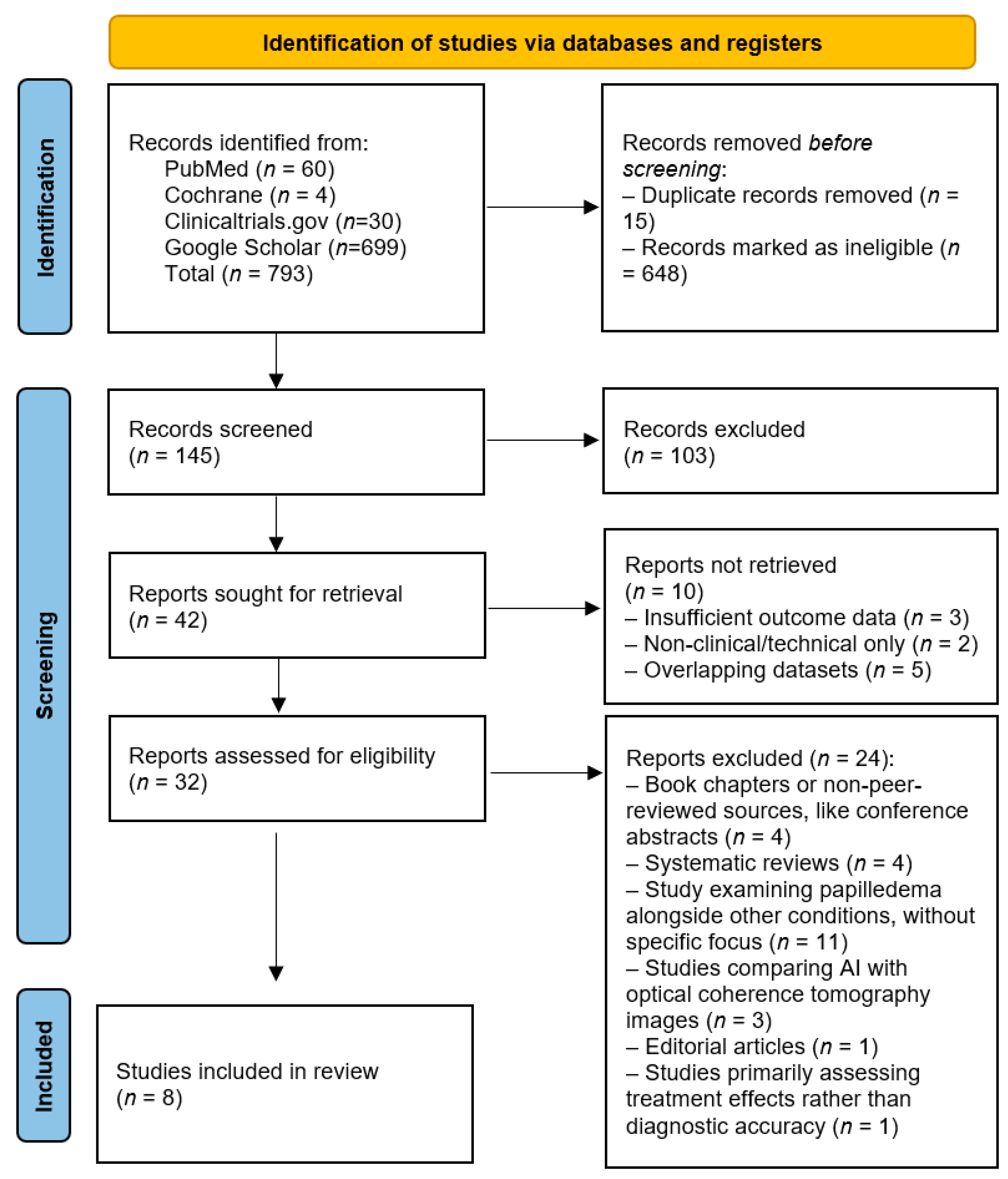



| No, Reference | Authors | Title | Year | Reason for Exclusion |
|---|---|---|---|---|
| 1 [31] | Branco J, Wang JK, Elze T, Garvin MK, Pasquale LR, Kardon R et al. | Classifying and quantifying changes in papilloedema using machine learning | 2024 | Focuses on treatment of papilledema rather than diagnostic accuracy, as well as on longitudinal monitoring and quantification of papilledema progression, rather than primary diagnostic detection or classification |
| 2 [32] | Grzybowski A, Jin K, Zhou J, Pan X, Wang M, Ye J et al. | Retina Fundus Photograph-Based Artificial Intelligence Algorithms in Medicine: A Systematic Review | 2024 | Systematic review; does not present new primary data on papilledema diagnostic accuracy |
| 3 [33] | Nam Y, Kim J, Kim K, Park KA, Kang M, Cho BH et al. | Deep Learning-Based Optic Disc Classification Is Affected by the Presence of Tilted Disc | 2024 | Focuses on the impact of optic disc tilt on deep learning model performance rather than specifically assessing papilledema |
| 4 [34] | Rambabu L, Smith BG, Tumpa S, Kohler K, Kolias AG, Hutchinson PJ et al. | Artificial intelligence-enabled ophthalmoscopy for papilledema: a systematic review protocol | 2024 | Systematic review protocol; does not present primary data on papilledema |
| 5 [35] | Salaheldin AM, Abdel Wahed M, Talaat M, and Saleh N | Deep Learning-Based Automated Detection and Grading of Papilledema From OCT Images: A Promising Approach for Improved Clinical Diagnosis and Management | 2024 | Primarily a model-development study based on OCT imaging, without direct clinical validation using fundus photograph-based papilledema diagnosis |
| 6 [36] | Sathianvichitr K, Najjar RP, Tang Z, Fraser JA, Yau CWL, Girard MJA et al. | A Deep Learning Approach for Accurate Discrimination Between Optic Disc Drusen and Papilledema on Fundus Photographs | 2024 | Focused primarily on differentiation between papilledema and optic disc drusen (pseudopapilledema) rather than standalone papilledema detection or grading |
| 7 [37] | Yanoff M | Advances in Ophthalmology and Optometry, 2024 | 2024 | Book; general overview |
| 8 [38] | Anandi L, Budihardja BM, Anggraini E, Badjrai RA, and Nusanti S | The use of artificial intelligence in detecting papilledema from fundus photographs | 2023 | Systematic review; does not present new primary data on papilledema |
| 9 [39] | Chan E, Tang Z, Najjar RP, Narayanaswamy A, Sathianvichitr K, Newman NJ et al. | A Deep Learning System for Automated Quality Evaluation of Optic Disc Photographs in Neuro-Ophthalmic Disorders | 2023 | Focuses on image quality assessment rather than directly evaluating papilledema |
| 10 [40] | Melissa W. Ko | Tele-Neuro-Ophthalmology | 2023 | Book chapter; general overview of telemedicine |
| 11 [41] | Sathianvichitr K, Lamoureux O, Nakada S, Tang Z, Schmetterer L, Chen C et al. | Through the Eyes into the Brain, Using Artificial Intelligence | 2023 | Focuses on multiple neurological abnormalities, not exclusively papilledema |
| 12 [42] | Sun G, Wang X, Xu L, Li C, Wang W, Yi Z et al. | Deep Learning for the Detection of Multiple Fundus Diseases Using Ultra-widefield Images | 2023 | Focuses on detecting multiple fundus diseases rather than specifically assessing papilledema |
| 13 [43] | Vasseneix C, Nusinovici S, Xu X, Hwang JM, Hamann S, Chen JJ et al. | Deep Learning System Outperforms Clinicians in Identifying Optic Disc Abnormalities | 2023 | Focused on broad optic disc abnormality classification, with papilledema included as one component rather than the primary outcome |
| 14 [44] | Biousse V, Najjar R, Sathianvichitr K, Tang Z, Hamann S, Fraser C et al. | Deep Learning Can Accurately Distinguish Between True Papilledema and Optic Disc Drusen On Ocular Fundus Photographs | 2022 | Focused primarily on differentiation between papilledema and optic disc drusen (pseudopapilledema) rather than standalone papilledema detection or grading |
| 15 [45] | Li M and Wan C | The use of deep learning technology for the detection of optic neuropathy | 2022 | Systematic review; does not present new primary data on papilledema |
| 16 [46] | Wang Z, Keane PA, Chiang M, Cheung CY, Wong TY, and Ting DSW | Artificial Intelligence and Deep Learning in Ophthalmology | 2022 | Book chapter; general overview, not focused on papilledema diagnostic accuracy |
| 17 [47] | Wang C, Zhang Y, Xu S, Liu Y, Xie L, Wu C et al. | Research on Assistant Diagnosis of Fundus Optic Neuropathy Based on Deep Learning | 2022 | Focuses on differentiating various optic neuropathies, not exclusively papilledema |
| 18 [48] | Wang JK, Garvin MK, Kupersmith MJ, and Kardon RH | Quantifying Spatial Patterns of OCT Total Retinal Thickness (TRT) in Papilledema Over Time using a Deep Learning Variational AutoEncoder | 2022 | Focus on OCT imaging technique rather than diagnostic accuracy assessment |
| 19 [49] | Li B, Chen H, Zhang B, Yuan M, Jin X, Lei B et al. | Development and evaluation of a deep learning model for the detection of multiple fundus diseases based on colour fundus photography | 2021 | Focuses on detecting multiple fundus diseases rather than specifically assessing diagnostic accuracy for papilledema |
| 20 [50] | Biousse V, Newman NJ, Najjar RP, Vasseneix C, Xu X, Ting DSW et al. | Optic Disc Classification by Deep Learning versus Expert Neuro-Ophthalmologists | 2020 | Focused on multiclass optic disc abnormality classification rather than dedicated papilledema detection |
| 21 [51] | Islam MS, Wang JK, Johnson SS, Thurtell MJ, Kardon RH, and Garvin MK | A Deep-Learning Approach for Automated OCT En-Face Retinal Vessel Segmentation in Cases of Optic Disc Swelling Using Multiple En-Face Images as Input | 2020 | Focuses on OCT imaging technique development rather than assessing diagnostic accuracy in a clinical context |
| 22 [52] | Islam MS, Wang JK, Deng W, Thurtell MJ, Kardon RH, and Garvin MK | Deep-Learning-Based Estimation of 3D Optic-Nerve-Head Shape from 2D Color Fundus Photographs in Cases of Optic Disc Swelling | 2020 | Focused on 3D optic nerve head reconstruction and general optic disc swelling analysis rather than direct clinical detection of papilledema |
| 23 [53] | Leong YY, Vasseneix C, Finkelstein MT, Milea D, Najjar R P | Artificial Intelligence Meets Neuro-Ophthalmology | 2022 | Editorial; does not present original research or primary diagnostic accuracy data |
| 24 [54] | Newman N, Najjar R, Vasseneix C, Zhubo J, Ting D, Liu Y et al. | Human vs. Machine: The Brain and Optic Nerve Study with Artificial Intelligence (BONSAI) | 2020 | Supplement issue: Conference abstract; lacks full methodological details |
| No., Reference | Authors | Title | Journal, Year |
|---|---|---|---|
| 1 [55] | Biousse V, Najjar RP, Tang Z, Lin MY, Wright DW, Keahey MT et al. | Application of a Deep Learning System to Detect Papilledema on Nonmydriatic Ocular Fundus Photographs in an Emergency Department | Am J Ophthalmol, 2024 |
| 2 [56] | Chang MY, Heidary G, Beres S, Pineles SL, Gaier ED, Gise R et al. | Artificial Intelligence to Differentiate Pediatric Pseudopapilledema and True Papilledema on Fundus Photographs | Ophthalmol. Sci., 2024 |
| 3 [57] | Lin MY, Najjar RP, Tang Z, Cioplean D, Dragomir M, Chia A et al. | The BONSAI Deep Learning System for Pediatric Papilledema Detection | J AAPOS, 2024 |
| 4 [58] | Azarmina M, Mahmoudi Nejad Azar S, Naghibzadeh SK, Aminzadeh H, Bagheri M et al. | AI Accuracy in Papilledema Diagnosis in Fundus Photographs within 201 Eyes | Biomed J Sci & Tech Res, 2023 |
| 5 [59] | Saba T, Akbar S, Kolivand H, and Bahaj SA | Automatic detection of papilledema through fundus retinal images using deep learning | Microsc Res Tech, 2021 |
| 6 [60] | Vasseneix C, Najjar RP, Xu X, Tang Z, Loo JL, Singhal S et al. | Accuracy of a Deep Learning System for Classification of Papilledema Severity on Ocular Fundus Photographs | Neurology, 2021 |
| 7 [61] | Milea D, Najjar RP, Jiang Z, Ting D, Vasseneix C, Xu X et al. | Artificial Intelligence to Detect Papilledema from Ocular Fundus Photographs | N Engl J Med, 2020 |
| 8 [62] | Akbar S, Akram MU, Sharif M, Tariq A, and Yasin UU | Decision Support System for Detection of Papilledema through Fundus Retinal Images | J Med Syst, 2017 |
| Study | Methods | Participants, Aquisitions | Model Type | Centre, Study Type | External Validation, Method | Training/Validation Strategy | Outcomes |
|---|---|---|---|---|---|---|---|
| Biousse et al. (2024) [55] | AI-assisted triage in emergency department settings | 1608 fundus photographs, ED patients, non-mydriatic | BONSAI deep learning system | Single-centre, real-world implementation, prospective | Yes; expert panel | BONSAI model previously trained; prospective real-world validation in emergency department cohort | AUC 0.97; sensitivity 84.0%; specificity 98.9%; improved ED triage efficiency |
| Chang et al. (2024) [56] | Retrospective image-based classification of papilledema vs. pseudopapilledema using deep learning; 10-fold cross-validation | 235 pediatric patients (<18 years), 851 fundus photographs, mydriasis not specified (probable) | DenseNet-based tri-branch CNN | Multi-centre, retrospective clinical study | Yes; separate external test set | 10-fold cross-validation with independent external test set | AUC 0.81 (external); sensitivity 90.4%; specificity 56–67%; higher sensitivity than experts, particularly for mild cases |
| Lin et al. (2024) [57] | Pediatric AI model validated across multiple centres | 898 fundus photographs, pediatric patients from three centres, mydriatic | BONSAI deep learning system optimized for pediatric use | Multi-centre, retrospective | Yes; multi-centre external validation | Multi-centre training and external validation across three pediatric cohorts | AUC 0.98; sensitivity 98.0%; specificity 94.1% in pediatric cases |
| Azarmina et al. (2023) [58] | Retrospective study; AI vs. clinician comparison | 201 eyes, adult population, mydriatic | CAD system vs. expert evaluation | Single-centre, retrospective | No; clinician comparison | Retrospective comparison with clinician grading; validation strategy not fully reported | 85% agreement with neuro-ophthalmologists |
| Saba et al. (2021) [59] | AI-based optic disc segmentation and vessel analysis | 100 fundus photographs, mydriatic | DenseNet + U-Net segmentation for grading severity | Single-centre, retrospective | No; expert panel | Internal validation on limited dataset; no external validation | Accuracy 99.17%; superior vessel segmentation |
| Vasseneix et al. (2021) [60] | Deep learning applied for severity grading | 2103 fundus photographs, neuroophthalmology clinic, mydriatic | BONSAI deep learning system for severity classification | Single-centre, retrospective | No; expert panel | Internal validation with expert comparison; no external validation | AUC 0.93; effective papilledema severity classification |
| Milea et al. (2020) [61] | Deep learning validation on large multiethnic dataset | 15,846 fundus photographs, multiethnic dataset, mydriatic | Deep learning models (DenseNet, U-Net) for classification | Multi-centre, retrospective | Yes; external testing | Development and external validation on large multiethnic datasets | AUC 0.99; sensitivity 96.4%; specificity 84.7% |
| Akbar et al. (2017) [62] | Machine learning classification with cross-validation | 160 fundus images, hospital-based dataset, mydriatic | SVM classifier using handcrafted papilledema-related image features | Single-centre, retrospective | No; cross-validation | Cross-validation on hospital-based dataset; no external validation | Accuracy 92.86% in classifying papilledema |
| No. | Study | Patient Selection Bias | Index Test Bias | Reference Standard Bias | Flow & Timing Bias | Overall Risk of Bias |
|---|---|---|---|---|---|---|
| 1 | Biousse et al. (2024) [55] | Low | Low | Low | Low | Low |
| 2 | Chang et al. (2024) [56] | Moderate | Low | Low | Moderate | Low |
| 3 | Lin et al. (2024) [57] | Moderate | Moderate | Low | Moderate | Moderate |
| 4 | Azarmina et al. (2023) [58] | Low | Low | Low | High | Moderate |
| 5 | Saba et al. (2021) [59] | High | Moderate | High | High | High |
| 6 | Vasseneix et al. (2021) [60] | Moderate | Low | Moderate | Moderate | Moderate |
| 7 | Milea et al. (2020) [61] | Low | Low | Low | Low | Low |
| 8 | Akbar et al. (2017) [62] | High | Moderate | High | High | High |
| No. | Study | Did It Assess Grading/Severity? | Grading Approach | Key Comment |
|---|---|---|---|---|
| 1 | Biousse et al. (2024) [55] | No/limited | Mainly detection of papilledema and optic disc abnormalities | Focused on emergency department detection using non-mydriatic fundus photographs, not formal severity grading. |
| 2 | Chang et al. (2024) [56] | Indirectly | Differentiation of true papilledema vs. pseudopapilledema; attention to mild cases | Useful for detecting mild pediatric papilledema, but not primarily a severity-grading study. |
| 3 | Lin et al. (2024) [57] | Limited/yes | Pediatric detection; some grading-related analysis | The BONSAI system was mainly evaluated for pediatric papilledema detection, with relevance to grading, but less focused on full severity stratification than Vasseneix et al. |
| 4 | Azarmina et al. (2023) [58] | Yes | Frisén score assigned to fundus photographs | Evaluated fundus photographs using Frisén grading and CAD assessment. |
| 5 | Saba et al. (2021) [59] | Yes | Mild vs. severe papilledema | Used DenseNet for detection and U-Net-derived vascular indices for grading; reported mild/severe classification. |
| 6 | Vasseneix et al. (2021) [60] | Yes | Papilledema severity classification, Frisén-based | The most directly relevant grading study; classified severity on mydriatic fundus photographs and performed comparably to neuro-ophthalmologists. |
| 7 | Milea et al. (2020) [61] | No/limited | Multiclass detection: normal, papilledema, other optic disc abnormality | Landmark detection study, but not primarily designed for severity grading. |
| 8 | Akbar et al. (2017) [62] | Yes | Mild vs. severe papilledema | Early decision-support system for both detection and grading; reported high accuracy for mild vs. severe classification. |
Disclaimer/Publisher’s Note: The statements, opinions and data contained in all publications are solely those of the individual author(s) and contributor(s) and not of MDPI and/or the editor(s). MDPI and/or the editor(s) disclaim responsibility for any injury to people or property resulting from any ideas, methods, instructions or products referred to in the content. |
© 2026 by the authors. Licensee MDPI, Basel, Switzerland. This article is an open access article distributed under the terms and conditions of the Creative Commons Attribution (CC BY) license.
Share and Cite
Samoilă, O.; Antonoupoulou, V.; Samoilă, L. Artificial Intelligence in the Detection of Papilledema: A Systematic Review. J. Clin. Med. 2026, 15, 4878. https://doi.org/10.3390/jcm15134878
Samoilă O, Antonoupoulou V, Samoilă L. Artificial Intelligence in the Detection of Papilledema: A Systematic Review. Journal of Clinical Medicine. 2026; 15(13):4878. https://doi.org/10.3390/jcm15134878
Chicago/Turabian StyleSamoilă, Ovidiu, Vasiliki Antonoupoulou, and Lăcrămioara Samoilă. 2026. "Artificial Intelligence in the Detection of Papilledema: A Systematic Review" Journal of Clinical Medicine 15, no. 13: 4878. https://doi.org/10.3390/jcm15134878
APA StyleSamoilă, O., Antonoupoulou, V., & Samoilă, L. (2026). Artificial Intelligence in the Detection of Papilledema: A Systematic Review. Journal of Clinical Medicine, 15(13), 4878. https://doi.org/10.3390/jcm15134878