Case Report

Congenital Pericardial Agenesis: An Innocent Finding or Clinically Significant Condition? A Case Series and Literature Review


1
UMHAT “St. Ekaterina” EAD, Medical University of Sofia, 1431 Sofia, Bulgaria
2
Medical Faculty, Medical University of Sofia, 1431 Sofia, Bulgaria
3
Department of Internal Diseases “Prof. St. Kirkovich”, Medical University of Sofia, 1431 Sofia, Bulgaria
*
Author to whom correspondence should be addressed.
J. Clin. Med. 2026, 15(11), 4394; https://doi.org/10.3390/jcm15114394
Submission received: 5 May 2026 / Revised: 29 May 2026 / Accepted: 3 June 2026 / Published: 5 June 2026
(This article belongs to the Section Cardiovascular Medicine)

Abstract

Congenital pericardial agenesis (CPA) is a rare anomaly that is often considered a benign incidental finding but may present with nonspecific symptoms and mimic structural heart disease. Its clinical relevance remains incompletely defined, particularly regarding the distinction between a harmless anatomical variant and a clinically significant condition. We present a retrospective two-center case series of four patients with imaging-confirmed CPA, combined with a narrative review of the literature aiming to evaluate the clinical spectrum, diagnostic challenges, and management implications of CPA. The clinical presentation of our patients was heterogeneous, ranging from incidental findings to chest discomfort and dyspnea. In all cases, initial echocardiography suggested alternative diagnoses, including right ventricular cardiomyopathy, atrial septal defect, or pericardial disease, leading to diagnostic uncertainty. Definitive diagnosis was established using multimodality imaging, particularly cardiac magnetic resonance and computed tomography, which demonstrated characteristic features such as cardiac levoposition and interposition of lung parenchyma. Three patients had complete left pericardial agenesis and one had a partial defect. All patients were managed conservatively, without complications during follow-up.

1. Introduction

Congenital pericardial agenesis (CPA) is a rare developmental anomaly characterized by complete or partial absence of the pericardial sac, with an estimated prevalence of 0.002–0.004% in the general population [1,2,3]. It results from defective embryologic development of the pleuropericardial membranes, most commonly due to premature atrophy of the left common cardinal vein (duct of Cuvier), leading to incomplete fusion during early gestation [2,4]. As a consequence, the heart lacks its normal fibrous restraint and may shift freely within the thoracic cavity, most often toward the left hemithorax [2,5,6].
CPA encompasses a spectrum ranging from complete absence of the pericardium—most frequently left-sided—to partial defects, which are considerably less common but clinically more significant. In the majority of cases, particularly in complete agenesis, the condition remains asymptomatic and is detected incidentally during imaging or surgical procedures performed for unrelated indications [5,7,8]. However, the absence of pericardial support may lead to characteristic anatomical and functional alterations, including cardiac levoposition, abnormal cardiac rotation, and interposition of lung tissue between mediastinal structures [5,9,10].
Despite its often benign course, CPA may present with nonspecific clinical manifestations such as chest pain, dyspnea, palpitations, or reduced exercise tolerance, frequently leading to misdiagnosis as structural heart disease or pericardial pathology [5,11,12]. Importantly, partial pericardial defects carry a risk of potentially life-threatening complications, including cardiac herniation, strangulation of cardiac chambers or appendages, and compression of coronary arteries, which may result in myocardial ischemia or sudden cardiac death [13,14,15]. These risks highlight the clinical relevance of distinguishing between complete and partial forms of the disease [8,16,17].
The diagnosis of CPA remains challenging due to the nonspecific nature of routine investigations such as electrocardiography, chest radiography, and transthoracic echocardiography [8,14,16]. Multimodality imaging plays a pivotal role, with cardiac computed tomography (CT) and particularly cardiac magnetic resonance (CMR) considered the gold standard for definitive diagnosis, allowing precise anatomical characterization and exclusion of differential diagnoses [5,8,14].
Given the rarity of the condition and the predominance of isolated case reports in the literature, the clinical significance of CPA remains incompletely defined, particularly regarding its distinction as a benign incidental finding versus a condition with potential clinical consequences. In this context, we present a series of four patients with congenital pericardial agenesis alongside a narrative review of the literature, aiming to clarify whether this rare anomaly represents an innocent incidental finding or a clinically relevant condition with potential implications for patient management.

2. Materials and Methods

2.1. Study Design and Case Selection

This study represents a two-center case series combined with a narrative literature review. Cases were retrospectively collected between January 2018 and December 2025.
Patients were eligible for inclusion if they were adults with a diagnosis of congenital pericardial agenesis confirmed by advanced cardiac imaging, including cardiac CT and/or CMR, and had sufficient clinical, electrocardiographic, echocardiographic, and imaging data available for retrospective analysis. Both complete and partial pericardial defects were considered eligible.
Patients were excluded if the diagnosis of congenital pericardial agenesis was only suspected but not confirmed by CT or CMR, if imaging data were insufficient to characterize the type of pericardial defect, or if clinical information and follow-up data were incomplete. Patients with acquired pericardial defects related to previous cardiac surgery, trauma, infection, or pericardiectomy were also excluded.
Four patients diagnosed with CPA were identified during routine clinical practice and imaging evaluation at tertiary cardiology and radiology centers in Sofia, Bulgaria. The cases were collected retrospectively based on the availability of complete clinical and imaging data.
All patients included in this series had a confirmed diagnosis of CPA established by advanced cardiac imaging modalities. Clinical data were obtained from medical records, including patient history, physical examination findings, instrumental and laboratory data, and follow-up information.

2.2. Diagnostic Evaluation

All patients underwent a stepwise diagnostic evaluation. Initial assessment included clinical examination, electrocardiography (ECG), routine laboratory investigations and transthoracic echocardiography (TTE), which in several cases raised suspicion for structural cardiac abnormalities but did not allow definitive diagnosis.
Definitive diagnosis was established using multimodality imaging, including contrast-enhanced cardiac computed tomography (CT) with ECG synchronization and cardiac magnetic resonance imaging (CMR). Characteristic imaging findings used to confirm CPA included:
  • Absence of the pericardium, most commonly along the left cardiac border;
  • Marked leftward displacement (levoposition) and rotation of the heart;
  • Interposition of lung parenchyma between the ascending aorta and pulmonary artery;
  • Separation of the heart from the diaphragm;
  • Absence of pericardial effusion despite apparent “free” cardiac contour.
These features were consistently demonstrated across cases. For example, CT imaging revealed absence of the left pericardium with lung interposition between the great vessels and adjacent to the left ventricular wall, as well as cardiac rotation with the apex directed upward. CMR further confirmed the absence of the pericardial layer and allowed detailed assessment of cardiac morphology and function, which remained preserved in all patients.

2.3. Data Collection and Analysis

Clinical and imaging data were systematically reviewed and analyzed. The following variables were collected for each patient:
-
Demographic characteristics (age, sex);
-
Clinical presentation and symptoms;
-
ECG findings;
-
TTE findings;
-
Imaging characteristics on CT and/or CMR;
-
Type of pericardial defect (complete or partial);
-
Management strategy and clinical outcome.
Given the descriptive nature of this study and the small number of cases, no statistical analysis was performed. The cases were analyzed qualitatively to identify common clinical and imaging patterns.

2.4. Literature Review

This review was designed as a narrative, non-systematic literature review intended to contextualize the presented cases rather than provide exhaustive systematic evidence synthesis. Accordingly, no formal PRISMA-based methodology or quality assessment was applied.
A structured search of the PubMed and Scopus databases was performed using combinations of the following keywords: “congenital pericardial agenesis”, “pericardial absence”, “partial pericardial defect”, “congenital pericardial anomaly”, “congenital pericardial malformation” and “cardiac herniation”.
Articles published in English and reporting adult patients with imaging-confirmed congenital CPA were considered eligible. Priority was given to case reports, case series, and review articles describing clinical presentation, imaging findings, complications, and management strategies. Relevant references from selected articles were also screened to ensure comprehensive coverage of the topic. The literature search included publications available up to March 2026 and identified 14 relevant publications, the majority consisting of isolated case reports and small case series.

2.5. Ethical Considerations

Our research was conducted in accordance with the principles of the Declaration of Helsinki. Patient consent was waived due to the retrospective and non-interventional nature of this case series and the use of fully anonymized clinical data, in accordance with applicable national regulations and institutional policies.
No artificial intelligence-assisted tools were used in the preparation of this manuscript.

3. Results

3.1. Case Series

A total of four patients diagnosed with CPA were included in this case series. The clinical presentations ranged from incidental findings during imaging to evaluation for suspected structural heart disease. In all cases, the diagnosis was confirmed using advanced imaging modalities, primarily CT and/or CMR.
  • Clinical case 1
A 54-year-old male presented with progressive exertional dyspnea and fatigue, without associated chest pain, syncope, or palpitations. In February 2025, he experienced an episode of acute respiratory distress requiring short-term hospitalization, although no definitive cardiopulmonary cause was identified at that time. His medical history was notable for hypothyroidism, adequately controlled with levothyroxine, and dyslipidemia treated with low-dose atorvastatin.
On physical examination, the patient was in good general condition. Vital signs were within normal limits (heart rate 78 bpm, blood pressure 120/70 mmHg, respiratory rate 16/min, SpO2 98% on room air). Cardiovascular examination revealed regular heart rhythm and a soft holosystolic murmur at the cardiac apex. No peripheral edema or signs of heart failure were present. Laboratory investigations, including NT-proBNP (36.1 pg/mL), were within normal limits, effectively excluding decompensated heart failure.
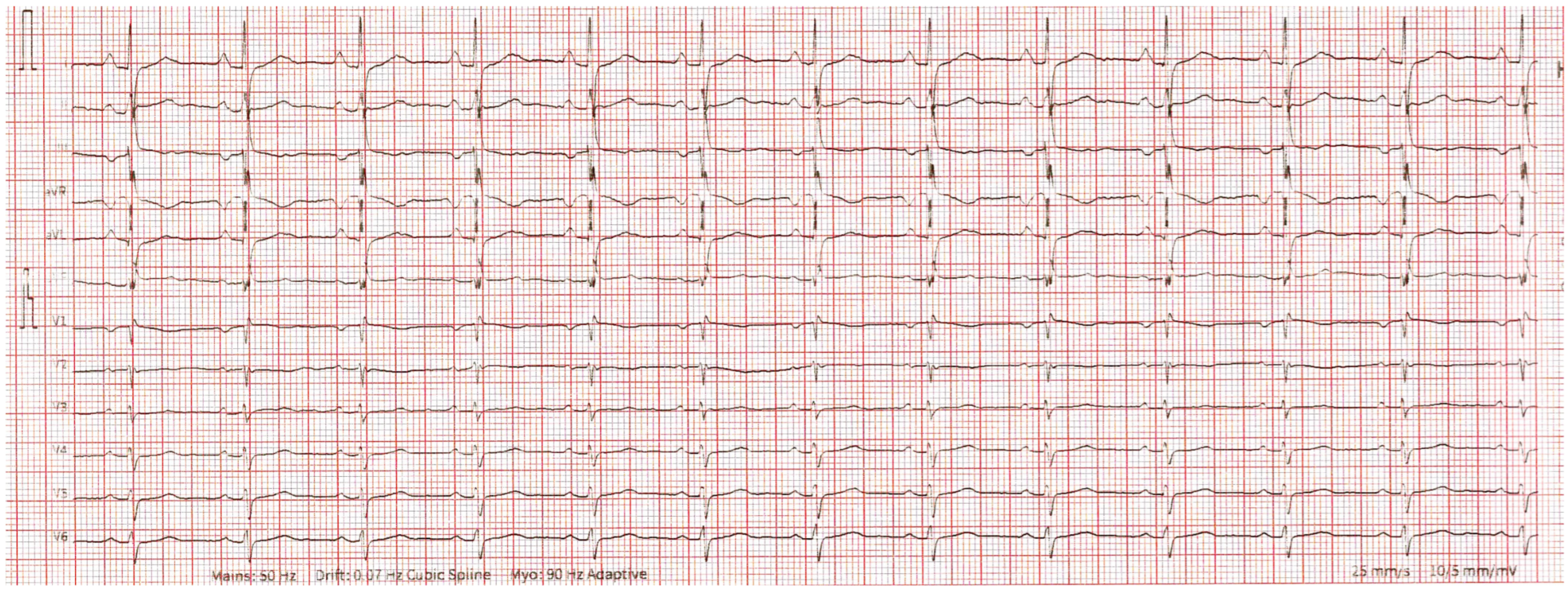
Electrocardiography demonstrated sinus rhythm with right axis deviation and incomplete right bundle branch block, without ischemic changes (Figure 1).
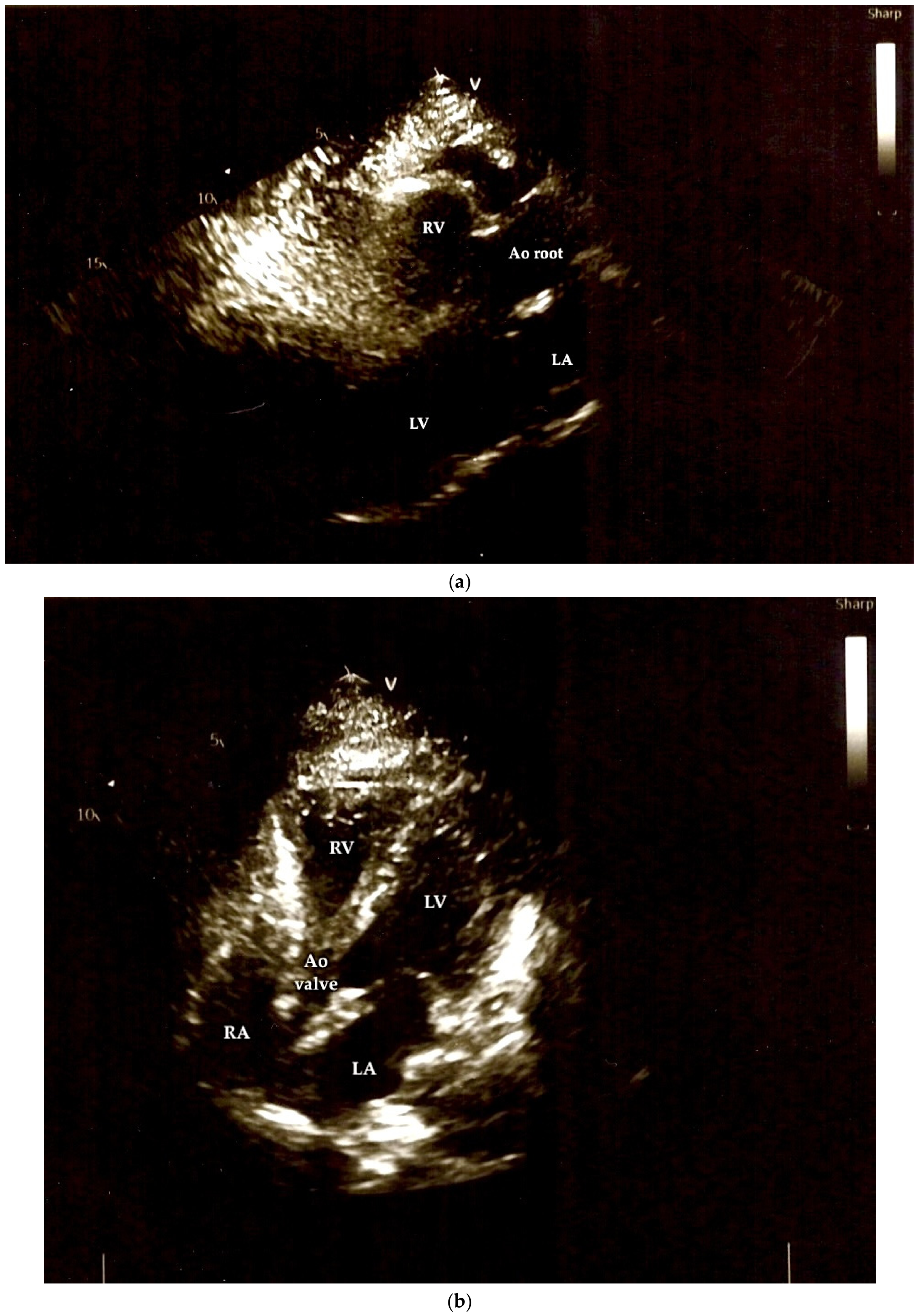
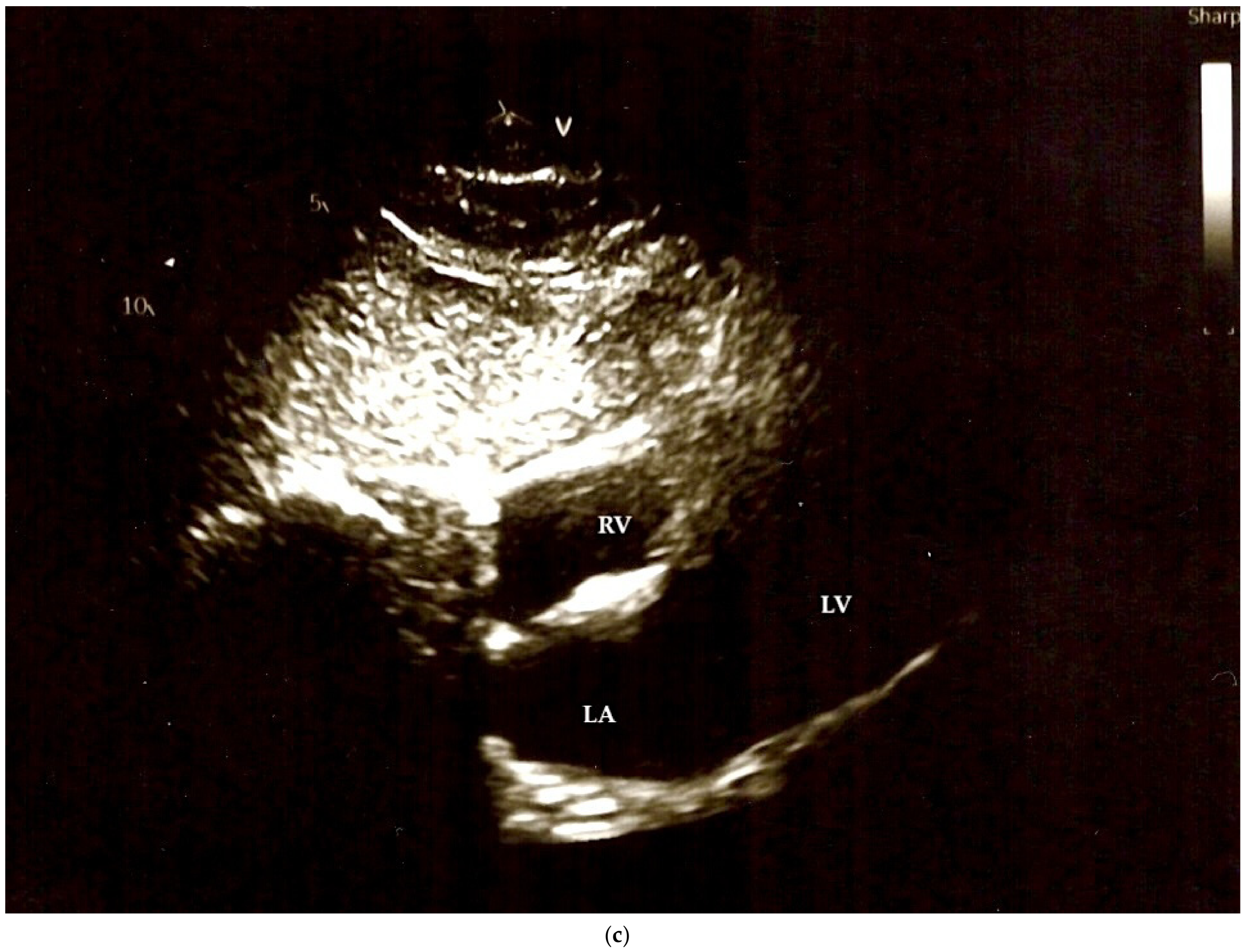
TTE was technically challenging due to the marked leftward displacement of the heart, resulting in poor acoustic windows and limited visualization. Standard parasternal and apical views could not be reliably obtained, and image interpretation was significantly restricted. Only subcostal views provided satisfactory image quality. From these, ventricular wall thickness was assessed as normal for both the left and right ventricles, with preserved left ventricular dimensions and systolic function, including normal ejection fraction estimated by the Teichholz method (Figure 2a–c).
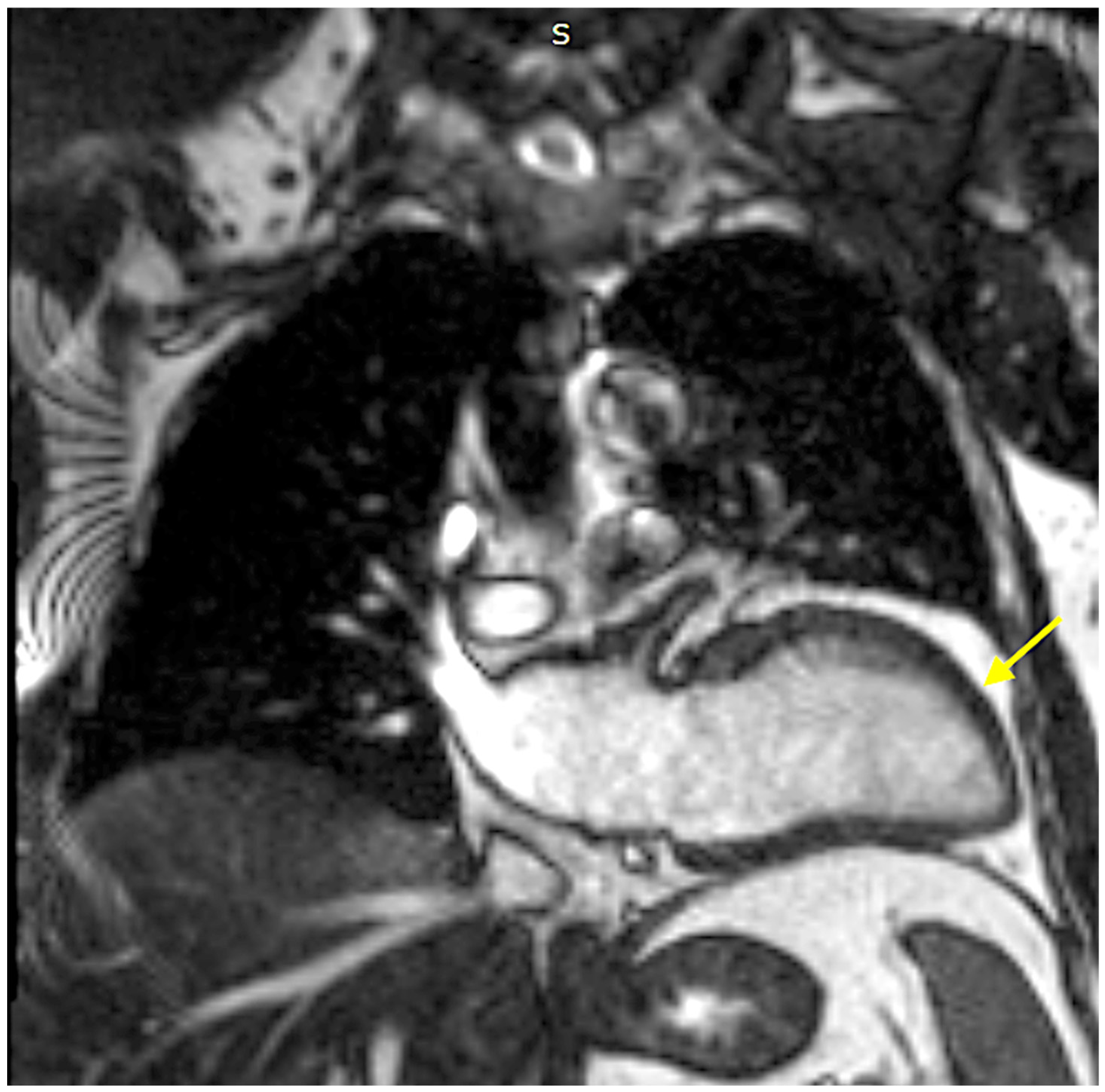
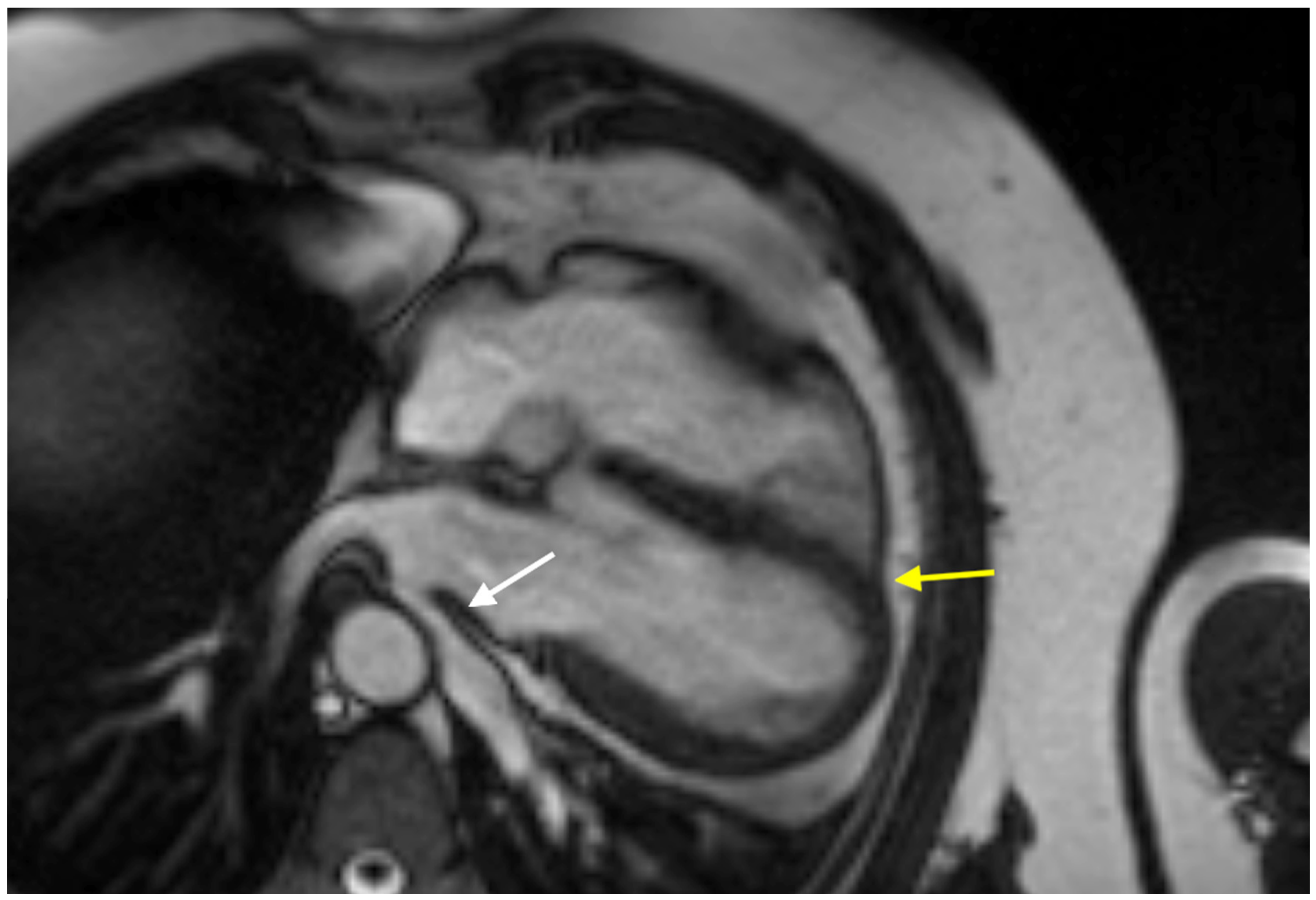
The abnormal cardiac orientation on TTE, together with its limited diagnostic yield and the persistence of symptoms, raised suspicion for an underlying structural or positional abnormality and prompted further evaluation with CMR, which ultimately established the diagnosis. It revealed complete congenital absence of the left pericardium, pronounced cardiac levoposition and clockwise rotation of the heart, with posterolateral displacement of the apex and approximately two-thirds of the atrial mass located within the left hemithorax. A key diagnostic feature was the interposition of lung parenchyma between the ascending aorta and the pulmonary artery, forming the characteristic “lingular sign”. Additionally, there was extension of epicardial fat into the mediastinum (Figure 3 and Figure 4).
Cardiac morphology and function were otherwise preserved, with normal biventricular volumes and systolic function and no evidence of myocardial edema or fibrosis. Incidental findings included mild mitral valve prolapse without significant regurgitation and a bovine aortic arch variant with a common origin of the brachiocephalic trunk and left common carotid artery. No pleural effusion, lymphadenopathy, or pulmonary pathology was identified.
Based on the absence of high-risk features and preserved cardiac function, a conservative management strategy was adopted. The patient continued his baseline medical therapy and was enrolled in a structured follow-up program, including clinical evaluation every six months, periodic laboratory monitoring, and annual electrocardiography and transthoracic echocardiography to assess cardiac position and exclude potential complications such as herniation. At 12-month follow-up, the patient remained clinically stable without new symptoms or imaging evidence of complications.
This case emphasizes the diagnostic value of CMR in confirming congenital pericardial agenesis and differentiating it from other structural cardiac abnormalities when initial clinical and echocardiographic findings are inconclusive.
  • Clinical case 2
A 22-year-old male with a history of a cardiac murmur detected at birth, which had not been fully investigated, presented with intermittent chest discomfort described as stabbing pain in the precordial region. He had no history of syncope, palpitations, or exercise intolerance.
On physical examination, the patient was in good general condition, with a hypersthenic habitus and increased body mass. Vital signs were stable, with blood pressure of 100/70 mmHg. Cardiovascular examination revealed regular heart rhythm with clear heart sounds and a grade 2/6 systolic murmur best heard at the pulmonary area. No signs of heart failure were present. Laboratory investigations were within normal limits.
Electrocardiography demonstrated atrial rhythm with a heart rate of 84 bpm, a semi-horizontal electrical axis, and an incomplete right bundle branch block, considered a physiological finding for age.
Initial transthoracic echocardiography (TTE) raised suspicion for structural heart disease, suggesting right ventricular dilatation, possible secundum atrial septal defect, and the presence of pericardial effusion. These findings led to diagnostic uncertainty. Repeat TTE evaluation revealed abnormal cardiac orientation, with leftward rotation of the cardiac apex, apparent right ventricular enlargement, and signal drop-out in the region of the fossa ovalis without evidence of interatrial shunting.
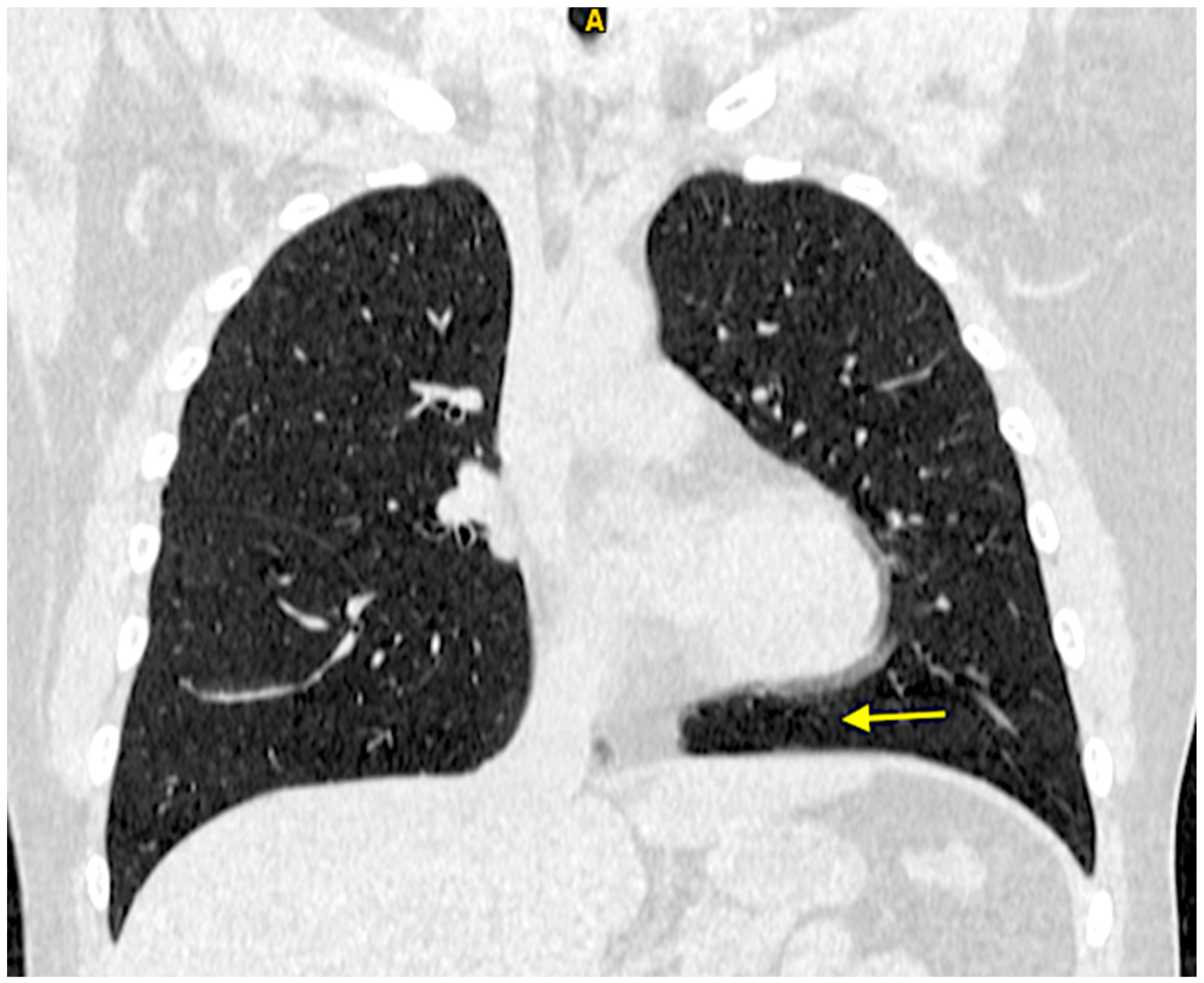
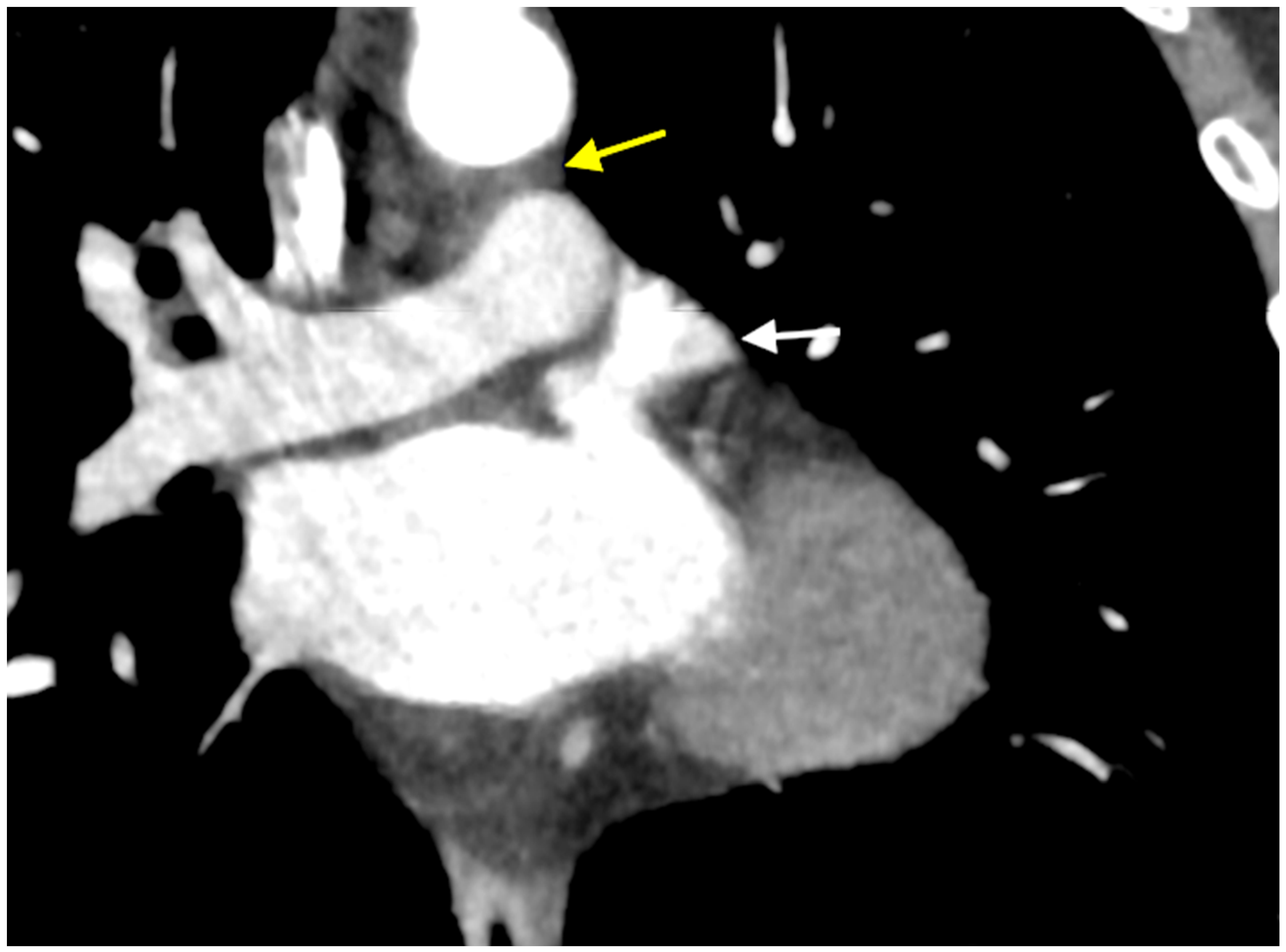
To further clarify these findings, contrast-enhanced multislice cardiac CT with ECG synchronization was performed. CT imaging demonstrated significant lateral displacement (levoposition) and rotation of the heart, with the apex directed superiorly. There was absence of the pericardium along the left cardiac border, accompanied by interposition of lung parenchyma between the ascending aorta and the pulmonary artery, a characteristic diagnostic feature. In addition, coronal reconstructions showed an apparent “suspension” of the heart away from the diaphragm. Importantly, cardiac chambers, septa, valves, and coronary anatomy were normal, and no true pericardial effusion was identified (Figure 5 and Figure 6).
These imaging findings established the diagnosis of congenital absence of the left pericardium.
The patient remained hemodynamically stable and largely asymptomatic, apart from mild intermittent chest discomfort. Given the absence of high-risk features or complications, no specific cardiologic treatment was indicated. A conservative management strategy with regular clinical and imaging follow-up was recommended. At 24-month follow-up, the patient remained clinically stable without new symptoms or imaging evidence of complications.
This case demonstrates the diagnostic challenges posed by congenital pericardial agenesis, which may mimic structural heart disease, including atrial septal defect, right ventricular cardiomyopathy, and pericardial effusion. It underscores the importance of considering this rare anomaly in patients with atypical echocardiographic findings and highlights the pivotal role of advanced imaging in achieving an accurate diagnosis.
  • Clinical case 3
A 29-year-old woman was initially evaluated in 2018 after cardiomegaly was incidentally identified during assessment for bronchopneumonia. At that time, TTE suggested dilatation of the right ventricular outflow tract (up to 42 mm) with suspected right ventricular systolic dysfunction, raising concern for possible arrhythmogenic right ventricular cardiomyopathy.
Subsequent echocardiographic examinations demonstrated persistent apparent enlargement of the right ventricular outflow tract (approximately 36 mm), with the cardiac apex appearing to be predominantly formed by the right ventricle. Left ventricular systolic function remained preserved (66%), and no significant valvular abnormalities were detected. Despite these findings, the clinical picture remained inconclusive.
In April 2023, the patient presented with an episode of panic attack accompanied by sinus tachycardia (heart rate 102 bpm) and mildly elevated blood pressure (130/80–85 mmHg), without chest pain or syncope. In 2024, she sought further medical evaluation following an episode of vertigo associated with transient blood pressure elevation.
On physical examination, the patient was in good general condition, with normal pulmonary findings. Cardiovascular examination revealed tachycardia and a grade 2/6 systolic murmur best heard at the second left intercostal space. Blood pressure was initially elevated (155/90 mmHg) but normalized spontaneously to 121/80 mmHg as heart rate decreased to 90 bpm.
Electrocardiography showed sinus tachycardia (123 bpm), a vertical electrical axis, and incomplete right bundle branch block. Twenty-four-hour Holter monitoring demonstrated sinus rhythm with rare supraventricular extrasystoles and no ventricular arrhythmias, pauses, or ischemic changes.
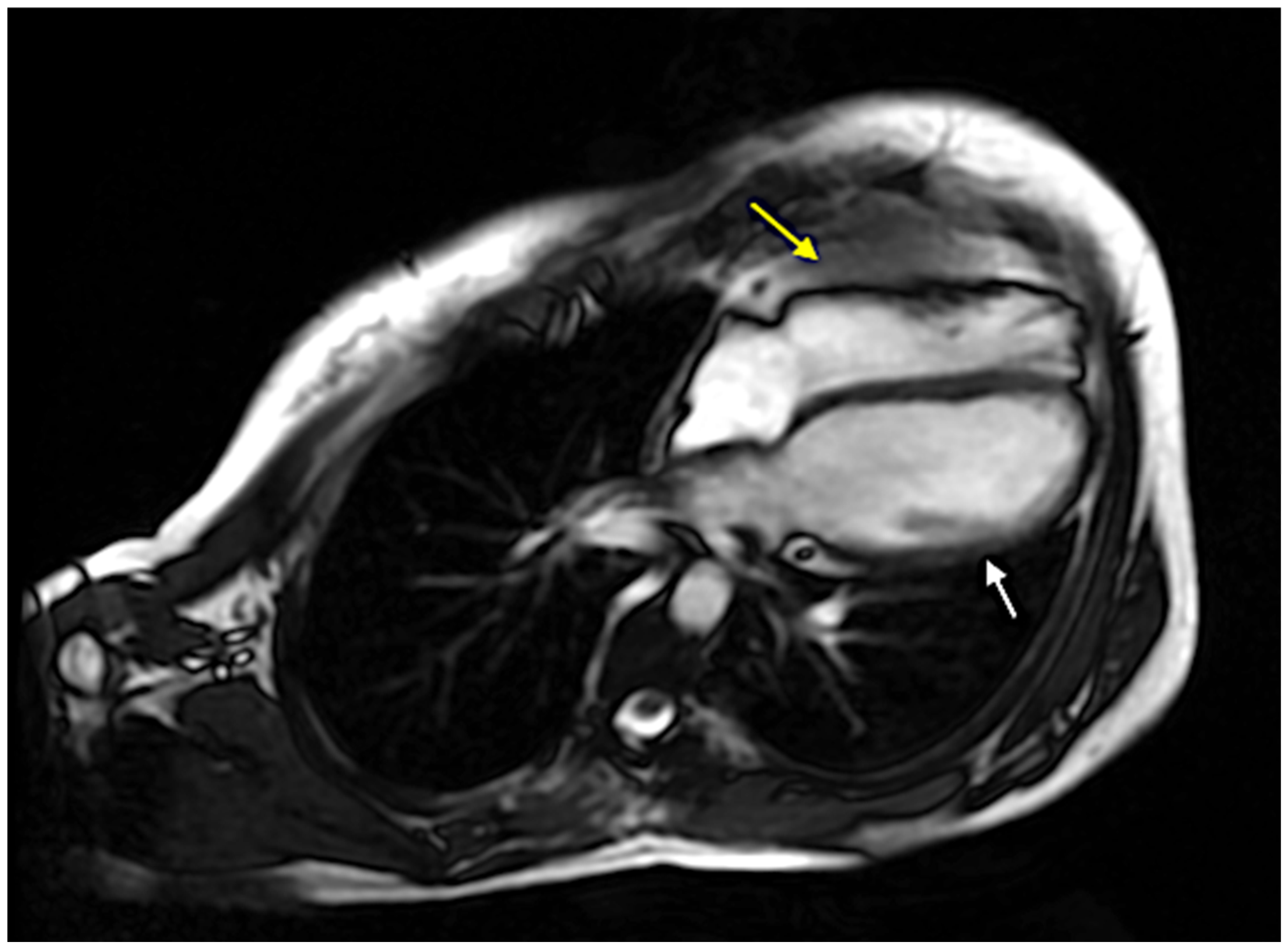
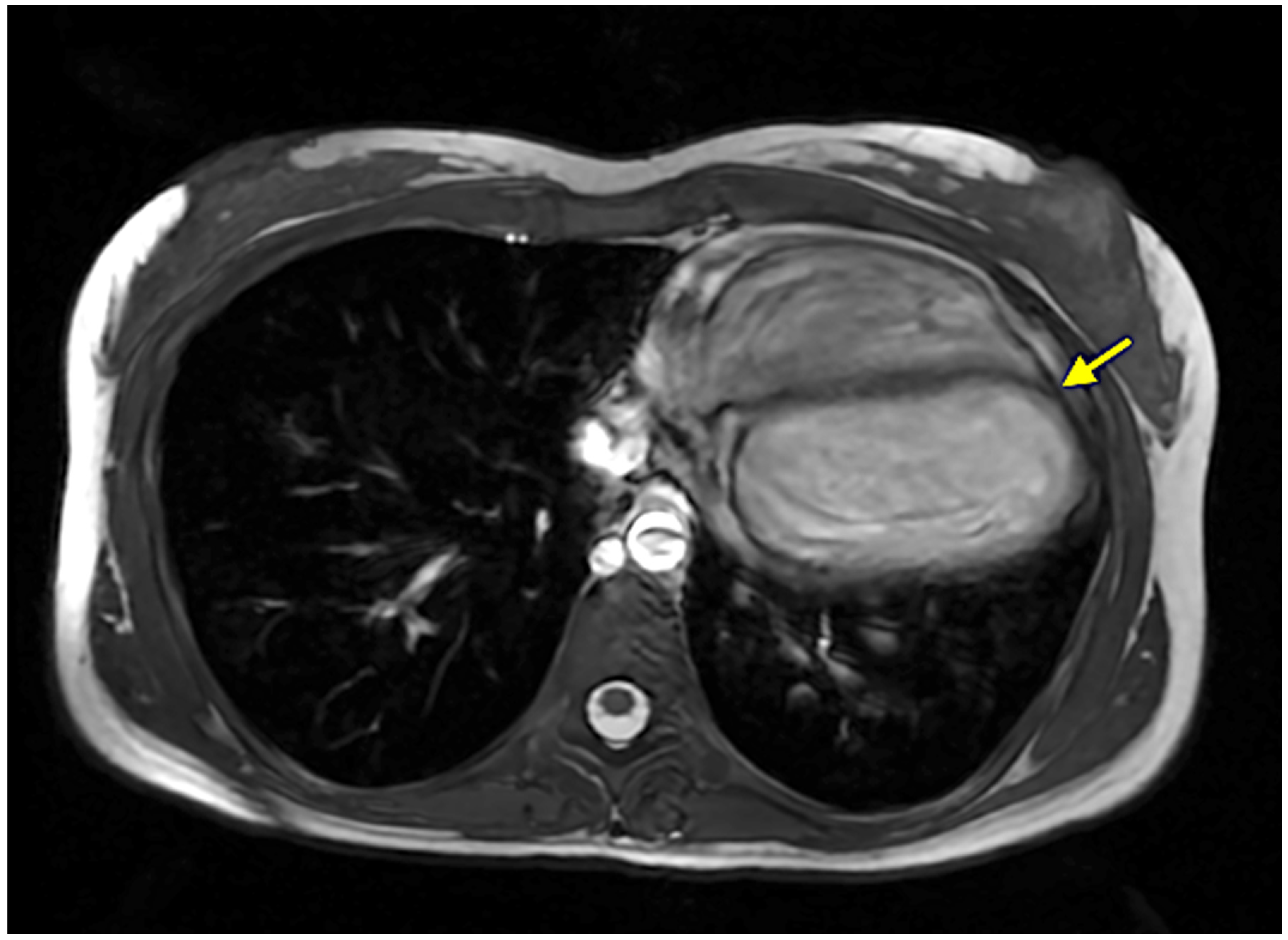
Given the persistent diagnostic uncertainty, CMR was performed. Imaging revealed complete congenital absence of the left pericardium, with pronounced leftward shift in the heart into the left hemithorax and clockwise rotation, resulting in leftward displacement of the apex. A key diagnostic feature was the interposition of lung parenchyma between the ascending aorta and the pulmonary artery. Despite the altered cardiac position, ventricular size, morphology, and systolic function were preserved, and no myocardial fibrosis or structural abnormalities were identified (Figure 7 and Figure 8).
These findings established the diagnosis of complete left pericardial agenesis, explaining the apparent right ventricular enlargement observed on echocardiography as a positional artifact rather than true pathology.
Given the absence of symptoms attributable to structural heart disease and the lack of high-risk features, no specific treatment was required. The patient was managed conservatively with regular clinical and imaging follow-up. She remained asymptomatic throughout the 18-month follow-up period.
This case illustrates how congenital pericardial agenesis may mimic right ventricular cardiomyopathy and other structural cardiac abnormalities on echocardiography, leading to diagnostic uncertainty. It underscores the pivotal role of CMR in establishing a definitive diagnosis and avoiding misclassification, while also demonstrating the generally benign clinical course of isolated complete pericardial agenesis in the absence of high-risk features.
  • Clinical case 4
A 62-year-old male patient was referred for cardiologic evaluation due to episodic, mild, transient chest oppression, predominantly left-sided, which had developed approximately two months prior to presentation. His medical history included arterial hypertension and mild dyslipidemia, both well controlled with medical therapy.
On physical examination, the patient was in good general condition. Vital signs were within normal limits (blood pressure 128/76 mmHg, heart rate 64 bpm). Cardiovascular and pulmonary examinations revealed no pathological findings. Laboratory investigations were unremarkable.
ECG demonstrated sinus rhythm at 67 bpm with left anterior fascicular block, without evidence of ischemic changes.
Initial TTE raised suspicion for pericardial pathology; however, image quality was limited due to a suboptimal acoustic window, precluding definitive assessment. In view of the persistent symptoms and inconclusive echocardiographic findings, the patient was referred for advanced imaging with CMR.
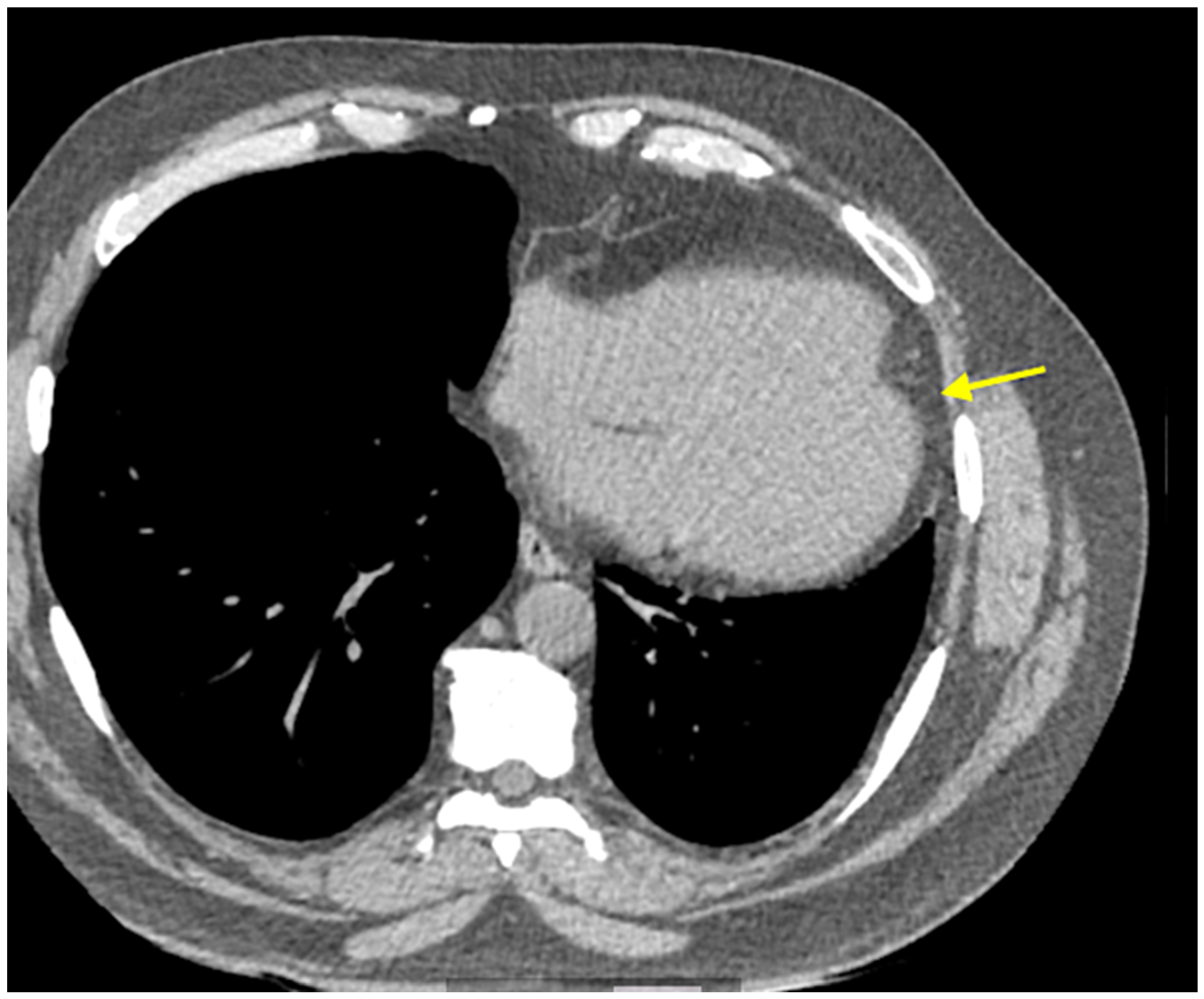
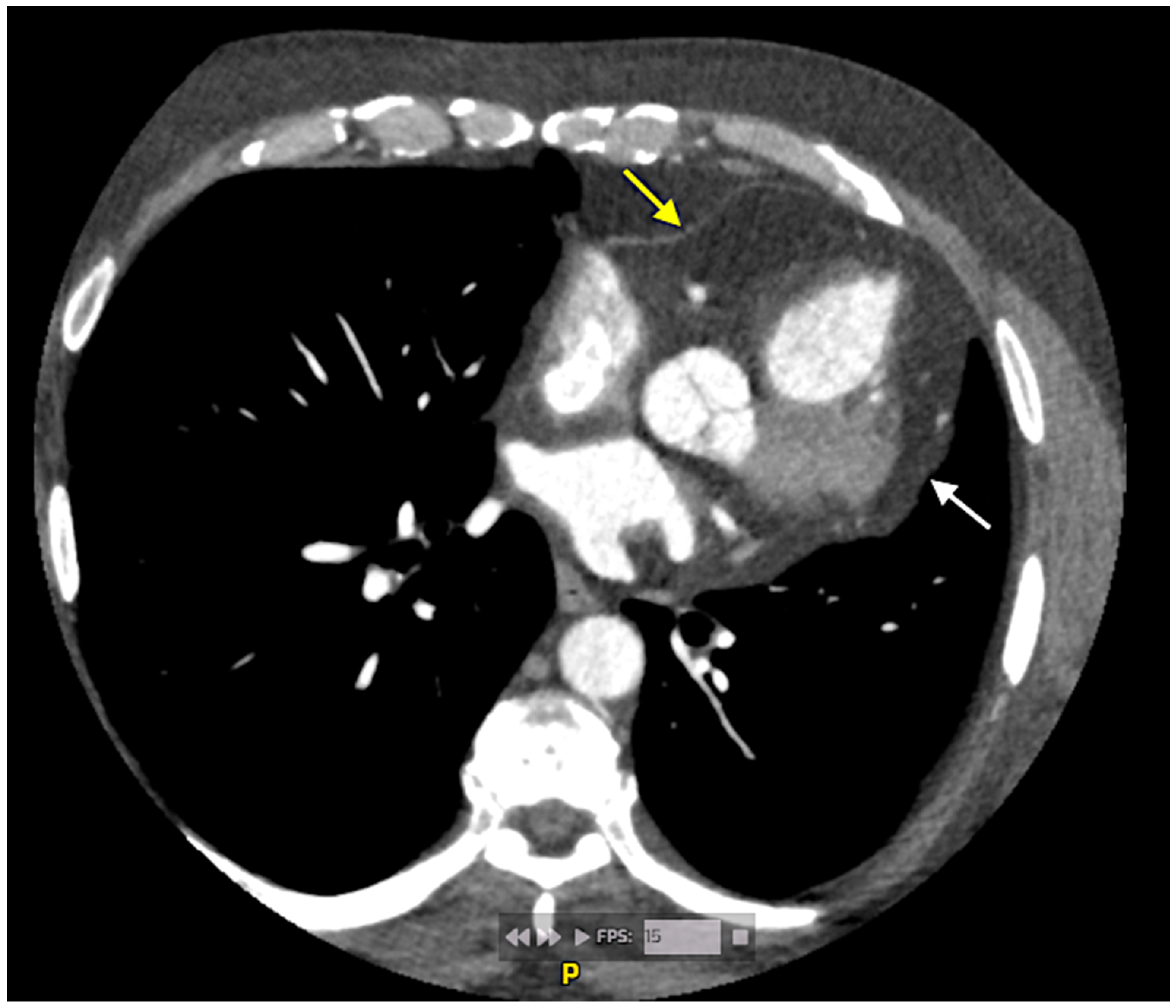
This investigation demonstrated partial congenital absence of the left pericardium. Imaging revealed marked leftward displacement and clockwise rotation of the heart, with the apex directed superiorly. Characteristic findings included interposition of lung parenchyma between the great vessels and beneath the heart, as well as prominent lobulated paracardiac fat along the left cardiac border. The right ventricle was positioned parallel to the sternum without evidence of dilatation.
Cardiac chambers, valvular structures, and ventricular systolic function were preserved, and no evidence of myocardial ischemia, fibrosis, or pericardial effusion was identified (Figure 9 and Figure 10).
Based on the absence of high-risk features such as herniation or myocardial compromise, a conservative management approach was adopted. The patient was advised to undergo regular clinical and imaging follow-up, with particular attention to the potential development of symptoms suggestive of mechanical complications. He remained asymptomatic throughout the 6-month follow-up period.
Unlike the preceding cases, this patient presented with a partial pericardial defect, a subtype associated with a greater risk of mechanical complications, highlighting the importance of accurate anatomical characterization and individualized follow-up.

3.2. Summary of Findings

Three of our patients had complete left pericardial agenesis, while one patient demonstrated a partial defect. Across all four cases, CPA was confirmed by multimodality imaging. The most consistent findings included:
  • Leftward displacement (levoposition) of the heart;
  • Clockwise cardiac rotation;
  • Interposition of lung parenchyma between mediastinal structures;
  • Absence of pericardial tissue, predominantly on the left side.
Clinical presentation was variable and nonspecific, ranging from asymptomatic cases to mild symptoms such as chest discomfort or dyspnea. Importantly, CPA mimicked other cardiac conditions on TTE in several cases, including cardiomyopathy, atrial septal defect, and pericardial effusion. ECG findings were nonspecific, typically showing sinus rhythm or tachycardia with axis deviation and minor conduction abnormalities (incomplete RBBB or left anterior fascicular block), without ischemic changes or significant arrhythmias.
None of the patients developed complications or required surgical intervention during the observation period. Table 1 summarizes the clinical and imaging characteristics of the presented clinical cases.

4. Discussion

Congenital pericardial agenesis is a rare developmental anomaly with a wide clinical spectrum, ranging from completely asymptomatic incidental findings to presentations mimicking structural or ischemic heart disease [5,12,18]. The present case series illustrates this variability and provides insight into the ongoing debate of whether CPA represents a benign anatomical variant or a clinically relevant condition.
In our cohort, all four patients demonstrated typical imaging features of left-sided pericardial absence, including cardiac levoposition, clockwise rotation, and interposition of lung parenchyma between mediastinal structures. Despite heterogeneous clinical presentations—ranging from exertional dyspnea and chest discomfort to incidental findings—none of the patients exhibited hemodynamic compromise or required surgical intervention. These observations are consistent with previous reports suggesting that complete pericardial agenesis is most often a benign condition, particularly when not associated with structural abnormalities [5,19,20].
The embryological basis of CPA is attributed to premature atrophy of the common cardinal veins (ducts of Cuvier), leading to defective formation and fusion of the pleuropericardial membranes during early gestation [2]. This results in absence of the pericardial sac and loss of its stabilizing function, allowing excessive cardiac mobility and displacement into the left hemithorax [12,14]. The predominance of left-sided defects is explained by the earlier regression of the left duct of Cuvier, impairing vascular supply to the developing pericardial membrane [5,17,21].
A key finding of our series is the high rate of initial misdiagnosis or diagnostic uncertainty, particularly on TTE. Importantly, three of the four patients in our series were initially suspected of having alternative structural cardiac conditions, including right ventricular cardiomyopathy, atrial septal defect, or pericardial disease. This observation is consistent with previous reports describing CPA as a frequent diagnostic mimic due to altered cardiac orientation, atypical echocardiographic windows, and apparent right ventricular dilatation [5,22,23]. Our findings therefore reinforce the importance of considering CPA in the differential diagnosis of unexplained right ventricular dilatation, abnormal cardiac rotation, or inconclusive echocardiographic findings, particularly when imaging findings appear discordant with the clinical presentation.
Multimodality imaging plays a central role in the diagnosis [5,20,24]. While chest radiography and ECG findings are nonspecific, cardiac CT and especially CMR provide definitive anatomical characterization [5,20,24]. The most reliable imaging features include marked leftward displacement of the heart, absence of the pericardial lining, and interposition of lung tissue between the ascending aorta and pulmonary artery—a hallmark diagnostic feature of CPA characterization [5,20,24]. In our series, CMR was particularly valuable in resolving diagnostic ambiguity and confirming normal cardiac morphology and function. These observations align with current evidence identifying CMR as the gold standard for non-invasive diagnosis [5,14,16].
The clinical relevance of CPA largely depends on the type of defect [5,12,23]. Complete pericardial agenesis, as observed in three of our patients, is generally well tolerated and rarely associated with complications. In contrast, partial defects carry a significantly higher risk, due to the presence of residual pericardial rims that may entrap cardiac structures [5,11,23]. Complications such as cardiac herniation, strangulation of the left atrial appendage or ventricular free wall, coronary artery compression, myocardial ischemia, and even sudden cardiac death have been reported [5,7,23].
Partial congenital pericardial defects deserve particular clinical attention because, unlike complete agenesis, residual fibrous pericardial rims may create sites of mechanical constriction [11,23]. This anatomical configuration predisposes to cardiac herniation and strangulation of mobile cardiac structures, most commonly the left atrial appendage or ventricular free wall [12,19,23]. Several reports have described severe complications including coronary artery compression, myocardial ischemia, ventricular incarceration, and sudden cardiac death associated predominantly with partial defects [4,13,23]. The risk appears to be highest in small or moderate-sized defects where incomplete pericardial margins may entrap protruding myocardial tissue during cardiac motion [16,23,25]. Consequently, symptomatic patients with partial CPA, evidence of cardiac hypermobility, imaging signs of focal myocardial constriction, or suspected coronary compression may warrant surgical evaluation [4,17,23]. Surgical approaches typically include pericardioplasty, patch enlargement of the defect, or defect closure, depending on anatomical characteristics and surgical expertise. In contrast, asymptomatic complete left-sided agenesis generally carries a substantially more benign prognosis and is usually managed conservatively [11,15,25].
Our fourth case is particularly illustrative in this regard. The patient presented with chest discomfort and was found to have a partial pericardial defect. Although no complications were observed, the symptomatic presentation highlights that CPA cannot always be considered an innocent finding, especially in the presence of partial defects. This reinforces the need for careful evaluation and follow-up in such patients.
Management strategies remain individualized due to the rarity of the condition and lack of large-scale studies [5,14,23]. Asymptomatic patients with complete pericardial agenesis generally require no specific treatment beyond periodic monitoring [5,14,23]. In contrast, symptomatic patients or those with partial defects may require closer surveillance or surgical intervention. Surgical options, including pericardioplasty or defect closure, are typically reserved for patients with significant symptoms or evidence of mechanical complications [5,7,14,23].
An important clinical implication of our study is that CPA should be actively considered in the differential diagnosis of unexplained cardiac displacement, suspected right ventricular pathology, or inconclusive echocardiographic findings. Failure to recognize this entity may lead to unnecessary diagnostic procedures, misdiagnosis, or inappropriate treatment.
Taken together, our findings support the concept that CPA represents a spectrum rather than a uniform entity. While complete forms are predominantly benign, partial defects may carry clinically relevant risks. Therefore, the question posed in the title—whether CPA is an innocent finding or a real clinical problem—does not have a binary answer. Instead, the clinical significance depends on the anatomical subtype, symptomatology, and presence of complications.
To place our findings into clinical context, we summarized representative published case reports and case series of congenital pericardial agenesis. The literature demonstrates substantial heterogeneity in clinical presentation, imaging findings, and management strategies, particularly between complete and partial defects (Table 2a,b and Table 3).
This representative overview highlights the heterogeneous clinical spectrum of congenital pericardial agenesis, ranging from incidental complete left-sided defects with benign outcomes to partial defects associated with mechanical complications and potential need for surgical intervention. Across reports, multimodality imaging—particularly CT and CMR—was essential for confirming the diagnosis, defining the defect type, and guiding management.
The key imaging characteristics that support the diagnosis of CPA across modalities are summarized in Table 4.
Given the absence of dedicated society guidelines for congenital pericardial agenesis, surveillance strategies remain largely individualized. Based on our case series and the available literature, we propose a pragmatic follow-up approach stratified according to defect type, symptom burden, and presence of high-risk features. This proposal is intended as a practical clinical framework rather than a formal guideline recommendation (Table 5).
Patients with complete asymptomatic defects generally have a favorable prognosis and may require only periodic surveillance. In contrast, partial defects warrant closer follow-up due to the potential risk of cardiac herniation, coronary compression, or ischemic complications.

Limitations

The present study has several limitations. First, the literature review component was designed to provide clinical context for the presented cases rather than to constitute a formal systematic review. Consequently, no PRISMA-based methodology or formal quality assessment was applied. Second, the currently available evidence on congenital pericardial agenesis is inherently limited and predominantly based on isolated case reports and small case series, making the evidence base relatively heterogeneous and vulnerable to publication bias. Third, the retrospective nature of our case series and the limited number of patients restrict the generalizability of our observations. Finally, due to the retrospective nature of the study and the fact that several CMR examinations were performed as part of routine clinical practice at different institutions, detailed sequence-level protocol information was not uniformly available for all patients. Nevertheless, all CMR studies included comprehensive anatomical and functional assessment sufficient to establish the diagnosis and exclude major structural abnormalities. Importantly, this heterogeneity also reflects real-world clinical imaging practice in patients with rare congenital pericardial defects, where diagnostic evaluation is frequently individualized according to local expertise and clinical presentation.

5. Conclusions

In conclusion, congenital pericardial agenesis represents a heterogeneous clinical entity ranging from incidental benign findings to clinically significant defects associated with mechanical complications. Complete left-sided agenesis is generally associated with a favorable prognosis and conservative management, whereas partial defects warrant closer surveillance because of their potential association with cardiac herniation, strangulation, and ischemic complications. Multimodality imaging, particularly CMR, plays a pivotal role not only in establishing the diagnosis but also in risk stratification and longitudinal follow-up. An individualized approach integrating defect type, symptom burden, and imaging findings remains essential for optimal clinical decision-making.

Author Contributions

Conceptualization, V.G. and S.N.; methodology, V.G., M.R., A.J. and S.N.; validation, V.G. and S.N.; formal analysis, S.N. and M.R.; investigation, V.G., M.R., A.J. and S.N.; resources, V.G., M.R., A.J. and S.N.; data curation, V.G. and S.N.; writing—original draft preparation, M.R. and A.J.; writing, S.N.; visualization, V.G., A.J. and S.N.; supervision, S.N.; project administration, V.G. All authors have read and agreed to the published version of the manuscript.

Funding

This research received no external funding.

Institutional Review Board Statement

The study was conducted in accordance with the Declaration of Helsinki. Ethical review and approval were waived due to the retrospective and non-interventional nature of our research and the use of fully anonymized clinical data, in accordance with applicable national regulations and institutional policies.

Informed Consent Statement

Patient consent was waived due to the retrospective and non-interventional nature of this case series and the use of fully anonymized clinical data, in accordance with applicable national regulations and institutional policies.

Data Availability Statement

The data supporting the results of this research are available from Stefan Naydenov (snaydenov@gmail.com) upon reasonable request, subject to applicable ethical and privacy restrictions.

Conflicts of Interest

The authors declare no conflicts of interest.

Abbreviations

The following abbreviations are used in this manuscript:
AoAortic
CMRCardiac magnetic resonance
CPACongenital pericardial agenesis
CTComputed tomography
ECGElectrocardiography
LALeft atrium
LVLeft ventricle
RARight atrium
RVRight ventricle
TTETransthoracic echocardiography

References

  1. Omole, A.E.; Nassereddin, A.; Launico, M.V. Anatomy, Thorax, Pericardium; StatPearls Publishing: Treasure Island, FL, USA, 2026. Available online: https://www.ncbi.nlm.nih.gov/books/NBK482256/ (accessed on 4 May 2026).
  2. Tubbs, O.S.; Yacoub, M.H. Congenital pericardial defects. Thorax 1968, 23, 598–607. [Google Scholar] [CrossRef] [PubMed]
  3. Weerakkody, Y.; Silverstone, L.; Campos, A. Pericardial Agenesis. Radiopaedia.org. 2013. Available online: https://radiopaedia.org/articles/22931 (accessed on 25 April 2026).
  4. Iglesias, P.C.; Pascual, E.A.; De La Torre, L.A.; Robinot, D.C.; Vázquez, B.T.; Ruiz, M.A.G. Pericardial agenesis. Ann. Pediatr. Cardiol. 2021, 14, 119–121. [Google Scholar] [CrossRef]
  5. Shah, A.B.; Kronzon, I. Congenital defects of the pericardium: A review. Eur. Heart J. Cardiovasc. Imaging 2015, 16, 821–827. [Google Scholar] [CrossRef]
  6. Trimarchi, G.; Zito, C.; Pelaggi, G.; Carerj, S.; Di Bella, G. Pericardial agenesis: A case report of a rare congenital heart disease. Eur. Heart J. Case Rep. 2024, 8, ytae200. [Google Scholar] [CrossRef]
  7. Bouchard, M.; Hoschtitzky, A.; Gatzoulis, M. Diagnosis and management of congenital absence of pericardium: A case report. Eur. Heart J. Case Rep. 2019, 3, 1–5. [Google Scholar] [CrossRef] [PubMed]
  8. Bernardinello, V.; Cipriani, A.; Perazzolo Marra, M.; Motta, R.; Barchitta, A. Congenital Pericardial Agenesis in Asymptomatic Individuals: Tips for the Diagnosis. Circ. Cardiovasc. Imaging 2020, 13, e010169. [Google Scholar] [CrossRef] [PubMed]
  9. Marzullo, R.; Capestro, A.; Cosimo, R.; Fogante, M.; Aprile, A.; Balardi, L.; Giordano, M.; Gaio, G.; Gauderi, G.; Russo, M.G.; et al. Congenital Absence of Pericardium: The Swinging Heart. J. Imaging 2024, 10, 199. [Google Scholar] [CrossRef]
  10. Gatzoulis, M.A.; Munk, M.-D.; Merchant, N.; Van Arsdell, G.S.; McCrindle, B.W.; Webb, G.D. Isolated congenital absence of the pericardium: Clinical presentation, diagnosis, and management. Ann. Thorac. Surg. 2000, 69, 1209–1215. [Google Scholar] [CrossRef]
  11. Khayata, M.; Alkharabsheh, S.; Shah, N.P.; Verma, B.R.; Gentry, J.L.; Summers, M.; Xu, B.; Asher, C.; Klein, A.L. Case series, contemporary review and imaging guided diagnostic and management approach of congenital pericardial defects. Open Heart 2020, 7, e001103. [Google Scholar] [CrossRef]
  12. Drury, N.E.; De Silva, R.J.; Hall, R.M.O.; Large, S.R. Congenital Defects of the Pericardium. Ann. Thorac. Surg. 2007, 83, 1552–1553. [Google Scholar] [CrossRef] [PubMed]
  13. Jafari, F.; Taheri, M.; Ebrahimi, P.; Soflaee, M.; Rafie, R.A.; Anafje, M. Congenital unilateral pericardial agenesis presenting as an isolated chest pain in an adolescent: A case report and comprehensive review. J. Cardiothorac. Surg. 2025, 20, 127. [Google Scholar] [CrossRef] [PubMed]
  14. Xu, B.; Betancor, J.; Asher, C.; Rosario, A.; Klein, A. Congenital Absence of the Pericardium: A Systematic Approach to Diagnosis and Management. Cardiology 2017, 136, 270–278. [Google Scholar] [CrossRef]
  15. Imazio, M.; Collini, V.; Aimo, A.; Autore, C.; Bauce, B.; Biagini, E.; Cappelli, F.; Castelletti, S.; D’Ascenzi, F.; De Gregorio, C.; et al. Update on the diagnosis and treatment of pericardial diseases: A position paper of the Italian Society of Cardiology in collaboration with the study group on cardiomyopathies and pericardial diseases. J. Cardiovasc. Med. 2025, 26, 29–37. [Google Scholar] [CrossRef] [PubMed]
  16. Gupta, M.; Butler, T.; Appaji, A.; Kwok, C.S. Pericardial Agenesis: The Significance of Multimodality Imaging in Diagnosis. Cureus 2025, 17, e82718. [Google Scholar] [CrossRef]
  17. Kalaydzhiev, P.; Partenova, A.; Ilieva, R.; Genova, K.; Kinova, E. Complete Left-Sided Pericardial Congenital Absence. Reports 2024, 7, 48. [Google Scholar] [CrossRef] [PubMed]
  18. D’Arma, G.M.A.; Chieppa, D.R.R.; Forte, V.; Masino, F.; Bartolomucci, F.; Guglielmi, G. Complete agenesis of pericardium in a young asymptomatic woman. Radiol. Case Rep. 2024, 19, 3062–3065. [Google Scholar] [CrossRef]
  19. Klein, A.L.; Wang, T.K.M.; Cremer, P.C.; Abbate, A.; Adler, Y.; Asher, C.; Brucato, A.; Chetrit, M.; Hoit, B.; Jellis, C.L.; et al. Pericardial Diseases. JACC Cardiovasc. Imaging 2024, 17, 937–988. [Google Scholar] [CrossRef]
  20. Oryshchyn, N.; Ivaniv, Y.; Yevtukh, V.; Oryshchyn, A. Multimodality cardiovascular imaging in the complete congenital absence of the pericardium: Case report and brief literature review. Heart Vessel. Transplant. 2025, 9, 577. [Google Scholar] [CrossRef]
  21. Mekonnen, S.; Farris, H.; Azmeraw, D. Complete Congenital Absence of the Left Pericardium in Elderly Patient: A Case Report. Int. Med. Case Rep. J. 2024, 17, 347–352. [Google Scholar] [CrossRef]
  22. Gupta, S.; Kumar, D. Complete Congenital Absence of Left Pericardium: A Case Report. Cureus 2025, 17, e92125. [Google Scholar] [CrossRef]
  23. Bassareo, P.P.; Secinaro, A.; Ciliberti, P.; Chessa, M.; Perrone, M.A.; Walsh, K.P.; Mcmahon, C.J. Congenital Absence of Pericardium: The Largest Systematic Review in the Field on 247 Worldwide Cases (1977-Now). Congenit. Heart Dis. 2023, 18, 595–610. [Google Scholar] [CrossRef]
  24. Koo, C.W.; Newburg, A. Congenital Absence of the Right Pericardium: Embryology and Imaging. J. Clin. Imaging Sci. 2015, 5, 12. [Google Scholar] [CrossRef]
  25. Scheuermann-Freestone, M.; Orchard, E.; Francis, J.; Petersen, M.; Friedrich, M.; Rashid, A.; Shore, D.; Myerson, S.; Neubauer, S. Partial Congenital Absence of the Pericardium. Circulation 2007, 116, e126–e129. [Google Scholar] [CrossRef]
  26. Nasser, W.K.; Helmen, C.; Tavel, M.E.; Feigenbaum, H.; Fisch, C. Congenital Absence of the Left Pericardium: Clinical, Electrocardiographic, Radiographic, Hemodynamic, and Angiographic Findings in Six Cases. Circulation 1970, 41, 469–478. [Google Scholar] [CrossRef] [PubMed]
  27. Van Son, J.A.M.; Danielson, G.K.; Schaff, H.V.; Mullany, C.J.; Julsrud, P.R.; Breen, J.F. Congenital Partial and Complete Absence of the Pericardium. Mayo Clin. Proc. 1993, 68, 743–747. [Google Scholar] [CrossRef] [PubMed]
  28. Connolly, H.M.; Click, R.L.; Schattenberg, T.T.; Seward, J.B.; Tajik, A.J. Congenital absence of the pericardium: Echocardiography as a diagnostic tool. J. Am. Soc. Echocardiogr. 1995, 8, 87–92. [Google Scholar] [CrossRef]
  29. Abbas, A.E.; Appleton, C.P.; Liu, P.T.; Sweeney, J.P. Congenital absence of the pericardium: Case presentation and review of literature. Int. J. Cardiol. 2005, 98, 21–25. [Google Scholar] [CrossRef] [PubMed]
  30. Brulotte, S.; Roy, L.; Larose, E. Congenital absence of the pericardium presenting as acute myocardial necrosis. Can. J. Cardiol. 2007, 23, 909–912. [Google Scholar] [CrossRef]
  31. Garnier, F.; Eicher, J.; Philip, J.; Lalande, A.; Bieber, H.; Voute, M.; Brenot, R.; Brunotte, F.; Wolf, J. Congenital Complete Absence of the Left Pericardium: A Rare Cause of Chest Pain or Pseudo-right Heart Overload. Clin. Cardiol. 2010, 33, E52–E57. [Google Scholar] [CrossRef]
  32. Wilson, S.R.; Kronzon, I.; Machnicki, S.C.; Ruiz, C.E. A Constrained Heart: A Case of Sudden Onset Unrelenting Chest Pain. Circulation 2014, 130, 1625–1631. [Google Scholar] [CrossRef]
  33. Kalekar, T.; Reddy, L.P.; Koganti, D.; Soman, N. Pericardial agenesis–The wandering heart. Egypt. Heart J. 2023, 75, 79. [Google Scholar] [CrossRef] [PubMed]
Figure 1. ECG of Clinical Case 1: Sinus rhythm (heart rate 75 bpm) with right axis deviation (+120°). Low QRS voltage in the precordial leads with leftward displacement of the R/S transition zone. Incomplete right bundle branch block pattern. Right ventricular hypertrophy cannot be excluded.
Jcm 15 04394 g001
Figure 2. (a) TTE of Clinical Case 1: The parasternal long-axis view is suboptimal due to marked cardiac levoposition with interposition of aerated lung tissue, precluding adequate visualization and reliable assessment of cardiac structures and function from this window; Ao—aortic; LA—left atrium; LV—left ventricle; RV—right ventricle. (b) TTE of Clinical Case 1: An apical five-chamber view was the only view obtainable from the apical window and was of suboptimal quality despite maximal left lateral positioning of the transducer. Standard apical four-, two-, and three-chamber views could not be acquired. Ao—aortic; LA—left atrium; LV—left ventricle; RA—right atrium; RV—right ventricle. (c) TTE of Clinical Case 1: The subcostal view provided satisfactory image quality and most closely approximated standard imaging planes; LA—left atrium; LV—left ventricle; RV—right ventricle.
Jcm 15 04394 g002aJcm 15 04394 g002b
Figure 3. Cardiac MRI in the coronal plane with a large field of view, demonstrating abnormal leftward displacement of the cardiac silhouette within the left hemithorax (yellow arrow).
Jcm 15 04394 g003
Figure 4. Cardiac magnetic resonance imaging (MRI) in the four-chamber cine view, confirming abnormal cardiac position with pronounced levorotation (yellow arrow). The inferior pulmonary vein is compressed between the descending aorta and the left atrium, likely as a consequence of the altered cardiac orientation (white arrow).
Jcm 15 04394 g004
Figure 5. CT of the chest in the coronal plane using a lung window, demonstrating interposition of lung parenchyma between the left hemidiaphragm and the cardiac base (yellow arrow).
Jcm 15 04394 g005
Figure 6. Contrast-enhanced CT of the heart in the coronal plane, demonstrating interposition of lung parenchyma between the aorta and the pulmonary artery (yellow arrow). A prominent left atrial appendage is also noted (white arrow).
Jcm 15 04394 g006
Figure 7. Cardiac MRI in the four-chamber cine view, demonstrating the pericardium as a thin linear structure anterior to the right ventricle, interposed between epicardial and mediastinal fat tissue (yellow arrow). The pericardium is not visualized posterior to the left-sided cardiac chambers (white arrow).
Jcm 15 04394 g007
Figure 8. Cardiac MRI in the axial plane, demonstrating leftward displacement of the heart with rotation of the cardiac apex (yellow arrow).
Jcm 15 04394 g008
Figure 9. Non-contrast CT of the chest, demonstrating leftward displacement of the heart with associated rotation (yellow arrow).
Jcm 15 04394 g009
Figure 10. ECG-gated CT in the axial plane, demonstrating partial visualization of the pericardium anterior to the right-sided cardiac chambers (yellow arrow), with absence of the pericardium along the left side (white arrow).
Jcm 15 04394 g010
Table 1. Summary of clinical and imaging characteristics.
CaseAge/SexPresentationInitial SuspicionImaging ModalityType of CPAKey Imaging FindingsManagementFollow-Up
154/MDyspnea, fatigueNo definite diagnosisCMRCompleteLevoposition, lung interposition, normal functionConservative12 months
222/MChest painASD, RV dilation, effusionCTCompleteCardiac rotation, absent pericardium, lung interpositionConservative24 months
329/FIncidental cardiomegalyRV cardiomyopathyCMRCompleteLeft displacement, normal functionConservative18 months
462/MMild chest discomfortNo definite diagnosisCTPartialLung interposition, partial defectConservative6 months
Table 2. (a) Representative published case reports and case series of congenital pericardial agenesis. (b) Representative published case reports and case series of congenital pericardial agenesis (continued).
(a)
Author/YearNo. of PatientsType of DefectClinical PresentationMain Imaging ModalityComplicationsManagementOutcome
Nasser et al., 1970 [26].6Complete left-sidedChest pain, abnormal ECG/radiographic findingsECG, chest X-ray, angiographyNo major complications reportedConservativeFavorable
Van Son et al., 1993 [27].Case seriesPartial and completeVariable; symptomatic and incidental casesCT/MRI/surgical findingsHerniation risk in partial defectsSurgical or conservative depending on anatomyGenerally favorable
Connolly et al., 1995 [28].10Congenital absence of pericardiumVariable; abnormal echocardiographic findingsEchocardiography, CT/MRICardiac hypermobility, abnormal septal motionMostly conservativeDiagnosis clarified by imaging
Gatzoulis et al., 2000 [10].Case seriesIsolated congenital absenceChest pain or incidental findingCT/MRI/echocardiographyRisk mainly in partial defectsIndividualized; surgery in selected casesGood prognosis in complete defects
Abbas et al., 2005 [29].1 + reviewCongenital absenceChest pain/incidental imaging findingEchocardiography, CT/MRINone in index caseConservativeStable
Scheuermann-Freestone et al., 2007 [25].1Partial left-sidedChest painCMRPotential ventricular/appendage entrapmentConservative/surgical considerationSymptom-guided
Brulotte et al., 2007 [30].1Complete left-sidedAcute chest pain with myocardial necrosisCMRMyocardial injuryConservative after exclusion of other causesStable
(b)
Author/YearNo. of PatientsType of DefectClinical PresentationMain Imaging ModalityComplicationsManagementOutcome
Garnier et al., 2010 [31].3Complete left-sidedChest pain or pseudo-right-heart overloadEchocardiography, CT/CMRNo major complicationsConservativeFavorable
Wilson et al., 2014 [32].1Partial defectSudden severe chest painMultimodality imagingConstrained cardiac motion/mechanical complicationSurgical evaluation/interventionSymptom-focused management
Xu et al., 2017 [14].1 + management reviewComplete/partial frameworkDiagnostic uncertainty, symptoms or incidental findingEcho, CT, CMRRisk stratified by defect typeProposed systematic diagnostic/management approachPractical management framework
Bouchard et al., 2019 [7].1Complete absenceSymptomatic, marked cardiac mobilityCMR/CTGross cardiac mobilityPericardial reconstructionSymptom improvement
Khayata et al., 2020 [11].8Partial and completeIncidental, chest pain, dyspnea, abnormal TTEEcho, CT, CMROne patient required surgeryImaging-guided conservative or surgical managementMost stable during follow-up
Kalekar et al., 2023 [33].1Pericardial agenesis“Wandering heart”; diagnostic uncertaintyCMRCardiac hypermobilityConservativeStable
Marzullo et al., 2024 [9].1Complete absenceIncidental/“swinging heart”CMRNo major complicationConservativeFavorable
Table 3. Complete versus partial CPA: clinical implications.
FeatureComplete CPAPartial CPA
SymptomsOften asymptomaticMore frequently symptomatic
Main riskUsually benignHerniation/strangulation
Imaging concernLevopositionFocal constriction
TreatmentConservativeSometimes surgical
Follow-upPeriodicCloser surveillance
Table 4. Key imaging features of congenital pericardial agenesis.
Imaging ModalityKey Findings in CPADiagnostic ValueLimitations
Chest X-rayLeftward cardiac displacement, elongated left heart border (“Snoopy sign”).Initial suspicionNonspecific
ECGAxis deviation, incomplete RBBB, poor R-wave progression.Supportive onlyLow specificity
EchocardiographyApparent RV enlargement, abnormal cardiac orientation, unusual acoustic windows.Raises suspicionFrequently misleading
Cardiac CTAbsence of pericardium, lung interposition, cardiac levoposition, anatomy of coronaries.High spatial resolutionRadiation exposure
CMR (Gold standard)Direct visualization of absent pericardium, lung interposition, cardiac mobility, preserved function.Definitive diagnosisLimited availability
Table 5. Suggested clinical follow-up approach in congenital pericardial agenesis.
Clinical ScenarioSuggested Follow-Up
Asymptomatic complete CPAClinical review + ECG/TTE every 1–2 years
Symptomatic complete CPAPeriodic CMR/CT + symptom reassessment
Partial CPA without complicationsAnnual clinical and imaging follow-up
Partial CPA with suspected herniation/compressionSurgical evaluation
Arrhythmias or ischemic symptomsHolter + ischemia assessment
Disclaimer/Publisher’s Note: The statements, opinions and data contained in all publications are solely those of the individual author(s) and contributor(s) and not of MDPI and/or the editor(s). MDPI and/or the editor(s) disclaim responsibility for any injury to people or property resulting from any ideas, methods, instructions or products referred to in the content.