Hyaluronic Acid vs. PRP, PRF, and Collagen in Regenerative Endodontics: A Systematic Review

 , and
, and 
Abstract
1. Introduction
2. Aim
3. Objectives
4. Materials and Methods
4.1. Study Design
- P (population): patients with immature necrotic permanent teeth.
- I (intervention): endodontic regeneration with hyaluronic acid scaffold.
- C (comparison): endodontic regeneration with PRP, PRF, and collagen scaffolds.
- O (outcome): endodontic treatment outcomes: Root development, Apical closure, Periapical healing.
4.2. Eligibility Criteria
4.3. Search Strategy and Study Selection
4.4. Data Extraction
- Study characteristics: author, year of publication, country, study design.
- Sample characteristics: age, sample size, number of teeth treated, and tooth type.
- Intervention characteristics: scaffold material used and disinfection protocol (irrigant).
- Follow-up characteristics: follow-up duration.
- Outcome measures: root length increase, dentinal wall thickening, apical closure, periapical healing, return of vitality, and clinical success.
- Radiographic assessment details: imaging method, radiographic standardisation protocol, and measurement software/tool used.
- Methodological characteristics: blinding procedures, statistical tests used, and reported significance levels.
4.5. Data Synthesis
4.6. Quality and Risk of Bias Assessment
5. Results
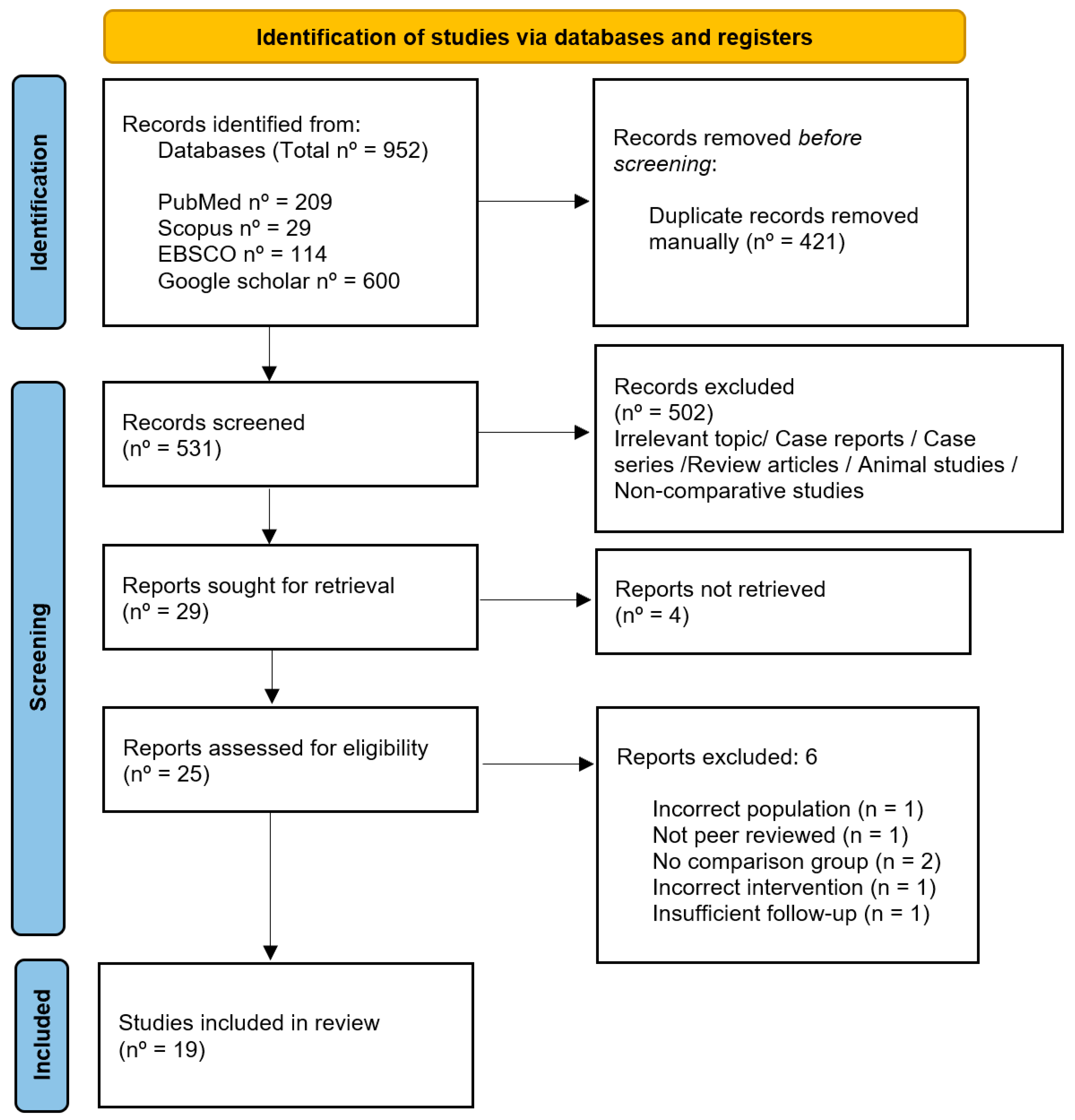
5.1. Selection of Studies Using PRISMA Flow Chart
5.2. Characteristics of Included Studies
5.3. Measurement Characteristics of Included Studies
5.4. Clinical and Radiographic Outcome Assessment
5.4.1. Root Length Increase
5.4.2. Dentinal Wall Thickening
5.4.3. Apical Closure
5.4.4. Periapical Healing
5.4.5. Return of Vitality
5.4.6. Clinical Success
5.5. Outcome Summary
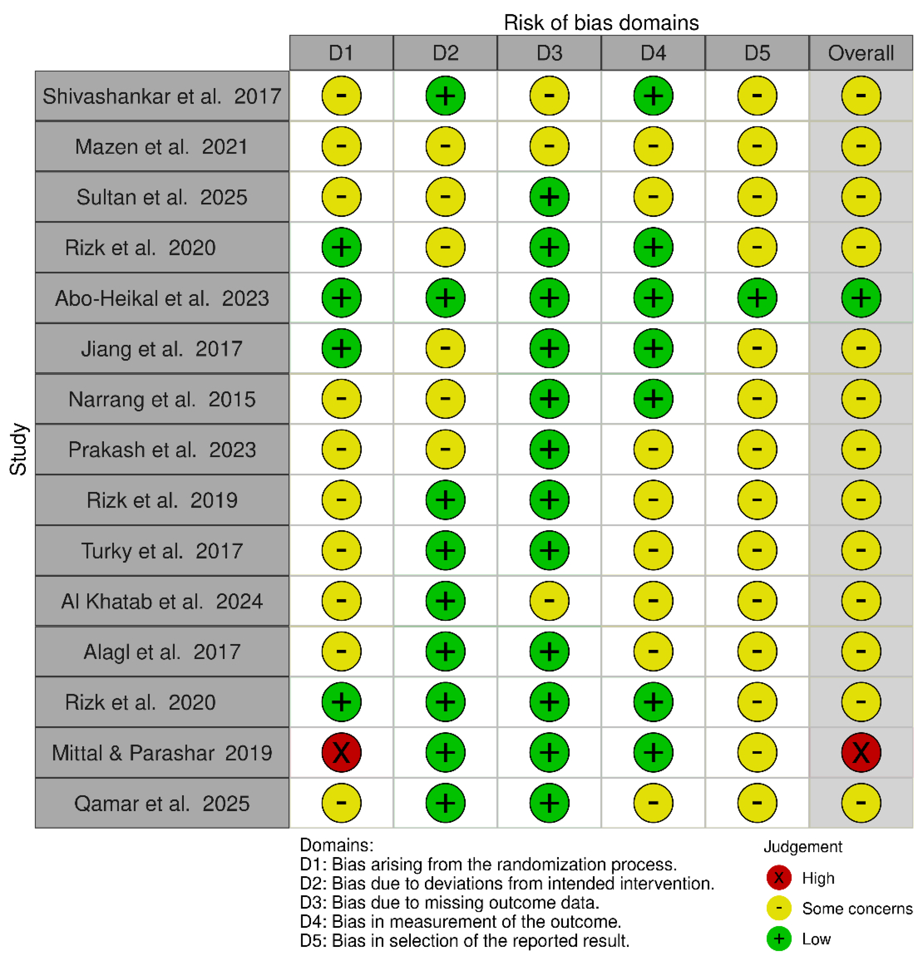
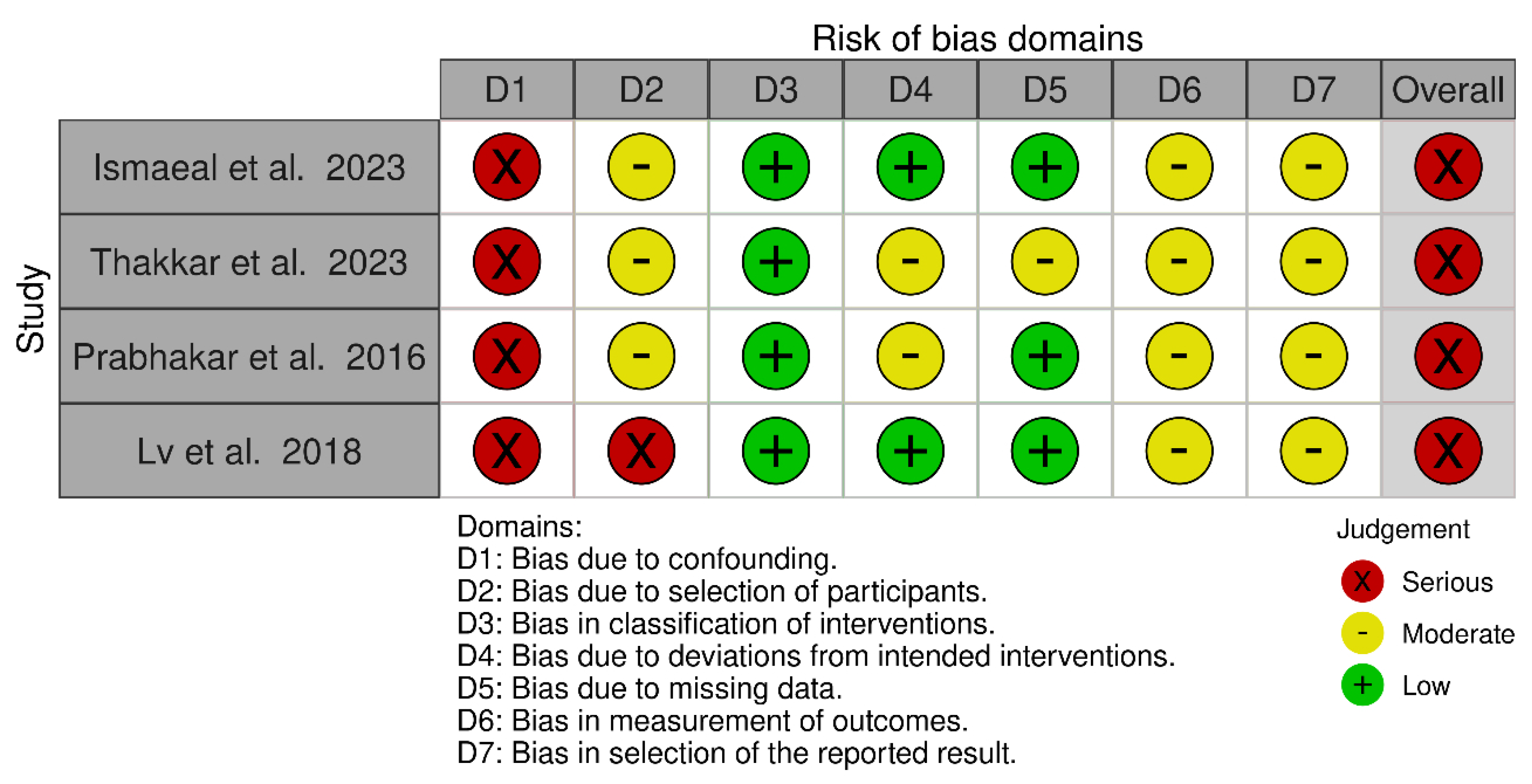
5.6. Risk of Bias Assessment
6. Discussion
6.1. Root Development Outcomes
6.2. Apical Closure and Periapical Healing Outcomes
6.3. Clinical Outcomes
6.4. Consistency and Statistical Significance
6.5. Limitations
6.6. Future Research
7. Conclusions
Supplementary Materials
Author Contributions
Funding
Institutional Review Board Statement
Informed Consent Statement
Data Availability Statement
Conflicts of Interest
References
- Abdulwahab, M.A.; Alqahtani, M.S.; Alshammari, A.A.; Jiffri, S.E.; Alasim, A.M.; Alsharidah, F.M.; Alqunfuthi, N.I.; Alnamnakani, B.A.; Alshahrani, R.S.; Alshahrani, N.A.; et al. Etiologies, risk factors and outcomes of dental pulp necrosis. Int. J. Community Med. Public Health 2021, 9, 348. [Google Scholar] [CrossRef] [Scilit]
- Flanagan, T.A. What can cause the pulps of immature, permanent teeth with open apices to become necrotic and what treatment options are available for these teeth. Aust. Endod. J. 2014, 40, 95–100. [Google Scholar] [CrossRef] [Scilit]
- Harlamb, S.C. Management of incompletely developed teeth requiring root canal treatment. Aust. Dent. J. 2016, 61, 95–106. [Google Scholar] [CrossRef] [Scilit]
- Levin, L.; Day, P.F.; Hicks, L.; O’COnnell, A.; Fouad, A.F.; Bourguignon, C.; Abbott, P.V. International Association of Dental Traumatology guidelines for the management of traumatic dental injuries: General introduction. Dent. Traumatol. 2020, 36, 309–313. [Google Scholar] [CrossRef] [Scilit]
- Koç, S.; Del Fabbro, M. Does the Etiology of Pulp Necrosis Affect Regenerative Endodontic Treatment Outcomes? A Systematic Review and Meta-analyses. J. Evid. Based Dent. 2020, 20, 101400. [Google Scholar] [CrossRef] [Scilit]
- Shabahang, S. Treatment options: Apexogenesis and apexification. Pediatr. Dent. 2013, 35, 125–128. [Google Scholar] [CrossRef] [Scilit]
- Murray, P.E.; Garcia-Godoy, F.; Hargreaves, K.M. Regenerative Endodontics: A Review of Current Status and a Call for Action. J. Endod. 2007, 33, 377–390. [Google Scholar] [CrossRef] [Scilit]
- Etezadkeyhan, P. Recent Advances in Regenerative Endodontics: Clinical Applications and Challenges. J. Oral Dent. Health Nexus 2024, 1, 29–42. [Google Scholar] [CrossRef] [Scilit]
- Hargreaves, K.M.; Diogenes, A.; Teixeira, F.B. Treatment options: Biological basis of regenerative endodontic procedures. J. Endod. 2013, 39, S30–S43. [Google Scholar] [CrossRef] [Scilit] [PubMed]
- Tong, H.J.; Rajan, S.; Bhujel, N.; Kang, J.; Duggal, M.; Nazzal, H. Regenerative Endodontic Therapy in the Management of Nonvital Immature Permanent Teeth: A Systematic Review—Outcome Evaluation and Meta-analysis. J. Endod. 2017, 43, 1453–1464. [Google Scholar] [CrossRef] [Scilit] [PubMed]
- American Association of Endodontists. AAE Clinical Considerations for a Regenerative Procedure; AAE: Chicago, IL, USA, 2021. [Google Scholar]
- Yan, H.; De Deus, G.; Kristoffersen, I.M.; Wiig, E.; Reseland, J.E.; Johnsen, G.F.; Silva, E.J.L.; Haugen, H.J. Regenerative Endodontics by Cell Homing: A Review of Recent Clinical trials. J. Endod. 2023, 49, 4–17. [Google Scholar] [CrossRef] [Scilit] [PubMed]
- Liu, H.; Lu, J.; Jiang, Q.; Haapasalo, M.; Qian, J.; Tay, F.R.; Shen, Y. Biomaterial scaffolds for clinical procedures in endodontic regeneration: Biomaterial scaffolds in endodontic regeneration. Bioact. Mater. 2022, 12, 257–277. [Google Scholar] [CrossRef] [Scilit]
- Yang, F.; Sheng, K.; Yu, L.; Wang, J. Does the use of different scaffolds have an impact on the therapeutic efficacy of regenerative endodontic procedures? A systematic evaluation and meta-analysis. BMC Oral Health 2024, 24, 319. [Google Scholar] [CrossRef] [Scilit]
- Raja, S.; Naidu, M. Platelet-rich fibrin: Evolution of a second-generation platelet concentrate. Indian J. Dent. Res. 2008, 19, 42. [Google Scholar] [CrossRef] [Scilit] [PubMed]
- Huang, R.; Chen, W.; Fang, M.; Peters, O.A.; Hosseinpour, S. Clinical and Radiographic Outcomes of Autologous Platelet-Rich Products in Regenerative Endodontics: A Systematic Review and Meta-Analysis. Dent. J. 2025, 13, 236. [Google Scholar] [CrossRef] [Scilit]
- Sugiaman, V.K.; Jeffrey Naliani, S.; Pranata, N.; Djuanda, R.; Saputri, R.I. Polymeric Scaffolds Used in Dental Pulp Regeneration by Tissue Engineering Approach. Polymers 2023, 15, 1082. [Google Scholar] [CrossRef] [Scilit]
- Raddall, G.; Mello, I.; Leung, B.M. Biomaterials and Scaffold Design Strategies for Regenerative Endodontic Therapy. Front. Bioeng. Biotechnol. 2019, 7, 317. [Google Scholar] [CrossRef] [Scilit]
- Al-Masoody, A.H.; Hodjat, M.; Kashi, T.S.J. Hyaluronic acid as a biomaterial for dental pulp regeneration: A systematic review of preclinical studies. Front. Bioeng. Biotechnol. 2025, 13, 1661188. [Google Scholar] [CrossRef] [Scilit]
- AlHowaish, N.A.; AlSudani, D.I.; AlMuraikhi, N.A. Evaluation of a hyaluronic acid hydrogel (Restylane Lyft) as a scaffold for dental pulp regeneration in a regenerative endodontic organotype model. Odontology 2022, 110, 726–734. [Google Scholar] [CrossRef] [Scilit] [PubMed]
- Astudillo-Ortiz, E.; Babo, P.S.; Reis, R.L.; Gomes, M.E. Evaluation of injectable hyaluronic acid-based hydrogels for endodontic tissue regeneration. Materials 2021, 14, 7325. [Google Scholar] [CrossRef] [Scilit] [PubMed]
- Khatab, A.M.; Abozena, N.I.; Elsaid, A. Evaluation of platelet-rich fibrin (PRF) as a scaffold in regeneration of pulp in young permanent anterior teeth. Al-Azhar Assiut Dent. J. 2024, 7, 177–188. [Google Scholar] [CrossRef] [Scilit]
- Mazen, E.A.M.; Batoty KMAl Nagy, M.M. Regeneration ability of immature teeth with necrotic pulp using hyaluronic acid a scaffold (clinical study). Ain Shams Dent. J. 2021, 22, 21–27. [Google Scholar] [CrossRef] [Scilit]
- Rizk, H.M.; Salah Al-Deen, M.S.M.; Emam, A.A. Comparative evaluation of Platelet Rich Plasma (PRP) versus Platelet Rich Fibrin (PRF) scaffolds in regenerative endodontic treatment of immature necrotic permanent maxillary central incisors: A double blinded randomized controlled trial. Saudi Dent. J. 2020, 32, 224–231. [Google Scholar] [CrossRef] [Scilit]
- Prakash, A.J.; Naik, S.V.; Attiguppe, P. Comparative Evaluation of the Regenerative Potential of Blood Clot and Platelet-rich Fibrin in Young Permanent Teeth Based on the Revised American Academy of Endodontics Clinical Considerations for Regenerative Procedure: 2016. Int. J. Clin. Pediatr. Dent. 2023, 16, S149–S154. [Google Scholar] [CrossRef] [Scilit] [PubMed]
- Rizk, H.M.; Salah Al-Deen, M.S.; Emam, A.A. Pulp revascularization/revitalization of bilateral upper necrotic immature permanent central incisors with blood clot vs. platelet-rich fibrin scaffolds—A split-mouth double-blind randomized controlled trial. Int. J. Clin. Pediatr. Dent. 2020, 13, 337–343. [Google Scholar] [CrossRef] [Scilit]
- Mittal, N.; Parashar, V. Regenerative evaluation of immature roots using PRF and artificial scaffolds in necrotic permanent teeth: A clinical study. J. Contemp. Dent. Pract. 2019, 20, 720–726. [Google Scholar] [CrossRef] [Scilit]
- Alagl, A.; Bedi, S.; Hassan, K.; AlHumaid, J. Use of platelet-rich plasma for regeneration in non-vital immature permanent teeth: Clinical and cone-beam computed tomography evaluation. J. Int. Med. Res. 2017, 45, 583–593. [Google Scholar] [CrossRef] [Scilit] [PubMed]
- Qamar, S.; Mondal, A.; Singh, S.; Deep, A.; Kakran, A.; Tanwar, A.S. Comparative Evaluation of Advanced Platelet-Rich Fibrin and Blood Clot as Scaffolds in Regenerative Endodontics for Immature Necrotic Teeth: A Randomized Clinical Study. J. Pharm. Bioallied Sci. 2025, 17, S1559–S1561. [Google Scholar] [CrossRef] [Scilit]
- Prabhakar, A.; Rani Ns Yavagal, C. Revascularization of immature necrotic teeth with platelet-rich fibrin and blood clot. Int. J. Oral Health Sci. 2016, 6, 4. [Google Scholar] [CrossRef] [Scilit]
- Lv, H.; Chen, Y.; Cai, Z.; Lei, L.; Zhang, M.; Zhou, R.; Huang, X. The efficacy of platelet-rich fibrin as a scaffold in regenerative endodontic treatment: A retrospective controlled cohort study. BMC Oral Health 2018, 18, 139. [Google Scholar] [CrossRef] [Scilit]
- Turky, M.; Kataia, M.A.; Ali, M.M.M.; Hassan, R.E. Revascularization Induced Maturogenesis of Human Non-Vital Immature Teeth via Platelets-Rich Plasma (PRP): Radiographic Study [Internet]. Available online: https://www.researchgate.net/publication/326294408 (accessed on 21 April 2026).
- Rizk, H.M.; Al-Deen, M.S.S.; Emam, A.A. Regenerative Endodontic Treatment of Bilateral Necrotic Immature Permanent Maxillary Central Incisors with Platelet-rich Plasma versus Blood Clot: A Split Mouth Double-blinded Randomized Controlled Trial. Int. J. Clin. Pediatr. Dent. 2019, 12, 332–339. [Google Scholar] [CrossRef] [Scilit]
- Thakkar, S.; Naik, S.; Nadig, B.; Bellal, S.; Thaliyil, A. A comparative clinico-radiographic analysis of regenerative endodontic procedure on immature necrotic permanent teeth using blood clot and PRF as scaffold: A retrospective study. Saudi Dent. J. 2023, 35, 753–759. [Google Scholar] [CrossRef] [Scilit]
- Jiang, X.; Liu, H.; Peng, C. Clinical and Radiographic Assessment of the Efficacy of a Collagen Membrane in Regenerative Endodontics: A Randomized, Controlled Clinical Trial. J. Endod. 2017, 43, 1465–1471. [Google Scholar] [CrossRef] [Scilit]
- Narang, I.; Mittal, N.; Mishra, N. A comparative evaluation of the blood clot, platelet-rich plasma, and platelet-rich fibrin in regeneration of necrotic immature permanent teeth: A clinical study. Contemp. Clin. Dent. 2015, 6, 63–68. [Google Scholar] [CrossRef] [Scilit]
- Abo-Heikal, M.M.; El-Shafei, J.M.; Shouman, S.A.; Roshdy, N.N. Evaluation of the efficacy of injectable platelet-rich fibrin versus platelet-rich plasma in the regeneration of traumatized necrotic immature maxillary anterior teeth: A randomized clinical trial. Dent. Traumatol. 2024, 40, 61–75. [Google Scholar] [CrossRef] [Scilit] [PubMed]
- Ismaeal, S.H.; Elhendawy, F.A.A.; Khattab, A. Evaluation of blood clot and collagen sponge in regeneration of nonvital immature permanent anterior teeth. Tanta Dent. J. 2023, 20, 54–59. [Google Scholar] [CrossRef] [Scilit]
- Sultan, S.E.S.; Alsharaan, M.A.; Alzhrani, A.A.R.; Alruwaili, M.M.; Kamel, M.M. Comparative Analysis of Regenerative Endodontic Procedures with Different Scaffold Materials. J. Pharm. Bioallied Sci. 2025, 17, S1246–S1248. [Google Scholar] [CrossRef] [Scilit] [PubMed]
- Shivashankar, V.Y.; Johns, D.A.; Maroli, R.K.; Sekar, M.; Chandrasekaran, R.; Karthikeyan, S.; Renganathan, S.K. Comparison of the effect of PRP, PRF and induced bleeding in the revascularization of teeth with necrotic pulp and open apex: A triple blind randomized clinical trial. J. Clin. Diagn. Res. 2017, 11, ZC34–ZC39. [Google Scholar] [CrossRef] [Scilit] [PubMed]
- Murray, P.E. Platelet-rich plasma and platelet-rich fibrin can induce apical closure more frequently than blood-clot revascularization for the regeneration of immature permanent teeth: A meta-analysis of clinical efficacy. Front. Bioeng. Biotechnol. 2018, 6, 139. [Google Scholar] [CrossRef] [Scilit]
- Shahoon, H.; Dehghani Soltani, A.; Dehghani Soltani, H.; Salmani, Z.; Sabzevari, B.; Sajedi, S.M. Clinical Outcomes of Biomaterial Scaffolds in Regenerative Endodontic Therapy: A Systematic Review and Meta-analysis. Eur. Endod. J. 2025, 10, 83–93. [Google Scholar] [CrossRef] [Scilit]



| Database | Keywords |
|---|---|
| PubMed | (“Regenerative Endodontics” OR revascularization OR revitalisation) AND (“Hyaluronic Acid” OR hyaluronan OR hyaluronate OR “Platelet-Rich Plasma” OR “Platelet-Rich Fibrin” OR “collagen scaffold” AND (“Pulp necrosis OR “necrotic pulp” OR immature OR “open apex”)) |
| Scopus | (Regenerative endodontics) AND (Hyaluronic acid OR PRF OR PRP OR collagen) AND (Immature necrotic teeth) |
| EBSCO | (Regenerative endodontics OR Immature necrotic teeth OR Revascularization or Revitalisation) AND (Hyaluronic acid OR PRF OR PRP OR Collagen) |
| Google Scholar | “Regenerative endodontics” AND (Hyaluronic acid OR PRF OR PRP OR collagen) AND (immature teeth OR necrotic teeth) |
| # | Author (s) | Year | Country | Study Design | Sample Size | Age Range | Tooth Type | Scaffold (s) Investigated | Follow-Up (Months) |
|---|---|---|---|---|---|---|---|---|---|
| 1 | Shivashankar et al. [40] | 2017 | India | Randomised controlled trial (triple-blind) | 54 (PRF = 20, IB = 15, PRP = 19) | 6–28 years | Immature necrotic permanent anterior teeth | PRP, PRF, induced bleeding | 3, 6, 9, 12 |
| 2 | Mazen et al. [23] | 2021 | Egypt | Randomised controlled clinical trial (2 arms) | 30 (BCs = 15, BCsC and HA = 15) | Not reported | Immature necrotic permanent teeth | Group I: BCs (classic REP); Group II: BC and crosslinked HA gel | 3, 6, 9, 12 |
| 3 | El Sayed Sultan et al. [39] | 2025 | Egypt and Saudi Arabia | Randomised clinical trial | 45 (PRF = 15, HA = 15, collagen = 15) | 10–18 years | Immature necrotic permanent teeth | PRF, HA, collagen | 3, 6, 12 |
| 4 | Rizk et al. [24] | 2020 | Egypt | Randomised controlled trial (double-blind) | 25 patients (PRP n = 13, PRF n = 12) | 8–14 years | Immature necrotic permanent maxillary central incisors | PRP, PRF | 3, 6, 9, 12 |
| 5 | Ismaeal et al. [38] | 2023 | Egypt | Prospective clinical trial | 30 teeth (BCs = 15, BCsC + Collagen = 15) | 7–9 years | Immature necrotic permanent anterior teeth (trauma) | Group I:BCsCs; Group II: BC + collagen sponge | 3, 6, 9, 12, 15, 18 |
| 6 | Abo-Heikal et al. [37] | 2023 | Egypt | Randomised clinical trial | 24 patients (i-PRF = 12, PRP = 12) | 9–24 years | Immature necrotic maxillary central incisors (Trauma) | Group 1: Injectable PRF; Group 2: PRP | 6, 12 |
| 7 | Jiang et al. [35] | 2017 | China | Randomised controlled trial | 43 teeth (40 patients) Control n = 22, experimental n = 21 | Mean age: ~9.8–10.3 years | Immature necrotic permanent anterior teeth and premolars (pulp necrosis/apical periodontitis) | Group I: BCs (control); Group II: BC + Bio-Gide collagen membrane | 6+ (control: Range of 8–28; Experimental range: 7–26) |
| 8 | Thakkar et al. [34] | 2023 | India | Retrospective comparative study | 28 cases (BCs = 14, PRF = 14) | 7–15 years | Immature necrotic permanent anterior teeth (trauma) | Group I: BCs; Group II: PRF | 3, 6, 12 |
| 9 | Narang et al. [36] | 2015 | India | Pilot randomised controlled trial | 20 patients (5/group) | <20 years | Immature necrotic permanent teeth (caries/trauma) | Group I: MTA apexification (control); Group II: BCs; Group III: PRF; Group IV: PRP + collagen | 6, 18 |
| 10 | Prakash et al. [25] | 2023 | India | Randomised clinical trial | 20 patients (10/group) | 7–12 years | Immature necrotic permanent teeth (trauma/caries) | Group I: BC; Group II: PRF | 1, 3, 6 |
| 11 | Rizk et al. [33] | 2019 | Egypt | Split-mouth randomised controlled trial (double-blinded) | 13 patients, 26 teeth (BCs = 13, PRP = 13) | 8–14 years | Bilateral immature necrotic permanent maxillary central incisors | Group I: BC; Group II: PRP | 3, 6, 9, 12 |
| 12 | Turky et al. [32] | 2017 | Egypt | Randomised clinical trial | 20 patients (BCs = 10, PRP = 10) | 9–20 years | Immature non-vital maxillary anterior teeth | Group I: BC; Group II: PRP | 6, 12 |
| 13 | Khatab et al. [22] | 2024 | Egypt | Randomised clinical trial | 14 patients (7/group) | 9–12 years | Immature necrotic permanent anterior teeth | Group I: PRF; Group II: BCs + MTA | 3, 6, 9,12 |
| 14 | Prabhakar et al. [30] | 2016 | India | Non-randomised controlled clinical trial | 14 patients (7/group) | 10–12 years | Immature necrotic permanent teeth | Group I: BC; Group II: PRF | 3, 6 |
| 15 | Lv et al. [31] | 2018 | China | Retrospective controlled cohort study | 10 patients (5 PRF, 5 BCs) | 9–14 years | Immature incisors & premolars with apical periodontitis | Group I: BC; Group II: PRF | 3, 6, 9, 12 |
| 16 | Alagl et al. [28] | 2017 | Saudi Arabia | Randomised clinical trial (split-mouth design) | 30 teeth (PRP = 15, BCs = 15) | 8–11 years | Non-vital immature permanent teeth (24 maxillary incisors—trauma & 6 premolars—caries) | Group I: BC; Group II: PRP | 3, 6, 9, 12 |
| 17 | Rizk et al. [26] | 2020 | Egypt | Split-mouth randomised controlled trial (double-blinded) | 24 teeth (PRF = 12, BCs = 12) | 8–14 years | Bilateral immature necrotic maxillary central incisors | Group I: BC; Group II: PRF | 3, 6, 9, 12 |
| 18 | Mittal et al. [27] | 2019 | India | Randomised clinical study | 16 teeth (4/group) | Not specified (paediatric cohort, immature teeth) | Immature necrotic permanent maxillary incisors with open apex | Group I: PRF; Group II: collagen; Group III: Placentrex; Group IV: Chitosan | 3, 6, 12 |
| 19 | Qamar et al. [29] | 2025 | India | Randomised clinical trial | 28 teeth (A-PRF = 14; BCs = 14) | 8–12 years | Non-vital immature maxillary incisors (trauma) | Group I: advanced PRF; Group II: BCs | 3, 6, 12 |
| Scaffold | Root Length Increase | Dentinal Wall Thickening | Apical Closure | Periapical Healing | Evidence Strength |
|---|---|---|---|---|---|
| PRF | Most consistent improvement; frequently superior to PRP and BCs. | Most consistent improvement; commonly greater than PRP and BCs. | Favourable and frequently superior to PRP and BCs. | Generally favourable; often positive, although not always statistically significant. | Low—strongest trend among scaffolds but limited by heterogeneity and risk of bias. |
| PRP | Moderate improvement; often superior to BCs but less consistent than PRF. | Moderate improvement; several studies showed better results than BCs. | Positive closure outcomes; generally better than blood clots in some studies. | Good healing outcomes; one study reported superior healing compared with PRF. | Low—more evidence than HA/collagen, but inconsistent significance. |
| Hyaluronic acid | Promising but limited; one study showed PRF > HA > collagen, while another found no significant difference vs. BCs. | Promising; one study showed greater thickening than BCs, another found PRF > HA > collagen. | Favourable; significant results reported in limited studies. | Positive healing reported, mainly quantitatively. | Very low—evidence extremely limited, preventing definitive conclusions. |
| Collagen | Some positive outcomes, but generally less effective than PRF and HA when directly compared. | Improvement reported but limited and generally lower than PRF and HA. | Positive closure outcomes; some evidence of greater closure than BCs. | Positive healing reported but evidence remains limited. | Very low—limited number of studies and weak comparative evidence. |
Disclaimer/Publisher’s Note: The statements, opinions and data contained in all publications are solely those of the individual author(s) and contributor(s) and not of MDPI and/or the editor(s). MDPI and/or the editor(s) disclaim responsibility for any injury to people or property resulting from any ideas, methods, instructions or products referred to in the content. |
© 2026 by the authors. Licensee MDPI, Basel, Switzerland. This article is an open access article distributed under the terms and conditions of the Creative Commons Attribution (CC BY) license.
Share and Cite
Ahmad, A.M.F.; Hijazi Alsadi, T.; Rodríguez, A.M.; Hijazi Muwaquet, K.; Muwaquet Rodriguez, S. Hyaluronic Acid vs. PRP, PRF, and Collagen in Regenerative Endodontics: A Systematic Review. J. Clin. Med. 2026, 15, 4257. https://doi.org/10.3390/jcm15114257
Ahmad AMF, Hijazi Alsadi T, Rodríguez AM, Hijazi Muwaquet K, Muwaquet Rodriguez S. Hyaluronic Acid vs. PRP, PRF, and Collagen in Regenerative Endodontics: A Systematic Review. Journal of Clinical Medicine. 2026; 15(11):4257. https://doi.org/10.3390/jcm15114257
Chicago/Turabian StyleAhmad, Aneeqah Maryam Farah, Tawfiq Hijazi Alsadi, Agustina Muñoz Rodríguez, Kais Hijazi Muwaquet, and Susana Muwaquet Rodriguez. 2026. "Hyaluronic Acid vs. PRP, PRF, and Collagen in Regenerative Endodontics: A Systematic Review" Journal of Clinical Medicine 15, no. 11: 4257. https://doi.org/10.3390/jcm15114257
APA StyleAhmad, A. M. F., Hijazi Alsadi, T., Rodríguez, A. M., Hijazi Muwaquet, K., & Muwaquet Rodriguez, S. (2026). Hyaluronic Acid vs. PRP, PRF, and Collagen in Regenerative Endodontics: A Systematic Review. Journal of Clinical Medicine, 15(11), 4257. https://doi.org/10.3390/jcm15114257