Comparison of High-Risk Breast Cancer Screenings with Concurrent Versus Staggered Breast MRI and Mammogram Schedules

 , , , ,
Abstract
1. Introduction
2. Methods
3. Results
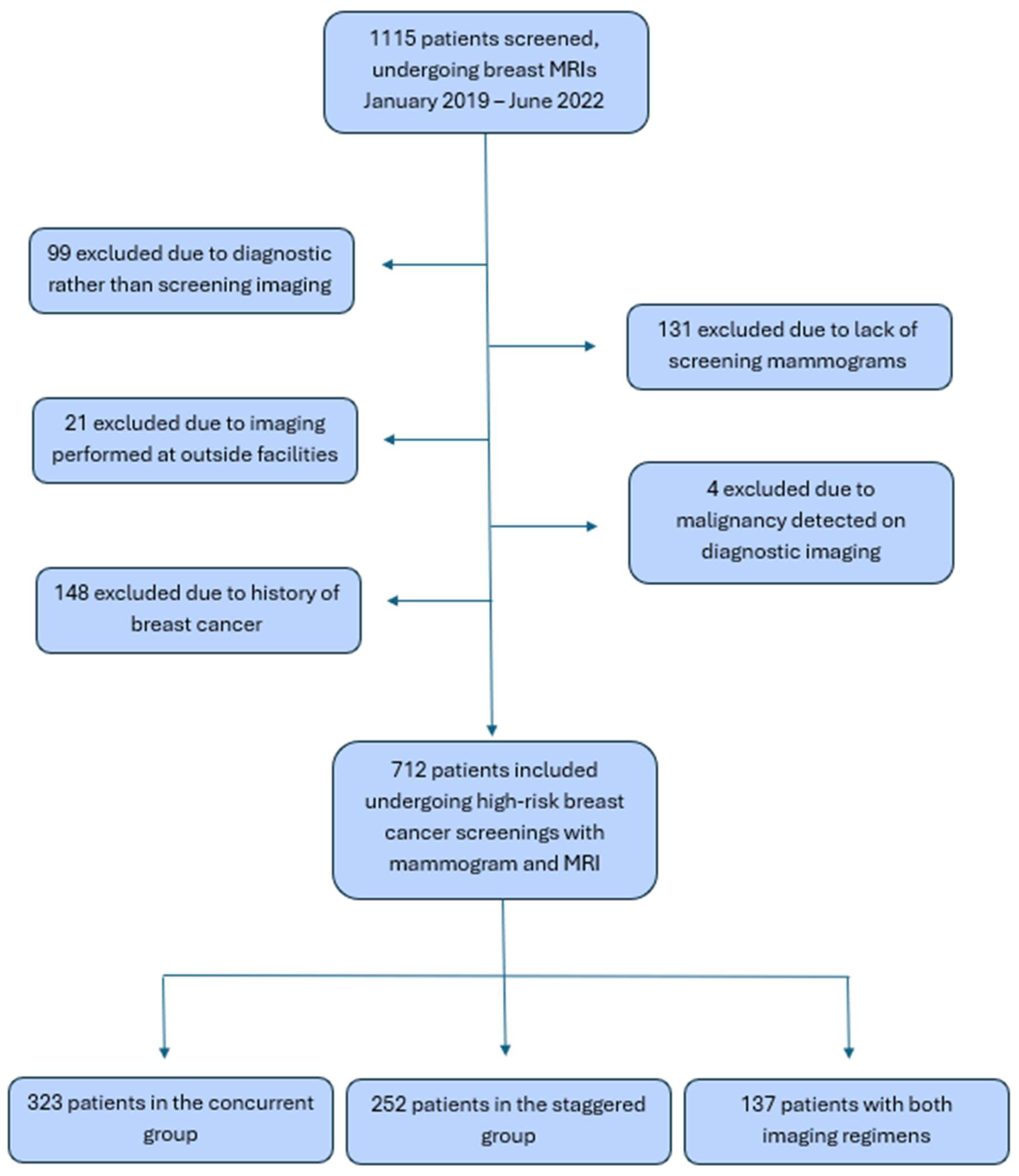
3.1. Study Population
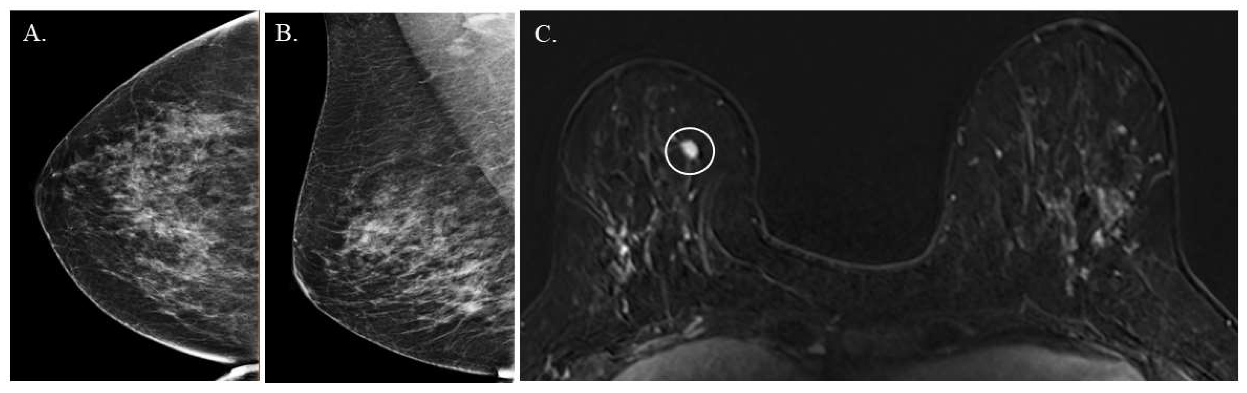
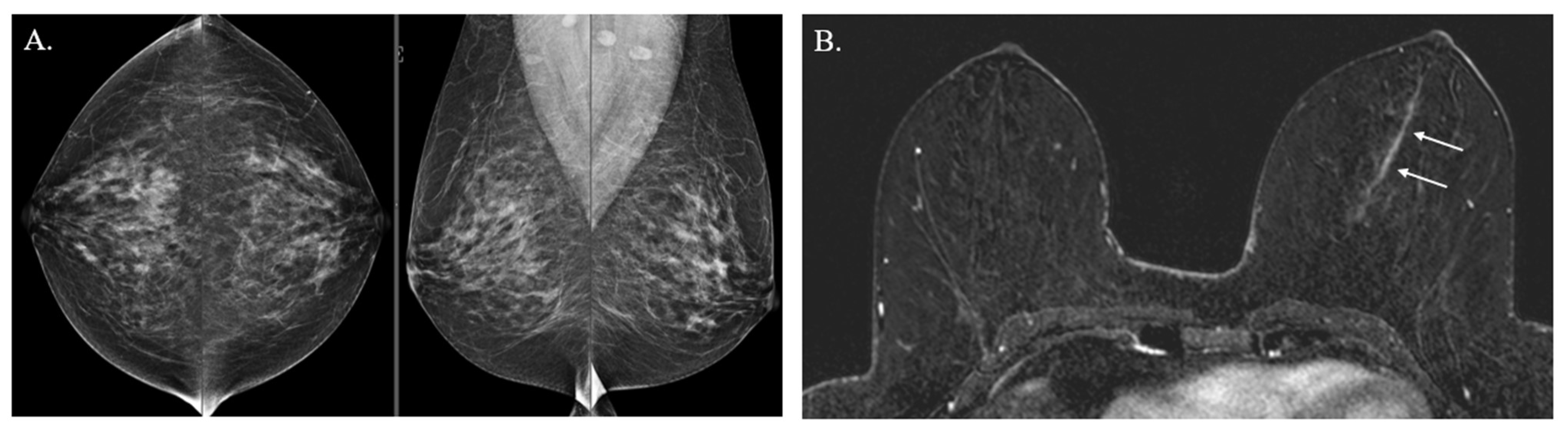
3.2. Clinical Outcomes
4. Discussion
Author Contributions
Funding
Institutional Review Board Statement
Informed Consent Statement
Data Availability Statement
Conflicts of Interest
References
- American Cancer Society. Breast Cancer Statistics: How Common Is Breast Cancer? 2023. Available online: https://www.cancer.org/cancer/types/breast-cancer/about/how-common-is-breast-cancer.html (accessed on 9 January 2023).
- Saslow, D.; Boetes, C.; Burke, W.; Harms, S.; Leach, M.O.; Lehman, C.D.; Morris, E.; Pisano, E.; Schnall, M.; Sener, S.; et al. American Cancer Society guidelines for breast screening with MRI as an adjunct to mammography. CA Cancer J. Clin. 2007, 57, 75–89. [Google Scholar]
- National Comprehensive Cancer Network (NCCN). NCCN Clinical Practice Guidelines in Oncology. Genetic/Familial High-Risk Assessment: Breast, Ovarian, and Pancreatic Version 1. 2024. Available online: https://www.nccn.org/professionals/physician_gls/pdf/genetics_bopp.pdf (accessed on 9 January 2023).
- Li, S.; Silvestri, V.; Leslie, G.; Rebbeck, T.R.; Neuhausen, S.L.; Hopper, J.L.; Nielsen, H.R.; Lee, A.; Yang, X.; McGuffog, L.; et al. Cancer Risks Associated with BRCA1 and BRCA2 Pathogenic Variants. J. Clin. Oncol. 2022, 40, 1529–1541. [Google Scholar] [CrossRef] [Scilit]
- Chen, J.; Bae, E.; Zhang, L.; Hughes, K.; Parmigiani, G.; Braun, D.; Rebbeck, T.R. Penetrance of Breast and Ovarian Cancer in Women Who Carry a BRCA1/2 Mutation and Do Not Use Risk-Reducing Salpingo-Oophorectomy: An Updated Meta-Analysis. JNCI Cancer Spectr. 2020, 4, pkaa029. [Google Scholar] [CrossRef] [Scilit]
- Hendrick, R.E.; Baker, J.A.; Helvie, M.A. Breast cancer deaths averted over 3 decades. Cancer 2019, 125, 1482–1488. [Google Scholar] [CrossRef] [Scilit]
- Duffy, S.; Tabar, L.; Vitak, B.; Day, N.; Smith, R.; Chen, H.; Yen, M. The relative contributions of screen-detected in situ and invasive breast carcinomas in reducing mortality from the disease. Eur. J. Cancer 2003, 39, 1755–1760. [Google Scholar] [CrossRef] [Scilit] [PubMed]
- Sankatsing, V.D.; van Ravesteyn, N.T.; Heijnsdijk, E.A.; Looman, C.W.; van Luijt, P.A.; Fracheboud, J.; Heeten, G.J.D.; Broeders, M.J.; de Koning, H.J. The effect of population-based mammography screening in Dutch municipalities on breast cancer mortality: 20 years of follow-up. Int. J. Cancer 2017, 141, 671–677. [Google Scholar] [CrossRef] [Scilit]
- Morrell, S.; Taylor, R.; Roder, D.; Robson, B.; Gregory, M.; Craig, K. Mammography service screening and breast cancer mortality in New Zealand: A National Cohort Study 1999–2011. Br. J. Cancer 2017, 116, 828–839. [Google Scholar] [CrossRef] [Scilit] [PubMed]
- Warner, E.; Messersmith, H.; Causer, P.; Eisen, A.; Shumak, R.; Plewes, D. Systematic review: Using magnetic resonance imaging to screen women at high risk for breast cancer. Ann. Intern Med. 2008, 148, 671–679. [Google Scholar] [CrossRef] [Scilit] [PubMed]
- Lord, S.; Lei, W.; Craft, P.; Cawson, J.; Morris, I.; Walleser, S.; Griffiths, A.; Parker, S.; Houssami, N. A systematic review of the effectiveness of magnetic resonance imaging (MRI) as an addition to mammography and ultrasound in screening young women at high risk of breast cancer. Eur. J. Cancer 2007, 43, 1905–1917. [Google Scholar] [CrossRef] [Scilit]
- Zhang, Z.; Berg, W.A.; Lehrer, D.; Jong, R.A.; Pisano, E.D.; Barr, R.G.; Böhm-Vélez, M.; Mahoney, M.C.; Evans, W.P.; Larsen, L.H.; et al. Detection of breast cancer with addition of annual screening ultrasound or a single screening MRI to mammography in women with elevated breast cancer risk. JAMA 2012, 307, 1394–1404. [Google Scholar] [CrossRef] [Scilit]
- Radhakrishna, S.; Agarwal, S.; Parikh, P.M.; Kaur, K.; Panwar, S.; Sharma, S.; Dey, A.; Saxena, K.K.; Chandra, M.; Sud, S. Role of magnetic resonance imaging in breast cancer management. South Asian J. Cancer 2018, 7, 69–71. [Google Scholar] [CrossRef] [Scilit] [PubMed]
- Chiarelli, A.M.; Prummel, M.V.; Muradali, D.; Majpruz, V.; Horgan, M.; Carroll, J.C.; Eisen, A.; Meschino, W.S.; Shumak, R.S.; Warner, E.; et al. Effectiveness of screening with annual magnetic resonance imaging and mammography: Results of the initial screen from the ontario high risk breast screening program. J. Clin. Oncol. 2014, 32, 2224–2230. [Google Scholar] [CrossRef] [Scilit]
- Phi, X.-A.; Saadatmand, S.; De Bock, G.H.; Warner, E.; Sardanelli, F.; Leach, M.O.; Riedl, C.C.; Trop, I.; Hooning, M.J.; Mandel, R.; et al. Contribution of mammography to MRI screening in BRCA mutation carriers by BRCA status and age: Individual patient data meta-analysis. Br. J. Cancer 2016, 114, 631–637. [Google Scholar] [CrossRef] [Scilit]
- Port, E.R.; Park, A.; Borgen, P.I.; Morris, E.; Montgomery, L.L. Results of MRI screening for breast cancer in high-risk patients with LCIS and atypical hyperplasia. Ann. Surg. Oncol. 2007, 14, 1051–1057. [Google Scholar] [CrossRef] [Scilit]
- Hartmann, L.C.; Degnim, A.C.; Santen, R.J.; Dupont, W.D.; Ghosh, K. Atypical hyperplasia of the breast—Risk assessment and management options. N. Engl. J. Med. 2015, 372, 78–89. [Google Scholar] [CrossRef] [Scilit]
- Niraula, S.; Biswanger, N.; Hu, P.; Lambert, P.; Decker, K. Incidence, Characteristics, and Outcomes of Interval Breast Cancers Compared With Screening-Detected Breast Cancers. JAMA Netw. Open 2020, 3, e2018179. [Google Scholar] [CrossRef] [Scilit] [PubMed]
- Le-Petross, H.T.; Whitman, G.J.; Atchley, D.P.; Yuan, Y.; Gutierrez-Barrera, A.; Hortobagyi, G.N.; Litton, J.K.; Arun, B.K. Effectiveness of alternating mammography and magnetic resonance imaging for screening women with deleterious BRCA mutations at high risk of breast cancer. Cancer 2011, 117, 3900–3907. [Google Scholar] [CrossRef] [Scilit] [PubMed]
- Smith, R.A.; Saslow, D.; Sawyer, K.A.; Burke, W.; Costanza, M.E.; Evans, W.P.; Foster, R.S.; Hendrick, E.; Eyre, H.J.; Sener, S. American Cancer Society guidelines for breast cancer screening: Update 2003. CA Cancer J. Clin. 2003, 53, 141–169. [Google Scholar] [CrossRef] [Scilit]
- Heijnsdijk, E.A.; Warner, E.; Gilbert, F.J.; Tilanus-Linthorst, M.M.; Evans, G.; Causer, P.A.; Eeles, R.A.; Kaas, R.; Draisma, G.; Ramsay, E.A.; et al. Differences in natural history between breast cancers in BRCA1 and BRCA2 mutation carriers and effects of MRI screening-MRISC, MARIBS, and Canadian studies combined. Cancer Epidemiol. Biomark. Prev. 2012, 21, 1458–1468. [Google Scholar] [CrossRef] [Scilit]
- Othman, E.; Wang, J.; Sprague, B.L.; Rounds, T.; Ji, Y.; Herschorn, S.D.; E Wood, M. Comparison of false positive rates for screening breast magnetic resonance imaging (MRI) in high risk women performed on stacked versus alternating schedules. Springerplus 2015, 4, 77. [Google Scholar] [CrossRef] [Scilit]
- Hirsch, B.R.; Lyman, G.H. Breast cancer screening with mammography. Curr. Oncol. Rep. 2011, 13, 63–70. [Google Scholar] [CrossRef] [Scilit] [PubMed]
- Onitilo, A.A.; Engel, J.M.; Liang, H.; Stankowski, R.V.; Miskowiak, D.A.; Broton, M.; Doi, S.A. Mammography utilization: Patient characteristics and breast cancer stage at diagnosis. AJR Am. J. Roentgenol. 2013, 201, 1057–1063. [Google Scholar] [CrossRef] [Scilit]
- Castaldi, M.; Smiley, A.; Kechejian, K.; Butler, J.; Latifi, R. Disparate access to breast cancer screening and treatment. BMC Womens Health 2022, 22, 249. [Google Scholar] [CrossRef] [Scilit] [PubMed]
- Ehsani, S.; Strigel, R.M.; Pettke, E.; Wilke, L.; Tevaarwerk, A.J.; DeMartini, W.B.; Wisinski, K.B. Screening magnetic resonance imaging recommendations and outcomes in patients at high risk for breast cancer. Breast J. 2015, 21, 246–253. [Google Scholar] [CrossRef] [Scilit]
- Christensen, E.W.; Rosenblatt, R.B.; Patel, A.G.; Rula, E.Y.; Carlos, R.C.; Narayan, A.K.; Patel, B.K. Differential Access to Breast Magnetic Resonance Imaging Compared with Mammography and Ultrasound. Am. J. Prev. Med. 2024, 67, 897–905. [Google Scholar] [CrossRef] [Scilit] [PubMed]
- Ooi, S.L.; Martinez, M.E.; Li, C.I. Disparities in breast cancer characteristics and outcomes by race/ethnicity. Breast Cancer Res. Treat. 2011, 127, 729–738. [Google Scholar] [CrossRef] [Scilit]



| Entire Cohort, N = 712 1 | ||||
|---|---|---|---|---|
| Concurrent, N = 323 N (%) | Staggered, N = 252 N (%) | Both, N =137 N (%) | p Value | |
| Age, median (range) 2 | 52 years (28–80) | 52 years (30–74) | 51 years (34–71) | 0.27 |
| Race/Ethnicity | ||||
| White | 279 (86.4) | 214 (84.9) | 114 (83.2) | 0.64 |
| African American | 19 (5.9) | 20 (7.9) | 12 (8.8) | 0.43 |
| Asian | 16 (5.0) | 11 (4.4) | 35 (4.9) | 0.79 |
| Pacific Islander | 8 (2.5) | 4 (1.6) | 2 (1.5) | 0.77 |
| Hispanic | 10 (3.1) | 10 (4.0) | 4 (2.9) | 0.86 |
| American Indian | 0 | 2 (0.8) | 0 | 0.16 |
| Other | 1 (0.3) | 1 (0.4) | 1 (0.7) | 1.00 |
| Tyrer–Cuzick mean lifetime percentage risk | N = 250, 27.2 | N = 192, 28.0 | N = 110, 28.0 | 0.66, 0.80 |
| History of high-risk intraepithelial lesions | 69 (21.8) | 43 (17.3) | 21 (15.4) | 0.22 |
| Types of high-risk intraepithelial lesions present | ||||
| Atypical ductal hyperplasia (ADH) | 36 (11.1) | 22 (8.7) | 12 (8.8) | 0.59 |
| Atypical lobular hyperplasia (ALH) | 36 (11.1) | 24 (9.5) | 7 (5.1) | 0.12 |
| Lobular carcinoma in situ (LCIS) | 12 (3.7) | 6 (2.4) | 6 (4.4) | 0.52 |
| Presence of hereditary cancer genes | 17 (5.3) | 30 (11.9) | 16 (11.7) | 0.007 |
| Hereditary breast cancer genes present | ||||
| BRCA1 | 3 (17.6) | 4 (13.3) | 2 (12.5) | 0.95 |
| BRCA2 | 6 (35.3) | 6 (20.0) | 3 (18.8) | |
| CHEK2 | 3 (17.6) | 9 (30.0) | 4 (25.0) | |
| ATM | 1 (5.9) | 2 (6.7) | 1 (6.2) | |
| PALB2 | 1 (5.9) | 2 (6.7) | 3 (18.8) | |
| Other 3 | 3 (17.6) | 7 (23.3) | 3 (18.8) | 0.096 |
| Any use of breast cancer prevention medications | 39 (12.1) | 26 (10.3) | 19 (13.9) | 0.56 |
| Current use of prevention medications | 23 (59.0) | 20 (76.9) | 16 (84.2) | 0.11 |
| Last year of prevention medication use, median (range) | 2019 (2004–2022) | 2017 (2005–2022) | 2014 (2012–2021) | 0.78 |
| Prevention medications used | ||||
| Tamoxifen | 14 (4.3) | 8 (3.2) | 4 (2.9) | 0.75 |
| Raloxifene | 10 (3.1) | 7 (2.8) | 7 (5.1) | 0.46 |
| Exemestane | 11 (3.4) | 7 (2.8) | 7 (5.1) | 0.49 |
| Anastrozole | 8 (2.5) | 9 (3.6) | 3 (2.2) | 0.70 |
| MRI Exams | ||||
|---|---|---|---|---|
| Concurrent (N = 784) | Staggered (N = 571) | Total (N = 1355) | p Value | |
| BI-RADS score | 0.16 | |||
| 0 | 41 (5.2%) | 26 (4.6%) | 67 (4.9%) | |
| 1/2 | 602 (76.8%) | 456 (79.9%) | 1058 (78.1%) | |
| 3 | 60(7.7%) | 51 (8.9%) | 111 (8.2%) | |
| 4/5 | 81 (10.3%) | 38 (6.7%) | 119 (8.8%) | |
| Mammogram Exams | ||||
| Concurrent (N = 780) | Staggered (N = 516) | Total (N = 1296) | p Value | |
| BI-RADS score | 0.097 | |||
| 0 | 77 (9.9%) | 49 (9.5%) | 126 (9.7%) | |
| 1 | 260 (33.3%) | 192 (37.2%) | 452 (34.9%) | |
| 2 | 417 (53.5%) | 268 (51.9%) | 685 (52.9%) | |
| 3 | 21 (2.7%) | 4 (0.8%) | 25 (1.9%) | |
| 4 | 5 (0.6%) | 3 (0.6%) | 8 (0.6%) | |
| Total MRI Exams (N = 1355) 1 | |||
|---|---|---|---|
| Concurrent, N = 784 N (%, 95%CI) | Staggered, N = 571 N (%, 95%CI) | p Value | |
| Additional imaging performed | 98 (12.5, 10.3–15.0) | 55 (9.6, 7.3–12.4) | 0.12 |
| MRI prompting biopsy | 77 (9.8, 7.8–12.1) | 40 (7.0, 5.1–9.4) | 0.08 |
| Total biopsies performed | 97 | 46 | |
| Biopsies done per MRI | 0.14 | ||
| 1 | 60 (77.9, 67.0–86.6) | 36 (90.0, 76.3–97.2) | |
| 2 | 14 (18.2, 10.3–28.6) | 2 (5.0, 0.6–16.9) | |
| 3 | 3 (3.9, 0.8–11.0) | 2 (5.0, 0.6–16.9) | |
| Malignancies detected | 4 (0.5, 0.1–1.3) | 3 (0.5, 0.1–1.5) | 1.0 |
| Incremental cancer detection rate (CDR) | 5/1000 | 5/1000 | |
| High-risk intraepithelial lesions | 13 (1.5) | 7 (1.1, 0.4–2.3) | 0.48 |
| Median size of cancer detected, mm | 5 (3–20) | 4 (3–12) | 0.72 |
Disclaimer/Publisher’s Note: The statements, opinions and data contained in all publications are solely those of the individual author(s) and contributor(s) and not of MDPI and/or the editor(s). MDPI and/or the editor(s) disclaim responsibility for any injury to people or property resulting from any ideas, methods, instructions or products referred to in the content. |
© 2026 by the authors. Licensee MDPI, Basel, Switzerland. This article is an open access article distributed under the terms and conditions of the Creative Commons Attribution (CC BY) license.
Share and Cite
Cornell, L.F.; Plante, M.; Pai, T.; Li, Z.; Maimone, S.; Morozov, A.; Maxwell, R.; Advani, P.; Robinson, K. Comparison of High-Risk Breast Cancer Screenings with Concurrent Versus Staggered Breast MRI and Mammogram Schedules. J. Clin. Med. 2026, 15, 4239. https://doi.org/10.3390/jcm15114239
Cornell LF, Plante M, Pai T, Li Z, Maimone S, Morozov A, Maxwell R, Advani P, Robinson K. Comparison of High-Risk Breast Cancer Screenings with Concurrent Versus Staggered Breast MRI and Mammogram Schedules. Journal of Clinical Medicine. 2026; 15(11):4239. https://doi.org/10.3390/jcm15114239
Chicago/Turabian StyleCornell, Lauren Foster, Marie Plante, Tanmayi Pai, Zhuo Li, Santo Maimone, Andrey Morozov, Robert Maxwell, Pooja Advani, and Kristin Robinson. 2026. "Comparison of High-Risk Breast Cancer Screenings with Concurrent Versus Staggered Breast MRI and Mammogram Schedules" Journal of Clinical Medicine 15, no. 11: 4239. https://doi.org/10.3390/jcm15114239
APA StyleCornell, L. F., Plante, M., Pai, T., Li, Z., Maimone, S., Morozov, A., Maxwell, R., Advani, P., & Robinson, K. (2026). Comparison of High-Risk Breast Cancer Screenings with Concurrent Versus Staggered Breast MRI and Mammogram Schedules. Journal of Clinical Medicine, 15(11), 4239. https://doi.org/10.3390/jcm15114239