Article

Recurrence of Non-Melanoma Skin Cancers in the Head and Neck Area—A Single-Center Retrospective Analysis


1
Student Scientific Circle of Plastic Surgery, Faculty of Medicine, Medical University of Gdańsk, 80-210 Gdańsk, Poland
2
Plastic Surgery Department, Medical University of Gdańsk, 80-210 Gdańsk, Poland
3
Department of Algorithms and Systems Modelling, Faculty of Electronics, Telecommunications and Informatics, Gdańsk University of Technology, 80-233 Gdańsk, Poland
4
Laboratory of Translational Oncology, Intercollegiate Faculty of Biotechnology of the University of Gdańsk, Medical University of Gdańsk, 80-210 Gdańsk, Poland
5
Centre of Biostatistics and Bioinformatics, Medical University of Gdańsk, 80-210 Gdańsk, Poland
*
Author to whom correspondence should be addressed.
J. Clin. Med. 2026, 15(11), 4196; https://doi.org/10.3390/jcm15114196
Submission received: 2 April 2026 / Revised: 15 May 2026 / Accepted: 25 May 2026 / Published: 29 May 2026
(This article belongs to the Section Dermatology)

Abstract

Background: Non-melanoma skin cancers (NMSCs) of the head and neck represent a therapeutic challenge due to the region’s complex anatomy, functional considerations, and frequent involvement of high-risk anatomical zones. Local recurrence remains a clinically significant concern, however real-world data regarding recurrence patterns and associated risk factors in facial NMSCs are limited. Objectives: To evaluate the incidence of local recurrence of facial skin cancers after surgical treatment and to determine clinicopathological and anatomical actors associated with an increased risk of recurrence. Methods: In this single-center retrospective cohort study, consecutive patients undergoing surgical excision of facial NMSC were included. The treatment of choice was always surgical excision under general or local anesthesia, with an adequate margin of macroscopically unchanged tissue. Mohs surgery was not used, and none of the patients received immunosuppression. Clinical and pathological data were extracted from medical records. Histopathological examination constituted the basis for establishing the final clinical diagnosis and thus was not verified otherwise. The primary outcome was histologically confirmed local recurrence defined as the reappearance of a tumor of the same histopathological type at the same anatomical site as the previously excised lesion. Patients in the non-recurrence group were defined as those who did not experience any recurrence within a 5-year follow-up period after the initial surgical treatment. Fisher’s exact test and the Mann–Whitney U test were used for statistical analysis. Logistic regression was performed to explore factors associated with recurrence. Due to incomplete follow-up data for the non-recurrent group, we limited the timing analysis to recurrent cases only, as these limitations precluded the use of standard survival analysis. Results: A total of 302 lesions were analyzed, with recurrence status available for 291 tumors. The overall recurrence rate was 28.52%. Basal cell carcinoma (BCC) was the most common histopathological subtype. Recurrences occurred more frequently in anatomically high-risk areas, particularly the scalp, temple and nose. Infiltrative BCC subtypes demonstrated higher recurrence rates than nodular and superficial subtypes. Patients with recurrent tumors were younger than those without recurrence. A history of prior skin radiotherapy was associated with increased odds of recurrence. Tumor size and surgical margin width were not significantly associated with recurrence. Multivariate models showed limited discriminatory ability, suggesting that additional unmeasured factors contribute to recurrence risk. Conclusions: Local recurrence of non-melanoma skin cancers in the head and neck region remains a substantial clinical concern, particularly in high-risk anatomical sites and tumors with aggressive histopathological features. These findings highlight the importance of long-term follow-up and support further prospective studies to improve recurrence risk assessment and treatment strategies.

1. Introduction

Non-melanoma skin cancers (NMSC) represent the most common malignancies worldwide [1]. Over recent decades, a continuous increase in their incidence has been observed [2,3,4,5], which is attributed to prolonged life expectancy and increasing exposure to ultraviolet (UV) radiation [6,7].
The main subtypes of non-melanoma skin cancers are basal cell carcinoma (BCC), accounting for approximately 80% of cases, and squamous cell carcinoma (SCC), representing about 20%. Together, these entities constitute nearly 99% of all NMSC cases [8]. Although they rarely lead to distant metastases or death, if left untreated they may infiltrate and destroy adjacent tissues [9,10,11]. Exposure to UV radiation is the principal risk factor for the development of NMSC, which explains their predominant localization in sun-exposed areas such as the face, neck, and extremities [8,9]. Due to the frequent involvement of the head and neck region, these tumors significantly affect not only patients’ health but also their quality of life, aesthetic appearance, and social functioning.
Surgical excision with histologically clear margins remains the gold standard in the treatment of NMSC [10]. A surgical margin of 2–4 mm is typically considered adequate to ensure complete tumor removal. However, surgical management of facial skin cancers often carries a risk of permanent disfigurement and functional impairment of critical anatomical structures, including the eyelids, oral commissure, or nose. Consequently, additional reconstructive procedures are frequently required to restore function and improve patients’ quality of life. However, over the past few years minimally invasive methods, such as electrochemotherapy (ECT) and topical chemotherapy, have gained increasing attention. Electrochemotherapy has been reported as effective, especially in selected cases of NMSC [12]. Local chemotherapy with use of 5-fluorouracil demonstrates efficacy mainly in treating superficial BCC and SCC in situ [13]. Systematic reviews suggest comparable short-term effectiveness in carefully selected superficial lesions, whereas its effectiveness is limited in deeper tumors, which are associated with higher risk of recurrence [12,13].
Despite surgical treatment, recurrence rates for BCC range from 0.3% to 6.5% [11], while those for SCC vary between 5% and 20% [14]. Determining the precise recurrence rate of NMSC remains challenging due to the lack of a standardized patient follow-up protocol and unified criteria for defining recurrence. The risk of recurrence is influenced by multiple factors, including the surgical technique used, history of prior recurrence, tumor size and anatomical location, depth of invasion, and histopathological grade [2,9,15,16].
Previous studies on recurrent NMSC have predominantly evaluated lesions occurring across the entire body [11,14]. In contrast, analyses specifically addressing recurrence rates in the facial region remain limited. Lesions located in the head and neck area, particularly within the so-called H-zone, are classified as high-risk skin cancers, due to their increased propensity for deep invasion, recurrence, and, in selected cases, metastasis [17]. The H-zone includes the nose, eyelids, eyebrows, periorbital region, ears, preauricular area, and central face, and is defined by the proximity of critical anatomical structures as well as considerable variability in skin thickness across different subregions [18].
The aim of the present study was to evaluate the recurrence rate of skin cancers of the head and neck in patients treated surgically at the Department of Plastic Surgery, University Clinical Center, between 2020 and 2024.

2. Materials and Methods

This retrospective cohort study analyzed clinical and pathological factors associated with non-melanoma skin cancer recurrence following surgical excision at a single institution. A study was conducted of patients hospitalized for head and neck skin cancers who underwent surgical treatment at the Department of Plastic Surgery, University Clinical Center, between 2020 and 2024. Histopathological data, including final diagnosis and information about completeness of excision and/or margins were obtained from original pathology reports. No independent re-evaluation of the pathology findings was performed for the purposes of this study.
A total of 302 lesions were included in the analysis. The primary outcome was histologically confirmed local recurrence defined as the reappearance of a tumor of the same histopathological type at the same anatomical site as the previously excised lesion, with 291 lesions having known outcome status. Patients classified as non-recurrent were those without evidence of recurrence during a minimum 5-year follow-up period.
Categorical variables were summarized as frequencies and percentages; continuous variables as median with interquartile range (IQR). Group comparisons used Fisher’s exact test for categorical variables (with Cramér’s V as effect size measure) and Mann–Whitney U test for continuous variables (with rank-biserial correlation as effect size). Logistic regression estimated odds ratios (OR) with 95% confidence intervals (CI). Time to documented recurrence among recurrent lesions was visualized as the cumulative distribution of months from surgery to recurrence. Considering the lack of complete follow-up data for non-recurrent lesions, we could not perform a standard Kaplan–Meier recurrence-free survival analysis. Instead, we focused on the timing of relapses among recurrent cases only. These data should not be misinterpreted as survival estimates for the entire cohort.
Multiple lesions per patient may exist in this dataset. Standard statistical tests assume independence between observations; therefore, standard errors may be underestimated and p-values should be interpreted cautiously. All analyses were performed using R version 4.3.2 (31 October 2023).

3. Results

A total of 302 lesions were included in the analysis, with complete recurrence data available for 291 lesions. Local recurrence was identified in 83 cases, corresponding to an overall recurrence rate of 28.52%.
Median patient age was 73 years (IQR 66–81; range 33–97) and sex distribution was balanced (F:M ratio 149:153).
Basal cell carcinoma constituted the majority of the lesions (BCC; 74.2%, 224/302), followed by squamous cell carcinoma (SCC; 16.6%, 50/302), basosquamous carcinoma (BSC; 4.0%, 12/302), SCC in situ (4.6%, 14/302), basosquamous cell carcinoma (BSC; 3.0%, 10/336), and mixed BCC/SCC (<1%, 2/302).
The most common anatomical location was the nose. Complete excision was documented in 72.1% (207/287) of cases with clear excision status. Characteristic of the study population are shown in Table 1.
In recurrence group comparisons, younger age was associated with recurrence—the median age in the recurrence group was 70 years (IQR 63–78) vs. 75 years in the non-recurrence group (IQR 67–81). Cancer type did not differ significantly between recurrence and non-recurrence groups (p = 0.1058), although SCC showed a higher recurrence rate than the overall cohort (38.78%, 19/49). Among BCC lesions with available subtype data, infiltrative variants demonstrated higher recurrence rates (27.10%, 29/107) compared with nodular (19.61%, 10/51) and superficial (13.64%, 3/22) subtypes.
Sex, smoking history, and immunosuppression were not associated with recurrence (all p > 0.05). Prior radiotherapy was associated with recurrence (58.33%, 7/12; p = 0.0418). Complete excision was not associated with recurrence (p = 0.2198), with recurrence observed in 31.58% (24/76) of incompletely excised lesions. Multiple-tumour status was associated with recurrence (p = 0.0164). Tumor dimension, excised specimen area, and smallest surgical margin width did not differ significantly between groups (p = 0.9458, p = 0.6866, and p = 0.4468, respectively).
Univariate logistic regression showed lower odds of recurrence with increasing age (OR 0.97 per year, 95% CI 0.950–0.993; p = 0.0110) and for age > 65 years (OR 0.41, 95% CI 0.226–0.737; p = 0.0028).
Prior radiotherapy was associated with higher odds of recurrence (OR 3.84, 95% CI 1.190–13.329; p = 0.0252). High-risk histology was not significantly associated with recurrence (OR 1.54, 95% CI 0.911–2.624; p = 0.1111). In a multivariable model restricted to BCC and SCC (n = 253), associations were attenuated (SCC vs. BCC OR 1.88, 95% CI 0.876–3.955; p = 0.0981; incomplete excision OR 1.62, 95% CI 0.866–2.982; p = 0.1265). Model discrimination was modest (AUC 0.61, 95% CI 0.54–0.69).
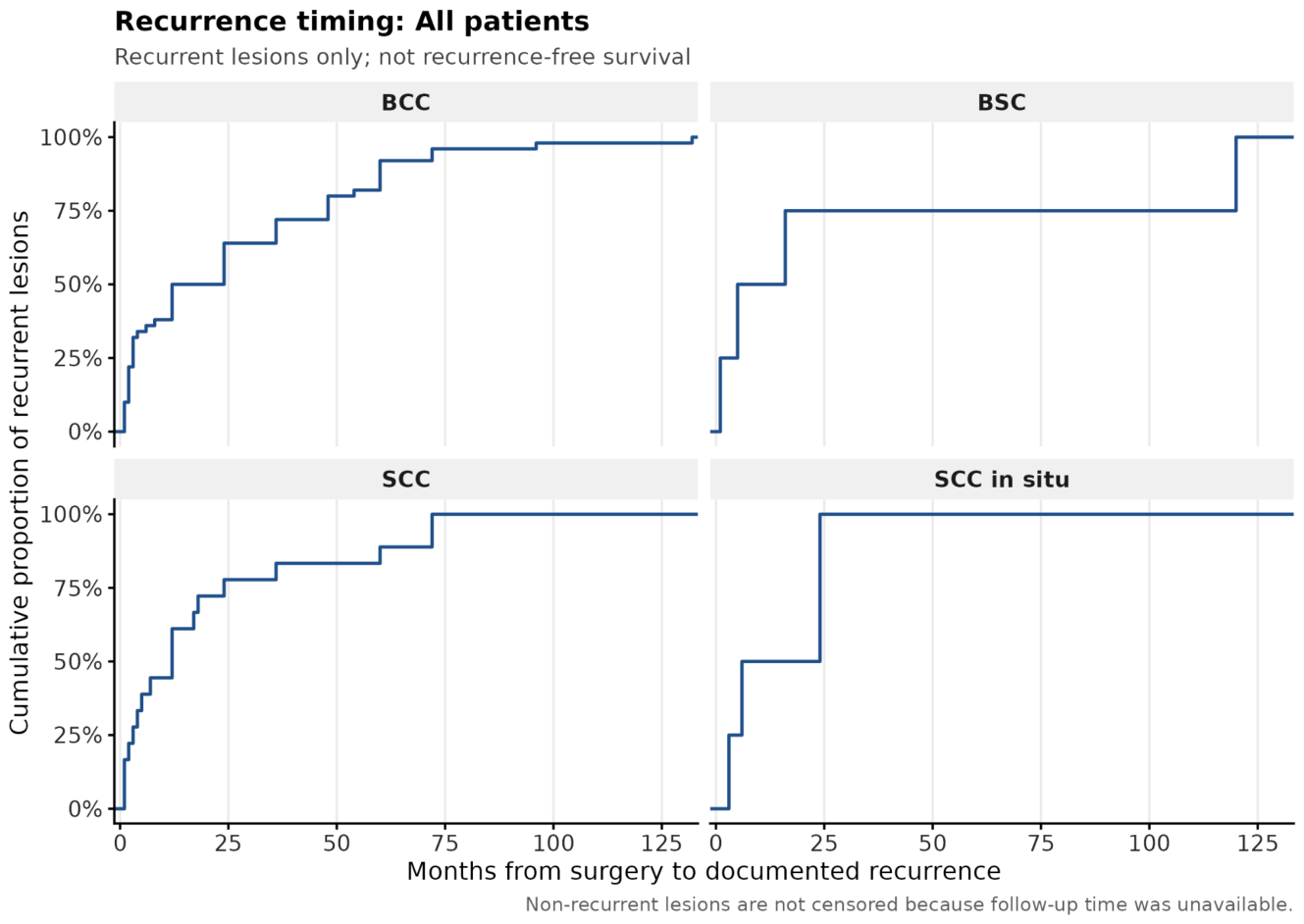
Among recurrent lesions with available timing data, time to recurrence varied across diagnostic groups. The median time to recurrence was 18.0 months for BCC (n = 50; IQR 3.0–48.0), 12.0 months for SCC (n = 18; IQR 3.2–22.5), and 15.0 months for SCC in situ (n = 4; IQR 5.2–24.0). For BSC (n = 4), the median time to recurrence was 10.5 months (IQR 4.0–42.0). Most recurrences occurred within the first two years following surgery, although late events were observed, with maximum times reaching up to 132 months for BCC and 72 months for SCC. The distribution of time to recurrence among recurrent lesions is presented in Figure 1.
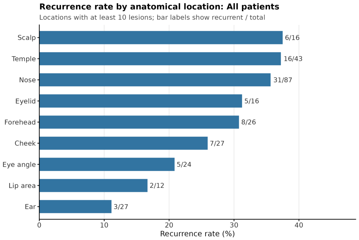
Recurrence rates varied by anatomical site, with the highest rate observed on the scalp (6/16, 37.50%), followed by the temple (16/43, 37.21%) and nose (31/87, 35.63%). Intermediate rates were seen on the eyelid (5/16, 31.25%) and forehead (8/26, 30.77%), while lower rates were observed on the ear (3/27, 11.11%). Missing data were substantial for tumor dimension (58/302) and smallest surgical margin (71/302). Recurrence was higher among lesions with missing tumor dimension data (42.11% vs. 25.21%; p = 0.0142). The association between tumor recurrence and analyzed variables are shown it Table 2. Recurrence rates by anatomical location are presented in Figure 2.

4. Discussion

In this retrospective, single-center study, the recurrence patterns of surgically treated non-melanoma head and neck skin cancers were assessed. Risk factors associated with an increased risk of local recurrence have also been identified. The overall recurrence rate of 28.52% observed in the present study is higher than many previous studies, which typically report recurrence rates of less than 10% for BCC and up to 20% for SCC [1,15,17,19,20,21]. This difference may result from the inclusion of only patients with head and neck skin cancers, which are considered high-risk locations for recurrence. The high recurrence rate in this retrospective is directly correlated with the center’s reference status. High-reference centers treat advanced lesions that are anatomically more challenging, therefore more difficult to treat with complete excision and predisposing to possible recurrence. Patients with less demanding cases of facial skin cancer, qualified for surgical excision, are referred to smaller centers, which likely accounts for the lower recurrence rate among patients at that center.
Basal cell carcinoma represented the largest group of cancers, whereas squamous cell carcinoma formed a smaller but more aggressive subgroup. This is consistent with epidemiological data indicating that BCC accounts for approximately 70–80% of non-melanoma skin cancers [8,11,22,23,24]. SCC demonstrated a higher recurrence rate than the general population, in accordance with previous reports describing its increased local invasiveness and recurrence potential [16,25,26,27].
Among the subtypes, in our cohort infiltrative (morpheaform) BCC subtypes exhibited higher recurrence rates compared with nodular and superficial variants. This matched previous findings suggesting that aggressive histological growth patterns strongly predicted recurrence [28,29,30,31,32].
The risk of recurrence was strongly correlated with tumor location. The highest recurrence rate was observed for lesions located on the scalp, temple and nose, followed by tumors of the eyelids, and forehead. These locations significantly overlap with the facial H-zone, which is commonly described as an area of increased cancer risk due to embryonic fusion planes, thin or variable dermis, and proximity to critical anatomical structures [14,33,34,35,36]. Structural integrity within the H-zone-nasal, periorbital and auricular regions remains crucial for both functional and aesthetic causes. In these anatomically challenging areas, wider surgical margins may substantially increase wound tension, thereby rendering closure and reconstruction more difficult. This often limits the extent of excision that can be safely achieved. As a result, maintaining an adequate margin in these locations can be limited. Combined with the technical difficulty of the surgical procedure, it may increase the likelihood of incomplete excision and therefore microscopic residual disease, thereby contributing to increased risk of cancer recurrence [16,37,38]. For functional and aesthetic reasons, maintaining an adequate margin in these locations can be limited. This, combined with the technical difficulty of the surgical procedure, may increase the risk of microscopic residual disease and lead to the cancer recurrence [16,37,38].
Interestingly, despite older age being a well-established risk factor for skin cancer development, a higher recurrence rate was observed in younger individuals. This is supported by previous studies showing more aggressive tumor behavior and higher recurrence rates in younger patients. Such findings may reflect underlying biological differences, genetic predisposition, or cumulative patterns of ultraviolet radiation exposure [33,39,40,41]. Regarding older patients, cancer is primarily correlated with cumulative exposure to UV radiation for many years. Younger people have less exposure to UV radiation, so the occurrence of skin cancers at an early age may be related to a genetic predisposition. Therefore, younger patients are more likely to have a more aggressive course of cancer, and these patients should be monitored closely, as the onset of cancer at a younger age predisposes them to developing further cancers due to genetic factors.
Prior radiotherapy was a significant predictor of recurrence. This correlation is aligned with previous reports demonstrating that irradiated skin shows impaired healing capacity, dysfunctional immune surveillance, and an increased tendency for aggressive tumor behavior and radioresistance [3,42,43,44]. These reports emphasize the importance of careful follow-up of patients with a history of skin radiotherapy.
Notably, tumor size, surgical margin width, and completeness of excision were not significantly associated with recurrence, despite the fact that incompletely excised tumors showed a numerical trend toward higher recurrence. These observations were consistent with those reported in other retrospective studies, suggesting that margin status alone may not be sufficient to predict recurrence in high-risk facial tumors [5,6,45,46]. However, in cases where data on tumor dimensions and margins were missing, the recurrence rate was substantially higher (42.11% vs. 25.21%; p = 0.0142). This likely reflects an overrepresentation of the most complex and difficult cases within the subgroup with missing data, particularly in lesions for which histopathological reports focused on the assessment of surgical radicality rather than exact measurements. Therefore, the prognostic value of tumor dimensions and surgical margins in our statistical models may be underestimated as high-risk recurrent cases were insufficiently represented in the evaluated variables. Consequently, the lack of statistical significance for these variables in our model should be interpreted with caution, and their potential predictive role as risk factors for recurrence should remain clinically relevant.
Considering that multivariate models based solely on clinical and histopathological variables showed only moderate discriminatory ability in this and in previous studies, it can be concluded that additional factors, i.e., surgeon experience, reconstruction method, molecular characteristics of the tumor, and patient compliance, also contribute to the increased risk of recurrence [8,47,48,49,50].
Diagnosis-specific median times to recurrence ranged from 10.5 to 18 months, with most events occurring within the first two years following surgery; this pattern is consistent with the literature and supports the need for close, early surveillance of patients with a history of skin cancer [11,35,36,51].

Limitations

Retrospective design: Subject to selection bias, information bias, and unmeasured confounding. Associations should not be interpreted causally.
Missing data not at random: Higher recurrence rate when tumor dimension is missing (42.11% vs. 25.21% (p = 0.0142). These missing measurements likely relate to the most difficult cases, where reporting was less standardized. As a result, the predictive value of tumor size and margins may be underestimated in our analysis.
No external validation: Findings are specific to this institution and patient population. External validation in independent cohorts is essential before clinical application.
Confounding by indication: Radiotherapy and “multiple tumors” associations likely reflect treatment selection rather than causal effects. These variables should not be used to guide clinical decisions without accounting for confounding.
Time-to-event analysis limitation: Only recurrent lesions contributed time data; follow-up time for non-recurrent lesions was not available, precluding recurrence-free survival analysis with censoring. The analysis therefore reflects the distribution of time to recurrence among events rather than a Kaplan–Meier estimate.
Outcome definition: “Recurrence” was defined based on histopathological confirmation of the same tumor type occurring at the identical anatomical site after excision; however, despite this definition, it cannot be completely excluded that some cases classified as recurrence may in fact represent new primary tumors arising within a field of cancerization in the same region. Nevertheless, such situations are considered extremely rare and are unlikely to have meaningfully influenced the results of the present study.

5. Conclusions

Recurrence of non-melanoma skin cancers in the head and neck region remains frequent in clinical practice, especially in high-risk anatomical locations and aggressive histological subtypes. These findings highlight the importance of long-term follow-up and support further prospective studies to improve recurrence risk assessment and treatment strategies. Due to the high risk of recurrence of skin cancers on the head and neck area and their more aggressive course, it should be remembered that such patients require intensified screening to detect possible recurrence at an early stage. It is important to remember to also include people who had previously undergone radiotherapy in this follow-up.

Author Contributions

M.W.: Data curation, Writing—Original draft, Conceptualization, Methodology, Project administration; K.M.: Data curation, Writing—Original draft, Conceptualization, Methodology; P.B.: Data curation, Writing—Original draft, Conceptualization, Methodology; M.G.: Data curation, Writing—Original draft, Conceptualization, Methodology; A.M.G.: Data curation, Writing—Original draft, Conceptualization, Methodology; S.K.: Data curation, Writing—Original draft, Conceptualization, Methodology; G.R.: Data curation, Writing—Original draft, Conceptualization, Methodology; M.T.: Data curation, Writing—Original draft, Conceptualization, Methodology; A.W.: Data curation, Writing—Original draft, Conceptualization, Methodology; K.P.: Formal analysis, Methodology, Resources; J.J.: Writing—Original draft, Methodology, Conceptualization, Supervision. All authors have read and agreed to the published version of the manuscript.

Funding

This research received no external funding.

Institutional Review Board Statement

Ethical review and approval were waived due to the use of anonymized survey data, the non-interventional nature of the study, and the absence of risk to participants, with informed consent obtained from all individuals involved.

Informed Consent Statement

Informed consent was obtained from all subjects involved in the study.

Data Availability Statement

The data underlying this article cannot be shared publicly due to the privacy of individuals that participated in the study. The data will be shared on reasonable request to the corresponding author.

Conflicts of Interest

The authors declare no conflicts of interest.

References

  1. Lomas, A.; Leonardi-Bee, J.; Bath-Hextall, F. A systematic review of worldwide incidence of nonmelanoma skin cancer. Br. J. Dermatol. 2012, 166, 1069–1080. [Google Scholar] [CrossRef]
  2. Sordi, E.; Piscitelli, P.; Albanese, C.; Melcarne, A.; Tardio, A.; Quarta, F.; Greco, E.; Miani, A.; Falco, A.; De Matteis, E.; et al. Incidence of Non-Melanoma Skin Cancers in Salento (Southern Italy): A 15-Year Retrospective Analysis from the Cancer Registry of Lecce. Epidemiologia 2024, 5, 1–10. [Google Scholar] [CrossRef]
  3. Ibrahim, N.; Ali, S.R.; Dobbs, T.D.; Gibson, J.A.G.; Hutchings, H.A.; Whitaker, I.S. The incidence of non-melanoma skin cancer in the UK and the Republic of Ireland: A systematic review. Eur. J. Dermatol. 2023, 33, 218–229. [Google Scholar] [CrossRef]
  4. Perera, E.; Gnaneswaran, N.; Staines, C.; Win, A.K.; Sinclair, R. Incidence and prevalence of non-melanoma skin cancer in Australia: A systematic review. Australas. J. Dermatol. 2015, 56, 258–267. [Google Scholar] [CrossRef]
  5. Aggarwal, P.; Knabel, P.; Fleischer, A.B., Jr. United States burden of melanoma and non-melanoma skin cancer from 1990 to 2019. J. Am. Acad. Dermatol. 2021, 85, 388–395. [Google Scholar] [CrossRef] [PubMed]
  6. Watson, M.; Holman, D.M.; Maguire-Eisen, M. Ultraviolet Radiation Exposure and Its Impact on Skin Cancer Risk. Semin. Oncol. Nurs. 2016, 32, 241–254. [Google Scholar] [CrossRef]
  7. Asgari, M.M.; Moffet, H.H.; Ray, G.T.; Quesenberry, C.P. Trends in Basal Cell Carcinoma Incidence and Identification of High-Risk Subgroups, 1998–2012. JAMA Dermatol. 2015, 151, 976–981. [Google Scholar] [CrossRef]
  8. Roland, N.; Memon, A. Non-melanoma skin cancer of the head and neck. Br. J. Hosp. Med. 2023, 84, 1–10. [Google Scholar] [CrossRef]
  9. Armstrong, L.T.D.; Magnusson, M.R.; Guppy, M.P.B. Risk factors for recurrence of facial basal cell carcinoma after surgical excision: A follow-up analysis. J. Plast. Reconstr. Aesthetic Surg. 2017, 70, 1738–1745. [Google Scholar] [CrossRef]
  10. Sathe, N.C.; Zito, P.M. Skin Cancer. In StatPearls; StatPearls Publishing: Treasure Island, FL, USA, 2025. [Google Scholar]
  11. Yildizdal, S.; Kucukguven, A.; Calis, M.; Vargel, I. Evaluation of histopathological margin and other recurrence parameters in basal cell carcinoma: A retrospective analysis of 8821 lesions. J. Plast. Reconstr. Aesthetic Surg. 2022, 75, 3390–3397. [Google Scholar] [CrossRef] [PubMed]
  12. Franczak, A.; Zmijewska, A.; Drzewiecka, E.M.; Kozłowska, W.; Wydorski, P.; Paukszto, Ł.; Brevini, T.L. Effect of electromagnetic field radiation on transcriptomic profile and DNA methylation level in pig conceptuses during the peri-implantation period. Sci. Rep. 2025, 15, 14025. [Google Scholar] [CrossRef] [PubMed]
  13. Prince, G.T.; Cameron, M.C.; Fathi, R.; Alkousakis, T. Topical 5-fluorouracil in dermatologic disease. Int. J. Dermatol. 2018, 57, 1259–1264. [Google Scholar] [CrossRef] [PubMed]
  14. Schmults, C.D.; Karia, P.S.; Carter, J.B.; Han, J.; Qureshi, A.A. Factors predictive of recurrence and death from cutaneous squamous cell carcinoma: A 10-year, single-institution cohort study. JAMA Dermatol. 2013, 149, 541–547. [Google Scholar] [CrossRef]
  15. Bourlidou, E.; Vahtsevanos, K.; Kyrgidis, A.; Tilaveridis, I.; Patsatsi, A.; Andreadis, D.; Cheva, A.; Patrikidou, A.; Kitikidou, K.; Boboridis, K. Risk factors for local recurrence of basal cell carcinoma and cutaneous squamous cell carcinoma of the middle third of the face: A 15-year retrospective analysis based on a single centre. Eur. J. Dermatol. 2019, 29, 490–499. [Google Scholar] [CrossRef]
  16. Wysong, A.; Higgins, S.; Blalock, T.W.; Ricci, D.; Nichols, R.; Smith, F.L.; Kossintseva, I. Defining skin cancer local recurrence. J. Am. Acad. Dermatol. 2019, 81, 581–599. [Google Scholar] [CrossRef] [PubMed]
  17. Madan, V.; Lear, J.T.; Szeimies, R.M. Non-melanoma skin cancer. Lancet 2010, 375, 673–685. [Google Scholar] [CrossRef]
  18. Gogineni, E.; Cai, H.; Carillo, D.; Rana, Z.; Bloom, B.; Potters, L.; Gaballa, H.; Ghaly, M. Computed tomography-based flap brachytherapy for non-melanoma skin cancers of the face. J. Contemp. Brachyther. 2021, 13, 51–58. [Google Scholar] [CrossRef]
  19. Pazdrowski, J.; Dańczak-Pazdrowska, A.; Golusiński, P.; Szybiak, B.; Silny, W.; Golusiński, W. Wznowy raka podstawnokomórkowego (BCC) twarzy w materiale Oddziału Chirurgii Głowy i Szyi i Onkologii Laryngologicznej Wielkopolskiego Centrum Onkologii w latach 2007–2010. Otolaryngol. Pol. 2012, 66, 185–190. [Google Scholar] [CrossRef]
  20. Brantsch, K.D.; Meisner, C.; Schönfisch, B.; Trilling, B.; Wehner-Caroli, J.; Röcken, M.; Breuninger, H. Analysis of risk factors determining prognosis of cutaneous squamous-cell carcinoma: A prospective study. Lancet. Oncol. 2008, 9, 713–720. [Google Scholar] [CrossRef]
  21. Gualdi, G.; Monari, P.; Crotti, S.; Damiani, G.; Facchetti, F.; Calzavara-Pinton, P.; Fantini, F. Matter of margins. J. Eur. Acad. Dermatol. Venereol. 2015, 29, 255–261. [Google Scholar] [CrossRef]
  22. Russell, E.; Udkoff, J.; Knackstedt, T. Basal cell carcinoma with bone invasion: A systematic review and pooled survival analysis. J. Am. Acad. Dermatol. 2022, 86, 621–627. [Google Scholar] [CrossRef] [PubMed]
  23. Pogorzelska-Dyrbuś, J.; Salwowska, N.; Bergler-Czop, B. Dermoscopic Pattern of Basal Cell Carcinoma in H- and Non-H-zones. Dermatol. Pract. Concept. 2023, 13, e2023125. [Google Scholar] [CrossRef] [PubMed]
  24. Bal Avci, E.; Koku Aksu, A.E.; Ilhan Erdil, D.; Erdemir, V.A.; Leblebici, C. Clinical Characteristics of Local Recurrent Basal Cell Carcinoma After Surgical Excision: A Retrospective Study of the Patients from a Tertiary Clinical Center. Cureus 2024, 16, e66668. [Google Scholar] [CrossRef] [PubMed]
  25. Fukumoto, T.; Fukumoto, R.; Oka, M.; Horita, N. Comparing treatments for basal cell carcinoma in terms of long-term treatment-failure: A network meta-analysis. J. Eur. Acad. Dermatol. Venereol. 2019, 33, 2050–2057. [Google Scholar] [CrossRef]
  26. Waldman, A.; Schmults, C. Cutaneous Squamous Cell Carcinoma. Hematol./Oncol. Clin. N. Am. 2019, 33, 1–12. [Google Scholar] [CrossRef]
  27. Ahmed, O.A.; Kelly, C. Head and neck melanoma (excluding ocular melanoma): United Kingdom National Multidisciplinary Guidelines. J. Laryngol. Otol. 2016, 130, S133–S141. [Google Scholar] [CrossRef]
  28. Mehanna, H.; Kong, A.; Ahmed, S.K. Recurrent head and neck cancer: United Kingdom National Multidisciplinary Guidelines. J. Laryngol. Otol. 2016, 130, S181–S190. [Google Scholar] [CrossRef]
  29. Burton, K.A.; Ashack, K.A.; Khachemoune, A. Cutaneous Squamous Cell Carcinoma: A Review of High-Risk and Metastatic Disease. Am. J. Clin. Dermatol. 2016, 17, 491–508. [Google Scholar] [CrossRef]
  30. Stratigos, A.; Garbe, C.; Lebbe, C.; Malvehy, J.; del Marmol, V.; Pehamberger, H.; Peris, K.; Becker, J.C.; Zalaudek, I.; Saiag, P.; et al. Diagnosis and treatment of invasive squamous cell carcinoma of the skin: European consensus-based interdisciplinary guideline. Eur. J. Cancer 2015, 51, 1989–2007. [Google Scholar] [CrossRef]
  31. Andrade, P.; Brites, M.M.; Vieira, R.; Mariano, A.; Reis, J.P.; Tellechea, O.; Figueiredo, A. Epidemiology of basal cell carcinomas and squamous cell carcinomas in a Department of Dermatology: A 5 year review. An. Bras. Dermatol. 2012, 87, 212–219. [Google Scholar] [CrossRef]
  32. Leiter, U.; Eigentler, T.; Garbe, C. Epidemiology of skin cancer. Adv. Exp. Med. Biol. 2014, 810, 120–140. [Google Scholar] [CrossRef]
  33. Nolan, G.S.; Kiely, A.L.; Totty, J.P.; Wormald, J.C.R.; Wade, R.G.; Arbyn, M.; Jain, A. Incomplete surgical excision of keratinocyte skin cancers: A systematic review and meta-analysis. Br. J. Dermatol. 2021, 184, 1033–1044. [Google Scholar] [CrossRef]
  34. Hasan, A.; Rabie, A.; Elhussiny, M.; Nasr, M.; Kamel, M.I.; Hegab, A.; El-Kady, A.S.; Nagaty, M.E.; Seleem, A.; Abbas, M.; et al. Recurrent cutaneous basal cell carcinoma after surgical excision: A retrospective clinicopathological study. Ann. Med. Surg. 2022, 78, 103877. [Google Scholar] [CrossRef] [PubMed]
  35. Russell, E.; Udkoff, J.; Knackstedt, T. Squamous Cell Carcinoma with Bone Invasion: A Systematic Review and Pooled Survival Analysis. Dermatol. Surg. 2022, 48, 1025–1028. [Google Scholar] [CrossRef] [PubMed]
  36. Pisano, C.E.; Trager, M.H.; Fan, W.; Samie, F.H. Surgical margins and outcomes for eyelid melanoma: A systematic review and meta-analysis. Arch. Dermatol. Res. 2024, 316, 106. [Google Scholar] [CrossRef]
  37. Paolino, G.; Donati, M.; Didona, D.; Mercuri, S.R.; Cantisani, C. Histology of Non-Melanoma Skin Cancers: An Update. Biomedicines 2017, 5, 71. [Google Scholar] [CrossRef] [PubMed]
  38. Kundnani, N.R.; Tirziu, R.V.; Borza, C.; Tirziu, C.; Sharma, A.; Rosca, C.I.; Baderca, F.; Paul, C.; Solovan, C.S.; Blidisel, A. Implication of wide surgical excision in minimizing positive margins and consequential secondary excision—A retrospective comparative study involving 106 basal cell carcinoma cases. Eur. Rev. Med. Pharmacol. Sci. 2021, 25, 4283–4288. [Google Scholar] [CrossRef]
  39. Bittar, P.G.; Bittar, J.M.; Etzkorn, J.R.; Brewer, J.D.; Aizman, L.; Shin, T.M.; Sobanko, J.F.; Higgins, H.W.; Giordano, C.N.; Cohen, J.V.; et al. Systematic review and meta-analysis of local recurrence rates of head and neck cutaneous melanomas after wide local excision, Mohs micrographic surgery, or staged excision. J. Am. Acad. Dermatol. 2021, 85, 681–692. [Google Scholar] [CrossRef]
  40. Campbell, E.H.; Crum, O.M.; Chelf, C.J.; Demer, A.M.; Brewer, J.D. Local Recurrence Rates of Malignant Melanoma After Mohs Micrographic Surgery Are Lowest With 5- to 10-mm Initial Margins: A Systematic Review and Meta-Analysis. Dermatol. Surg. 2023, 49, 119–123. [Google Scholar] [CrossRef]
  41. Ran, N.A.; Veerabagu, S.; Miller, C.J.; Elenitsas, R.; Chu, E.Y.; Krausz, A.E. Local Recurrence Rates After Excision of Desmoplastic Melanoma: A Systematic Review and Meta-Analysis. Dermatol. Surg. 2023, 49, 330–337. [Google Scholar] [CrossRef]
  42. Ciuciulete, A.R.; Stepan, A.E.; Andreiana, B.C.; Simionescu, C.E. Non-Melanoma Skin Cancer: Statistical Associations between Clinical Parameters. Curr. Health Sci. J. 2022, 48, 110–115. [Google Scholar] [CrossRef]
  43. Newlands, C.; Currie, R.; Memon, A.; Whitaker, S.; Woolford, T. Non-melanoma skin cancer: United Kingdom National Multidisciplinary Guidelines. J. Laryngol. Otol. 2016, 130, S125–S132. [Google Scholar] [CrossRef]
  44. Gerring, R.C.; Ott, C.T.; Curry, J.M.; Sargi, Z.B.; Wester, S.T. Orbital exenteration for advanced periorbital non-melanoma skin cancer: Prognostic factors and survival. Eye 2017, 31, 379–388. [Google Scholar] [CrossRef] [PubMed]
  45. Schmidt, S.A.; Schmidt, M.; Mehnert, F.; Lemeshow, S.; Sørensen, H.T. Use of antihypertensive drugs and risk of skin cancer. J. Eur. Acad. Dermatol. Venereol. 2015, 29, 1545–1554. [Google Scholar] [CrossRef] [PubMed]
  46. Angeles, C.V.; Kang, R.; Shirai, K.; Wong, S.L. Meta-analysis of completion lymph node dissection in sentinel lymph node-positive melanoma. Br. J. Surg. 2019, 106, 672–681. [Google Scholar] [CrossRef]
  47. Gandini, S.; Sera, F.; Cattaruzza, M.S.; Pasquini, P.; Zanetti, R.; Masini, C.; Boyle, P.; Melchi, C.F. Meta-analysis of risk factors for cutaneous melanoma: III. Family history, actinic damage and phenotypic factors. Eur. J. Cancer 2005, 41, 2040–2059. [Google Scholar] [CrossRef]
  48. Didona, D.; Paolino, G.; Bottoni, U.; Cantisani, C. Non Melanoma Skin Cancer Pathogenesis Overview. Biomedicines 2018, 6, 6. [Google Scholar] [CrossRef]
  49. Griffin, L.L.; Lear, J.T. Photodynamic Therapy and Non-Melanoma Skin Cancer. Cancers 2016, 8, 98. [Google Scholar] [CrossRef]
  50. Hanna, S.; Lo, S.N.; Saw, R.P. Surgical excision margins in primary cutaneous melanoma: A systematic review and meta-analysis. Eur. J. Surg. Oncol. 2021, 47, 1558–1574. [Google Scholar] [CrossRef] [PubMed]
  51. Morelló-Vicente, A.; Espejo-Marín, M.; Oteiza-Rius, I.; Antoñanzas, J.; Vélez, A.; Salido-Vallejo, R. Increased Risk of Local Recurrence in Cutaneous Squamous Cell Carcinoma Arising in Sun-Exposed Skin: A Retrospective Cohort Study. Cancers 2023, 15, 5037. [Google Scholar] [CrossRef]
Figure 1. Cumulative distribution of time (months) from surgery to documented recurrence among recurrent lesions, stratified by diagnosis. The figure includes only lesions with documented recurrence and available timing data and does not represent a Kaplan–Meier recurrence-free survival estimate.
Jcm 15 04196 g001
Figure 2. Recurrence rate by anatomical location. Bars represent the proportion of recurrent lesions for each site, with labels indicating recurrent/total counts. Only locations with at least 10 lesions are shown.
Jcm 15 04196 g002
Table 1. Table highlighting the characteristics of the study population.
Study Population Summary, All Patients
ParameterValue
Eligible lesions (NMSC and SCC in situ)302
Patients contributing eligible lesions132
Lesions with known recurrence outcome291
Recurrent lesions83
Recurrence rate28.52% (83/291)
Median age, years (IQR)73.0 (66.0–81.0)
Female:Male (lesions)149:153
Most common diagnosisBCC
Most common anatomical locationNose
Baseline Characteristics, Categorical, All Patients
VariableLevelN (%)
SexFemale149 (49.3%)
Male153 (50.7%)
DiagnosisBCC224 (74.2%)
BSC12 (4.0%)
Mixed BCC/SCC2 (0.7%)
SCC50 (16.6%)
SCC in situ14 (4.6%)
Location risk groupStandard-risk84 (27.9%)
High-risk217 (72.1%)
Complete excisionNo80 (27.9%)
Yes207 (72.1%)
Nerve or vessel invasionNo287 (97.6%)
Yes7 (2.4%)
MetastasesNo294 (99.7%)
Yes1 (0.3%)
Multiple tumorsNo200 (67.3%)
Yes97 (32.7%)
History of skin cancerNo121 (40.7%)
Yes176 (59.3%)
SmokingNo211 (69.9%)
Yes91 (30.1%)
Prior radiotherapyNo286 (95.7%)
Yes13 (4.3%)
ImmunosuppressionNo279 (93.6%)
Yes19 (6.4%)
Age group≤5012 (4.0%)
51–6552 (17.3%)
66–75104 (34.6%)
>75133 (44.2%)
Table 2. Table showing the association between tumor recurrence and analyzed variables.
VariableLevelTotal Lesions at LevelRecurrentNon-RecurrentRecurrence Rate (%)Cramer’s
V
p Value
SexFemale1444110328.470.00110.000
Male1474210528.57
Anatomical locationNose87315635.630.2270.1456
Temple43162737.21
Cheek2772025.93
Ear2732411.11
Forehead2681830.77
Eye angle2451920.83
Eyelid1651131.25
Scalp1661037.50
Lip area1221016.67
Chin/Jaw8080.00
Neck4040.00
Location risk groupStandard-risk81216025.930.0370.5652
High-risk2096214729.67
DiagnosisBCC2165416225.000.1500.1058
SCC49193038.78
SCC in situ1441028.57
BSC105550.00
Mixed BCC/SCC21150.00
Complete excisionNo76245231.580.0780.2198
Yes2014815323.88
Nerve or vessel invasionNo2777420326.710.1060.0929
Yes74357.14
MetastasesNo2847720727.110.0970.2737
Yes110100.00
Multiple tumorsNo1956313232.310.1440.0164
Yes92177518.48
History of skin cancerNo118328627.120.0140.8937
Yes1694812128.40
SmokingNo2035514827.090.0480.4799
Yes88286031.82
Prior radiotherapyNo2777420326.710.1400.0418
Yes127558.33
ImmunosuppressionNo2707719328.520.0410.6034
Yes1941521.05
Age group≤50124833.330.1980.0100
51–6549232646.94
66–7599287128.28
>751302810221.54
Smallest margin category<1 mm53163730.190.0880.6589
1-<2 mm61154624.59
2-<5 mm89256428.09
≥5 mm1931615.79
Fisher’s exact test. Cramér’s V = effect size (0.1 small, 0.3 medium, 0.5 large).
Disclaimer/Publisher’s Note: The statements, opinions and data contained in all publications are solely those of the individual author(s) and contributor(s) and not of MDPI and/or the editor(s). MDPI and/or the editor(s) disclaim responsibility for any injury to people or property resulting from any ideas, methods, instructions or products referred to in the content.