1. Introduction
Epiretinal membrane (ERM) is a common macular disorder that can lead to visual distortions, including aniseikonia, which is characterized by a perceived difference in image size between the two eyes [
1]. Aniseikonia encompasses both micropsia, in which objects appear smaller than their actual size, and macropsia, in which objects appear larger than they actually are. When mechanical forces applied to the retina stretch the photoreceptors, a given image activates a reduced number of receptors, resulting in the perception of a smaller image (micropsia). In contrast, the compression of photoreceptors leads to the activation of a greater number of receptors in the same image, producing the perception of an enlarged image (macropsia) [
2]. Patients with ERM predominantly experience macropsia, although micropsia has also been reported [
1].
Several methods are available for measuring aniseikonia, including the New Aniseikonia Test (NAT; Handaya Co., Ltd., Tokyo, Japan), Aniseikonia Inspector (AI; Optical Diagnostics, Culemborg, The Netherlands), Brecher test, Miles test, and Space Eikonometer. Among these, the NAT and AI are commercially available and have well-established validity and reliability [
1]. The NAT is administered using a booklet containing adjacent calibrated pairs of red and green half-circular targets of varying sizes. Using red/green analytical filters for dissociation, one half-circle is perceived by the right eye and the other by the left eye, enabling the assessment of perceived image size differences between the two eyes [
3]. The AI is a computerized test that quantifies aniseikonia by presenting red/green rectangular targets for direct size comparison. Patients indicate which target appears larger, and their responses are used to generate a psychometric function that reflects perceived image size differences [
4].
Pars plana vitrectomy (PPV) is the standard surgical treatment for ERM; however, its effectiveness in improving aniseikonia remains controversial. Although some studies have reported no significant improvement in aniseikonia after PPV, others have demonstrated significant changes. For example, Fukuyama et al. [
5] reported no significant improvements in aniseikonia 6 months after PPV in patients with ERM when idiopathic and secondary ERM were grouped together. Similarly, Ichikawa et al. [
6] and Nakashizuka et al. [
7] observed no statistically significant changes in aniseikonia 3 and 12 months after PPV, respectively, in studies that included both idiopathic and secondary ERM. Okamoto et al. [
8] also reported minimal improvements in aniseikonia 6 months after PPV, suggesting that heterogeneity in the pathogenesis of ERM may influence outcomes. In contrast, Kim et al. [
9] found significant improvements in aniseikonia only in cases of idiopathic ERM, whereas cases of secondary ERM, such as those associated with retinal breaks, showed no meaningful changes 6 months after PPV. These findings highlight the importance of focusing specifically on idiopathic ERM when analyzing aniseikonia outcomes because pooling data from heterogeneous ERM populations may obscure the therapeutic effects of PPV.
Idiopathic ERM is primarily associated with proliferation of glial cells, including Müller cells, and typically presents as a uniform circular global attachment to the macula [
10,
11]. This even distribution facilitates a more stable macular microstructure after surgery, likely enabling photoreceptor realignment and improving visual symmetry, which contributes to aniseikonia reduction. Secondary ERM often arises from retinal pigment epithelial cells and exhibits irregular finger-like projections with focal attachment patterns [
12]. This morphology may account for the limited improvement observed in secondary ERM, as irregularities introduce persistent structural disruptions. Additionally, idiopathic ERM tends to develop in older individuals with gradual macular changes, whereas secondary ERM is often associated with traumatic or inflammatory events in younger patients [
11]. These conditions introduce chronic retinal damage, edema, or scarring, further limiting postoperative functional recovery.
This meta-analysis aimed to determine whether PPV leads to statistically significant improvements in aniseikonia in idiopathic ERM and to examine the trajectory of these changes over time. By analyzing data exclusively from studies including idiopathic ERM, this study aimed to clarify the effectiveness of PPV in improving aniseikonia and to define an appropriate duration for postoperative monitoring.
2. Materials and Methods
2.1. Search Strategy and Study Selection
A systematic search was conducted using PubMed, EMBASE, and the Cochrane Library from inception to March 2024. This study aimed to identify studies that have evaluated the effect of PPV on aniseikonia in patients with idiopathic ERM. Search terms included combinations of Medical Subject Headings, Embase subject headings (Emtree), and keywords such as “epiretinal membrane,” “macular pucker,” “vitrectomy,” “aniseikonia,” and “metamorphopsia.” The full search terms were ((“Epiretinal Membrane”[MeSH] OR “Epiretinal Membranes”[TW] OR “Membrane, Epiretinal”[TW] OR “Cellophane Maculopathy”[TW] OR “Cellophane Maculopathies”[TW] OR “Maculopathy, Cellophane”[TW] OR “Premacular Fibrosis”[TW] OR “Fibrosis, Premacular”[TW] OR “Premacular Fibroses”[TW] OR “Macular Puckers”[TW] OR “Macular Pucker”[TW] OR “Pucker, Macular”[TW] OR “Preretinal Membrane”[TW] OR “Membrane, Preretinal”[TW] OR “Preretinal Membranes”[TW] OR “Epimacular Membrane”[TW] OR “Epimacular Membranes”[TW] OR “Membrane, Epimacular”[TW] OR “Surface-Wrinkling Retinopathy”[TW] OR “Retinopathy, Surface-Wrinkling”[TW] OR “Surface Wrinkling Retinopathy”[TW] OR “Surface-Wrinkling Retinopathies”[TW] OR “Preretinal Macular Fibrosis”[TW] OR “Fibrosis, Preretinal Macular”[TW] OR “Macular Fibrosis, Preretinal”[TW] OR “Preretinal Macular Fibroses”[TW]) OR ((Epiretinal membrane) OR (Epiretinal membranes) OR (Macular Puckers) OR (Macular Pucker))) AND (((“Aniseikonia”[MeSH] OR “Aniseikonia”[TW]) OR (Aniseikonia)) OR ((“Metamorphopsia”[TW] OR “Metamorphopsias”[TW]) OR (Metamorphopsia OR Metamorphopsias))).
The inclusion criteria were as follows: (1) diagnosis of idiopathic ERM; (2) treatment with PPV; and (3) reporting of outcomes related to aniseikonia. The exclusion criteria were: (1) studies involving secondary ERM or where idiopathic and secondary ERM were not clearly differentiated, (2) studies not including PPV as an intervention, and (3) studies that did not assess aniseikonia.
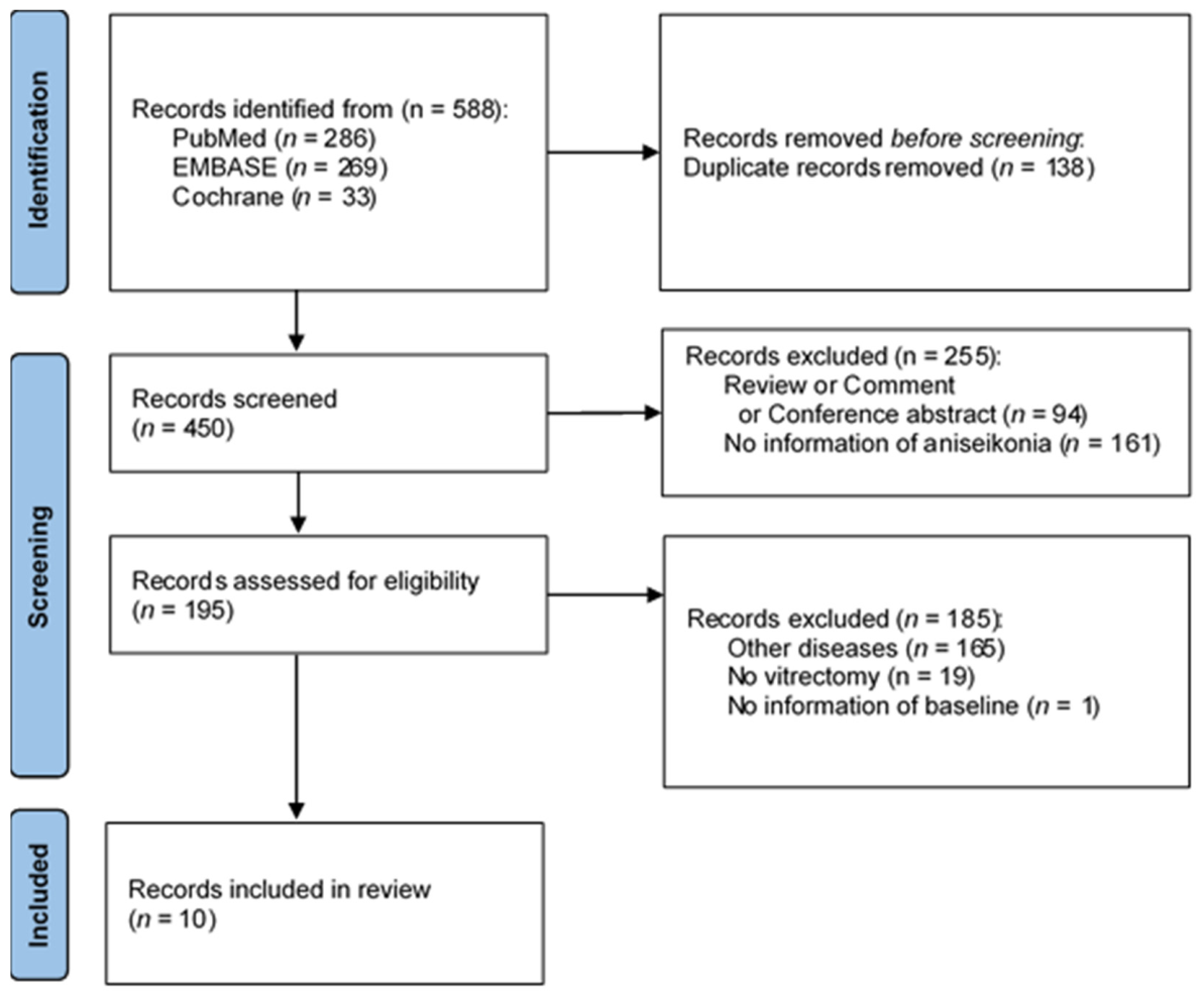
Of the 588 articles initially identified, duplicates were removed and titles and abstracts were screened independently by two investigators, resulting in 433 potentially relevant studies. Full-text reviews further narrowed the selection to 10 eligible studies that focused exclusively on idiopathic ERM (
Figure 1). Any discrepancies in the study selection were resolved through discussion.
We have retrospectively registered this study in a public registry, PROSPERO. The registration record number is 1336412.
2.2. Data Extraction
The data extracted from each study are presented in
Table 1. The key attributes included the first author, publication year, country of study, study design, number of eyes included, follow-up duration, aniseikonia measurement methods, and outcomes assessed. Additional methodological details, such as the gauge of PPV instrumentation and analyses of macular configurations, including central macular thickness (CMT), foveal avascular zone (FAZ), ellipsoid zone disruption (EZD), ectopic inner foveal layer (EIFL), inner retinal layers including the inner nuclear layer (INL), and ganglion cell-inner plexiform layer (GCIPL), were also extracted.
The results were categorized based on the statistical significance of aniseikonia changes, PPV gauge used, and follow-up duration. For studies reporting best-corrected visual acuity (BCVA) in the Snellen format, values were converted into logarithm of the minimum angle of resolution (logMAR) using the formula (logMAR = −1 × log10 [Snellen fraction]). Numerical data presented only in figure format were extracted using a Web Plot Digitizer (version 4.4; Automeris LLC, Austin, TX, USA). The term “end of the study” refers to the completion of each trial or the final follow-up point.
2.3. Quality Assessment
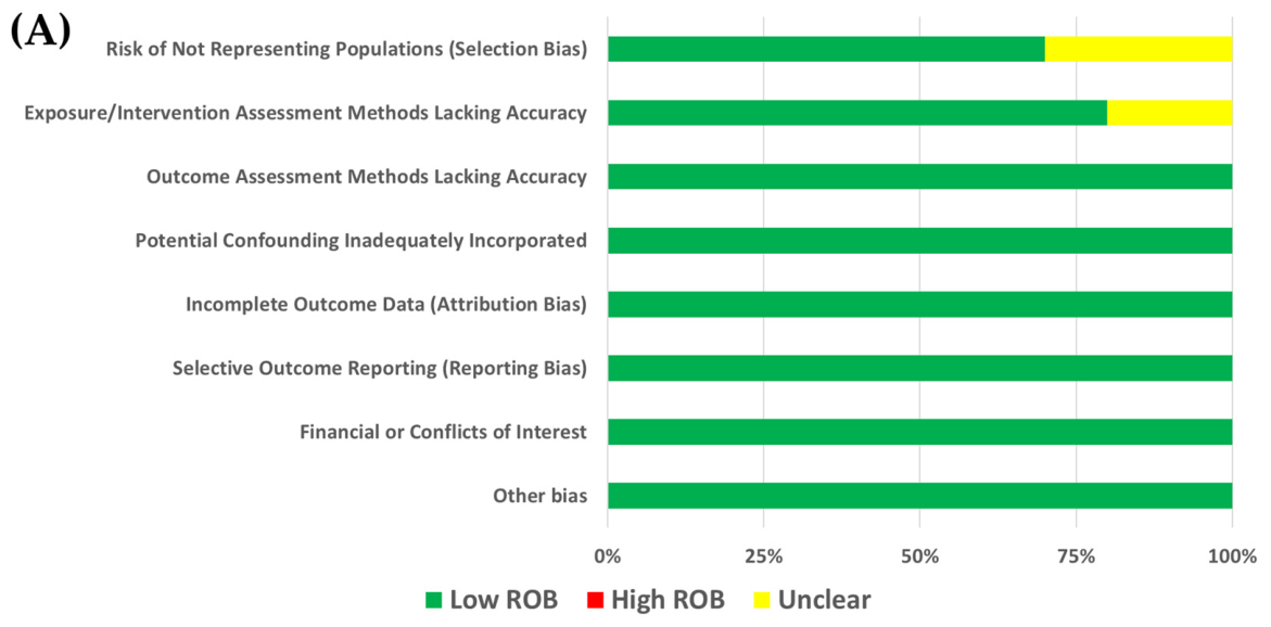
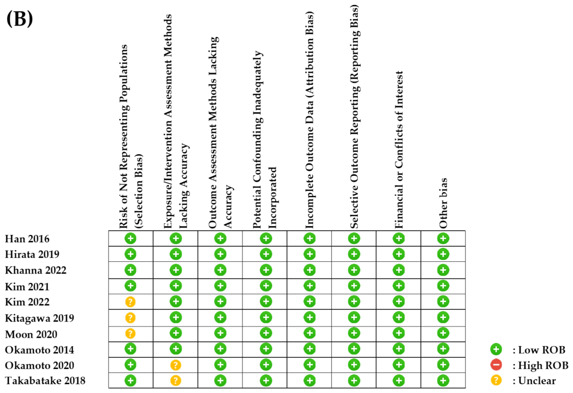
The Newcastle–Ottawa Scale was used to assess the quality of the included studies. Each study was evaluated for participant selection, cohort comparability, and outcomes. Studies that lacked comparative designs or did not clearly report participant retention rates were rated as having an unclear risk in specific Newcastle–Ottawa Scale domains. Two investigators independently performed quality assessment and resolved disagreements by consensus. A summary of the quality assessment is shown in
Figure 2.
2.4. Statistical Analyses
The statistical analyses adhered to the Preferred Reporting Items for Systematic Reviews and Meta-Analyses (PRISMA) guidelines. Data were analyzed using RevMan version 5.3 (Cochrane, London, UK). Continuous outcomes were synthesized using a random-effects model based on the inverse variance method to calculate pooled mean differences with 95% confidence intervals. The random-effects model was implemented to account for between-study variability. Preoperative and postoperative mean values, standard deviations, and sample sizes reported in each study were directly entered into RevMan for the analysis of aniseikonia outcomes. Heterogeneity was quantified using the I2 statistic, with low, moderate, and severe heterogeneity defined as I2 values of ≤25%, 25–75%, and >75%, respectively.
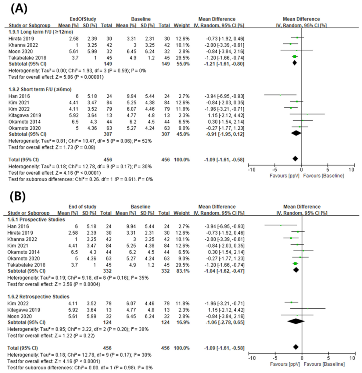
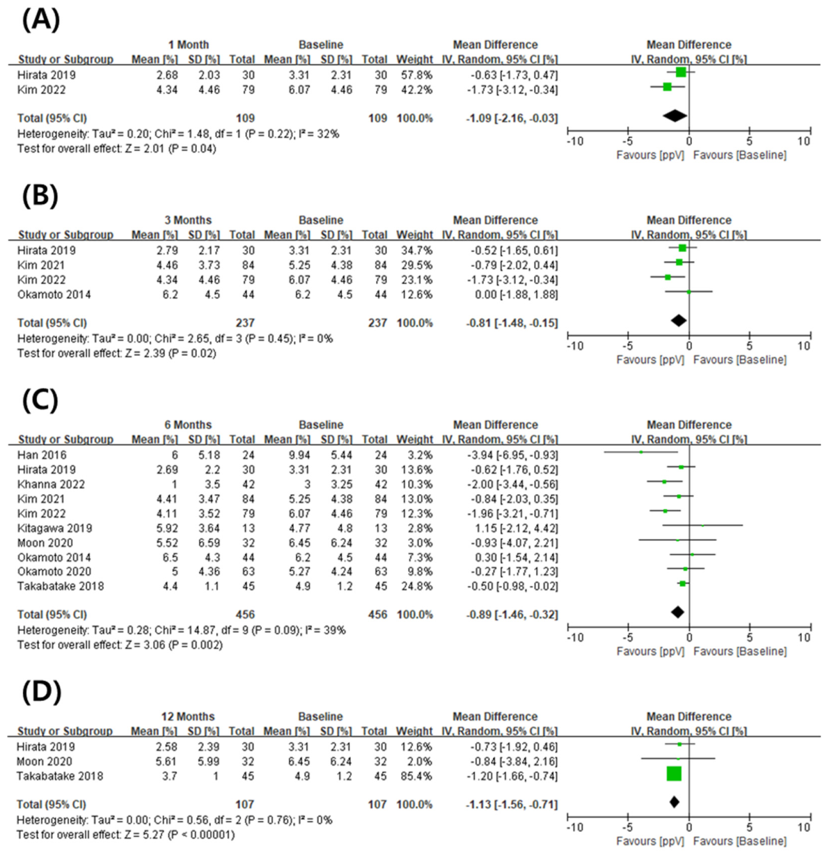
Subgroup analyses were performed to evaluate the impact of the follow-up duration, study design (prospective vs. retrospective), and other methodological differences on aniseikonia outcomes. Forest plots were generated to illustrate the pooled effect sizes and subgroup analyses (
Figure 3,
Figure 4,
Figure 5 and
Figure 6). Correlation analyses between changes in aniseikonia and structural/functional parameters, such as BCVA and CMT, were performed using Spearman’s rank correlation. All statistical tests were two-sided, and the threshold for statistical significance was set to
p < 0.05.
4. Discussion
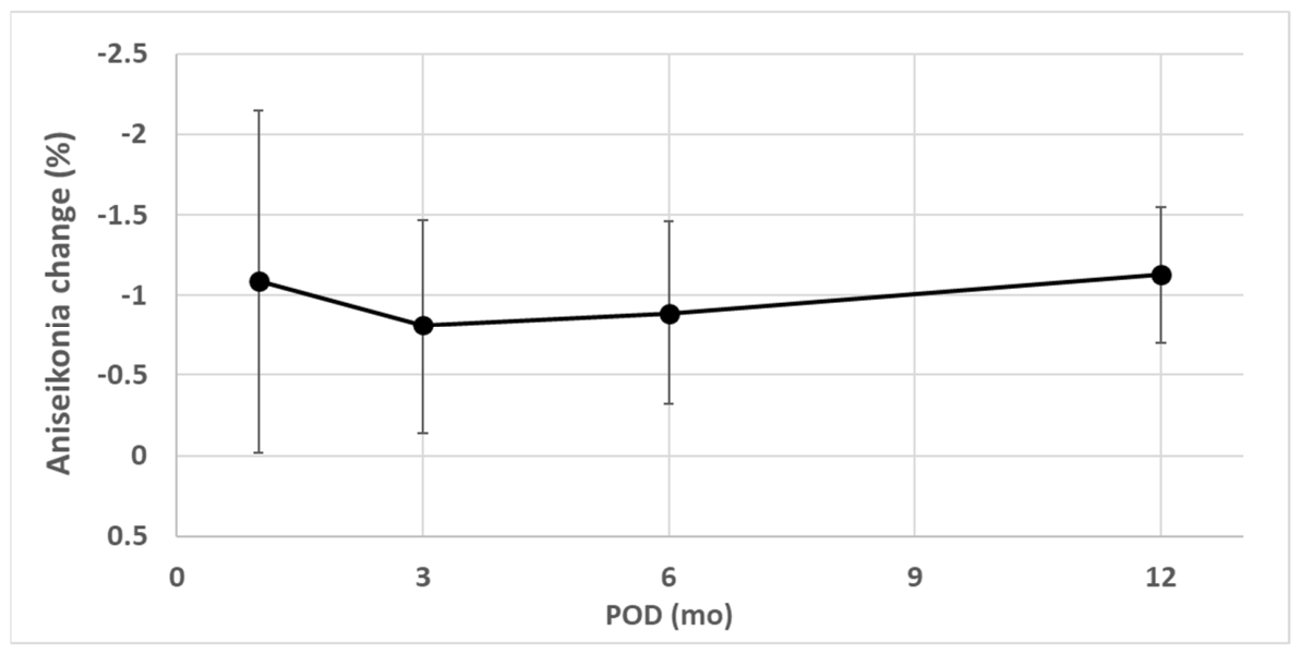
This meta-analysis evaluated longitudinal changes in aniseikonia following PPV for idiopathic ERM and demonstrated a statistically significant but modest reduction over time. Notably, the improvement in aniseikonia appeared to be gradual and nonlinear, with more pronounced changes observed at longer follow-up periods. These findings highlight the importance of long-term follow-up when assessing functional outcomes after PPV. Nevertheless, the magnitude and consistency of this improvement varied across studies.
Because the pooled reduction in aniseikonia was approximately 1%, the clinical significance of this magnitude requires careful interpretation. Previous studies have reported that aniseikonia of 1–2% may be present in normal individuals, whereas levels exceeding 3% are associated with impaired binocular vision and levels above 5% may result in severely compromised binocular function [
22]. In the present analysis, baseline aniseikonia in most included studies ranged from approximately 3% to 6%, suggesting that even modest reductions may shift patients toward a less symptomatic or subclinical range. In addition, recent evidence indicates that even small degrees of aniseikonia may affect stereoacuity [
23], supporting the potential functional relevance of relatively small changes. Even a ~1% reduction may be meaningful for some patients given baseline values of 3–6%. Nevertheless, the clinical impact of a reduction of approximately 1% remains uncertain and should be interpreted with caution.
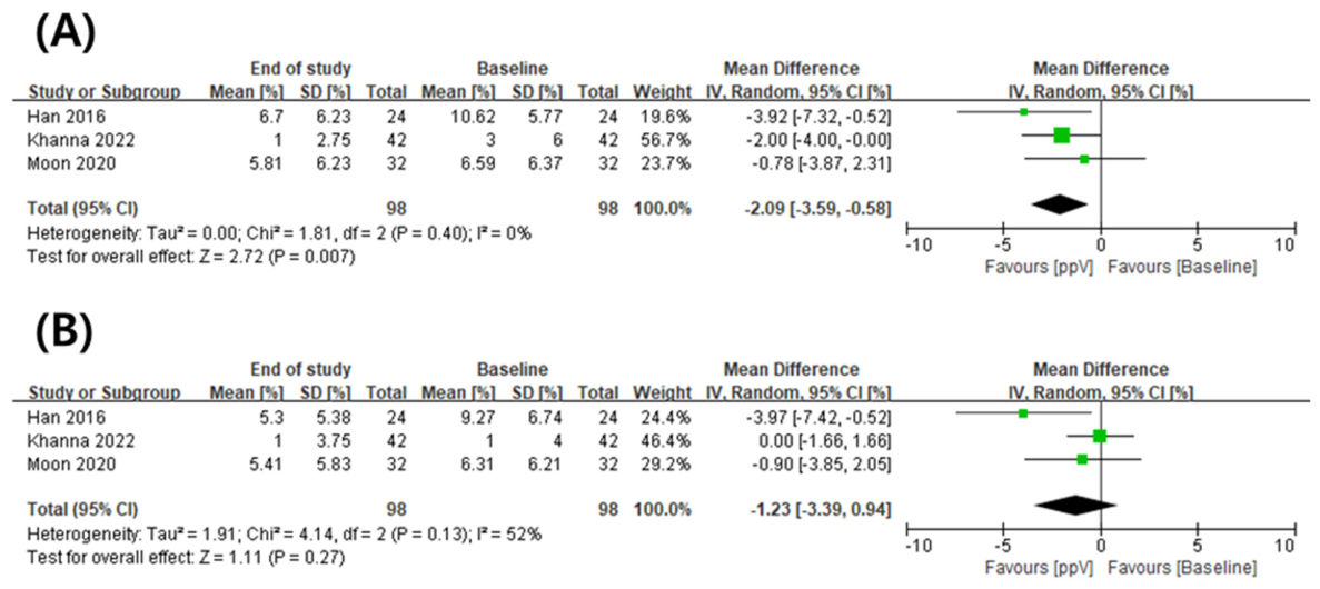
In this context, subgroup analysis based on study design showed that statistically significant improvements in aniseikonia were observed only in prospective studies, whereas retrospective studies did not demonstrate significant changes (
Figure 5B). Given that prospective studies are generally less susceptible to bias, this finding suggests that study design may have influenced the observed effect and should be considered when interpreting the results.
Subgroup analyses based on follow-up duration provided further insight into the temporal pattern of gradual improvement. Statistically significant improvements in aniseikonia were observed only in studies with long-term follow-up (≥12 months), whereas studies with short-term follow-up (≤6 months) did not demonstrate significant changes (
Figure 5A). These findings suggest that a minimum follow-up period of at least 12 months may be required to capture the full extent of aniseikonia improvement after PPV for idiopathic ERM. This delayed recovery pattern is consistent with previous reports indicating that, while metamorphopsia may improve relatively early after surgery, aniseikonia tends to recover more slowly and progressively over time [
21]. However, these subgroup analyses should also be interpreted cautiously. The results may partly reflect differences in study design, baseline severity, or statistical power rather than a true delayed recovery effect.
The mechanisms underlying aniseikonia improvement after PPV are likely multifactorial and remain incompletely understood. Structural factors such as retinal displacement and photoreceptor realignment following membrane removal may play an important role. In addition, cortical adaptation may contribute to the progressive improvement observed over time, as the visual system adjusts to altered retinal input after surgery. These mechanisms may help explain the delayed and nonlinear recovery pattern identified in this study; however, the proposed pathophysiological mechanisms remain speculative and were not directly supported by the included data.
The present analysis also revealed a distinct improvement in vertical aniseikonia postoperatively, whereas horizontal aniseikonia did not show significant changes (
Figure 6). This finding contrasts with the metamorphopsia outcomes reported in previous studies, where horizontal metamorphopsia showed statistically significant improvement following PPV in patients with ERM and vertical metamorphopsia did not demonstrate meaningful changes during a 12-month follow-up period [
21,
24,
25]. Additionally, the study by Ichikawa et al. [
6] suggested that aniseikonia could be influenced by retinal displacement in both the tangential and axial planes, potentially differentially affecting directional improvement in individual cases. Predominant tangential traction vectors in ERM may also contribute to directional differences in retinal displacement and postoperative visual perception changes. This directional discrepancy suggests that mechanisms underlying aniseikonia improvement are likely complex and cannot be fully explained by uniform structural recovery alone. Nevertheless, the conclusion regarding vertical versus horizontal aniseikonia may be premature given the limited number of available studies and the potential variability in measurement methodologies.
Interestingly, despite clear improvements in both BCVA and CMT, this meta-analysis did not identify significant correlations between changes in these parameters and changes in aniseikonia (
Supplementary Figures S1 and S2,
Table 2). Similarly, preoperative BCVA and CMT were not associated with postoperative changes in aniseikonia. These findings suggest that anatomical recovery and visual acuity improvement do not necessarily translate into proportional improvements in perceptual image size disparity. This dissociation highlights the complexity of aniseikonia and indicates that it may represent a distinct visual outcome that cannot be fully explained by conventional structural or functional metrics.
In line with the lack of correlation between aniseikonia and conventional parameters, several macular microstructural parameters, including INL thickness, GCIPL thickness, and EZD, showed significant improvements at the final follow-up (
Supplementary Figure S3B–D). However, no statistically significant associations were found between changes in aniseikonia and changes in these parameters (Spearman’s rank correlation analysis;
p = 0.167, 0.167, and 0.667, respectively). In addition, FAZ did not show a significant change over time (
Supplementary Figure S3A), nor was it correlated with aniseikonia changes (Spearman’s rank correlation analysis;
p = 0.333). These analyses were based on a limited number of studies (n = 3 for each parameter), which may have reduced the statistical power to detect potential associations. Taken together, these findings suggest that currently measurable structural parameters may not fully account for the mechanisms underlying aniseikonia improvement.
However, these analyses were conducted at the study level and are therefore subject to potential ecological bias and limited statistical power. Accordingly, these findings should be interpreted cautiously and considered exploratory.
Differences between idiopathic and secondary ERM may also contribute to variability in reported outcomes. Studies that did not differentiate between idiopathic and secondary ERM, such as those by Fukuyama et al. [
5], Ichikawa et al. [
6], Nakashizuka et al. [
7], and Okamoto et al. [
8], consistently reported no significant postoperative improvement in aniseikonia. Although these studies did not meet the inclusion criteria, their findings suggest that differences between idiopathic and secondary ERM may influence postoperative aniseikonia outcomes. In contrast, Kim et al. [
9] demonstrated that significant improvements occurred only in cases of idiopathic ERM, whereas cases of secondary ERM, such as those associated with retinal breaks, showed no significant changes. This may be related to differences in underlying pathophysiology, as idiopathic ERM is typically associated with posterior vitreous detachment and relatively uniform traction, whereas secondary ERM may involve more complex and heterogeneous retinal changes [
10,
17,
21]. However, the hypothesis that idiopathic ERM may permit more stable microstructural recovery than secondary ERM remains speculative and was not directly supported by the included data.
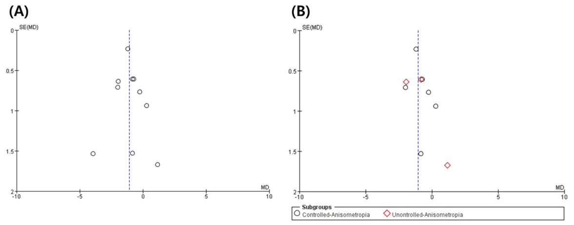
Despite these findings, the underlying mechanisms explaining why aniseikonia improves after ERM surgery remain incompletely understood. No specific macular configuration was identified as being consistently associated with changes in aniseikonia, and half of the included studies did not demonstrate statistically significant improvement, highlighting the variability and uncertainty of the observed effect. In addition, the inclusion of studies with varying methodological rigor, particularly those that did not adequately control for anisometropia, may have influenced the reliability of the pooled estimates. As such, the results of this meta-analysis should be interpreted with caution and considered exploratory.
From a clinical perspective, these findings suggest that improvements in aniseikonia may evolve over time and may not be fully apparent within the early postoperative period. Therefore, clinicians should consider long-term follow-up when evaluating functional outcomes and should counsel patients regarding the expected timeline of recovery.
Several limitations of this study should be acknowledged. First, the number of included studies was relatively small, and the absence of randomized controlled trials limits the strength of the evidence. Second, most studies were conducted in East Asia, which may limit the generalizability of the findings. Third, variability in measurement methods (e.g., NAT vs. AI) and surgical techniques such as internal limiting membrane peeling, differences in PPV gauge size and baseline severity of aniseikonia, and the inclusion of combined procedures such as cataract surgery may have contributed to heterogeneity and may significantly influence visual perception outcomes. Finally, anisometropia is a major confounding factor in aniseikonia assessment, yet control for anisometropia was inconsistent across the included studies and may have influenced the reliability and comparability of the pooled estimates. These factors should be considered when interpreting the pooled estimates.
Sensitivity analyses were performed to address some of these concerns, including stratification based on anisometropia control and measurement methods (
Figure 7B and
Supplementary Figure S4). Although the overall direction of the findings remained broadly similar, methodological differences—including variability in measurement methods, surgical techniques, and anisometropia control—likely contributed substantially to inter-study heterogeneity and warrant careful consideration when interpreting the pooled estimates.
Future studies with larger sample sizes and standardized methodologies are needed to better elucidate the mechanisms underlying aniseikonia improvement and to identify reliable predictors of postoperative outcomes. In particular, further research is needed to clarify the relationship between macular microstructural changes and aniseikonia, which remains poorly understood.




 ,
,