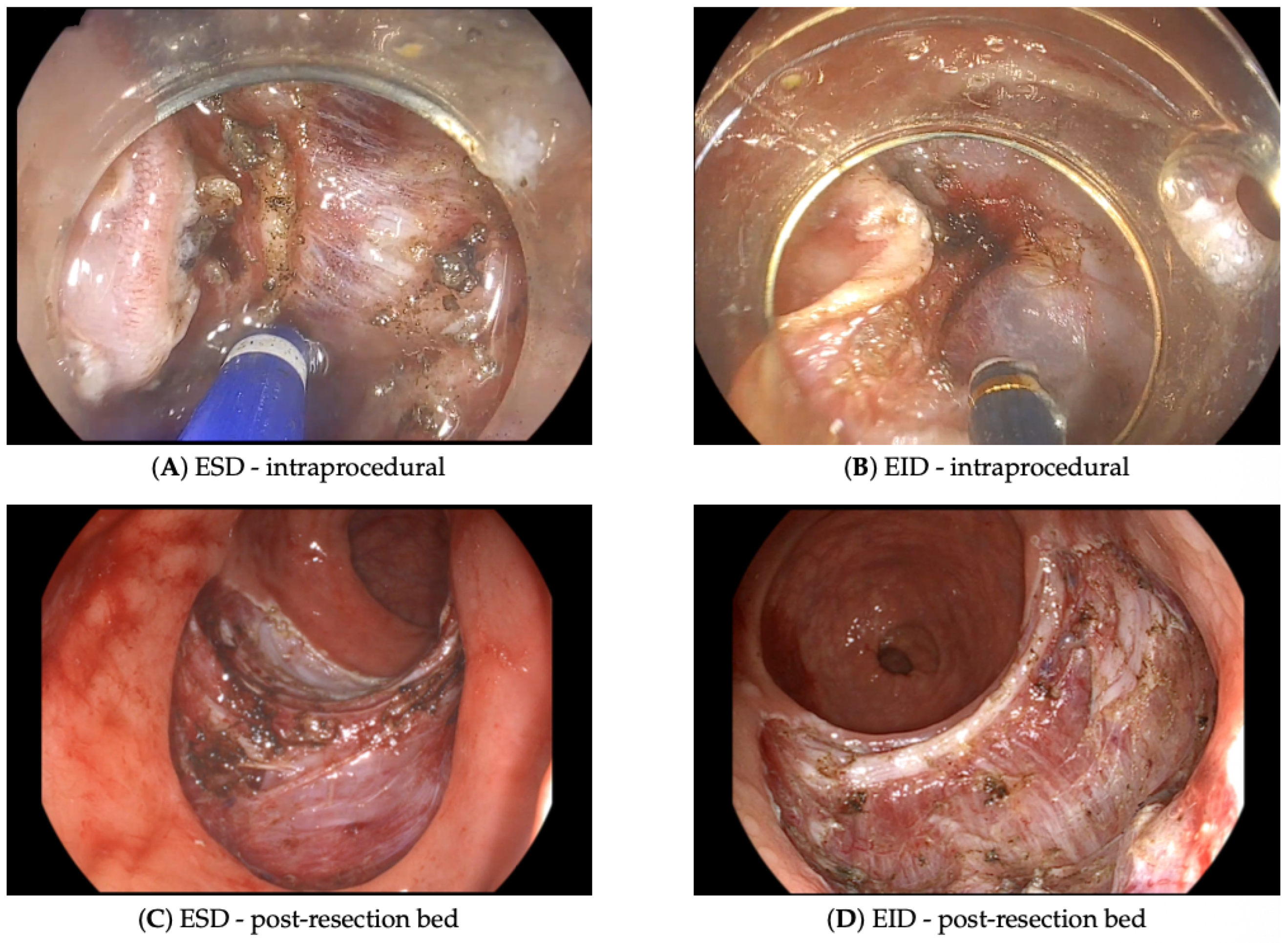
Endoscopic Resection of Rectal Neuroendocrine Tumors: How Deep Should We Go?
 , , , , , and
Abstract
1. Introduction
2. Materials and Methods
3. Results
4. Discussion
5. Conclusions
Author Contributions
Funding
Institutional Review Board Statement
Informed Consent Statement
Data Availability Statement
Acknowledgments
Conflicts of Interest
Abbreviations
| NET | Neuroendocrine tumor |
| r-NET | Rectal neuroendocrine tumor |
| ESD | Endoscopic submucosal dissection |
| EID | Endoscopic intermuscular dissection |
| EMR | Endoscopic mucosal resection |
| L-EMR | Ligation-assisted endoscopic mucosal resection |
| P-EMR | Endoscopic mucosal resection after circumferential precutting |
| ENETS | European Neuroendocrine Tumor Society |
| UICC | Union for International Cancer Control |
| AJCC | American Joint Committee on Cancer |
| MRI | Magnetic resonance imaging |
| Ki-67 | Proliferation marker Ki-67 |
| R0 | Microscopically margin-negative resection |
| R1 | Microscopically margin-positive resection |
| G1/G2/G3 | Tumor grade 1/2/3 |
References
- Dasari, A.; Shen, C.; Halperin, D.; Zhao, B.; Zhou, S.; Xu, Y.; Shih, T.; Yao, J.C. Trends in the Incidence, Prevalence, and Survival Outcomes in Patients with Neuroendocrine Tumors in the United States. JAMA Oncol. 2017, 3, 1335. [Google Scholar] [CrossRef]
- Wang, X.Y.; Chai, N.L.; Linghu, E.Q.; Li, H.K.; Zhai, Y.Q.; Feng, X.X.; Zhang, W.G.; Zou, J.L.; Li, L.S.; Xiang, J.Y. Efficacy and safety of hybrid endoscopic submucosal dissection compared with endoscopic submucosal dissection for rectal neuroendocrine tumors and risk factors associated with incomplete endoscopic resection. Ann. Transl. Med. 2020, 8, 368. [Google Scholar] [CrossRef]
- Gallo, C.; Rossi, R.E.; Cavalcoli, F.; Barbaro, F.; Boškoski, I.; Invernizzi, P.; Massironi, S. Rectal neuroendocrine tumors: Current advances in management, treatment, and surveillance. World J. Gastroenterol. 2022, 28, 1123–1138. [Google Scholar] [CrossRef] [PubMed]
- Kaminski, M.; Polkowski, M.; Regula, J. Prevalence and endoscopic features of rectal neuroendocrine tumors (carcinoids) among 50148 participants of the Polish colorectal-cancer screening programme. Gut 2007, 56, A310. [Google Scholar]
- Moon, C.M.; Huh, K.C.; Jung, S.A.; Park, D.I.; Kim, W.H.; Jung, H.M.; Koh, S.J.; Kim, J.O.; Jung, Y.; Kim, K.O.; et al. Long-Term Clinical Outcomes of Rectal Neuroendocrine Tumors According to the Pathologic Status After Initial Endoscopic Resection: A KASID Multicenter Study. Am. J. Gastroenterol. 2016, 111, 1276–1285. [Google Scholar] [CrossRef]
- Fine, C.; Roquin, G.; Terrebonne, E.; Lecomte, T.; Coriat, R.; Do Cao, C.; de Mestier, L.; Coffin, E.; Cadiot, G.; Nicolli, P.; et al. Endoscopic management of 345 small rectal neuroendocrine tumours: A national study from the French group of endocrine tumours (GTE). United Eur. Gastroenterol. J. 2019, 7, 1102–1112. [Google Scholar] [CrossRef]
- Folkert, I.W.; Sinnamon, A.J.; Concors, S.J.; Bennett, B.J.; Fraker, D.L.; Mahmoud, N.N.; Metz, D.C.; Stashek, K.M.; Roses, R.E. Grade is a Dominant Risk Factor for Metastasis in Patients with Rectal Neuroendocrine Tumors. Ann. Surg. Oncol. 2019, 27, 855–863. [Google Scholar] [CrossRef] [PubMed]
- Fields, A.C.; McCarty, J.C.; Ma-Pak, L.; Lu, P.; Irani, J.; Goldberg, J.E.; Bleday, R.; Chan, J.; Melnitchouk, N. New lymph node staging for rectal neuroendocrine tumors. J. Surg. Oncol. 2018, 119, 156–162. [Google Scholar] [CrossRef] [PubMed]
- Ramage, J.; De Herder, W.; Delle Fave, G.; Ferolla, P.; Ferone, D.; Ito, T.; Ruszniewski, P.; Sundin, A.; Weber, W.; Zheng-Pei, Z.; et al. ENETS Consensus Guidelines Update for Colorectal Neuroendocrine Neoplasms. Neuroendocrinology 2016, 103, 139–143. [Google Scholar] [CrossRef] [PubMed]
- Son, H.J.; Sohn, D.K.; Hong, C.W.; Han, K.S.; Kim, B.C.; Park, J.W.; Choi, H.S.; Chang, H.J.; Oh, J.H. Factors associated with complete local excision of small rectal carcinoid tumor. Int. J. Color. Dis. 2012, 28, 57–61. [Google Scholar] [CrossRef]
- Pan, J.; Zhang, X.; Shi, Y.; Pei, Q. Endoscopic mucosal resection with suction vs. endoscopic submucosal dissection for small rectal neuroendocrine tumors: A meta-analysis. Scand. J. Gastroenterol. 2018, 53, 1139–1145. [Google Scholar] [CrossRef]
- Matsuhashi, N.; Takahashi, T.; Tomita, H.; Araki, H.; Ibuka, T.; Tanaka, K.; Tanahashi, T.; Matsui, S.; Sasaki, Y.; Tanaka, Y.; et al. Evaluation of treatment for rectal neuroendocrine tumors sized under 20 mm in comparison with the WHO 2010 guidelines. Mol. Clin. Oncol. 2017, 7, 476–480. [Google Scholar] [CrossRef][Green Version]
- Zhou, X.; Xie, H.; Xie, L.; Li, J.; Cao, W.; Fu, W. Endoscopic resection therapies for rectal neuroendocrine tumors: A systematic review and meta-analysis. J. Gastroenterol. Hepatol. 2014, 29, 259–268. [Google Scholar] [CrossRef]
- Yu, Q.; Zhang, Y.; Su, Y.; Zhao, Q.; Xiong, K.; Zhang, L.; Fang, H. Optimization of Endoscopic Submucosal Dissection and Endoscopic Mucosal Resection Strategies for Rectal Neuroendocrine Tumors Within 20 mm. Am. Surg. 2024, 90, 1176–1186. [Google Scholar] [CrossRef] [PubMed]
- Frydman, A.; Srirajaskanthan, R. An Update on the Management of Rectal Neuroendocrine Neoplasms. Curr. Treat. Options Oncol. 2024, 25, 1461–1470. [Google Scholar] [CrossRef] [PubMed]
- Sun, P.; Zheng, T.; Hu, C.; Gao, T.; Ding, X. Comparison of endoscopic therapies for rectal neuroendocrine tumors: Endoscopic submucosal dissection with myectomy versus endoscopic submucosal dissection. Surg. Endosc. 2021, 35, 6374–6378. [Google Scholar] [CrossRef]
- Liao, S.; Li, B.; Huang, L.; Qiu, Q.; Yang, G.; Ren, J.; Huang, S. Endoscopic intermuscular dissection in the management of a rectal neuroendocrine tumor. Endoscopy 2023, 55, E977–E979. [Google Scholar] [CrossRef] [PubMed]
- Dang, H.; Hardwick, J.C.; Boonstra, J.J. Endoscopic intermuscular dissection with intermuscular tunneling for local resection of rectal cancer with deep submucosal invasion. VideoGIE 2022, 7, 273–277. [Google Scholar] [CrossRef]
- Rahni, D.; Toyonaga, T.; Ohara, Y.; Lombardo, F.; Baba, S.; Takihara, H.; Tanaka, S.; Kawara, F.; Azuma, T. First reported case of per anal endoscopic myectomy (PAEM): A novel endoscopic technique for resection of lesions with severe fibrosis in the rectum. Endosc. Int. Open 2017, 5, E146–E150. [Google Scholar] [CrossRef]
- Yang, G.; Huang, S.; Li, B.; Deng, H.; Liao, S. Efficacy of endoscopic intermuscular dissection vs. endoscopic submucosal dissection in treating rectal neuroendocrine tumors <10 mm. Endosc. Int. Open 2025, 13, a26415725. [Google Scholar] [CrossRef]
- Han, L.; Hou, H.; Tan, Y.; Li, R.; Liang, C.; Lv, L.; Wang, Y.; Liu, D.; Min, L. Endoscopic intermuscular dissection and endoscopic submucosal dissection for treatment of rectal neuroendocrine tumors: A retrospective cohort study. Minim. Invasive Ther. Allied Technol. 2026, 1–9. [Google Scholar] [CrossRef] [PubMed]
- Deprez, P.H.; Moons, L.M.G.; O’Toole, D.; Gincul, R.; Seicean, A.; Pimentel-Nunes, P.; Fernández Esparrach, G.; Polkowski, M.; Vieth, M.; Borbath, I.; et al. Endoscopic management of subepithelial lesions including neuroendocrine neoplasms: European Society of Gastrointestinal Endoscopy (ESGE) Guideline. Endoscopy 2022, 54, 412–429. [Google Scholar] [CrossRef]
- Park, S.B.; Kim, D.J.; Kim, H.W.; Choi, C.W.; Kang, D.H.; Kim, S.J.; Nam, H.S. Is endoscopic ultrasonography essential for endoscopic resection of small rectal neuroendocrine tumors? World J. Gastroenterol. 2017, 23, 2037–2043. [Google Scholar] [CrossRef]
- Spychalski, M.; Włodarczyk, M.; Winter, K.; Włodarczyk, J.; Dąbrowski, I.; Dziki, A. Outcomes of 601 Colorectal Endoscopic Submucosal Dissections in a Single Western Center: Is Right Colon Location Still a Major Concern? Surg. Laparosc. Endosc. Percutan. Tech. 2021, 31, 578–583. [Google Scholar] [CrossRef] [PubMed]
- Spychalski, M.; Skulimowski, A.; Dziki, A.; Saito, Y. Colorectal endoscopic submucosal dissection (ESD) in the West – when can satisfactory results be obtained? A single-operator learning curve analysis. Scand. J. Gastroenterol. 2017, 52, 1442–1452. [Google Scholar] [CrossRef]
- Chen, J.; Ye, J.; Zheng, X.; Chen, J. Endoscopic treatments for rectal neuroendocrine tumors: A systematic review and network meta-analysis. J. Gastrointest. Surg. 2024, 28, 301–308. [Google Scholar] [CrossRef]
- Han, L.; Li, J.; Liang, C.; Chu, Y.; Wang, Y.; Lv, L.; Liu, D.; Tan, Y. Risk factors for positive resection margins after endoscopic resection for gastrointestinal neuroendocrine tumors. Surg. Endosc. 2024, 38, 2041–2049. [Google Scholar] [CrossRef] [PubMed]
- Binmoeller, K.F. Underwater EMR without submucosal injection: Is less more? Gastrointest. Endosc. 2019, 89, 1117–1119. [Google Scholar] [CrossRef]


| Variable | Category | N (%)/ Mean/Median | SD/IQR | Min | Max |
|---|---|---|---|---|---|
| Demographics and anthropometrics | |||||
| Age (years) | 57.48 a | 13.78 | 24 | 81 | |
| Sex | Male | 23 (54.76%) | – | – | – |
| Female | 19 (45.24%) | – | – | – | |
| Weight (kg) | 81.40 a | 15.44 | 53 | 125 | |
| Height (cm) | 169.36 a | 8.49 | 154 | 186 | |
| Body mass index (kg/m2) | 28.38 a | 5.12 | 20.05 | 43.25 | |
| Comorbidities and medications | |||||
| Nicotine dependence | 4 (9.52%) | – | – | – | |
| Arterial hypertension | 20 (47.62%) | – | – | – | |
| Diabetes mellitus | 4 (9.52%) | – | – | – | |
| Asthma or COPD | 3 (7.14%) | – | – | – | |
| Atrial fibrillation | 2 (4.76%) | – | – | – | |
| Other diseases | 23 (54.76%) | – | – | – | |
| Use of any medications | 34 (80.95%) | – | – | – | |
| Anticoagulants | 2 (4.76%) | – | – | – | |
| Surgical history | |||||
| Previous surgeries | 28 (66.67%) | – | – | – | |
| Abdominal surgeries | 17 (40.48%) | – | – | – | |
| Other surgeries | 15 (35.71%) | – | – | – | |
| Preoperative biopsy or polypectomy | 37 (88.10%) | – | – | – | |
| Procedure and perioperative course | |||||
| Procedure | ESD | 32 (76.19%) | – | – | – |
| EID | 10 (23.81%) | – | – | – | |
| Knife | Pentax M-knife | 20 (47.62%) | – | – | – |
| DualKnife | 10 (23.81%) | – | – | – | |
| FlushKnife | 7 (16.67%) | – | – | – | |
| Goldknife | 4 (9.52%) | – | – | – | |
| HybridKnife | 1 (2.38%) | – | – | – | |
| Distance from anal verge (cm) (n = 38) | 5.00 b | 5.00–8.00 | 2 | 12 | |
| Procedure time (min) | 20.00 b | 11.25–20.00 | 10 | 30 | |
| En bloc resection | 42 (100.00%) | – | – | – | |
| Postoperative day of discharge | 1.00 b | 1.00–1.00 | 0 | 2 | |
| Delayed discharge (non-ESD-related) | 1 (2.38%) | – | – | – | |
| Intraprocedural bleeding | 5 (11.90%) | – | – | – | |
| Endoscopic clip placement | 5 (11.90%) | – | – | – | |
| Coagulation | 1 (2.38%) | – | – | – | |
| Endoscopic hand suturing | 1 (2.38%) | – | – | – | |
| Lesion and histopathology | |||||
| Lesion status | Primary lesion | 26 (61.90%) | – | – | – |
| Scar | 16 (38.10%) | – | – | – | |
| Diagnosis | NET G1 (R0) | 28 (66.67%) | – | – | – |
| Scar without NET | 14 (33.33%) | – | – | – | |
| Scar outcome | Not applicable | 26 (61.90%) | – | – | – |
| Normal scar without NET | 14 (33.33%) | – | – | – | |
| NET recurrence | 2 (4.76%) | – | – | – | |
| Lesion size (mm) (n = 26) | 5.00 b | 5.00–8.00 | 2 | 15 | |
| Scar size (mm) (n = 4) | 4.00 a | 1.41 | 2 | 5 | |
| Width of mucosal layer (mm) (n = 27) | 17.33 a | 5.85 | 10 | 30 | |
| Deep margin (mm) (n = 15) | 0.70 b | 0.30–1.00 | 0.1 | 2.5 | |
| Variable | EID | ESD | p Value |
|---|---|---|---|
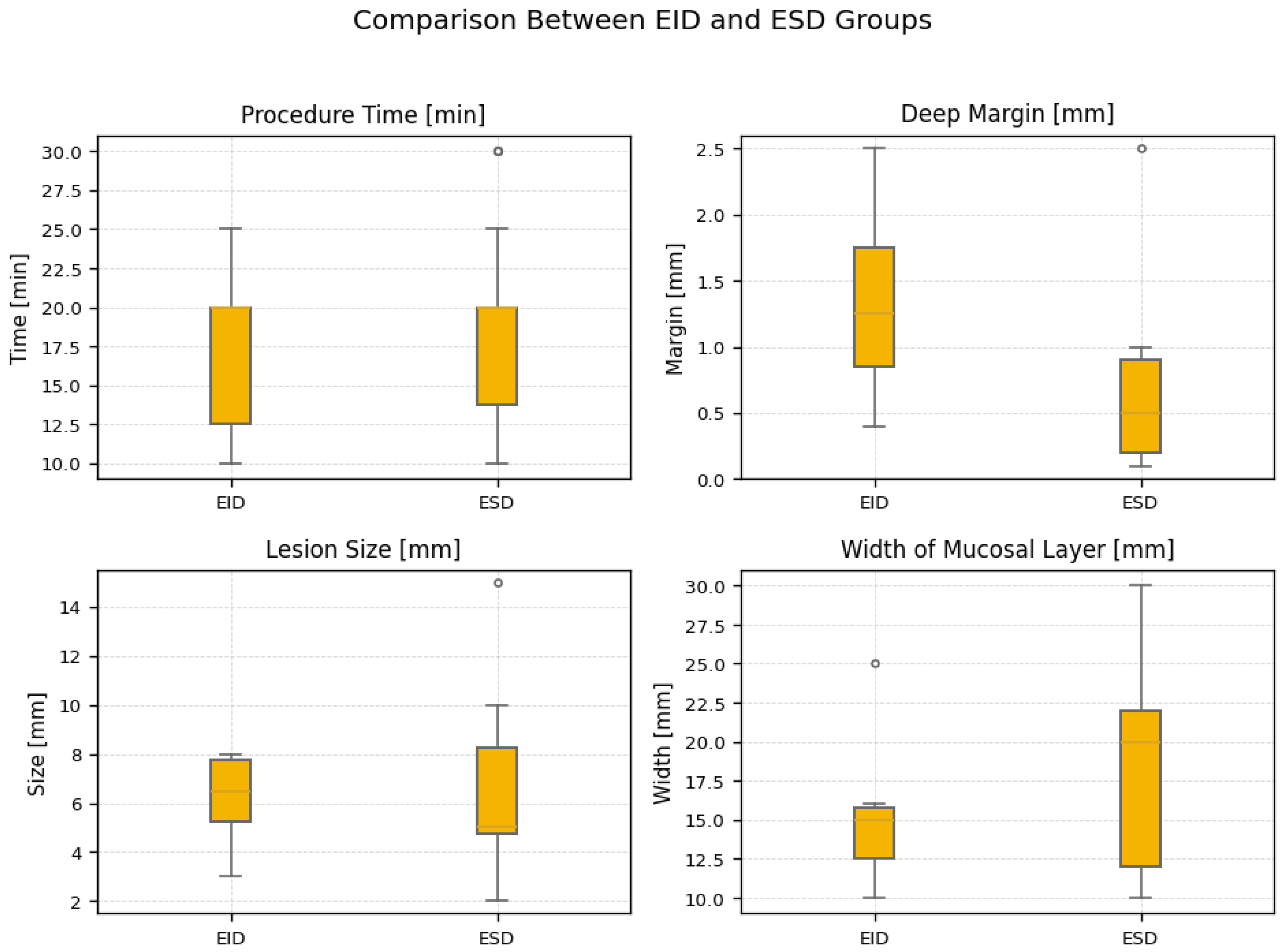
| Width of mucosal layer (mm) | 15.00 (12.50–15.75) | 20.00 (12.00–22.00) | 0.086 |
| Lesion size (mm) | 6.50 (5.25–7.75) | 5.00 (4.75–8.25) | 0.640 |
| Deep margin (mm) | 1.25 (0.85–1.75) | 0.50 (0.20–0.90) | 0.147 |
| Procedure time (min) | 20.00 (12.50–20.00) | 20.00 (13.75–20.00) | 0.658 |
| Category | EID | ESD | p Value |
|---|---|---|---|
| Diagnosis | |||
| NET G1 (R0) | 6 (60.0%) | 22 (68.8%) | 0.71 |
| Scar tissue only | 4 (40.0%) | 10 (31.3%) | |
| Lesion type | |||
| Primary | 6 (60.0%) | 20 (62.5%) | 1.00 |
| Scar | 4 (40.0%) | 12 (37.5%) | |
| Scar outcome | |||
| Not applicable | 6 (60.0%) | 20 (62.5%) | 0.67 |
| No recurrence | 4 (40.0%) | 10 (31.3%) | |
| NET recurrence | 0 (0.0%) | 2 (6.3%) | |
Disclaimer/Publisher’s Note: The statements, opinions and data contained in all publications are solely those of the individual author(s) and contributor(s) and not of MDPI and/or the editor(s). MDPI and/or the editor(s) disclaim responsibility for any injury to people or property resulting from any ideas, methods, instructions or products referred to in the content. |
© 2026 by the authors. Licensee MDPI, Basel, Switzerland. This article is an open access article distributed under the terms and conditions of the Creative Commons Attribution (CC BY) license.
Share and Cite
Maryńczak, K.; Kasprzyk, P.; Pierzchała, K.; Osielczak, A.; Orzeszko, Z.; Dziki, Ł.; Spychalski, M. Endoscopic Resection of Rectal Neuroendocrine Tumors: How Deep Should We Go? J. Clin. Med. 2026, 15, 4103. https://doi.org/10.3390/jcm15114103
Maryńczak K, Kasprzyk P, Pierzchała K, Osielczak A, Orzeszko Z, Dziki Ł, Spychalski M. Endoscopic Resection of Rectal Neuroendocrine Tumors: How Deep Should We Go? Journal of Clinical Medicine. 2026; 15(11):4103. https://doi.org/10.3390/jcm15114103
Chicago/Turabian StyleMaryńczak, Kasper, Przemysław Kasprzyk, Karol Pierzchała, Aleksandra Osielczak, Zofia Orzeszko, Łukasz Dziki, and Michał Spychalski. 2026. "Endoscopic Resection of Rectal Neuroendocrine Tumors: How Deep Should We Go?" Journal of Clinical Medicine 15, no. 11: 4103. https://doi.org/10.3390/jcm15114103
APA StyleMaryńczak, K., Kasprzyk, P., Pierzchała, K., Osielczak, A., Orzeszko, Z., Dziki, Ł., & Spychalski, M. (2026). Endoscopic Resection of Rectal Neuroendocrine Tumors: How Deep Should We Go? Journal of Clinical Medicine, 15(11), 4103. https://doi.org/10.3390/jcm15114103