
The Role of Periarticular Knee Muscle Torques in Ensuring the Body Balance of Older Adults with Balance Disturbances

 ,
,  and
and
Abstract
1. Introduction
2. Materials and Methods
2.1. Study Design
- Sample: 52 participants aged > 55 years.
- Recruitment: clinical and community sources in Bialystok, Poland.
- Assessments performed:
- General: weight, height, and BMI.
- Clinical: DHI, GDS, POMA, FRT, FES-I, and body composition (bioimpedance).
- Biomechanical: muscle torques of knee flexors and extensors (isometric and isokinetic conditions), and centre of pressure (COP) position changes.
- Group allocation:
- Balance disorders group: ≥ 1 fall in the past year and DHI ≥ 10.
- Control group: no falls and DHI < 10.
- Statistical analysis:
- Shapiro–Wilk and Levene’s tests.
- Group comparisons: t-test or Mann–Whitney U.
- Multiple regression to identify predictors of the COP path.
2.2. Detailed Description of Study Procedures
- Age of 55 years or older at the time of recruitment.
- Ability to ambulate independently without the use of mobility aids (e.g., walkers or wheelchairs).
- No history of acute orthopedic injuries or surgical interventions involving the lower limbs in the previous six months.
- No diagnosed neurological conditions significantly affecting balance (e.g., Parkinson’s disease, stroke, or multiple sclerosis).
- Consent to participate in clinical and biomechanical assessments.
- The Dizziness Handicap Inventory (DHI) is a scale designed to measure the disability perceived by someone complaining of dizziness, vertigo, or unsteadiness. It is a 25-item self-report questionnaire that quantifies the impact of dizziness on daily life by measuring self-perceived handicaps in functional, emotional, and physical categories. The patient is asked to answer each question regarding dizziness or unsteadiness problems, explicitly considering their condition during the last month (No = 0; Sometimes = 2; and Yes = 4 points). Scores above 10 points should be referred to balance specialists for further evaluation [35].
- The 15-item Geriatric Depression Scale (GDS) is an instrument used to assess the possibility of depression in older adults. The patient is asked to answer each question explicitly considering his or her condition during the previous two weeks (0 or 1 point is assigned to the answer depending on the question). Scores above 5 points should be referred to more in-depth clinical evaluation for depression [36].
- The Performance-Oriented Mobility Assessment (POMA) is the first clinical balance assessment tool [37]. It measures an individual’s gait and balance abilities using 16 items of balance and gait. Each item is scored on a 3-point ordinal scale (0–2) with 28 possible points. The balance portion (POMA-B) contains nine items for a maximum score of 16 and the gait portion (POMA-G) includes seven items for a maximum score of 12. The test is reliable, valid, and responsive [38,39,40]. The POMA has excellent test–retest reliability in people with dementia [41].
- The Functional Reach Test (FRT) evaluates the forward stability of a standing subject who voluntarily extends one arm as far forward as possible in the horizontal plane, while keeping both heels in contact with the ground. The score is obtained by measuring the distance between the fingertip’s starting and end positions. The FRT’s good reliability and low inter-examiner variability have been demonstrated in various studies [42,43]. The FRT score correlates with the anteroposterior excursion of the centre of pressure (COP) [42,44]. Furthermore, the FRT also shows good reproducibility over time [42,45] and is sensitive to change [46,47]. Lastly, the FRT score is known to decrease with age, and an abnormally low score is a good predictor of the risk of falls [46].
- The Falls Efficacy Scale—International (FES-I), developed and validated by the Prevention of Falls Network Europe (ProFaNE), has become a widely accepted tool for assessing concern about falling [48,49]. Previous studies indicate that the FES-I has excellent reliability and validity across different cultures and languages [50]. The psychometric properties of the FES-I have been evaluated using classical test theory [48,49]. The original questionnaire contains 16 items scored on a four-point scale (1 = not at all concerned to 4 = very concerned). The shortened questionnaire contains seven items [48].
- Body composition assessment with a Jawon Medical X-Contact 357S (Jawon Medical Co., Ltd., Gyeongsan, Republic of Korea) bioimpedance analyzer was used to assess skeletal muscle mass (SMM) and the derived parameter—the difference between the obtained SMM value and its reference value (Diff SMM).
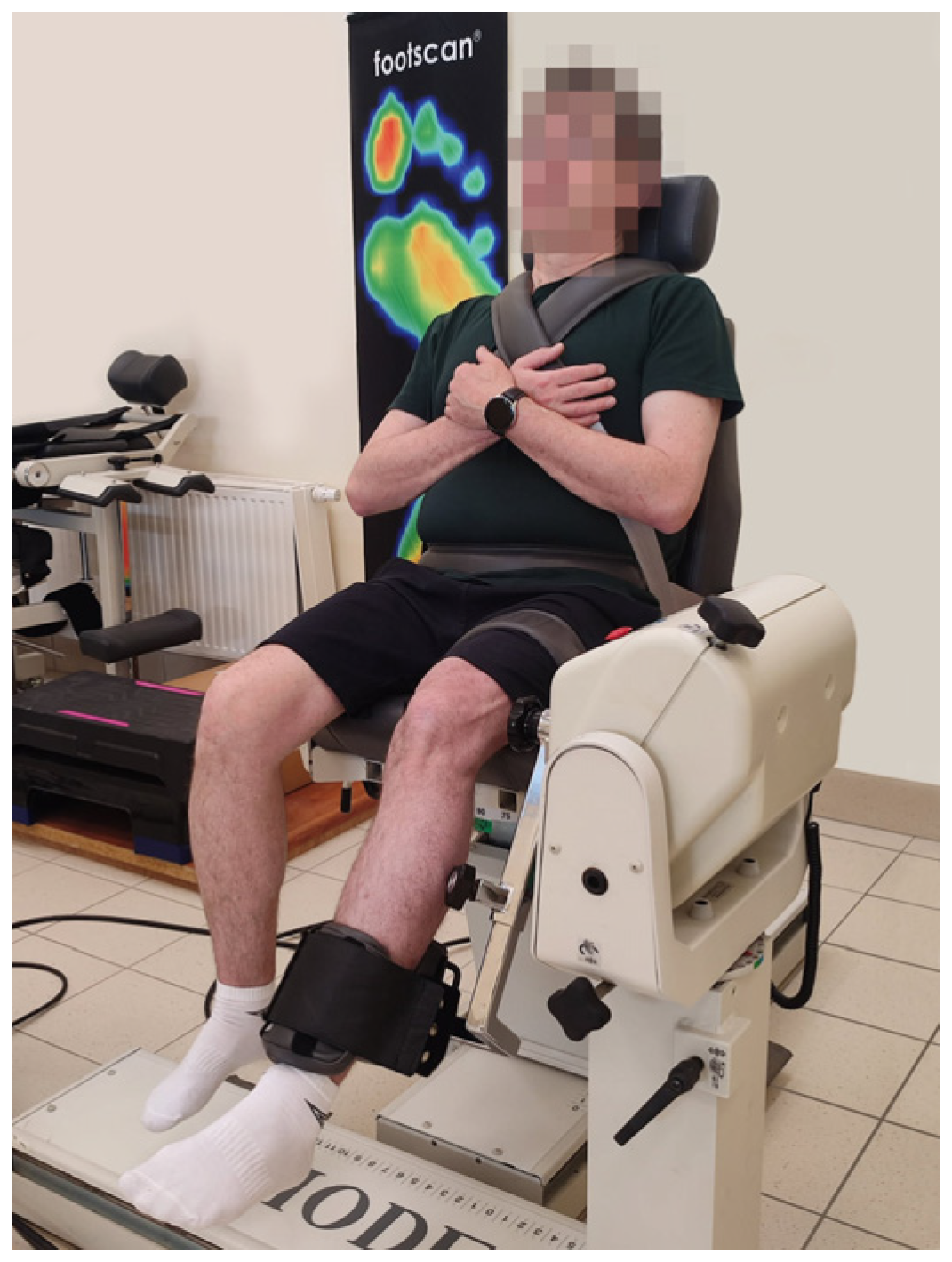
- Muscle torques of knee flexors and extensors developed in isometric and isokinetic conditions.
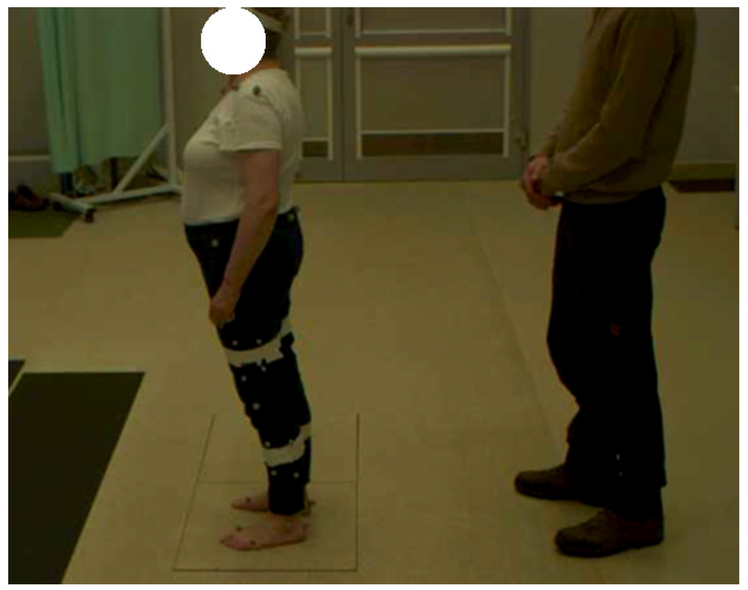
- The centre of pressure (COP) position changes while standing with eyes open and closed.
3. Results
4. Discussion
5. Limitations of the Study and Future Directions
6. Conclusions
Author Contributions
Funding
Institutional Review Board Statement
Informed Consent Statement
Data Availability Statement
Acknowledgments
Conflicts of Interest
Abbreviations
| DHI | Dizziness Handicap Inventory |
| GDS | Geriatric Depression Scale |
| POMA | Performance-Oriented Mobility Assessment |
| FRT | Functional Reach Test |
| FES-I | Falls Efficacy Scale—International |
| SMM | Skeletal muscle mass |
| Diff SMM | The difference between the obtained SMM value and its reference value |
| COP | Centre of pressure |
| WHO | World Health Organization |
| Ikin | Isokinetic |
| Imet | Isometric |
| Ext | Extensors |
| Flex | Flexors |
| Asym | Asymmetry |
| Amp | Amplitude |
| ML | Mediolateral direction |
| AP | Anteroposterior direction |
| EO | Eyes opened |
| EC | Eyes closed |
References
- World Health Organisation. World Report on Ageing and Health; World Health Organization: Geneva, Switzerland, 2015. [Google Scholar]
- World Health Organisation. Active Ageing: A Policy Framework; World Health Organization: Geneva, Switzerland, 2002. [Google Scholar]
- Crimmins, E.M. Lifespan and Healthspan: Past, Present, and Promise. Gerontologist 2015, 55, 901–911. [Google Scholar] [CrossRef]
- World Health Organisation. Aging and Health; World Health Organization: Geneva, Switzerland, 2024. [Google Scholar]
- Pang, L.; Liu, Y.; Shen, M.; Ye, J.; Chen, R.; Lan, Z.; Wu, Z.; Guo, Y.; Zhang, P. Influence of aging on deterioration of patients with COVID-19. Aging 2020, 12, 26248–26262. [Google Scholar] [CrossRef] [PubMed]
- Quijoux, F.; Vienne-Jumeau, A.; Bertin-Hugault, F.; Zawieja, P.; Lefèvre, M.; Vidal, P.-P.; Ricard, D. Center of pressure displacement characteristics differentiate fall risk in older people: A systematic review with meta-analysis. Ageing Res. Rev. 2020, 62, 101117. [Google Scholar] [CrossRef]
- Rubenstein, L.Z. Falls in older people: Epidemiology, risk factors and strategies for prevention. Age Ageing 2006, 35, ii37–ii41. [Google Scholar] [CrossRef]
- World Health Organisation. WHO Global Report on Falls Prevention in Older Age; World Health Organization: Geneva, Switzerland, 2007. [Google Scholar]
- Mak, T.C.T.; Ng, S.S.M.; Leung, M.C.Y.; Wong, T.W.L. Examining the role of attention focus walking training on conscious motor processing during rehabilitation by older adults at risk of falling: A randomized controlled trial. Arch. Gerontol. Geriatr. 2024, 121, 105352. [Google Scholar] [CrossRef]
- Martinez, P.S.; Lord, S.R.; Close, J.C.T.; Taylor, M.E. Associations between psychotropic and anti-dementia medication use and falls in community-dwelling older adults with cognitive impairment. Arch. Gerontol. Geriatr. 2023, 114, 105105. [Google Scholar] [CrossRef]
- Salari, N.; Darvishi, N.; Ahmadipanah, M.; Shohaimi, S.; Mohammadi, M. Global prevalence of falls in the older adults: A comprehensive systematic review and meta-analysis. J. Orthop. Surg. Res. 2022, 17, 334. [Google Scholar] [CrossRef] [PubMed]
- Deshpande, N.; Metter, E.J.; Lauretani, F.; Bandinelli, S.; Ferrucci, L. Interpreting fear of falling in the elderly: What do we need to consider? J. Geriatr. Phys. Ther. 2009, 32, 91–96. [Google Scholar] [CrossRef] [PubMed]
- Prabhakaran, K.; Gogna, S.; Pee, S.; Samson, D.J.; Con, J.; Latifi, R. Falling Again? Falls in Geriatric Adults-Risk Factors and Outcomes Associated with Recidivism. J. Surg. Res. 2020, 247, 66–76. [Google Scholar] [CrossRef]
- Petersen, N.; König, H.H.; Hajek, A. The link between falls, social isolation and loneliness: A systematic review. Arch. Gerontol. Geriatr. 2020, 88, 104020. [Google Scholar] [CrossRef]
- Meyer, M.; Constancias, F.; Vogel, T.; Kaltenbach, G.; Schmitt, E. Gait Disorder among Elderly People, Psychomotor Disadaptation Syndrome: Post-Fall Syndrome, Risk Factors and Follow-Up—A Cohort Study of 70 Patients. Gerontology 2021, 67, 17–24. [Google Scholar] [CrossRef] [PubMed]
- Pereira, C.L.; Vogelaere, P.; Baptista, F. Role of physical activity in the prevention of falls and their consequences in the elderly. Eur. Rev. Aging Phys. Act. 2008, 5, 51–58. [Google Scholar] [CrossRef]
- Ilic, I.; Ristic, B.; Stojadinovic, I.; Ilic, M. Epidemiology of Hip Fractures Due to Falls. Medicina 2023, 59, 1528. [Google Scholar] [CrossRef] [PubMed]
- Bui, M.; Nijmeijer, W.S.; Hegeman, J.H.; Witteveen, A.; Groothuis-Oudshoorn, C.G.M. Systematic review and meta-analysis of preoperative predictors for early mortality following hip fracture surgery. Osteoporos. Int. 2024, 35, 561–574. [Google Scholar] [CrossRef] [PubMed]
- Chantanachai, T.; Sturnieks, D.L.; Lord, S.R.; Payne, N.; Webster, L.; Taylor, M.E. Risk factors for falls in older people with cognitive impairment living in the community: Systematic review and meta-analysis. Ageing Res. Rev. 2021, 71, 101452. [Google Scholar] [CrossRef]
- Tamaki, C.; Maul, K.; Talian, D.S.; Sparks, S. Deaf Individuals Who Report Having Good Balance Function Present with Significant Vestibular Deficits. J. Am. Acad. Audiol. 2021, 32, 510–520. [Google Scholar] [CrossRef]
- Canever, J.B.; Danielewicz, A.L.; Leopoldino, A.A.O.; Corseuil, M.W.; de Avelar, N.C.P. Gender differentiated score on the Falls Efficacy Scale International (FES-I Brazil) to assess self-efficacy in falls in community-dwelling older adults. Aging Clin. Exp. Res. 2022, 34, 1341–1347. [Google Scholar] [CrossRef]
- Ferrer-Ramos, P.; Garnacho-Castaño, M.V.; Girabent-Farrés, M.; Faundez-Zanuy, M.; Serra-Payá, N. Physical performance tests for preliminary cognitive screening in older adults: A systematic review of strength, walking, and balance assessments. Arch. Gerontol. Geriatr. 2025, 130, 105722. [Google Scholar] [CrossRef]
- Magnuszewski, L.; Wojszel, A.; Kasiukiewicz, A.; Wojszel, Z.B. Falls at the geriatric hospital ward in the context of risk factors of falling detected in a comprehensive geriatric assessment. Int. J. Environ. Res. Public Health 2022, 19, 10789. [Google Scholar] [CrossRef]
- Omaña, H.; Bezaire, K.; Brady, K.; Davies, J.; Louwagie, N.; Power, S.; Santin, S.; Hunter, S.W. Functional Reach Test, Single-Leg Stance Test, and Tinetti Performance-Oriented Mobility Assessment for the Prediction of Falls in Older Adults: A Systematic Review. Phys. Ther. 2021, 101, pzab173. [Google Scholar] [CrossRef]
- Debruin, D.A.; Miksa, K.; Vogrin, S.; Duque, G.; Sales, M.; Hayes, A. Exploring new balance and gait factors that are associated with osteosarcopenia in patients with a previous fall and/or fracture history. Arch. Gerontol. Geriatr. 2024, 117, 105221. [Google Scholar] [CrossRef] [PubMed]
- Lunt, E.; Ong, T.; Gordon, A.L.; Greenhaff, P.L.; Gladman, J.R.F. The clinical usefulness of muscle mass and strength measures in older people: A systematic review. Age Ageing 2021, 50, 88–95. [Google Scholar] [CrossRef] [PubMed]
- Mehdizadeh, S.; van Ooteghem, K.; Gulka, H.; Nabavi, H.; Faieghi, M.; Taati, B.; Iaboni, A. A systematic review of center of pressure measures to quantify gait changes in older adults. Exp. Gerontol. 2021, 143, 111170. [Google Scholar] [CrossRef]
- Quijoux, F.; Nicolaï, A.; Chairi, I.; Bargiotas, I.; Ricard, D.; Yelnik, A.; Oudre, L.; Bertin-Hugault, F.; Vidal, P.-P.; Vayatis, N.; et al. A review of center of pressure (COP) variables to quantify standing balance in elderly people: Algorithms and open-access code. Physiol. Rep. 2021, 9, e15067. [Google Scholar] [CrossRef] [PubMed]
- Richmond, S.B.; Fling, B.W.; Lee, H.; Peterson, D.S. The assessment of center of mass and center of pressure during quiet stance: Current applications and future directions. J. Biomech. 2021, 123, 110485. [Google Scholar] [CrossRef]
- Silva, J.; Atalaia, T.; Martins, R.; Aleixo, P. Centre of pressure kinematics during the Timed Up and Go test in elderly fallers and non-fallers. Gait Posture 2022, 97, S315–S316. [Google Scholar] [CrossRef]
- Gafner, S.C.; Bastiaenen, C.H.G.; Ferrari, S.; Gold, G.; Trombetti, A.; Terrier, P.; Hilfiker, R.; Allet, L. The role of hip abductor strength in identifying older persons at risk of falls: A diagnostic accuracy study. Clin. Interv. Aging 2020, 15, 645–654. [Google Scholar] [CrossRef]
- Ko, S.; Jerome, G.J.; Simonsick, E.M.; Ferrucci, L. Investigating balance-related gait patterns and their relationship with maximum torques generated by the hamstrings and quadriceps in older adults—Results from the Baltimore longitudinal study of aging. Arch. Gerontol. Geriatr. 2024, 123, 105411. [Google Scholar] [CrossRef]
- Kozinc, Ž.; Smajla, D.; Šarabon, N. Relationship between hip abductor strength, rate of torque development scaling factor and medio-lateral stability in older adults. Gait Posture 2022, 95, 264–269. [Google Scholar] [CrossRef]
- Lanza, M.B.; Arbuco, B.; Ryan, A.S.; Shipper, A.G.; Gray, V.L.; Addison, O. Systematic Review of the Importance of Hip Muscle Strength, Activation, and Structure in Balance and Mobility Tasks. Arch. Phys. Med. Rehabil. 2022, 103, 1651–1662. [Google Scholar] [CrossRef]
- Jacobson, G.P.; Newman, C.W. The development of the Dizziness Handicap Inventory. Arch. Otolaryngol. Head Neck Surg. 1990, 116, 424–427. [Google Scholar] [CrossRef] [PubMed]
- Yesavage, J.A. Geriatric Depression Scale. Psychopharmacol. Bull. 1988, 24, 709–711. [Google Scholar] [PubMed]
- Tinetti, M.E. Performance-oriented assessment of mobility problems in elderly patients. J. Am. Geriatr. Soc. 1986, 34, 119–126. [Google Scholar] [CrossRef]
- Kegelmeyer, D.A.; Kloos, A.D.; Thomas, K.M.; Kostyk, S.K. Reliability and validity of the Tinetti Mobility Test for individuals with Parkinson disease. Phys. Ther. 2007, 87, 1369–1378. [Google Scholar] [CrossRef] [PubMed]
- Maki, B.E.; Holliday, P.J.; Topper, A.K. A prospective study of postural balance and risk of falling in an ambulatory and independent elderly population. J. Gerontol. 1994, 49, M72–M84. [Google Scholar] [CrossRef]
- Yang, C.; Mo, Y.; Cao, X.; Zhu, S.; Wang, X.; Wang, X. Reliability and validity of the Tinetti performance oriented mobility assessment in Chinese community-dwelling older adults. Geriatr. Nurs. 2023, 53, 85–89. [Google Scholar] [CrossRef]
- Sterke, C.S.; Huisman, S.L.; van Beeck, E.F.; Looman, C.W.N.; van der Cammen, T.J.M. Is the Tinetti Performance Oriented Mobility Assessment (POMA) a feasible and valid predictor of short-term fall risk in nursing home residents with dementia? Int. Psychogeriatr. 2010, 22, 254–263. [Google Scholar] [CrossRef]
- Duncan, P.W.; Weiner, D.K.; Chandler, J.; Studenski, S. Functional reach: A new clinical measure of balance. J. Gerontol. 1990, 45, M192–M197. [Google Scholar] [CrossRef]
- Hageman, P.A.; Leibowitz, J.M.; Blanke, D. Age and gender effects on postural control measures. Arch. Phys. Med. Rehabil. 1995, 76, 961–965. [Google Scholar] [CrossRef]
- Jonsson, E.; Henriksson, M.; Hirschfeld, H. Does the functional reach test reflect stability limits in elderly people? J. Rehabil. Med. 2003, 35, 26–30. [Google Scholar] [CrossRef]
- Lin, M.-R.; Hwang, H.-F.; Hu, M.-H.; Wu, H.-D.I.; Wang, Y.-W.; Huang, F.-C. Psychometric comparisons of the timed up and go, one-leg stand, functional reach, and Tinetti balance measures in community-dwelling older people. J. Am. Geriatr. Soc. 2004, 52, 1343–1348. [Google Scholar] [CrossRef] [PubMed]
- Okumiya, K.; Matsubayashi, K.; Wada, T.; Kimura, S.; Doi, Y.; Ozawa, T. Effects of exercise on neurobehavioral function in community-dwelling older people more than 75 years of age. J. Am. Geriatr. Soc. 1996, 44, 569–572. [Google Scholar] [CrossRef]
- Weiner, D.K.; Bongiorni, D.R.; Studenski, S.A.; Duncan, P.W.; Kochersberger, G.G. Does functional reach improve with rehabilitation? Arch. Phys. Med. Rehabil. 1993, 74, 796–800. [Google Scholar] [CrossRef]
- Kempen, G.I.J.M.; Yardley, L.; van Haastregt, J.C.M.; Zijlstra, G.A.R.; Beyer, N.; Hauer, K.; Todd, C. The Short FES-I: A shortened version of the falls efficacy scale-international to assess fear of falling. Age Ageing 2008, 37, 45–50. [Google Scholar] [CrossRef] [PubMed]
- Yardley, L.; Beyer, N.; Hauer, K.; Kempen, G.; Piot-Ziegler, C.; Todd, C. Development and initial validation of the Falls Efficacy Scale-International (FES-I). Age Ageing 2005, 34, 614–619. [Google Scholar] [CrossRef] [PubMed]
- Kempen, G.I.; Todd, C.J.; van Haastregt, J.C.M.; Zijlstra, G.A.R.; Beyer, N.; Freiberger, E.; Hauer, K.A.; Piot-Ziegler, C.; Yardley, L. Cross-cultural validation of the Falls Efficacy Scale International (FES-I) in older people: Results from Germany, the Netherlands and the UK were satisfactory. Disabil. Rehabil. 2007, 29, 155–162. [Google Scholar] [CrossRef]
- Avelino-Silva, T.J.; Farfel, J.M.; Curiati, J.A.E.; Amaral, J.R.G.; Campora, F.; Jacob-Filho, W. Comprehensive geriatric assessment predicts mortality and adverse outcomes in hospitalized older adults. BMC Geriatr. 2014, 14, 129. [Google Scholar] [CrossRef]
- Inouye, S.K.; Studenski, S.; Tinetti, M.E.; Kuchel, G.A. Geriatric syndromes: Clinical, research, and policy implications of a core geriatric concept. J. Am. Geriatr. Soc. 2007, 55, 780–791. [Google Scholar] [CrossRef]
- Köpke, S.; Meyer, G. Der Tinetti-Test–Babylon im geriatrischen Assessment: Babylon in geriatric assessment. Z. Für Gerontol. Und Geriatrie 2006, 39, 288–291. [Google Scholar] [CrossRef]
- Milisen, K.; Staelens, N.; Schwendimann, R.; de Paepe, L.; Verhaeghe, J.; Braes, T.; Boonen, S.; Pelemans, W.; Kressig, R.W.; Dejaeger, E. Fall prediction in inpatients by bedside nurses using the St. Thomas’s Risk Assessment Tool in Falling Elderly Inpatients (STRATIFY) instrument: A multicenter study. J. Am. Geriatr. Soc. 2007, 55, 725–733. [Google Scholar] [CrossRef]
- Li, Y.; Sun, X.; Hou, T.; Tang, S.; Szanton, S.L. Independent and synergistic effects of pain, insomnia, and depression on falls among older adults: A longitudinal study. BMC Geriatr. 2020, 20, 491. [Google Scholar] [CrossRef]
- Wang, Y.-C.; Lin, F.-G.; Yu, C.-P.; Tzeng, Y.-M.; Liang, C.-K.; Chang, Y.-W.; Chou, C.-C.; Chien, W.-C.; Kao, S. Depression as a predictor of falls amongst institutionalized elders. Aging Ment. Health 2012, 16, 763–770. [Google Scholar] [CrossRef] [PubMed]
- Reijnierse, E.M.; Verlaan, S.; Pham, V.K.; Lim, W.K.; Meskers, C.G.M.; Maier, A.B. Lower Skeletal Muscle Mass at Admission Independently Predicts Falls and Mortality 3 Months Post-discharge in Hospitalized Older Patients. J. Gerontol. A Biol. Sci. Med. Sci. 2019, 74, 1650–1656. [Google Scholar] [CrossRef] [PubMed]
- Szulc, P.; Beck, T.J.; Marchand, F.; Delmas, P.D. Low skeletal muscle mass is associated with poor structural parameters of bone and impaired balance in elderly men--the MINOS study. J. Bone Miner. Res. 2005, 20, 721–729. [Google Scholar] [CrossRef] [PubMed]
- Arai, H.; Nozoe, M.; Kamiya, K.; Matsumoto, S.; Morimoto, T. Association Between Skeletal Muscle Mass Index and Falls in Patients with Functional Impairment. Am. J. Phys. Med. Rehabil. 2023, 102, 913–918. [Google Scholar] [CrossRef]
- Hewson, D.J.; Singh, N.K.; Snoussi, H.; Duchene, J. Classification of Elderly as Fallers and Non-Fallers Using Centre of Pressure Velocity. In Proceedings of the Annual International Conference of the IEEE Engineering in Medicine and Biology, Buenos Aires, Argentina, 31 August–4 September 2010; pp. 3678–3681. [Google Scholar] [CrossRef]
- Wiśniowska-Szurlej, A.; Ćwirlej-Sozańska, A.; Wilmowska-Pietruszyńska, A.; Sozański, B. The Use of Static Posturography Cut-Off Scores to Identify the Risk of Falling in Older Adults. Int. J. Environ. Res. Public Health 2022, 19, 6480. [Google Scholar] [CrossRef]
- Pizzigalli, L.; Cremasco, M.M.; Mulasso, A.; Rainoldi, A. The contribution of postural balance analysis in older adult fallers: A narrative review. J. Bodyw. Mov. Ther. 2016, 20, 409–417. [Google Scholar] [CrossRef]


| Biomechanical Parameter | Description |
|---|---|
| Ikin-ext [Nm/kg] | The averaged value of max. muscle torques of knee extensors (from all repetitions) obtained in isokinetic conditions for angular velocities of 90°/s and 150°/s, normalized to the person’s weight. |
| Ikin-ext-asym | The ratio between the averaged values of muscle torques of the left and right knee extensors obtained in isokinetic conditions for angular velocities of 90°/s and 150°/s normalized to the person’s weight. |
| Imet-ext [Nm/kg] | The averaged values of max. muscle torques of knee extensors (from all repetitions) obtained in isometric conditions for joint angles of 75° and 90°, normalized to the person’s weight. |
| Imet-ext-asym | The ratio between the averaged values of muscle torques of the left and right knee extensors obtained in isometric conditions for joint angles of 75° and 90° normalized to the person’s weight. |
| Ikin-flex [Nm/kg] | The averaged value of muscle torques of knee flexors obtained in isokinetic conditions for angular velocities of 90°/s and 150°/s, normalized to the person’s weight. |
| Ikin-flex-asym | The ratio between the averaged values of muscle torques of the left and right knee flexors obtained in isokinetic conditions for angular velocities of 90°/s and 150°/s normalized to the person’s weight. |
| Imet-flex [Nm/kg] | The averaged values of muscle torques of knee flexors obtained in isometric conditions for joint angles of 75° and 90°, normalized to the person’s weight. |
| Imet-flex-asym | The ratio between the averaged values of muscle torques of the left and right knee flexors obtained in isometric conditions for joint angles of 75° and 90° normalized to the person’s weight. |
| Biomechanical Parameter | Description |
|---|---|
| COP Amp ML—EO [mm/cm] | The averaged amplitude of COP displacement in the mediolateral direction while standing with eyes opened, normalized to the person’s body height. |
| COP Amp AP—EO [mm/cm] | The averaged amplitude of COP displacement in the anteroposterior direction while standing with eyes opened, normalized to the person’s body height. |
| COP path—EO [mm/cm] | The averaged path length of the COP while standing with eyes open, normalized to the person’s body height. |
| COP Amp ML—EC [mm/cm] | The averaged amplitude of the COP displacement in the mediolateral direction while standing with eyes closed, normalized to the person’s body height. |
| COP Amp AP—EC [mm/cm] | The averaged amplitude of COP displacement in the anteroposterior direction while standing with eyes closed, normalized to the person’s body height. |
| COP path—EC [mm/cm] | The averaged path length of the COP while standing with eyes closed, normalized to the person’s body height. |
| Model No. | Group | Dependent Variable | Independent Variable |
|---|---|---|---|
| Model 1a | Control group (n = 26) | COP path—EO [mm/cm] | Ikin-ext [Nm/kg] Ikin-ext-asym Imet-ext [Nm/kg] Imet-ext-asym |
| Model 1b | Balance disorder group (n = 26) | ||
| Model 2a | Control group (n = 26) | COP path—EO [mm/cm] | Ikin-flex [Nm/kg] Ikin-flex-asym Imet-flex [Nm/kg] Imet-flex-asym |
| Model 2b | Balance disorder group (n = 26) | ||
| Model 3a | Control group (n = 26) | COP path—EC [mm/cm] | Ikin-ext [Nm/kg] Ikin-ext-asym Imet-ext [Nm/kg] Imet-ext-asym |
| Model 3b | Balance disorder group (n = 26) | ||
| Model 4a | Control group (n = 26) | COP path—EC [mm/cm] | Ikin-flex [Nm/kg] Ikin-flex-asym Imet-flex [Nm/kg] Imet-flex-asym |
| Model 4b | Balance disorder group (n = 26) |
| Total (n = 52) | Control Group (n = 26) | Balance Disorder Group (n = 26) | Levene’s Test | Test-t | Mann–Whitney U test | ||||||||||||||
|---|---|---|---|---|---|---|---|---|---|---|---|---|---|---|---|---|---|---|---|
| Mean | Min. | Max. | STD | S-W Test | Mean | Min. | Max. | STD | S-W Test | Mean | Min. | Max. | STD | S-W Test | |||||
| p | p | p | p | p | p | ||||||||||||||
| General Parameters | Age [years] | 67.58 | 55.00 | 82.00 | 7.30 | 0.28 | 67.77 | 55.00 | 78.00 | 5.76 | 0.55 | 67.38 | 55.00 | 82.00 | 8.68 | 0.57 | 0.08 | 0.85 | |
| Body weight [kg] | 75.10 | 47.00 | 99.00 | 13.42 | 0.32 | 79.85 | 55.00 | 99.00 | 13.28 | 0.17 | 70.35 | 47.00 | 91.00 | 12.01 | 0.26 | 0.30 | 0.009 | ||
| Body height [cm] | 163.92 | 151.00 | 187.00 | 8.80 | 0.03 | 165.50 | 151.00 | 183.00 | 8.51 | 0.15 | 162.35 | 151.00 | 187.00 | 8.98 | 0.54 | 0.99 | 0.20 | ||
| BMI [kg/m2] | 27.77 | 20.00 | 37.00 | 3.88 | 0.09 | 29.00 | 22.00 | 37.00 | 3.79 | 0.11 | 26.54 | 20.00 | 35.00 | 3.64 | 0.003 | 0.74 | 0.02 | ||
| Clinical Parameters | DHI | 17.50 | 0.00 | 58.00 | 19.34 | <0.001 | 0.77 | 0.00 | 6.00 | 1.97 | <0.001 | 34.23 | 10.00 | 58.00 | 13.29 | <0.001 | <0.001 | <0.001 | |
| FES-I | 10.13 | 0.00 | 22.00 | 4.41 | <0.001 | 7.81 | 7.00 | 12.00 | 1.41 | <0.001 | 12.46 | 0.00 | 22.00 | 5.13 | 0.08 | <0.001 | <0.001 | ||
| GDS | 3.36 | 0.00 | 14.00 | 3.09 | <0.001 | 2.54 | 0.00 | 8.00 | 2.34 | <0.001 | 4.25 | 0.00 | 14.00 | 3.58 | 0.22 | 0.20 | 0.04 | ||
| POMA | 25.59 | 10.00 | 28.00 | 4.51 | <0.001 | 27.58 | 23.00 | 28.00 | 1.10 | <0.001 | 23.35 | 10.00 | 28.00 | 5.76 | 0.72 | <0.001 | 0.002 | ||
| SMM [kg] | 26.94 | 18.40 | 39.20 | 5.47 | 0.01 | 28.12 | 19.70 | 39.20 | 5.46 | <0.001 | 25.72 | 18.40 | 38.50 | 5.30 | 0.02 | 0.61 | 0.12 | ||
| Diff SMM | 1.51 | −2.80 | 6.40 | 2.35 | 0.36 | 2.14 | −2.70 | 6.40 | 2.31 | 0.90 | 0.87 | −2.80 | 4.40 | 2.26 | 0.06 | 0.87 | 0.04 | ||
| Biomechanical Parameters | Ikin-ext [Nm/kg] | 109.34 | 41.07 | 194.41 | 34.61 | 0.88 | 108.77 | 41.07 | 171.65 | 32.72 | 0.92 | 109.91 | 47.43 | 194.41 | 37.06 | 0.002 | 0.63 | 0.91 | |
| Ikin-ext-asym | 0.12 | 0.00 | 0.38 | 0.10 | 0.002 | 0.14 | 0.00 | 0.38 | 0.10 | 0.11 | 0.11 | 0.00 | 0.37 | 0.09 | 0.03 | 0.52 | 0.27 | ||
| Imet-ext [Nm/kg] | 148.57 | 44.35 | 322.04 | 53.32 | 0.08 | 144.35 | 44.35 | 268.56 | 50.59 | 0.93 | 152.78 | 68.56 | 322.04 | 56.61 | 0.02 | 0.79 | 0.57 | ||
| Imet-ext-asym | 0.14 | 0.00 | 0.51 | 0.12 | <0.001 | 0.12 | 0.00 | 0.46 | 0.11 | <0.001 | 0.15 | 0.01 | 0.51 | 0.13 | 0.007 | 0.29 | 0.62 | ||
| Ikin-flex [Nm/kg] | 53.87 | 19.35 | 102.25 | 17.67 | 0.10 | 54.07 | 19.35 | 85.57 | 15.90 | 0.69 | 53.66 | 27.94 | 102.25 | 19.60 | 0.10 | 0.47 | 0.60 | ||
| Ikin-flex-asym | 0.13 | 0.00 | 0.52 | 0.11 | <0.001 | 0.09 | 0.00 | 0.29 | 0.08 | 0.01 | 0.16 | 0.00 | 0.52 | 0.13 | 0.26 | 0.07 | 0.033 | ||
| Imet-flex [Nm/kg] | 63.32 | 25.60 | 121.38 | 22.50 | 0.02 | 62.79 | 25.60 | 104.64 | 19.16 | 0.99 | 63.84 | 32.77 | 121.38 | 25.79 | 0.04 | 0.20 | 0.69 | ||
| Imet-flex-asym | 0.14 | 0.00 | 0.64 | 0.11 | <0.001 | 0.14 | 0.00 | 0.64 | 0.13 | <0.001 | 0.15 | 0.00 | 0.38 | 0.09 | 0.63 | 0.21 | 0.22 | ||
| COP Amp ML—EO [mm/cm] | 0.07 | 0.01 | 0.14 | 0.03 | 0.35 | 0.06 | 0.01 | 0.11 | 0.03 | 0.70 | 0.08 | 0.04 | 0.14 | 0.03 | 0.04 | 0.36 | 0.008 | ||
| COP Amp AP—EO [mm/cm] | 0.10 | 0.04 | 0.23 | 0.04 | 0.01 | 0.08 | 0.04 | 0.13 | 0.03 | 0.72 | 0.12 | 0.07 | 0.23 | 0.04 | 0.005 | 0.10 | <0.001 | ||
| COP path—EO [mm/cm] | 1.50 | 0.82 | 2.46 | 0.41 | 0.04 | 1.23 | 0.82 | 1.72 | 0.23 | 0.50 | 1.76 | 0.99 | 2.46 | 0.38 | 0.65 | 0.04 | <0.001 | ||
| COP Amp ML—EC [mm/cm] | 0.07 | 0.02 | 0.20 | 0.04 | 0.002 | 0.05 | 0.02 | 0.12 | 0.03 | 0.03 | 0.09 | 0.03 | 0.20 | 0.04 | 0.26 | 0.30 | 0.002 | ||
| COP Amp AP—EC [mm/cm] | 0.12 | 0.05 | 0.30 | 0.05 | <0.001 | 0.09 | 0.05 | 0.17 | 0.03 | 0.04 | 0.15 | 0.07 | 0.30 | 0.05 | 0.03 | 0.03 | <0.001 | ||
| COP path—EC [mm/cm] | 1.79 | 0.80 | 3.65 | 0.58 | 0.04 | 1.46 | 0.80 | 2.17 | 0.36 | 0.82 | 2.12 | 1.07 | 3.65 | 0.58 | 0.63 | 0.04 | <0.001 | ||
| Unstandardized Coefficients | Standardized Coefficients | t-Value | p | |||
|---|---|---|---|---|---|---|
| B | STD | Beta | ||||
| Model 1a | Constant | 1.302 | 0.173 | 7.521 | <0.001 | |
| Ikin-ext [Nm/kg] | 0.001 | 0.003 | 0.081 | 0.215 | 0.83 | |
| Ikin-ext-asym | 0.387 | 0.434 | 0.168 | 0.891 | 0.38 | |
| Imet-ext [Nm/kg] | −0.002 | 0.002 | −0.405 | −1.079 | 0.29 | |
| Imet-ext-asym | 0.667 | 0.383 | 0.327 | 1.739 | 0.10 | |
| Model 1b | Constant | 1.727 | 0.289 | 5.971 | <0.001 | |
| Ikin-ext [Nm/kg] | 0.007 | 0.005 | 0.703 | 1.470 | 0.047 | |
| Ikin-ext-asym | 0.941 | 0.934 | 0.224 | 1.007 | 0.03 | |
| Imet-ext [Nm/kg] | −0.005 | 0.003 | −0.761 | −1.589 | 0.13 | |
| Imet-ext-asym | −0.499 | 0.640 | −0.173 | −0.780 | 0.44 | |
| Model 2a | Constant | 1.462 | 0.200 | 7.301 | <0.001 | |
| Ikin-flex [Nm/kg] | −0.005 | 0.006 | −0.372 | −0.838 | 0.41 | |
| Ikin-flex-asym | 0.067 | 0.690 | 0.022 | 0.096 | 0.92 | |
| Imet-flex [Nm/kg] | 0.001 | 0.005 | 0.054 | 0.122 | 0.904 | |
| Imet-flex-asym | 0.112 | 0.398 | 0.064 | 0.281 | 0.782 | |
| Model 2b | Constant | 1.699 | 0.283 | 6.013 | <0.001 | |
| Ikin-flex [Nm/kg] | −0.008 | 0.011 | −0.404 | −0.711 | 0.49 | |
| Ikin-flex-asym | −0.168 | 0.739 | −0.056 | −0.227 | 0.82 | |
| Imet-flex [Nm/kg] | 0.005 | 0.009 | 0.344 | 0.597 | 0.56 | |
| Imet-flex-asym | 1.278 | 1.071 | 0.303 | 1.193 | 0.25 | |
| Model 3a | Constant | 0.269 | 5.284 | 5.284 | <0.001 | |
| Ikin-ext [Nm/kg] | 0.004 | 0.649 | 0.248 | 0.649 | 0.52 | |
| Ikin-ext-asym | 0.674 | 0.545 | 0.104 | 0.545 | 0.59 | |
| Imet-ext [Nm/kg] | 0.003 | −1.163 | −0.442 | −1.163 | 0.26 | |
| Imet-ext-asym | 0.595 | 2.051 | 0.391 | 2.051 | 0.05 | |
| Model 3b | Constant | 1.847 | 0.398 | 4.642 | <0.001 | |
| Ikin-ext [Nm/kg] | 0.014 | 0.007 | 0.879 | 2.024 | 0.046 | |
| Ikin-ext-asym | 2.810 | 1.286 | 0.442 | 2.185 | 0.04 | |
| Imet-ext [Nm/kg] | −0.009 | 0.004 | −0.893 | −2.053 | 0.05 | |
| Imet-ext-asym | −0.960 | 0.881 | −0.220 | −1.091 | 0.29 | |
| Model 4a | Constant | 1.924 | 0.310 | 6.207 | <0.001 | |
| Ikin-flex [Nm/kg] | −0.002 | 0.010 | −0.101 | −0.226 | 0.82 | |
| Ikin-flex-asym | −0.589 | 1.068 | −0.129 | −0.551 | 0.59 | |
| Imet-flex [Nm/kg] | −0.004 | 0.008 | −0.209 | −0.467 | 0.65 | |
| Imet-flex-asym | −0.268 | 0.616 | −0.101 | −0.435 | 0.67 | |
| Model 4b | Constant | 1.824 | 0.425 | 4.290 | <0.001 | |
| Ikin-flex [Nm/kg] | −0.004 | 0.017 | −0.149 | −0.264 | 0.79 | |
| Ikin-flex-asym | 0.282 | 1.111 | 0.063 | 0.254 | 0.802 | |
| Imet-flex [Nm/kg] | 0.003 | 0.013 | 0.148 | 0.259 | 0.80 | |
| Imet-flex-asym | 1.827 | 1.611 | 0.287 | 1.134 | 0.27 | |
Disclaimer/Publisher’s Note: The statements, opinions and data contained in all publications are solely those of the individual author(s) and contributor(s) and not of MDPI and/or the editor(s). MDPI and/or the editor(s) disclaim responsibility for any injury to people or property resulting from any ideas, methods, instructions or products referred to in the content. |
© 2025 by the authors. Licensee MDPI, Basel, Switzerland. This article is an open access article distributed under the terms and conditions of the Creative Commons Attribution (CC BY) license (https://creativecommons.org/licenses/by/4.0/).
Share and Cite
Prochor, P.; Magnuszewski, Ł.; Zalewska, P.; Świętek, M.; Wojszel, Z.B.; Piszczatowski, S. The Role of Periarticular Knee Muscle Torques in Ensuring the Body Balance of Older Adults with Balance Disturbances. J. Clin. Med. 2025, 14, 3251. https://doi.org/10.3390/jcm14093251
Prochor P, Magnuszewski Ł, Zalewska P, Świętek M, Wojszel ZB, Piszczatowski S. The Role of Periarticular Knee Muscle Torques in Ensuring the Body Balance of Older Adults with Balance Disturbances. Journal of Clinical Medicine. 2025; 14(9):3251. https://doi.org/10.3390/jcm14093251
Chicago/Turabian StyleProchor, Piotr, Łukasz Magnuszewski, Paulina Zalewska, Michał Świętek, Zyta Beata Wojszel, and Szczepan Piszczatowski. 2025. "The Role of Periarticular Knee Muscle Torques in Ensuring the Body Balance of Older Adults with Balance Disturbances" Journal of Clinical Medicine 14, no. 9: 3251. https://doi.org/10.3390/jcm14093251
APA StyleProchor, P., Magnuszewski, Ł., Zalewska, P., Świętek, M., Wojszel, Z. B., & Piszczatowski, S. (2025). The Role of Periarticular Knee Muscle Torques in Ensuring the Body Balance of Older Adults with Balance Disturbances. Journal of Clinical Medicine, 14(9), 3251. https://doi.org/10.3390/jcm14093251