Article

Effects of Age, Gender and Laterality on the sEMG of the Orbicularis Oculi in Healthy Adults

by
Larysa Krajewska-Węglewicz
1,* and
Małgorzata Dorobek
2
1
Department of Ophthalmology, National Institute of Medicine of the Ministry of Interior and Administration, 02-507 Warsaw, Poland
2
Department of Neurology, National Institute of Medicine of the Ministry of Interior and Administration, 02-507 Warsaw, Poland
*
Author to whom correspondence should be addressed.
J. Clin. Med. 2025, 14(9), 3119; https://doi.org/10.3390/jcm14093119
Submission received: 12 April 2025 / Revised: 22 April 2025 / Accepted: 28 April 2025 / Published: 30 April 2025
(This article belongs to the Section Ophthalmology)

Abstract

:
Background/Objective: The orbicularis oculi muscle (OOM) is crucial for eyelid function and ocular protection. Surface electromyography (sEMG) is a useful tool for assessing OOM activity, but interpretation remains limited due to a lack of standardized reference values. This study investigates the influence of gender, laterality, and age on OOM activity using sEMG. Methods: In total, 84 healthy adult participants (44 females and 40 males) underwent sEMG measurements for both eyes during gentle (GEC) and maximal (MVC) voluntary eyelid closure. Results: The average age was 62.5 years (ranging from 27 to 86; SD = 13.79) for males, and 63.12 years (ranging from 27 to 87; SD = 13.8) for females. The mean Root Mean Square (RMS) MVC values were 157.80 ± 56.43 for men and 146.45 ± 56.48 for women. The mean RMS GEC values were 7.42 ± 2.94 for men and 8.35 ± 4.86 for female. No significant gender-based differences were found (p-value = 0.195 and 0.138, respectively). No significant differences between sides were found: The mean RMS MVC values were 152.24 ± 57.73 for left eyes and 151.47 ± 55.74 for right eyes (p-value = 0.93). The mean RMS GEC values were 8.29 ± 5.04 for left eyes and 7.53 ± 2.79 for right eyes (p-value = 0.227). The age of participants correlated negatively with maximal amplitude, mean amplitude, RMS and the difference between RMS and RMS baseline in MVC (p-value < 0.001). Conclusions: While OOM function remains consistent across gender and between eyes, aging contributes to a decline in OOM performance. Age-adjusted reference values may be beneficial in clinical and research applications assessing facial muscle function.

1. Introduction

The orbicularis oculi muscle (OOM) is an essential component of facial musculature, playing a pivotal role in eyelid function and facial expression. It facilitates both voluntary and involuntary movements, including spontaneous blinking, gentle eyelid closure, and forceful eye closure [1]. It facilitates tear drainage through rhythmic contraction during blinking [2]. Functionally, the OOM is vital for ocular protection, as it maintains corneal hydration, prevents the intrusion of foreign bodies, and promotes the even distribution of the tear film [3]. Impairments in OOM function are associated with a range of clinical conditions, such as Bell’s palsy, blepharospasm, facial nerve paralysis, and neuromuscular disorders including amyotrophic lateral sclerosis and Parkinson’s disease [4,5,6]. Moreover, age-related degeneration of the OOM contributes to functional and esthetic eyelid disorders, such as lagophthalmos and dermatochalasis, with potential consequences for visual acuity and ocular surface health.
Surface electromyography (sEMG) is a widely used, non-invasive technique for assessing muscle activity by detecting electrical signals generated during muscle contractions [7]. While sEMG has been extensively applied in neuromuscular research and clinical diagnostics, the interpretation of OOM activity remains limited by a lack of standardized reference values. Several demographic and physiological factors, including gender, side dominance (laterality), and aging, may influence sEMG recordings and require further investigation.
The assumption of bilateral equivalence is crucial for clinical assessments and research studies that rely on unilateral sEMG measurements as a proxy for overall neuromuscular function. On the one hand, the assumption of symmetrical OOM activity may facilitate the development of standardized rehabilitation protocols. On the other hand, the presence of significant lateral differences would necessitate the consideration of factors such as ocular dominance or muscular asymmetry in the interpretation of sEMG findings.
Sex-related differences in neuromuscular performance have been well documented across various skeletal muscles, yet data specific to the OOM are sparse [8,9]. Hormonal and structural distinctions between males and females may influence muscle fiber composition, activation dynamics, and fatigue resistance [10]. Clarifying whether OOM activity varies by sex is therefore crucial for determining the necessity of sex-specific normative sEMG values.
Aging is associated with progressive declines in neuromuscular function due to factors such as motor unit remodeling, reduced muscle mass, and altered neuromuscular control. Studies have shown that the OOM undergoes structural changes with age, including a decrease in muscle thickness and an increase in orbital fat prolapse, which may contribute to diminished eyelid closure strength [11]. Additionally, electromyographic studies on other facial muscles have reported reductions in muscle activation with aging, but data specific to the OOM remain limited. Establishing how aging influences OOM sEMG activity is essential for distinguishing normal age-related changes from pathological conditions such as facial nerve palsy or blepharospasm.
The objective of this study is to evaluate the influence of gender and laterality on sEMG parameters using RMS-MVC and RMS-GEC values, and to analyze the effect of age on these measurements.

2. Materials and Methods

2.1. Participants

In total, 84 healthy subjects aged 27–87, 44 females and 40 males, participated in the study. Participants were divided by gender, and sEMG measurements were collected from both eyes.
Participants were excluded if they had any neurological conditions involving the neuromuscular junction, broader neurological disorders, or systemic illnesses with potential neurological complications, such as diabetes mellitus. Individuals with a family history of neuromuscular diseases were also excluded. Additional exclusion criteria included the use of medications influencing peripheral nervous system function, a history of substance abuse or alcohol dependency, and prior administration of botulinum toxin in any muscle. Subjects were also excluded if they had dermatological conditions affecting the eyelid skin, blepharochalasis, a history of trauma or surgical procedures involving the eyelids, current use of medications known to alter skin or muscle structure, and active smoking.

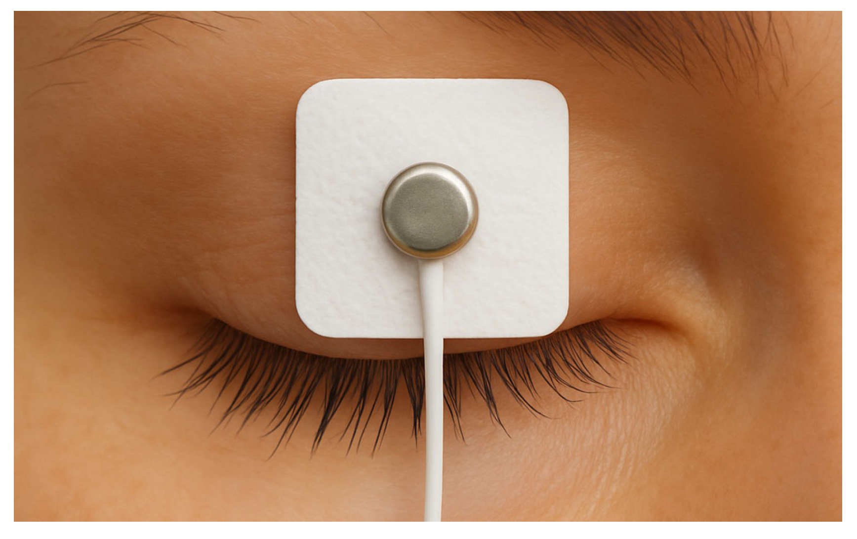
2.2. Electrode Placement

Surface electrodes were applied as follows: the active electrode was placed horizontally across the central portion of the upper eyelid (Figure 1), approximately 5 mm above the line of the eyelashes; the reference electrode was positioned on the forehead, while the ground electrode was secured over the left clavicle.

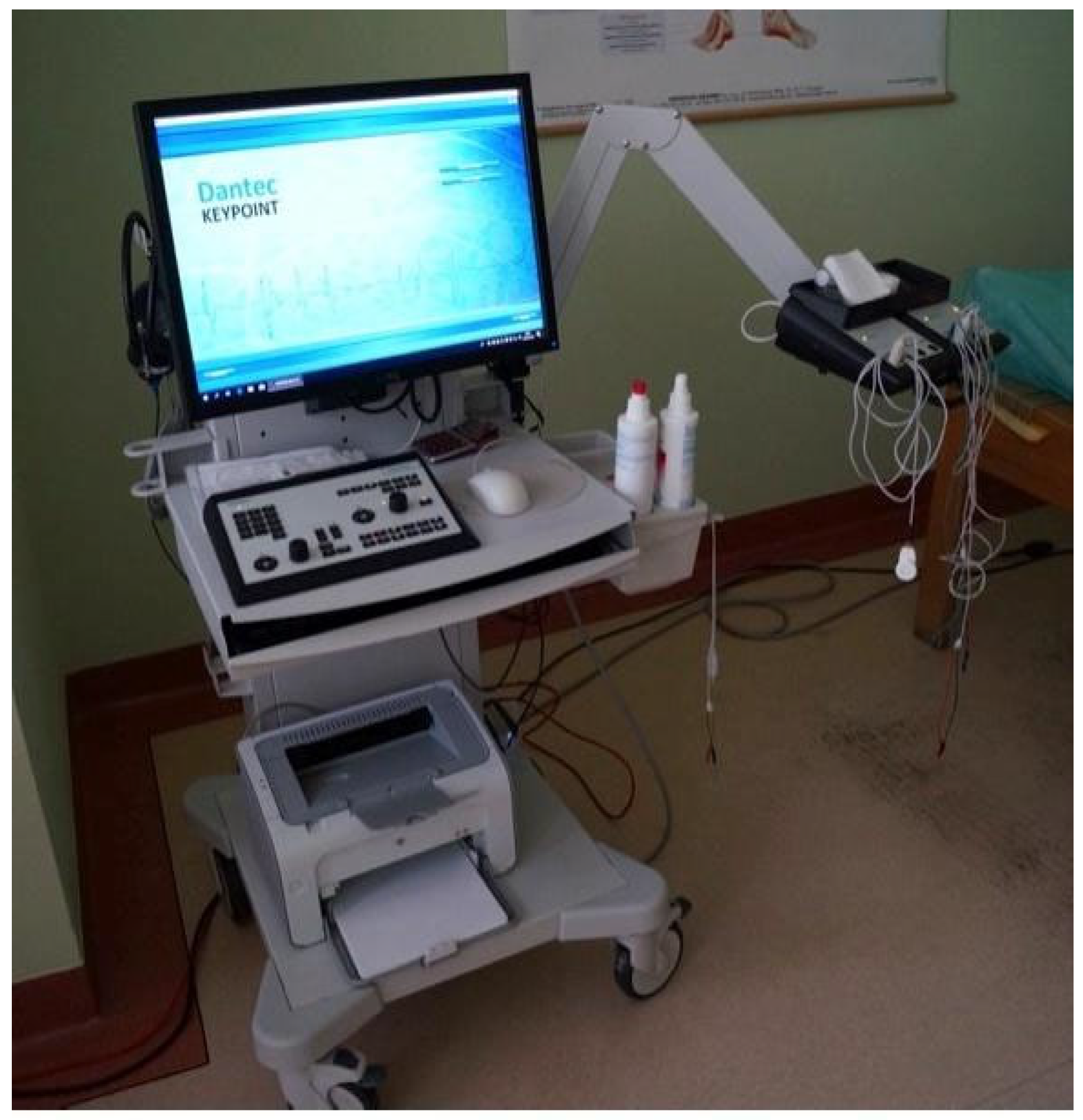
2.3. sEMG Measurement Protocol

Participants remained seated in an upright posture with their heads maintained in a neutral alignment throughout the procedure. Electromyographic signals were recorded under three conditions: (1) maximal voluntary contraction (MVC), achieved through deliberate and strong eyelid closure; (2) gentle eyelid closure (GEC), reflecting light muscular activation; and (3) resting state, recorded with the eyes open and focused in primary gaze.
Each condition was recorded over a five-second interval, and three consecutive trials were performed. To ensure sufficient muscular recovery and reduce the potential for fatigue, a one-minute rest period was observed between trials. For each condition, the average of the three measurements was calculated and used for further analysis (Figure 2).

2.4. Ethics Approval

This study involved human participants and received approval from the Ethics Committee of the National Institute of the Ministry of Interior and Administration (Approval No. 45/2024). All procedures adhered to the principles outlined in the Declaration of Helsinki. Written informed consent was obtained from all participants prior to their inclusion in this study.

2.5. Statistical Analysis

Independent samples t-tests assessed gender and laterality differences. Pearson correlation and regression analyses evaluated the relationship between age and sEMG parameters. The calculations were performed with the use of reflimR package working in R Statistics 4.3.3 open-source software environment. The qualitative findings were summarized narratively.

3. Results

A total of 84 individuals, ranging in age from 27 to 87 years (mean age = 63.12, SD = 13.80), were enrolled in the study. The sample included 44 female participants (aged 27–87, M = 63.12, SD = 13.80) and 40 male participants (aged 27–86, M = 62.50, SD = 13.79). Data were collected bilaterally from each participant, resulting in a total of 168 eye-specific observations.
All procedures were well tolerated, and no participants reported experiencing discomfort during the recordings.

3.1. Differences Between Males and Females

Table 1 depicts the mean values of the analyzed variables in the group females and in the group of males with the values of independent samples Student’s t test.
No statistically significant differences between males and females were detected.

3.2. Differences Between the Values for the Left and the Right Eye

Table 2 displays the average values of the examined parameters for the left and right eyes, accompanied by the corresponding p-values from the independent samples Student’s t-test assessing differences between sides.
No statistically significant differences between the values for the left eye and the values for the right eye were detected.

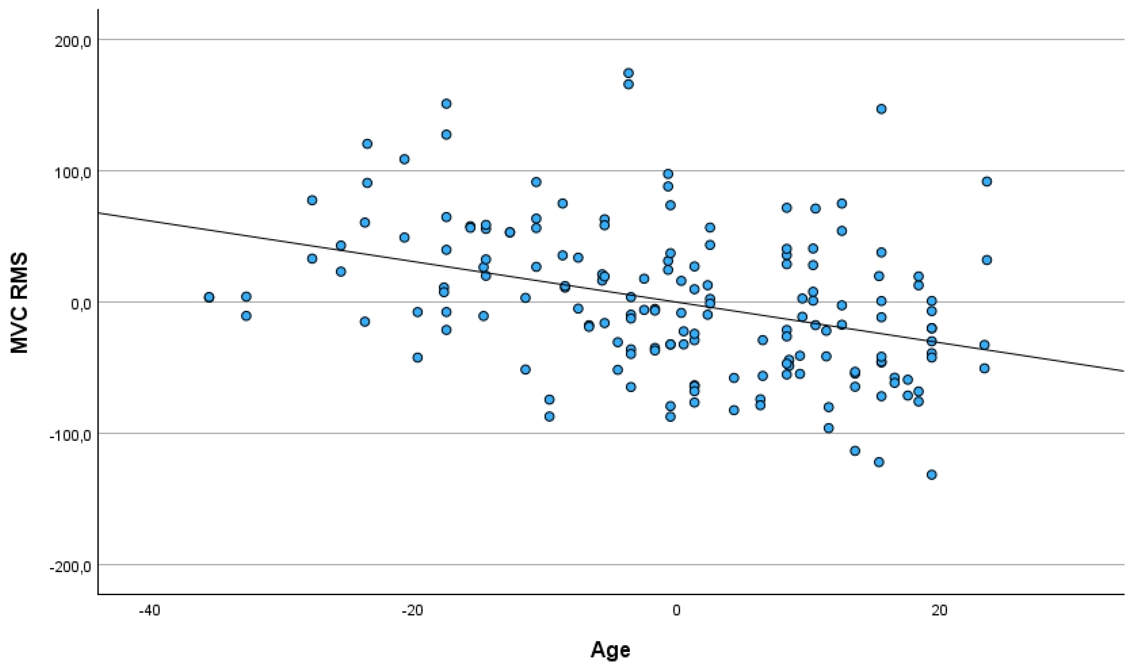
3.3. Age of Participants

The Pearson correlation coefficients between participants’ age and the analyzed variables are summarized in Table 3.
The age of participants correlated negatively with maximal amplitude, mean amplitude, RMS and the difference between RMS and RMS baseline in maximal voluntary contraction (MVC).

3.4. The Differences Between Males and Females, Between the Left and the Right Eye and the Age of Participants as Predictors of Analyzed Variables

The differences between males and females, between the left and the right eye and the age of participants as predictors of analyzed variables with the use of regression analysis based on the enter method. Each variable was analyzed in a separate regression model. The results are depicted in Table 4.
The age of participants related negatively to maximal amplitude, mean amplitude, RMS and the difference between RMS and RMS baseline in MVC. No statistically significant association between participants’ sex or the differences between the left and right eye were detected. The older participants had lower values of ampl. max, ampl. mean, RMS and the difference between RMS and RMS baseline in maximal voluntary contraction (see Figure 3 and Figure 4).

4. Discussion

This study evaluated the effect of aging on sEMG measurements of the OOM and investigated potential differences in sEMG results of the OOM between men and women and between the left and right side.

4.1. Age-Related Decline in Neuromuscular Function

Age was found to be inversely related to MVC-related parameters—namely, maximum amplitude, mean amplitude, and RMS values. This relationship is in line with earlier studies of other skeletal muscles that have documented a decline in neuromuscular function and muscle strength with advancing age. Piasecki, when comparing the vastus lateralis and tibialis anterior in young and old participants, found that older males had fewer but larger motor units. Additionally, these older motor units exhibited greater instability in neuromuscular transmission and lower firing rates, indicating a decline in neuromuscular function with age [12]. Age-related decrement in the sEMG response is indicative of a generalized decline in the motor performance of the OOM. These reductions are likely due to age-associated changes in motor unit recruitment, shifts in muscle fiber composition, and alterations in neuromuscular control.
The findings of the present study are consistent with our earlier research conducted on a separate cohort, in which we observed that patients with blepharochalasis over the age of 60 exhibited significantly reduced OOM activity during maximal voluntary contraction (MVC) compared to younger individuals. This age-related decline was evident in both the mean amplitude (p = 0.029) and RMS values (p = 0.045). Notably, ultrastructural analysis revealed morphological features characteristic of skeletal muscle aging within the OOM, irrespective of the participants’ age, suggesting that structural degeneration may precede or occur independently of functional decline [11]. In the present study, individuals with known neurological conditions were excluded. However, the potential influence of other systemic conditions that may affect muscle tone was not examined. Future research should aim to address this limitation by systematically evaluating the role of comorbidities in neuromuscular function.
Importantly, the age-related decline in OOM function observed with surface EMG has also been supported by findings from needle EMG studies. Needle EMG allows for direct recording of individual motor unit potentials, offering insight into motor unit number and firing characteristics. Studies employing needle EMG in aging populations have demonstrated similar patterns—such as increased motor unit potential duration and polyphasia, reduced recruitment, and decreased firing rates—consistent with motor unit remodeling and denervation–reinnervation processes that accompany aging. While needle EMG provides more detailed motor unit-level information, sEMG offers a non-invasive and functionally relevant assessment of muscle performance during natural movements such as blinking or voluntary eye closure. The convergence of findings from both techniques reinforces the evidence of age-related neuromuscular decline in periorbital muscles like the OOM [13].
Okuda et al. in a cross-sectional study involving both men and women revealed that older individuals exhibit a thinner OOM and a greater degree of orbital fat prolapse compared to younger subjects, with these two variables showing a negative correlation [14]. In this study, computed tomography was used to measure the OOM thickness in 34 adults aged between 20 and 79 years. The results indicated a significant reduction in muscle thickness with age.
Conversely, Fukuda and Kajiya [15] reported that while the mean myofiber cross-sectional area of the OOM did not differ significantly between young and older adults, the proportion of the muscle area composed of myofibers was lower in the older group. It is worth noting that in the study by Fukuda and Kajiya, the specific region measured was not detailed, and the exact age ranges defining the young and old groups (within the overall sample aged 19 to 62 years) were not provided.
In addition, in a study on blinking it has been reported that the percentage of incomplete eye closure increases in older adults (range 51–77 years) compared with young adults (range 20–30 years) [16].
Together, these results suggest that the function of OOM is affected by age and that change may be due to decreases in strength. Consequently, incorporating age-adjusted reference values into clinical assessments is advisable.

4.2. Gender Differences in Neuromuscular Function

In skeletal muscles differences between men and women in absolute strength in various muscle groups have been reported [17,18,19]. However, we found no statistically significant differences between male and female participants across any of the analyzed variables in OOM. Similarly, Bertozzi et al. found no statistically significant difference between male and female participants in the sEMG study of facial muscles that included OOM [20]. Kim et al. investigated RMS values of facial muscles as well, and reported no significant gender differences. However, their study of facial muscles did not include an analysis of the OOM [21]. This contrasts with findings from ultrasound studies, which have reported that OOM thickness is approximately 16% smaller in women than in men [22]. However, it is possible that muscle thickness differences do not translate directly to differences in sEMG amplitude or activation patterns. Additionally, Volk et al. observed an age-related decline in OOM size in men but not in women in ultrasound measurements, suggesting that sex differences in OOM function may become more apparent with aging [23].
In a study of lid power assessment, including 50 participants, based on the analysis of the maximum compression of the lid speculum, a weak correlation was found between the sex of the subjects and eyelid power [24].
Altogether, the influence of gender on OOM function needs further investigation, but it seems that there is no evident predominance of neither the sex.

4.3. Laterality: Left vs. Right Eye Measurements

Our analysis did not reveal significant differences between left and right eye measurements, indicating symmetry in neuromuscular function, which is consistent with previous study of Bertozzi et al. [20]. The study of Mueller et al. also noted that there was no significant difference between the left and right sides of the face in terms of muscle activation patterns, indicating a symmetrical activation during the tasks [25]. The issue of muscle symmetry in facial palsy has been addressed in a systematic review by Franz et al. By introducing an ‘asymmetry index’ in sEMG assessments, the authors aim to eliminate side predominance, using the comparison between the healthy and affected sides as the basis for normalization [26]. Volk et al. demonstrated in a MRI study on facial muscles volume that in healthy individuals the volume of the orbicularis oculi muscle is symmetrical, further supporting the assumption that neuromuscular function is bilaterally equivalent [27].

5. Conclusions

The results of the present study demonstrated a significant effect of age on the sEMG performance of the OOM and suggested that this variable should be taken into consideration in the interpretation of OOM sEMG in basic and clinical studies. No significant differences were observed between genders or between left and right side.

Author Contributions

Conceptualization, L.K.-W.; methodology, L.K.-W.; formal analysis, L.K.-W. and M.D.; investigation, L.K.-W.; resources, L.K.-W. and M.D.; data curation, L.K.-W.; writing—original draft preparation, L.K.-W.; writing—review and editing, L.K.-W. and M.D.; visualization, L.K.-W.; supervision, M.D.; project administration, L.K.-W.; funding acquisition, L.K.-W. and M.D. All authors have read and agreed to the published version of the manuscript.

Funding

This research received no external funding.

Institutional Review Board Statement

The study was conducted in accordance with the Declaration of Helsinki and approved by the Ethics Committee of the National Institute of the Ministry of Interior and Administration (Approval No. 45/2024, 4 June 2024).

Informed Consent Statement

Informed consent was obtained from all subjects involved in the study.

Data Availability Statement

All data relevant to the study are included in the article.

Conflicts of Interest

The authors declare no conflicts of interest.

Abbreviations

The following abbreviations are used in this manuscript:
OOMorbicularis oculi muscle
sEMGsurface electromyography
MVCmaximal voluntary contraction
GECgentle eyelid closure

References

  1. Tong, J.; Lopez, M.J.; Fakoya, A.O.; Patel, B.C. Anatomy, Head and Neck: Orbicularis Oculi Muscle. In StatPearls; StatPearls Publishing: Treasure Island, FL, USA, 2025. Available online: http://www.ncbi.nlm.nih.gov/books/NBK441907/ (accessed on 12 April 2025).
  2. Ali, M.J.; Zetzsche, M.; Scholz, M.; Hahn, D.; Gaffling, S.; Heichel, J.; Hammer, C.M.; Bräuer, L.; Paulsen, F. New insights into the lacrimal pump. Ocul. Surf. 2020, 18, 689–698. [Google Scholar] [CrossRef] [PubMed]
  3. May, P.J. The Eyelid: Anatomy, Neural Control and Pathology. In Reference Module in Neuroscience and Biobehavioral Psychology; Elsevier: Amsterdam, The Netherlands, 2024; p. B9780443138201000025. [Google Scholar] [CrossRef]
  4. Scherrer, E.; Chaloupka, K. Future treatment options for facial nerve palsy: A review on electrical stimulation devices for the orbicularis oculi muscle. Neurol. Sci. 2024, 45, 1969–1977. [Google Scholar] [CrossRef] [PubMed]
  5. Osaki, T.H.; Gameiro, G.R.; Osaki, M.H.; Osaki, T.; Campos, E.D.; Belfort, R.; Marie, S.K.N. Orbicularis Oculi Muscle Immunohistochemical, Metabolic, and Morphometric Differences in Affected and Nonaffected Sides in Hemifacial Spasm vs Healthy Subjects. J. Neuro-Ophthalmol. 2023, 43, 410–416. [Google Scholar] [CrossRef] [PubMed]
  6. Kimura, J. Alteration of the Orbicularis Oculi Reflex by Pontine Lesions: Study in Multiple Sclerosis. Arch. Neurol. 1970, 22, 156. [Google Scholar] [CrossRef]
  7. Radecka, A.; Lubkowska, A. The usefulness of surface electromyography in rehabilitation and physiotherapy: Systematic review. Pomeranian J. Life Sci. 2020, 66, 49–56. [Google Scholar] [CrossRef]
  8. Huang, H.-Y.; Hu, S. Sex Differences Found in Facial EMG Activity Provoked by Viewing Pleasant and Unpleasant Photographs. Percept. Mot. Skills 2009, 109, 371–381. [Google Scholar] [CrossRef]
  9. Olmos, A.A.; Sterczala, A.J.; Parra, M.E.; Dimmick, H.L.; Miller, J.D.; Deckert, J.A.; Sontag, S.A.; Gallagher, P.M.; Fry, A.C.; Herda, T.J.; et al. Sex-related differences in motor unit behavior are influenced by myosin heavy chain during high- but not moderate-intensity contractions. Acta Physiol. 2023, 239, e14024. [Google Scholar] [CrossRef]
  10. Haizlip, K.M.; Harrison, B.C.; Leinwand, L.A. Sex-Based Differences in Skeletal Muscle Kinetics and Fiber-Type Composition. Physiology 2015, 30, 30–39. [Google Scholar] [CrossRef]
  11. Krajewska-Węglewicz, L.; Felczak, P.; Dorobek, M. Effects of Aging on Orbicularis Oculi Muscle Strength and Ultrastructure in Dermatochalasis: A Pilot Study. J. Clin. Med. 2024, 14, 162. [Google Scholar] [CrossRef]
  12. Piasecki, M.; Ireland, A.; Jones, D.A.; McPhee, J.S. Age-dependent motor unit remodelling in human limb muscles. Biogerontology 2016, 17, 485–496. [Google Scholar] [CrossRef]
  13. Shinohara, M. Adaptations in motor unit behavior in elderly adults. Curr. Aging Sci. 2011, 4, 200–208. [Google Scholar] [CrossRef] [PubMed]
  14. Okuda, I.; Irimoto, M.; Nakajima, Y.; Sakai, S.; Hirata, K.; Shirakabe, Y. Using multidetector row computed tomography to evaluate baggy eyelid. Aesthetic Plast. Surg. 2012, 36, 290–294. [Google Scholar] [CrossRef] [PubMed]
  15. Fukada, K.; Kajiya, K. Age-related structural alterations of skeletal muscles and associated capillaries. Angiogenesis 2020, 23, 79–82. [Google Scholar] [CrossRef] [PubMed]
  16. Sforza, C.; Rango, M.; Galante, D.; Bresolin, N.; Ferrario, V.F. Spontaneous blinking in healthy persons: An optoelectronic study of eyelid motion. Ophthalmic Physiol. Opt. 2008, 28, 345–353. [Google Scholar] [CrossRef]
  17. Young, A.; Stokes, M.; Crowe, M. The size and strength of the quadriceps muscles of old and young men. Clin. Physiol. 1985, 5, 145–154. [Google Scholar] [CrossRef]
  18. Sepic, S.B.; Murray, M.P.; Mollinger, L.A.; Spurr, G.B.; Gardner, G.M. Strength and range of motion in the ankle in two age groups of men and women. Am. J. Phys. Med. 1986, 65, 75–84. [Google Scholar]
  19. Rice, C.L.; Cunningham, D.A.; Paterson, D.H.; Rechnitzer, P.A. Strength in an elderly population. Arch. Phys. Med. Rehabil. 1989, 70, 391–397. [Google Scholar]
  20. Bertozzi, N.; Bianchi, B.; Salvagni, L.; Raposio, E. Activity Evaluation of Facial Muscles by Surface Electromyography. Plast. Reconstr. Surg. Glob. Open 2020, 8, e3081. [Google Scholar] [CrossRef]
  21. Kim, B.-H.; Kim, K.H.; Kim, L.-H.; Kim, J.-U.; Yook, T.-H. Difference between Right and Left Facial Surface Electromyography in Healthy People. Evid.-Based Complement. Altern. Med. ECAM 2018, 2018, 4069530. [Google Scholar] [CrossRef]
  22. Abe, T.; Wong, V.; Spitz, R.W.; Viana, R.B.; Bell, Z.W.; Yamada, Y.; Chatakondi, R.N.; Loenneke, J.P. Influence of sex and resistance training status on orofacial muscle strength and morphology in healthy adults between the ages of 18 and 40: A cross-sectional study. Am. J. Hum. Biol. 2020, 32, e23401. [Google Scholar] [CrossRef]
  23. Volk, G.F.; Sauer, M.; Pohlmann, M.; Guntinas-Lichius, O. Reference values for dynamic facial muscle ultrasonography in adults. Muscle Nerve 2014, 50, 348–357. [Google Scholar] [CrossRef] [PubMed]
  24. Koschmieder, A.; Pisowocki, P.; Zietz, C.; Bader, R.; Stachs, O.; Jünemann, A. Measurement of the Strength of Human Musculus Orbicularis Oculi Using Video Analysis. Klin. Monatsbl. Augenheilkd. 2017, 234, 1451–1457. [Google Scholar] [CrossRef] [PubMed]
  25. Mueller, N.; Trentzsch, V.; Grassme, R.; Guntinas-Lichius, O.; Volk, G.F.; Anders, C. High-resolution surface electromyographic activities of facial muscles during mimic movements in healthy adults: A prospective observational study. Front. Hum. Neurosci. 2022, 16, 1029415. [Google Scholar] [CrossRef] [PubMed]
  26. Franz, L.; de Filippis, C.; Daloiso, A.; Biancoli, E.; Iannacone, F.P.; Cazzador, D.; Tealdo, G.; Marioni, G.; Nicolai, P.; Zanoletti, E. Facial surface electromyography: A systematic review on the state of the art and current perspectives. Am. J. Otolaryngol. 2024, 45, 104041. [Google Scholar] [CrossRef]
  27. Volk, G.F.; Karamyan, I.; Klingner, C.M.; Reichenbach, J.R.; Guntinas-Lichius, O. Quantitative magnetic resonance imaging volumetry of facial muscles in healthy patients with facial palsy. Plast. Reconstr. Surg. Glob. Open 2014, 2, e173. [Google Scholar] [CrossRef]
Figure 1. Schematic position of the active electrode.
Jcm 14 03119 g001
Figure 2. Outpatient setting. Dantec Keypoint EMG workstation.
Jcm 14 03119 g002
Figure 3. Partial regression plot depicting relationship between participants’ age and RMS in maximal voluntary contraction (MVC).
Jcm 14 03119 g003
Figure 4. Partial regression plot depicting relationship between participants’ age and the difference between RMS and RMS baseline in maximal voluntary contraction (MVC).
Jcm 14 03119 g004
Table 1. Mean values of the analyzed variables in the group females and in the group of males.
FemalesMales
VariableMSDMSDtdfp
RMS baseline11.205.2810.235.811.131660.261
MVCampl. max1197.26411.631283.51449.58−1.301660.196
ampl. Mean349.3791.03369.6795.44−1.411660.160
RMS146.4556.48157.8056.43−1.301660.195
ΔRMS135.2557.32147.5658.00−1.381660.169
GECampl. max52.1523.1549.2619.400.871660.385
RMS8.354.867.422.941.491660.138
ΔRMS−2.846.85−2.815.39−0.031660.974
M—mean value; SD—standard deviation; t—value of Student’s t test for independent samples; df—degrees of freedom; p—two-tailed statistical significance.
Table 2. Mean values of the analyzed variables for the left eye and the right eye.
LeftRight
VariableMSDMSDtdfp
RMS baseline10.406.3911.084.56−0.801660.426
MVCampl. max1221.26448.771255.41414.44−0.511660.609
ampl. Mean358.3896.45359.6990.89−0.091660.928
RMS152.2457.73151.4755.740.091660.930
ΔRMS141.8459.22140.3956.700.161660.871
GECampl. max52.3522.1649.1920.700.961660.340
RMS8.295.047.532.791.211660.227
ΔRMS−2.117.47−3.554.471.521660.130
M—mean value; SD—standard deviation; t—value of Student’s t test for independent samples; df—degrees of freedom; p—two-tailed statistical significance.
Table 3. Correlation coefficients between the age of participants and analyzed variables.
Age
Variablerp
RMS baseline0.0730.344
MVCampl. max−0.3440.001
ampl. Mean−0.3580.001
RMS−0.3800.001
ΔRMS−0.3790.001
GECampl. max−0.0660.393
RMS−0.0180.813
ΔRMS−0.0780.315
r—Pearson correlation coefficient; p—two-tailed statistical significance.
Table 4. The differences between males and females, between the left and the right eye and the age of participants as predictors of analyzed variables.
VariablesPredictorsBetatpFdfpR2
MVCampl. maxMales vs. females0.091.170.2437.9631640.0010.127
Eye left vs. right0.040.550.587
Age−0.34−4.660.001
ampl. meanMales vs. females0.091.290.1998.6831640.0010.137
Eye left vs. right0.010.100.923
Age−0.35−4.880.001
RMSMales vs. females0.081.170.2449.7531640.0010.151
Eye left vs. right−0.01−0.100.925
Age−0.38−5.230.001
ΔRMSMales vs. females0.091.260.2109.7931640.0010.152
Eye left vs. right−0.01−0.180.861
Age−0.38−5.210.001
GECampl. maxMales vs. females−0.07−0.910.3650.0831640.4830.015
Eye left vs. right−0.07−0.960.341
Age−0.07−0.900.372
RMSMales vs. females−0.12−1.500.1361.2631640.2910.022
Eye left vs. right−0.09−1.210.227
Age−0.02−0.300.762
ΔRMSMales vs. females0.00−0.010.9911.1131640.3490.020
Eye left vs. right−0.12−1.520.131
Age−0.08−1.010.315
Beta—standardized regression coefficient; t—the value of statistical test for a predictor; p—statistical significance; F—the value of the statistical test for a model; df—degrees of freedom; R2—determination coefficient.
Disclaimer/Publisher’s Note: The statements, opinions and data contained in all publications are solely those of the individual author(s) and contributor(s) and not of MDPI and/or the editor(s). MDPI and/or the editor(s) disclaim responsibility for any injury to people or property resulting from any ideas, methods, instructions or products referred to in the content.

