Review

Immunomodulation in Pediatric Sepsis: A Narrative Review

1
Pediatric Intensive Care Unit, Children Hospital Bambino Gesù, IRCSS, 00165 Rome, Italy
2
Department of Intensive Care, Hopital Universitaire de Bruxelles (HUB), Université Libre de Bruxelles (ULB), 1050 Brussels, Belgium
3
Pediatric Clinic, “Microcitemico—A. Cao” Pediatric Hospital, University of Cagliari, 09124 Cagliari, Italy
4
Academy of Pediatrics, Bambino Gesù Children’s Hospital, IRCCS, 00165 Rome, Italy
5
Université Paris Cité Sorbonne, 75006 Paris, France
6
Recherche Service Maladies Infectieuses, CHU de Nice, 06200 Nice, France
*
Author to whom correspondence should be addressed.
J. Clin. Med. 2025, 14(9), 2983; https://doi.org/10.3390/jcm14092983
Submission received: 9 March 2025 / Revised: 16 April 2025 / Accepted: 21 April 2025 / Published: 25 April 2025
(This article belongs to the Special Issue Sepsis: New Insights into Diagnosis and Treatment)

Abstract

:
Pediatric sepsis presents a unique clinical challenge due to the distinct characteristics of the developing immune system. The immune response in children differs significantly from that in adults, exhibiting a unique combination of resistance, disease tolerance, and resilience. These factors influence the clinical presentation and prognosis of pediatric patients with sepsis. Over the past few years, various studies have explored the role of immunomodulatory therapies in managing sepsis, including the use of immunoglobulins, corticosteroids, monoclonal antibodies, and immunostimulatory treatments. However, the heterogeneity of the clinical presentations and individual responses makes it difficult to identify universally effective treatments. Recent research has highlighted the importance of a personalized approach based on specific biomarkers and patient phenotyping. Extracorporeal blood purification techniques have emerged as promising strategies for the modulation of hyperinflammation. However, strong evidence supporting their routine use in pediatric sepsis is lacking. This review provides a comprehensive overview of the current knowledge of the immune response in pediatric sepsis and discusses the main immunomodulatory strategies and future perspectives for personalized therapy. A deeper understanding of the immunological differences between children and adults could improve the prognosis and treatment efficacy, paving the way for new approaches to pediatric sepsis management.

1. Background

The complexity of sepsis immunobiology explains the failure of randomized controlled trials to test immunomodulatory therapies for sepsis over the last 10 years [1,2].
The host response to sepsis is characterized by concurrent hyperinflammation and immunosuppression, with highly heterogeneous clinical presentations [2,3]. The incomplete understanding of the underlying pathophysiology continues to slow down therapeutic advances [1,2]. Both the severity of the host’s dysregulated response and the trajectory of the process over time exhibit extreme variability [1,2]. Hyperinflammation is driven by the uncontrolled activity of pro-inflammatory effector mechanisms involving activated leukocytes and endothelial cells, along with the overwhelming presence of inflammatory mediators [1]. This process can lead to collateral damage (immunopathology) and contribute to sepsis-induced organ failure [1]. Furthermore, it is followed by an immunosuppressive profile, which increases the risk of nosocomial infections and viral reactivation [1,2,3].
This scenario is even more complex in children because of the development of their immune systems. The Phoenix definition of 2024 has acknowledged sepsis in children as a life-threatening infection [4,5,6], aligning with the Sepsis-3 definition [7] but adapting it to reflect the distinct immunobiology of critically ill children. However, to understand the rationale behind therapies, it is important to recognize the key differences that distinguish the host response in adults from that in children. The aim of this review is to provide, based on the existing literature, a description of the main characteristics of immunobiology in children compared to adults. Moreover, for the first time, to the best of our knowledge, we offer a comprehensive overview of the main immunomodulatory therapies currently in use and the existing evidence in this field.

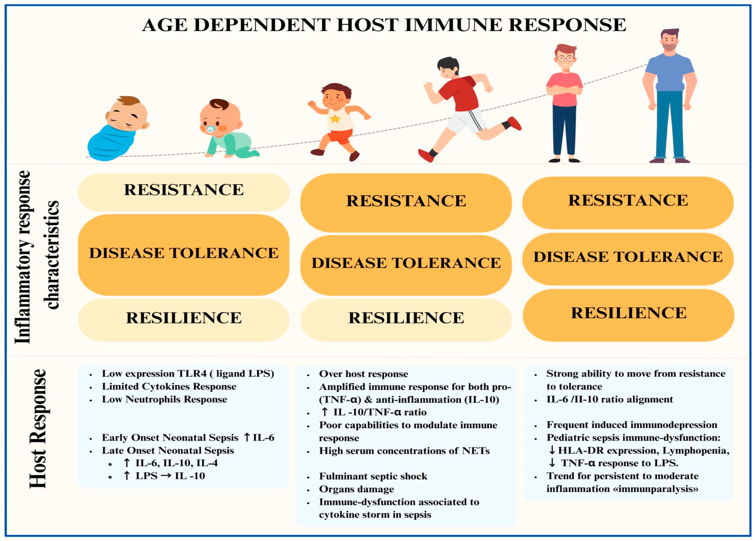
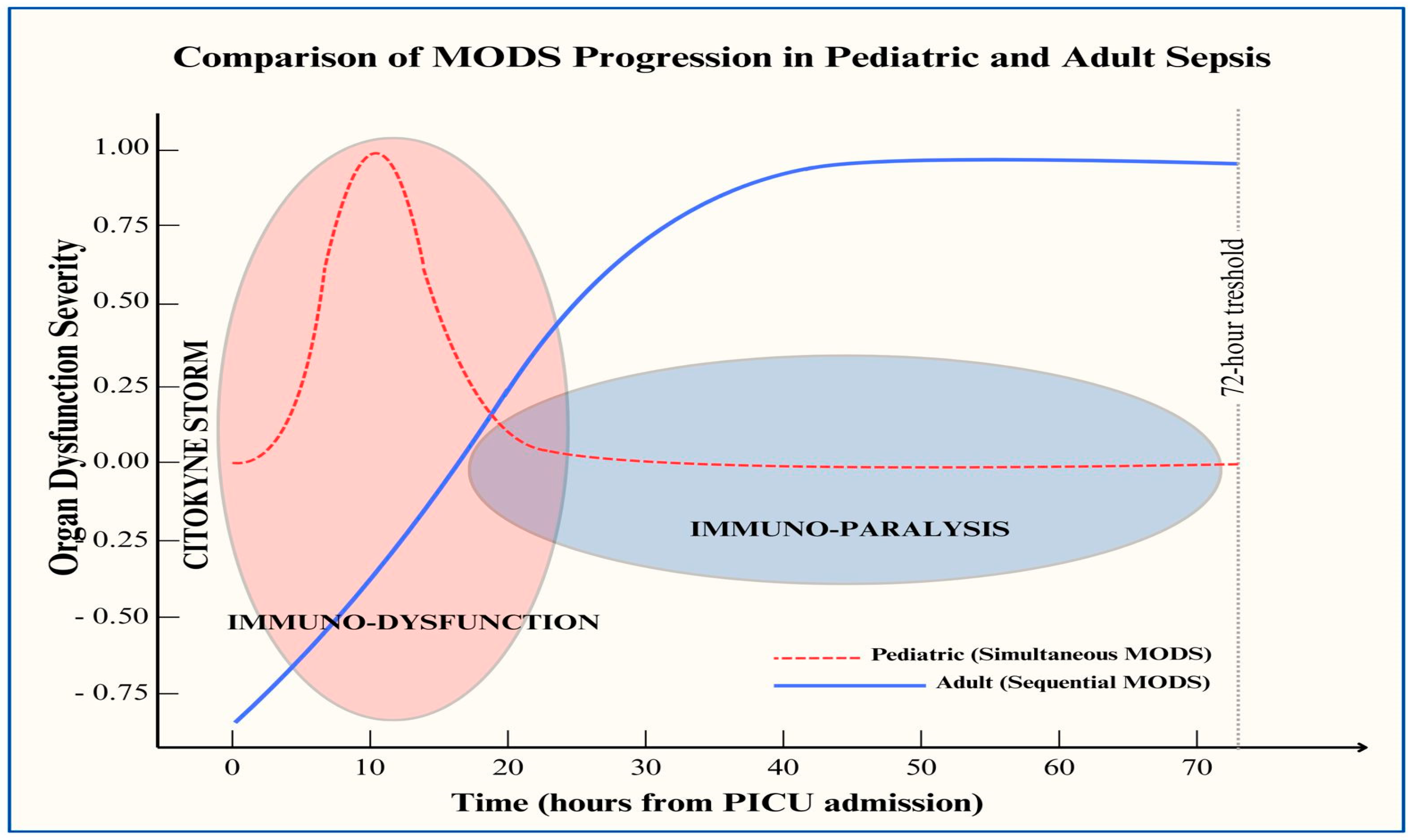
2. Host Response in Children with Sepsis and Differences from Adult Populations

Beyond the paradigms of hyperinflammation and immunosuppression, a recent review of the immunobiology of sepsis has extended the link between the immunological and metabolic phases. The initial phase aims to eliminate the pathogen (resistance), which is followed by a metabolic shift (catabolism) associated with an immunodepression profile that limits the host response-induced tissue damage (immunopathology) (disease tolerance). The third phase covers tissue repair (resilience), with the restoration of metabolic and immune function [8]. In the developing immune systems of children, resistance, disease tolerance, and resilience seem to have different capabilities according to their development as compared to adults. This maturation shapes specific patterns in the host response compared with adults. Preclinical models and clinical studies have confirmed that age and a history of microbial exposure can significantly influence immune biology [9] (Figure 1).
Compared with adults, newborns show diminished Toll-like receptor (TLR)-induced responses and reduced pro-inflammatory cytokine production [10]. Preclinical models have shown that the ability to produce cytokines in response to LPS stimulation increases with age [10]. Several neonatal immune cell types can directly regulate aberrant inflammation and promote tolerance. Newborns can produce and secrete cytokines and express markers of resident memory phenotypes [9]. Some authors have noted a significant difference in cytokine responses between early-onset sepsis (EOS) and late-onset sepsis (LOS), suggesting that the postnatal age may influence the disease [11]. Although both EOS and LOS patients showed a significant increase in serum IL-6 levels, only LOS patients released anti-inflammatory cytokines such as IL-10 and IL-4 [12].
During immune system development, a transition in T cell populations is observed. The predominant regulatory T cell populations in newborns shift to pro-inflammatory T cells in older children [13]. These T cells respond to antigens that may be under the control of IL-8 upregulation [13]. Similarly, the CD4 T helper (Th) cell population shifts from a Th2 phenotype (anti-inflammatory) to a Th1 phenotype (pro-inflammation), producing interferon-γ [13].
The neutrophils of newborns maintain low expression of TLR4, the ligand of LPS, on the surfaces of Gram-negative bacteria [13]. The expression of signal transducers immediately downstream of TLRs is also low in newborns, limiting cytokine responses and neutrophil chemotaxis [13]. However, there is evidence that pediatric patients with sepsis have significantly higher serum concentrations of neutrophil extracellular traps (NETs) than adults, and the severity of pediatric sepsis is positively correlated with the level of NETs [14,15,16].
Differences in the innate and adaptive immune responses between childhood and adulthood may explain the specificity of sepsis in children [17]. An imbalance in inflammatory and compensatory anti-inflammatory responses in children may lead to septic shock and multiple-organ dysfunction syndrome (MODS) more frequently than in adults. Macrophages from young children produce greater amounts of both pro-inflammatory TNF-α and anti-inflammatory IL-10 than those from adults, and the resulting IL-10/TNF ratio is higher in children [17]. Using an ex vivo model of children’s macrophages stimulated by LPS, a 15-fold higher IL-10/TNF-α ratio than that in adults was observed [18,19]. Similarly to adults, clinical studies have demonstrated that higher initial cytokine responses in children are followed by impairments in innate immunity, as assessed by decreased monocyte HLA-DR expression [17]. Immunological tests showed that immunosuppression occurred as early as at the first 72 h in a cohort of children with severe sepsis and septic shock [20,21]. These findings strongly support previous reports indicating that children with sepsis exhibit a different pattern of MODS than adults (simultaneous vs. sequential) (Figure 2) [18,19]. These observations fit well with epidemiological data showing a higher rate of mortality within the first 72 h after PICU admission than in adults [22,23,24,25].

3. Management of Pediatric Septic Shock: Exploring New Perspectives Beyond Standard Care

Despite advancements in clinical management, such as early identification, appropriate resuscitation, and timely antibiotic therapy, the mortality rate associated with pediatric sepsis remains high. While adjuvant therapies for pediatric septic shock have been proposed, few demonstrations of their clinical benefits have been reported [26].
In recent decades, immunomodulation during septic shock has been extensively tested [27], targeting different inflammatory pathways in sepsis. Figure 3 provides a graphical representation of the possible immunomodulatory strategies in sepsis.
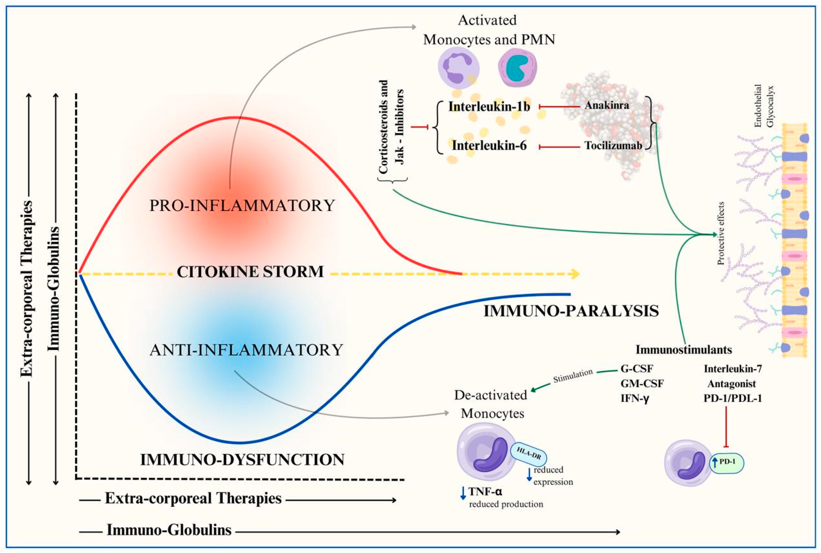
  • Cytokine modulation: Techniques such as extracorporeal blood purification therapies or the administration of immunoglobulins aim to control the overwhelming cytokine response.
  • Targeted immunomodulation: Selective drugs such as monoclonal antibodies block key mediators of septic shock.
  • Immune stimulation: Strategies to counteract immune paralysis through immunostimulatory therapies.
Targeting inflammatory pathways in sepsis is of increasing interest for the management of microcirculation alterations in sepsis. While the role of endothelial damage in pediatric inflammatory diseases is increasingly being recognized, the complex interplay between endothelial injury, the immune response, and therapeutic immunomodulation remains underexplored [28]. Endothelial damage, particularly involving the glycocalyx, initiates a cascade that includes the release of inflammatory mediators and immune cell recruitment, further perpetuating vascular injury. This bidirectional relationship contributes to microcirculatory dysfunction and increased microvascular permeability [29], which are hallmarks of severe pediatric conditions such as sepsis and multisystem inflammatory syndromes. Moreover, disruption of the endothelial glycocalyx not only compromises the vascular barrier but also alters cell signaling and leukocyte adhesion, exacerbating inflammation. Current evidence suggests that treatments aimed at restoring endothelial integrity, such as corticosteroids, anticoagulants, and biologics, play a crucial role in stabilizing the vascular barrier [30]. Therefore, future research and clinical guidelines should focus on how various therapies modulate endothelial function, with an emphasis on preserving or restoring vascular homeostasis in pediatric patients.
Although none of these approaches have demonstrated clear outcome benefits in sepsis, experts agree that this failure of clinical trials can be explained. Firstly, sepsis is a syndrome with multiple expressions, accounting for the great heterogeneity among patients [1,2]. Secondly, genetic susceptibility varies greatly within individuals and may change over time with environmental exposure, nutritional habits, and physical activity. Thirdly, age, comorbidities, chronic treatment, and pre-sepsis immunodepression may modify the host response. Different combinations of these features may naturally lead to distinct phenotypes with varying outcomes and responses to specific “trait-based” treatments [31].
Future clinical trial designs should better select the enrolled patients using parameters that reflect both clinical and biological characteristics, seeking to cluster patients more homogenously. This crucial step necessitates the use of validated biomarkers, especially those reflecting the major sub-endotypes based on molecular mechanisms.
As a consequence, the clinical strategy for therapies will be based on integrated inflammatory parameters such as coagulation, the longitudinal immune response, and cellular metabolic markers [32]. Selecting these parameters requires a deep understanding of the immunobiology, allowing one to intervene with a more personalized approach. Among these, adaptive interactions with immune and metabolic cellular changes lead to better results in sepsis.

4. Immunomodulation in Pediatric Septic Shock: Current Evidence

Evidence regarding immunomodulatory therapies for pediatric sepsis is limited. There is a lack of interventional studies conducted in an adequate pediatric population, and the validation and generalizability of the observational studies described need to be further explored.
The present authors, supported by two external librarians, electronically searched the literature using a combination of key medical terms related to immunomodulation AND septic shock; immunoglobulins AND septic shock; corticosteroids AND septic shock; monoclonal antibodies AND septic shock; immune stimulation AND septic shock; and extracorporeal blood purification therapies AND septic shock. The search was limited to pediatric patients (aged less than 18 years) and included randomized control trials (if available).

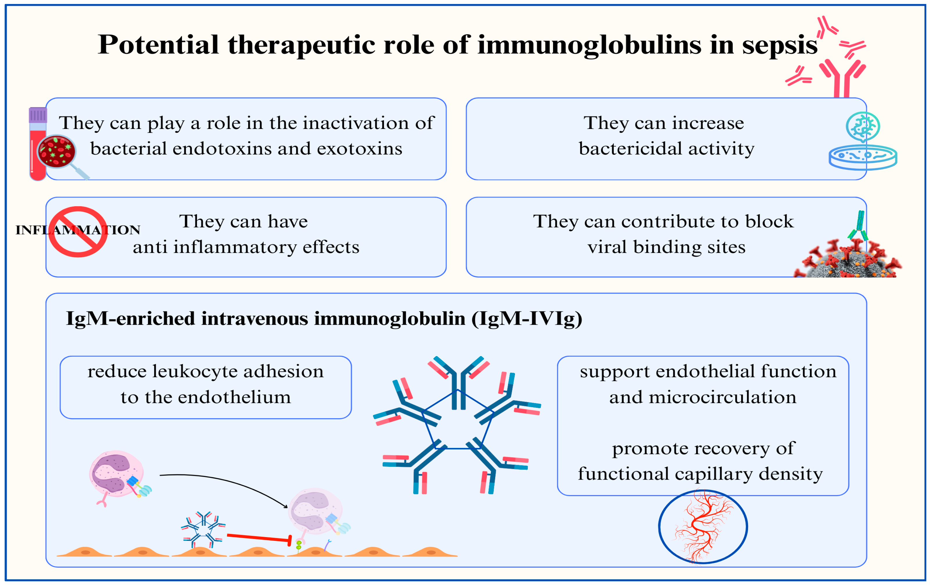
4.1. Immunoglobulins

Immunoglobulins have been used for decades to treat several infectious diseases and immunological disorders. Intravenous immunoglobulin (IVIG) could be a valuable adjunctive therapy in sepsis for several reasons: it can help to inactivate bacterial endotoxins and exotoxins, blocks viral binding sites, enhances bactericidal activity, and exhibits anti-inflammatory effects [33] (Figure 4).
The international Guidelines on the Management of Pediatric Septic Shock and Sepsis-Associated Organ Dysfunction do not support the routine use of IVIG in children with sepsis or septic shock. However, emerging evidence suggests that some patients may benefit from this treatment [34]. These patients include children with toxic shock syndrome (TSS), necrotizing fasciitis, or primary immunodeficiencies or immunocompromised children with low levels of immunoglobulins [34]. Additionally, the absence of specific immunoglobulins in newborns during the first few months of life is one reason for which the administration of IVIG during this period could theoretically be highly beneficial [35].
A recent retrospective cohort study conducted by Huang et al. (2023) involved 304 hospitalized children with septic shock in the PICU of the Children’s Hospital of Chongqing Medical University [36]. Of these, 29.3% received intravenous immunoglobulin (IVIG). This group showed a greater need for continuous renal replacement therapy (CRRT) (43% vs. 24%, p = 0.001), a longer duration of mechanical ventilation (6 days vs. 3, p = 0.002), a longer length of stay in the pediatric intensive care unit (PICU) (7 days vs. 4, p = 0.002), and a longer hospital stay (18 vs. 11 days, p = 0.001). Survival at 28 days showed better results in the IVIG group (p = 0.033), while the in-hospital mortality did not exhibit significant differences between the two groups, although it was lower once again in the IVIG group. After propensity score matching, no significant differences were found in terms of CRRT, the mechanical ventilation duration, the PICU and hospital LOS, in-hospital mortality, and 28-day survival [36].
The aforementioned guidelines highlight that the administration of immunoglobulin M- and A-enriched polyclonal IVIG could be useful in managing septic shock [34]. Several adult-based studies have investigated this aspect, with noteworthy results [37,38,39,40]. The few reported pediatric studies on this topic suggest the potential benefit of using IgM-enriched immunoglobulins. In their systematic review and meta-analysis, Pan et al. (2023) emphasized the role of IVIG in reducing mortality and the in-hospital length of stay (LOS) (mainly including adult-based studies), stressing that IgM-enriched IVIG can reduce neonatal sepsis mortality with the efficacy of normal IVIG in adults [41].
A prospective study conducted by El-Nawaway et al. (2005) [42] at the PICU of Alexandria University Children’s Hospital showed promising results. Their study involved 100 children aged 1–24 years with sepsis, divided into two groups. Group I received traditional therapy plus IVIG (polyclonal IgM-enriched immunoglobulins), whereas Group II received only the traditional treatment. The results showed a significantly greater number of discharged patients in Group I than in Group II (36 vs. 22, p = 0.0046) and lower mortality rates in Group I (28% vs. 56%). Other findings included a lower tendency for Group I patients to develop complications in the PICU (8% vs. 32%, p = 0.0027), particularly disseminated intravascular coagulation (DIC). Additionally, Group I had a shorter LOS compared to Group II (6 vs. 9 days), despite presenting with more severe clinical conditions based on the PRISM III score at admission [42].
A retrospective study (the PIGMENT study), published in 2020, focused on the duration of treatment. Conducted in Eskisehir Osmangazi University Medical Faculty Hospital, the study included 254 children with sepsis, septic shock, and multi-organ failure, admitted to the PICU from January 2010 to December 2017. Of these, 104 patients received treatment for three days, while 150 received treatment for five days. The mortality rate was lower in the group of patients who underwent 5-day-long treatment (20.6% vs. 40.3%; R:0.51, 95% CI 0.34–0.75; p < 0.001) [43]. Table 1 provides a summary of the main clinical studies on the use of immunoglobulins (IVIG) and IgM-enriched IVIG in pediatric sepsis.

4.2. Immunoglobulins and Toxic Shock Syndrome

As mentioned previously, the use of IVIG in children with TSS is a distinct consideration. TSS is a severe illness associated with infections caused by Staphylococcus aureus and Streptococcus pyogenes. Its pathogenesis involves massive cytokine release in response to bacterial toxins [44]. Wilkins et al. [45] outlined the steps in managing TSS, including IVIG and clindamycin as adjunctive therapies. IVIG appears to offer benefits in terms of antigen recognition, the activation of the innate immune system, and the neutralization of super-antigen toxin activity [45]. A 2018 systematic review and meta-analysis, although primarily focused on adults, found a reduction in mortality among patients with streptococcal TSS treated with IVIG in combination with clindamycin [46]. Interestingly, in the only pediatric study included, none of the patients treated with IVIG died, while the overall mortality rate was 16% [47].
Evidence of the beneficial effects of immunoglobulins in critically ill children with toxic shock syndrome is contradictory. A pediatric retrospective analysis showed promising results: 94% of the patients received IVIG and no deaths were recorded [48]. A multicenter retrospective cohort study involving children with streptococcal TSS reported a mortality rate of 4.2%, with no statistically significant differences between the IVIG-treated and control groups [49].

4.3. Corticosteroids

Cortisol plays different roles in septic shock: it directly decreases the reuptake of norepinephrine, enhances calcium availability in myocardial and vascular smooth muscle cells, promotes myocardial contractility and vasoconstriction, and inhibits prostacyclin and endogenous nitric oxide production, resulting in increased vascular tone, the modulation of capillary leaks, and the augmentation of the beta-adrenergic receptor in the heart [34]. On the other hand, adverse effects caused by corticosteroid therapy include hyperglycemia, catabolism-related diffuse neuromuscular weakness, and hospital-acquired infections [34] (Figure 5).
The use of corticosteroids as an adjunctive therapy for septic shock remains a topic of debate. The current Surviving Sepsis guidelines recommend hydrocortisone only for fluid-refractory, catecholamine-resistant, and suspected/proven adrenal insufficiency [34].
In adults, Annane et al. demonstrated that a 7-day treatment with low doses of hydrocortisone and fludrocortisone significantly reduced the mortality risk in patients with septic shock and relative adrenal insufficiency, without increasing the number of adverse events (hazard ratio 0.67; 95% confidence interval 0.47–0.95; p = 0.02) [50]. Conversely, another RCT involving 3800 adults with septic shock on mechanical ventilation found that continuous hydrocortisone infusion did not reduce 90-day mortality compared with a placebo (odds ratio 0.95; 95% confidence interval [CI] 0.82 to 1.10; p = 0.50) [51].
However, evidence in critically ill children with septic shock is limited. In a randomized control trial, Valoor et al. reported a trend toward earlier shock reversal (median 49.5 vs. 70 h, p = 0.65) and lower inotrope requirements (median inotrope score 20 vs. 50, p = 0.15) in hydrocortisone-treated patients, although the differences were not statistically significant. The mortality rates were similar between the groups (p = 1.0) [52]. El-Nawawy et al. found a significantly shorter shock reversal time in patients receiving corticosteroids at the start of treatment compared with those receiving them later (p = 0.046), although the mortality rates were similar (p = 0.734) [53]. Menon et al. analyzed 49 pediatric patients and found no significant differences in the time on vasopressors (p = 0.65), days of mechanical ventilation (p = 0.37), or PICU length of stay (p = 0.48) [54]. Alkhalf et al. observed a 42% lower risk of prolonged PICU stays in patients receiving steroids, but no difference in mortality (p = 0.492) [55].
Recent evidence suggests a need for a personalized approach that acknowledges the variable responses to steroids. Alder et al. found that children with different glucocorticoid receptor (GCR) and cortisol concentrations may respond differently to corticosteroids. In their study, pediatric patients with complicated septic shock (defined as two or more organ failures by day 7 or death by day 28) had lower GCR expression and higher cortisol levels. This subgroup showed a 75% rate of complicated outcomes, compared with 13–33% in other GCR and cortisol combinations (p < 0.05) [56].
Wong et al. developed a gene expression panel that segregates children with acute septic shock into endotypes based on the immune response and GCR signaling. Children with endotype A, characterized by the underactivation of adaptive immunity and GCR signaling, had increased mortality when treated with corticosteroids (OR = 4.1; CI95 = 1.4–12.0; p = 0.011) [57]. Approximately one-third of the children with septic shock exhibited a change in endotype assignment during the first 3 days, with those persisting as endotype A at the highest risk for poor outcomes. This evolving understanding highlights the importance of individualized treatment strategies in managing pediatric septic shock with corticosteroids [57,58]. Table 2 provides a summary of the main clinical studies on the use of corticosteroids for pediatric sepsis.

4.4. Monoclonal Antibodies

Human IL-1 receptor antagonist (rhIL-1ra). In 1994, Fisher et al. conducted a study that found no significant survival benefit from anakinra in sepsis (p = 0.22) [59]. However, a secondary analysis showed a dose-related increase in the survival time among patients with organ dysfunction [60]. These data were re-analyzed by Shakoory et al. in 2016, focusing on patients with hepatobiliary dysfunction and disseminated intravascular coagulation, which are features of macrophage activation syndrome (MAS), and found that anakinra treatment improved the 28-day survival rate (HR = 0.28, p = 0.0071) [60]. A trial by Opal et al. in 1997 was halted after an interim analysis revealed no significant difference in mortality at 28 days between the case (33.1%) and control (36.4%, p = 0.36) groups [61].
A recent narrative review by Manchikalapati et al. in 2023 highlighted the need for randomized controlled trials (RCTs) to study the potential role of anti-IL-1 in pediatric sepsis [62]. The review discussed five studies involving anakinra in children with secondary hemophagocytic lymphohistiocytosis (HLH), 72 of whom also had sepsis [63,64,65,66]. However, the results were inconclusive. Currently, there are two ongoing studies investigating the use of anakinra in sepsis. The first was ImmunoSep (personalized immunotherapy in sepsis), a double-blind, placebo-controlled, randomized phase II clinical trial (NCT04990232), which included 280 patients [67]. The second is a prospective, multicenter, double-blind, placebo-controlled clinical trial involving children with sepsis (NCT05267821) [68]. Table 3 provides a summary of the main clinical studies on the use of human IL-1 receptor antagonists (rhIL-1ra) in pediatric sepsis.
Anti-interleukin (IL)-6 antibodies: A systematic review in 2023 summarized its efficacy and safety in sepsis and SARS-CoV-2 infection [69]. Tocilizumab was associated with reduced 28-day mortality (RR 0.88, 95% CI 0.81–0.94), although its impact on 60-day mortality remains uncertain [70]. It also showed a favorable safety profile, with minimal differences in adverse effects compared with traditional treatments [71]. Regarding the use of tocilizumab in sepsis, only two papers have been published, involving a total of six adult patients, where the antibody showed promising results [72].
Janus kinase (JAK) inhibitors: Song et al.’s 2023 systematic review and meta-analysis explored the role of baricitinib in COVID-19 patients [73]. The study found that baricitinib reduced mortality (OR = 0.61, p = 0.008) and the need for mechanical ventilation (OR = 0.57, p = 0.002) [73]. There was no significant difference in the in-hospital length of stay or adverse effects between the baricitinib and control groups [73]. Currently, no data are available regarding the use of baricitinib in patients with sepsis or pediatric septic shock.

4.5. Immunostimulation

The dysregulated immune system in sepsis and septic shock may induce a phase of depression of the immune system (called “immunoparalysis”), inactivating immune cells and contributing to mortality and morbidity due to secondary infections [74].
Recently, criteria to detect signs of immune paralysis in children were established in the PODIUM consensus [75], potentially identifying those who might benefit from immunostimulatory therapies. These are different therapeutic approaches that aim to impact different molecular pathways, including cytokines, immune cells, and growth factors. Pediatric studies are a minority, but the trials are growing in number.
Granulocyte Colony-Stimulating Factor (G-CSF) and Granulocyte-Macrophage Colony-Stimulating Factor (GM-CSF). G-CSF and GM-CSF are hematopoietic stimulators that enhance the production, migration, survival, and activity of neutrophils, monocytes, and other immune cells. Because of these properties, they have been explored as supportive therapies for patients with sepsis [74]. A meta-analysis of adult patients with septic shock indicated that, while G-CSF and GM-CSF improved immune cell function, they did not significantly affect overall mortality at 14 or 28 days (16.6% vs. 17.6%; p = 0.44) or in-hospital mortality. However, they were associated with the greater resolution of infections (29.4% vs. 21.8%; p = 0.002) [76]. This analysis suggests that these drugs should not be routinely administered in sepsis [76], particularly without immune monitoring.
In pediatric patients with MODS and immune paralysis, GM-CSF prevented secondary infections and helped to recover immune system function more quickly than in the placebo group [77]. A retrospective study of 109 pediatric sepsis patients found that those receiving immunomodulatory therapy, including G-CSF, required more frequent PICU admission and invasive ventilation. However, they had shorter ventilator-free and PICU-free periods [78]. Studies on neonates with sepsis have demonstrated that G-CSF and GM-CSF could restore immune cell function, including increasing HLA-DR expression and monocyte numbers [79,80].
The literature remains divided on the use of G-CSF and GM-CSF in sepsis treatment, particularly because of the potential for adverse effects such as organ failure [81]. Based on a Cochrane meta-analysis [82], which showed no benefit of GM-CSF in newborns and infants who were not selected based on the presence of neutropenia, the need for personalized treatment approaches when using immune-stimulatory therapies in sepsis has been highlighted [82].
Interleukin-7. One of the main functions of IL-7 is to prevent a decrease in lymphocytes by stimulating their survival and proliferation, particularly CD4+ and CD8+ T cells. This action helps to increase pathogen clearance and protects against secondary infections by recruiting neutrophils to the peripheral tissue. In a study of adult septic patients after IL-7 treatment, lymphocytes showed increased numbers (p < 0.05) and glucose utilization (p < 0.05) via the better functioning of the mTOR pathway (p < 0.05), indicating their ability to reverse metabolic alterations [83]. The IRIS-7 trial, which was a randomized double-blind trial, investigated CYT107 (recombinant human IL-7) and reported an increase in lymphocyte numbers (p < 0.001), particularly CD4+ and CD8+ T cells, owing to its anti-apoptotic effect and impact on cell expansion. Although not powered for outcome evaluation, the trial did not affect the 28-day mortality rate (p = 0.687) [84]. Currently, there are no studies available on the efficacy of recombinant human IL-7 in pediatric septic shock.
Interferon-γ. Several studies have focused on the effects of exogenous IFN-γ on immunoparalysis in patients with sepsis. IFN-γ increases HLA-DR expression in circulating monocytes, enhances their activation, and stimulates neutrophil function to eliminate the microbes. It can only be used after the diagnosis of immunoparalysis; if so, treatment must be carefully monitored. Consequently, its early use during a cytokine storm is contraindicated, as recently reported [85]. A pioneering study involved nine adult septic patients with impaired HLA-DR expression, which increased after a subcutaneous injection of IFN-γ (100 µg). In parallel, INF-γ enhanced TNF production in vitro. Eight patients recovered after the treatment [86]. A remarkable study in 18 healthy volunteers induced a reduction in the monocyte expression of HLA-DR via the injection of small doses of LPS. Only delayed exogenous INF-γ administration prevented the reduction in TNF levels (p = 0.01), lowered the IL-10 levels, and restored normal HLA-DR expression (p = 0.02). GM-CSF treatment showed a similar trend but this did not reach statistical significance [87]. A tri-centric case series evaluated IFN-γ treatment in patients with septic shock: cohort 1 received IFN-γ based on criteria such as ICU hospitalization > 7 days, the presence of secondary infections despite antibiotics, and persistently low HLA-DR expression on immune cells. The first group showed a significant increase in monocyte HLA-DR expression and negative culture after IFN-γ treatment and was discharged from the ICU. Cohort 2 began INF-γ treatment when their norepinephrine doses were halved and showed a similar improvement in HLA-DR expression. Additionally, a pediatric liver transplant case with septic shock followed by deep immunoparalysis and untreatable infections recovered after IFN-γ therapy with a maintained protocol of anti-rejection drugs [88].
In premature newborns, who often have an immature immune system that deteriorates during sepsis, IFN-γ administration has been investigated to restore normal immune function. The observed increase in TNF-α and IL-6 levels was associated with a decrease in IL-10 levels. The pathogen receptors for bacteria in immune cells increase dramatically, and phagocytosis returned to normal [89]. However, the lack of pediatric trials suggests a need for further investigation. The general approach is to identify patient subgroups and test immunostimulation with a unique drug, or those in combination, in the late phase of sepsis [85].
Immune checkpoints. Some studies have explored the use of PD-1 and PDL-1 antagonists, known as “immune checkpoints”, to counteract immune cell apoptosis and paralysis during sepsis and restore immune function [85]. PD-1 is mainly expressed on T cells, whereas PDL-1 appears on antigen-presenting cells, and its activation inhibits T cell function and significantly reduces IL-2 and IFN-γ production [85,90].
A clinical trial involving adult septic patients administered anti-PDL-1 antibodies demonstrated a safe increase in HLA-DR expression. However, more studies are needed to understand its effects on the prevention of secondary infections and the overall survival rate [91]. A study involving 43 adult septic patients showed benefits when anti-PD-1 and anti-PDL-1 drugs were administered, including increased cytokine levels (e.g., IL-2; p < 0.01), reduced immune cell apoptosis (p < 0.01), and improved IFN-γ secretion (p < 0.01) [92].
Others—Glutathione Peroxidase 4 (GPX4): A recent study in children investigated GPX4, which plays a crucial role in sepsis diagnosis and is associated with the disruption of oxidative balance, lipid peroxide accumulation, and ferroptosis. The loss of GPX4 function is linked to organ failure in sepsis and worsens the prognosis by damaging mitochondria. This study also highlighted an imbalance in the immune system during sepsis, potentially due to altered GPX4 function [93]. Table 4 provides a summary of the main clinical studies on the use of immunostimulation therapy in pediatric sepsis.

4.6. Extracorporeal Blood Purification Techniques in Pediatric Septic Shock

Renal replacement therapies (RRTs) are used to support or replace kidney function in critically ill patients with acute kidney injury (AKI). These therapies help to maintain the fluid and chemical balance while eliminating waste products from the body. RRT is crucial for the management of AKI, including sepsis-associated AKI [94]. The “peak concentration” hypothesis, introduced in 2003, proposes that partially clearing cytokines from the bloodstream during the early stages of sepsis could lower their peak levels and help to regulate the inflammatory response [95]. The potential advantages of extracorporeal blood purification therapies have gained further attention with the recognition of “organ cross-talk” in sepsis, where dysfunction in one organ can negatively impact others, particularly in cases of multiple-organ dysfunction syndrome (MODS). This approach is based on the idea that malfunctioning organs remain perfused with blood, making this a viable target for therapeutic interventions [96,97,98,99]. Figure 6 provides a graphical representation of extracorporeal blood purification techniques according to removal target among mediators of the sepsis process.
Hemofilters with adsorption capacities. AN69ST is a hemofilter that can be mounted on normal CRRT devices with a polyacrylonitrile membrane on which a surface treatment (ST) has been applied by grafting a biocompatible polymer named polyethyleneimine (PEI). In a prospective cohort study, the use of AN69ST as a hemofilter in patients showed a trend of decreasing pro-inflammatory and anti-inflammatory cytokines, and membrane adsorption emerged as the main cytokine clearance mechanism [100]. The subsequent AN69 Oxiris® version is characterized by a three-layer structure, and the second layer consists of PEI at concentrations three-fold higher than those in the AN69ST filter, enabling the biologically active PEI surface to adsorb endotoxins. Recently, Morin reported the use of Oxiris® in a single-center prospective observational study. The cohort included pediatric patients with vasoplegic shock and acute kidney injury (seven of 11 patients affected by septic shock). The authors reported a 50%reduction in inotropic support within 24 h (considered a success in the treatment) in 5 of 11 patients (four of seven with septic shock) [101].
Polymyxin B (PMX-B) is a basic cyclic polypeptide that alters the permeability of the membranes of Gram-negative bacteria. PMX-B immobilized on polystyrene fibers was developed as an extracorporeal clearance system to remove endotoxins, with hemoadsorption cycles of 2 h for 2 days [101]. Interestingly, PMX-05R and PMX-01R are Toraymixin® cartridges (Toray Medical Co., Houston, TX, USA) with safe priming volumes in newborns and children [102]. The results of two large multicenter RCTs demonstrated a non-significant increase in mortality and no improvement in organ failure with PMX-HP treatment compared with the conventional treatment of peritonitis-induced septic shock [103,104]. Two ancillary studies have reported the effects of PMX-HP on the plasma levels of 21 cytokines [105] and the actual effect on the mass of plasma LPS [106]. Surprisingly, none of these studies demonstrated any effect of PMX-HP treatment on the plasma cytokine levels or on the plasma mass of LPS (mass spectrometry). These results shed light on the significant reduction in LPS mass caused by PMX-HP. It is therefore possible that the rate of LPS release into the plasma dominates the extraction capacity of the membrane.
However, the EUPHRATES post hoc analysis highlighted that, in a subgroup of patients with an indirect endotoxin assay value between 0.6 and 0.9, an improvement in survival was found; this provides future opportunities for a new study with a more appropriate population [107,108,109].
Clinical experiences in newborns with septic shock who were successfully treated with hemoadsorption using a PMX-B-immobilized fiber column have been reported in Japan [110,111]. Moroshita et al. described their experience using blood purification with PMX-B in a 13-month-old child with sepsis due to Pseudomonas aeruginosa, with the resolution of the clinical picture [112]. Nanishi et al. reported their use of PMX-B in an adolescent with toxic shock syndrome, with a positive outcome, thus questioning the potential benefit of PMX-B for Gram-positive bacterial infections [113].
Polymyxin B hemoadsorption with PMX-05 cartridges was applied to 15 children with septic shock. The authors reported a trend towards an improvement in hemodynamics after two sessions of therapy, although the inotropic agent requirements did not change over time [114]. Recently, Saetang et al. described the use of PMX-20R (a modified polymyxin-B cartridge circuit for pediatric use) in six children and reported a significant reduction in the PELOD-2 and VIS scores after two sessions of therapy [115].
Regarding plasma separation (PE) techniques, plasmapheresis is a technique used for the separation of plasma from blood cells, and it can be performed by centrifugation with cell separation to reach ≥80% hematocrit or by membrane separation with porous fibers. The latter technique is less efficient because blood cell separation using a membrane system can be hindered when very high hematocrit values are reached. In addition, the upper cutoff of the TPE membrane is 1000 kDa, suggesting that molecules with higher molecular weights, such as Von Willerbrand multimers, cannot be removed [116].
Nguyen T. [117] showed an improvement in mortality at 28 days and in organ dysfunction scores in pediatric patients treated with PE. The trial was conducted exclusively in children with septic shock and thrombocytopenia-associated multiple-organ failure (TAMOF). The study conducted by the above-mentioned authors showed that children with septic shock presenting a clinical picture of TAMOF had reduced ADAMTS13 activity, and autopsies of non-survivors revealed microcirculation thromboses with von Willebrand factor-rich microaggregates [117]. Therefore, PE was indicated as a therapeutic priority in this subgroup of patients [117,118].
A recent systematic review by Lee highlighted that PE use may be associated with harmful effects in pediatric patients with sepsis and septic shock. However, this has not been confirmed in studies including pediatric patients with septic shock and a TAMOF phenotype [119].
Adsorption columns are new options for adsorbent membranes. They are characterized by a very large surface area (8,500 m2) compared to traditional membranes (1.5 m2), which could make them a very effective tool for hypercytokinemia, with potential therapeutic efficacy in sepsis [120]. Recently, hemoadsorption with new sorbents customized to adsorb cytokines and other inflammatory mediators has emerged as the most investigated and clinically established procedure in the clinical context of sepsis [121].
CytoSorb® (Cytosorbents Europe GmbH, Berlin, Germany) is a hemocompatible cytokine adsorber, whose unique structure allows the generation of hydrophobic bonds with cytokines, which are adsorbed, while plasma proteins are filtered among micropores and returned to the systemic circulation [122]. In 2023, the first pediatric interventional study confirmed the benefit of Cytosorb® in combination with CRRT in pediatric patients with septic shock, in terms of reducing the need for vasopressors and inotropes, as well as 28-day mortality, compared to a historical cohort of pediatric patients with septic shock treated only with CRRT [123]. In addition to a secondary analysis, the authors showed a significant reduction in IL-6 and IL-10 levels, with evidence that, at 24 h after the end of hemoadsorption, no rebound effect was observed, suggesting that the extracorporeal therapeutic effect could substantially affect immune homeostasis [124]. Furthermore, longitudinal measurements of monocyte HLA-DR expression in the enrolled population showed that hemoadsorption prevented the expected downregulation of HLA-DR [124].
Jafron® cartridges (Guangdong, China) containing neutro-macroporous resin-adsorbing beads composed of a styrene–divinylbenzene copolymer have been used for the removal of a wide spectrum of molecules. HA330® cartridges have also been used to treat children [125,126]. Jafron recently released two novel devices specifically dedicated to the care of the smallest numbers of patients, i.e., HA60 and BS80. The first is devoted to the care of septic children with a priming volume of 60 mL, and the second is focused on hyperbilirubinemia with a priming volume of 50 mL. The current literature on these materials is limited to non-indexed publications, but the evolution of this technology to miniaturized devices will certainly help to improve the available evidence in pediatric and neonatal settings [127].

5. Knowledge Gaps and Research Opportunities in Pediatric Sepsis

Despite existing evidence suggesting the role of immunomodulatory therapies in pediatric sepsis, the widespread translation of these results into clinical practice is still far from being realized. In particular, there is a general need to determine how to implement the personalization of therapy. The heterogeneity of the immune responses in children complicates the identification of effective targeted treatments. The current research also lacks robust biomarkers to stratify patients based on their immune status, leading to challenges in clinical decision-making. Table 5 provides a summary of some key points related to the knowledge gaps and research opportunities for immunomodulatory therapies in pediatric sepsis.

6. Conclusions

Pediatric sepsis is a complex condition that requires a deep understanding of the specific immunobiology of children to develop effective therapeutic strategies. Research has shown that the immune system in children differs significantly from that of adults. These characteristics not only influence disease progression but also affect the responses to immunomodulatory treatments.
Therapeutic approaches for pediatric sepsis are continuously evolving. Current strategies include the use of immunoglobulins, corticosteroids, monoclonal antibodies, and blood purification techniques. However, regarding pediatric intensive care, there is still a relatively small number of studies on immunomodulation in pediatric sepsis and septic shock, leading to recommendations that are based largely on expert opinions rather than on published data. In this context, the difficult transition in the science of intensive care medicine—from evidence-based medicine to personalized medicine—becomes particularly evident.
A crucial aspect of the future management of pediatric sepsis will be the development of diagnostic tests capable of identifying specific biomarkers to stratify patients (phenotype, sub-endotypes) and monitor the drug response, avoid iatrogenicity, and fully implement the concept of personalized treatment.

Author Contributions

G.B. and F.S.T. conceptualized the manuscript. C.C. and D.P. reviewed the manuscript. G.B., A.C., M.I., P.C. and M.S. performed the bibliographic research and drafted the manuscript. G.B. and M.S. created the illustrations and finalized the design. All authors have read and agreed to the published version of the manuscript.

Funding

This research received no external funding.

Conflicts of Interest

G.B. has received honoraria for lectures from Jafron® and CytoSorbents®. The other authors have no conflicts to declare.

Abbreviations

AKIAcute Kidney Injury
CIConfidence Interval
CRRTContinuous Renal Replacement Therapy
DICDisseminated Intravascular Coagulation
ELBWExtremely Low Birth Weight
EOSEarly-Onset Sepsis
GCRGlucocorticoid Receptor
G-CSFGranulocyte Colony-Stimulating Factor
GLUT-1Glucose Transporter 1
GM-CSFGranulocyte-Macrophage Colony-Stimulating Factor
GPX4Glutathione Peroxidase 4
HBDHepatobiliary Disfunction
HFHemofiltration
HLAHuman Leukocyte Antigen
HLHHemophagocytic Lymphohistiocytosis
HRHazard Ratio
HVHFHigh-Volume Hemofiltration
ICUIntensive Care Unit
IFNInterferon
ILInterleukin
IQRInterquartile Range
IVIGIntravenous Immunoglobulins
JAKJanus Kinase
kDaKilodalton
LOSLate-Onset Sepsis, Length of Stay
LPSLipopolysaccharide
MASMacrophage Activation Syndrome
MODSMultiple-Organ Dysfunction Syndrome
MVMechanical Ventilation
NETsNeuthrophil Extracellular Traps
NONitric Oxide
OROdds Ratio
PD-1Programmed Cell Death Protein 1
PD-L1Programmed Death Ligand 1
PEPlasma Separation Techniques
PEIPolyethyleneimine
PICUPediatric Intensive Care Unit
PMXPolymyxin
PRISMPediatric Risk of Mortality
RCTRandomized Controlled Trial
rhIL-1raRecombinant Human IL-1 Receptor Antagonists
RR Relative Risk
RRTsRenal Replacement Therapies
STSurface Treatment
TAMOFThrombocytopenia-Associated Multiple-Organ Failure
ThT Helper
TLRToll-Like Receptor
TNFTumor Necrosis Factor
TPEPlasma Exchange
TSSToxic Shock Syndrome

References

  1. Cavaillon, J.M.; Singer, M.; Skirecki, T. Sepsis therapies: Learning from 30 years of failure of translational research to propose new leads. EMBO Mol. Med. 2020, 12, e10128. [Google Scholar] [CrossRef] [PubMed]
  2. Vincent, J.L. Improved survival in critically ill patients: Are large RCTs more useful than personalized medicine? No. Intensive Care. Med. 2016, 42, 1778–1780. [Google Scholar] [CrossRef] [PubMed]
  3. Vincent, J.L.; van der Poll, T.; Marshall, J.C. The End of “One Size Fits All” Sepsis Therapies: Toward an Individualized Approach. Biomedicines. 2022, 10, 2260. [Google Scholar] [CrossRef] [PubMed]
  4. Schlapbach, L.J.; Watson, R.S.; Sorce, L.R.; Argent, A.C.; Menon, K.; Hall, M.W.; Akech, S.; Albers, D.J.; Alpern, E.R.; Balamuth, F.; et al. Society of Critical Care Medicine Pediatric Sepsis Definition Task Force. International Consensus Criteria for Pediatric Sepsis and Septic Shock. JAMA. 2024, 331, 665–674. [Google Scholar] [CrossRef]
  5. Sanchez-Pinto, L.N.; Bennett, T.D.; DeWitt, P.E.; Russell, S.; Rebull, M.N.; Martin, B.; Akech, S.; Albers, D.J.; Alpern, E.R.; Balamuth, F.; et al. Development and Validation of the Phoenix Criteria for Pediatric Sepsis and Septic Shock. JAMA 2024, 331, 675–686. [Google Scholar] [CrossRef]
  6. Jabornisky, R.; Kuppermann, N.; González-Dambrauskas, S. Transitioning from SIRS to Phoenix with the updated pediatric sepsis criteria—the difficult task to simplyfing the complex. JAMA 2024, 331, 650–651. [Google Scholar] [CrossRef]
  7. Singer, M.; Deutschman, C.S.; Seymour, C.W.; Shankar-Hari, M.; Annane, D.; Bauer, M.; Bellomo, R.; Bernard, G.R.; Chiche, J.-D.; Coopersmith, C.M.; et al. The Third International Consensus Definitions for Sepsis and Septic Shock (Sepsis-3). JAMA 2016, 315, 801–810. [Google Scholar] [CrossRef]
  8. Shankar-Hari, M.; Calandra, T.; Soares, M.P.; Bauer, M.; Wiersinga, W.J.; Prescott, H.C.; Knight, J.C.; Baillie, K.J.; Bos, L.D.J.; Derde, L.P.G.; et al. Reframing sepsis immunobiology for translation: Towards informative subtyping and targeted immunomodulatory therapies. Lancet Respir. Med. 2024, 12, 323–336. [Google Scholar] [CrossRef]
  9. Greenfield, K.G.; Badovinac, V.P.; Griffith, T.S.; Knoop, K.A. Sepsis, Cytokine Storms, and Immunopathology: The Divide between Neonates and Adults. Immunohorizons 2021, 5, 512–522. [Google Scholar] [CrossRef]
  10. Zhang, X.; Zhivaki, D.; Lo-Man, R. Unique aspects of the perinatal immune system. Nat. Rev. Immunol. 2017, 17, 495–507. [Google Scholar] [CrossRef]
  11. Wynn, J.L.; Guthrie, S.O.; Wong, H.R.; Lahni, P.; Ungaro, R.; Lopez, M.C.; Baker, H.V.; Moldawer, L.L. Postnatal Age Is a Critical Determinant of the Neonatal Host Response to Sepsis. Mol. Med. 2015, 21, 496–504. [Google Scholar] [CrossRef]
  12. Khaertynov, K.S.; Boichuk, S.V.; Khaiboullina, S.F.; Anokhin, V.A.; Andreeva, A.A.; Lombardi, V.C.; Satrutdinov, M.A.; Agafonova, E.A.; Rizvanov, A.A. Comparative Assessment of Cytokine Pattern in Early and Late Onset of Neonatal Sepsis. J. Immunol. Res. 2017, 2017, 8601063. [Google Scholar] [CrossRef]
  13. Carter, M.J.; Carrol, E.D.; Ranjit, S.; Mozun, R.; Kissoon, N.; Watson, R.S.; Schlapbach, L.J. Susceptibility to childhood sepsis, contemporary management, and future directions. Lancet Child Adolesc. Health 2024, 8, 682–694. [Google Scholar] [CrossRef]
  14. Mandel, J.; Casari, M.; Stepanyan, M.; Martyanov, A.; Deppermann, C. Beyond homeostasis: Platelet innate immune interactions and thrombo-inflammation. Int. J. Mol. Sci. 2022, 23, 3868. [Google Scholar] [CrossRef]
  15. Meier, A.; Sakoulas, G.; Nizet, V.; Ulloa, E.R. Neutrophil extracellular traps: An emerging therapeutic target to improve infectious disease outcomes. J. Infect. Dis. 2024, 230, 514–521. [Google Scholar] [CrossRef]
  16. Colon, D.F.; Wanderley, C.W.; Franchin, M.; Silva, C.M.; Hiroki, C.H.; Castanheira, F.V.S.; Donate, P.B.; Lopes, A.H.; Volpon, L.C.; Kavaguti, S.K.; et al. Neutrophil extracellular traps (NETs) exacerbate severity of infant sepsis. Crit. Care 2019, 23, 113. [Google Scholar] [CrossRef]
  17. Mithal, L.B.; Arshad, M.; Swigart, L.R.; Khanolkar, A.; Ahmed, A.; Coates, B.M. Mechanisms and Modulation of Sepsis-Induced Immune Dysfunction in Children. Pediatr. Res. 2022, 91, 447–453. [Google Scholar] [CrossRef]
  18. Barsness, K.A.; Bensard, D.D.; Partrick, D.A.; Calkins, C.M.; Hendrickson, R.J.; Banerjee, A.; McIntyre, R.C., Jr. IL-1beta induces an exaggerated pro- and anti-inflammatory response in peritoneal macrophages of children compared with adults. Pediatr. Surg. Int. 2004, 20, 238–242. [Google Scholar] [CrossRef]
  19. Barsness, K.A.; Bensard, D.D.; Partrick, D.A.; Calkins, C.M.; Hendrickson, R.J.; McIntyre, R.C., Jr. Endotoxin Induces an Exaggerated Interleukin-10 Response in Peritoneal Macrophages of Children Compared with Adults. J. Pediatr. Surg. 2004, 39, 912–915. [Google Scholar] [CrossRef] [PubMed]
  20. Muszynski, J.A.; Nofziger, R. Early immune function and duration of organ dysfunction in critically ill children with sepsis. Am. J. Respir. Crit. Care Med. 2018, 198, 361–369. [Google Scholar] [CrossRef] [PubMed]
  21. Remy, S.; Koley-Descamps, K. Occurrence of marked sepsis-induced immunosuppression in pediatric septic shock: A pilot study. Ann. Intensive Care 2018, 13, 1–10. [Google Scholar] [CrossRef] [PubMed]
  22. Morin, L.; Ray, S.; Wilson, C.; Remy, S.; Benissa, M.R.; Jansen, N.J.G.; Javouhey, E.; Peters, M.J.; Kneyber, M.; De Luca, D.; et al. A European Society of Paediatric and Neonatal Intensive Care Definition. Intensive Care Med. 2016, 42, 1948–1957. [Google Scholar] [CrossRef]
  23. Schlapbach, L.J.; MacLaren, G.; Festa, M.; Alexander, J.; Erickson, S.; Beca, J.; Slater, A.; Schibler, A.; Pilcher, D.; Millar, J.; et al. Prediction of Pediatric Sepsis Mortality Within 1 Hour of Intensive Care Admission. Intensive Care Med. 2017, 43, 1085–1096. [Google Scholar] [CrossRef] [PubMed]
  24. Weiss, S.L.; Balamuth, F.; Hensley, J.; Fitzgerald, J.C.; Bush, J.; Nadkarni, V.M.; Thomas, N.J.; Hall, M.; Muszynski, J. The Epidemiology of Hospital Death Following Pediatric Severe Sepsis: When, Why, and How Children with Sepsis Die. Pediatr. Crit. Care Med. 2017, 18, 823–830. [Google Scholar] [CrossRef]
  25. Cvetkovic, M.; Lutman, D.; Ramnarayan, P.; Pathan, N.; Inwald, D.P.; Peters, M.J. Timing of death in children referred for intensive care with severe sepsis: Implications for interventional studies. Pediatr. Crit. Care Med. 2015, 16, 410–417. [Google Scholar] [CrossRef]
  26. Hanna, W.; Wong, H.R. Pediatric sepsis: Challenges and adjunctive therapies. Crit. Care Clin. 2013, 29, 203–222. [Google Scholar] [CrossRef] [PubMed]
  27. Venet, F.; Lukaszewicz, A.C.; Payen, D.; Hotchkiss, R.; Monneret, G. Monitoring the immune response in sepsis: A rational approach to administration of immunoadjuvant therapies. Curr. Opin. Immunol. 2013, 25, 477–483. [Google Scholar] [CrossRef]
  28. Puchwein-Schwepcke, A.; Genzel-Boroviczeny, O.; Nussbaum, C. The endothelial glycocalyx: Physiology and pathology in neonates, infants and children. Front. Cell Dev. Biol. 2021, 9, 733557. [Google Scholar] [CrossRef]
  29. Fernandez-Sarmiento, J.; Hernandez-Sarmiento, R.; Salazar, M.P.; Barrera, S.; Castilla, V.; Duque, C. The association between hypoalbuminemia and microcirculation, endothelium, and glycocalyx disorders in children with sepsis. Microcirculation 2023, 30, e12829. [Google Scholar] [CrossRef]
  30. Ying, J.; Zhang, C.; Wang, Y.; Liu, T.; Yu, Z.; Wang, K.; Chen, W.; Zhou, Y.; Lu, G. Sulodexide improves vascular permeability via glycocalyx remodeling in endothelial cells during sepsis. Front. Immunol. 2023, 14, 1172892. [Google Scholar] [CrossRef]
  31. Seymour, C.W.; Kennedy, J.N.; Wang, S.; Chang, C.H.; Elliott, C.F.; Xu, Z.; Berry, S.; Clermont, G.; Cooper, G.; Gomez, H.; et al. Derivation, validation, and potential treatment implications of novel clinical phenotypes for sepsis. JAMA 2019, 321, 2003–2017. [Google Scholar] [CrossRef]
  32. Willmann, K.; Moita, L.F. Physiologic disruption and metabolic reporgramming in infection and sepsis. Cell Metab. 2024, 36, 927–946. [Google Scholar] [CrossRef] [PubMed]
  33. Burgunder, L.; Heyrend, C.; Olson, J.; Stidham, C.; Lane, R.D.; Workman, J.K.; Larsen, G.Y. Medication and fluid management of pediatric sepsis and septic shock. Paediatr. Drugs 2022, 24, 193–205. [Google Scholar] [CrossRef]
  34. Weiss, S.L.; Peters, M.J.; Alhazzani, W.; Agus, M.S.D.; Flori, H.R.; Inwald, D.P.; Nadel, S.; Schlapbach, L.J.; Tasker, R.C.; Argent, A.C.; et al. Surviving sepsis campaign international guidelines for the management of septic shock and sepsis-associated organ dysfunction in children. Intensive Care Med. 2020, 46 (Suppl. S1), 10–67. [Google Scholar] [CrossRef]
  35. Ohlsson, A.; Lacy, J.B. Intravenous immunoglobulin for suspected or proven infection in neonates. Cochrane Database Syst. Rev. 2020, 1, CD001239. [Google Scholar] [CrossRef]
  36. Huang, H.; Chen, J.; Dang, H.; Liu, C.; Huo, J.; Fu, Y.Q. Effect of intravenous immunoglobulin on the outcome of children with septic shock in a PICU: A retrospective cohort study. Eur. J. Pediatr. 2023, 182, 5315–5323. [Google Scholar] [CrossRef]
  37. Berlot, G.; Vassallo, M.C.; Busetto, N.; Nieto Yabar, M.; Istrati, T.; Baronio, S.; Quarantotto, G.; Bixio, M.; Barbati, G.; Dattola, R.; et al. Effects of the timing of administration of IgM- and IgA-enriched intravenous polyclonal immunoglobulins on the outcome of septic shock patients. Ann. Intensive Care 2018, 8, 122. [Google Scholar] [CrossRef] [PubMed]
  38. Cavazzuti, I.; Serafini, G.; Busani, S.; Rinaldi, L.; Biagioni, E.; Buoncristiano, M.; Girardis, M. Early therapy with IgM-enriched polyclonal immunoglobulin in patients with septic shock. Intensive Care Med. 2014, 40, 1888–1896. [Google Scholar] [CrossRef]
  39. Kakoullis, L.; Pantzaris, N.-D.; Platanaki, C.; Lagadinou, M.; Papachristodoulou, E.; Velissaris, D. The use of IgM-enriched immunoglobulin in adult patients with sepsis. J. Crit. Care 2018, 47, 30–35. [Google Scholar] [CrossRef] [PubMed]
  40. Cui, J.; Wei, X.; Lv, H.; Li, Y.; Li, P.; Chen, Z.; Liu, G. The clinical efficacy of intravenous IgM-enriched immunoglobulin (pentaglobin) in sepsis or septic shock: A meta-analysis with trial sequential analysis. Ann. Intensive Care 2019, 9, 27. [Google Scholar] [CrossRef]
  41. Pan, B.; Sun, P.; Pei, R.; Lin, F.; Cao, H. Efficacy of IVIG therapy for patients with sepsis: A systematic review and meta-analysis. J. Transl. Med 2023, 21, 765. [Google Scholar] [CrossRef] [PubMed]
  42. El-Nawawy, A.; El-Kinany, H.; Hamdy El-Sayed, M.; Boshra, N. Intravenous polyclonal immunoglobulin administration to sepsis syndrome patients: A prospective study in a pediatric intensive care unit. J. Trop. Pediatr. 2005, 51, 271–278. [Google Scholar] [CrossRef] [PubMed]
  43. Abdullayev, E.; Kilic, O.; Bozan, G.; Kiral, E.; Iseri Nepesov, M.; Dinleyici, E.C. Clinical, laboratory features and prognosis of children receiving IgM-enriched immunoglobulin (3 days vs. 5 days) as adjuvant treatment for serious infectious disease in pediatric intensive care unit: A retrospective single-center experience (PIGMENT study). Hum. Vaccines Immunother. 2020, 16, 1997–2002. [Google Scholar]
  44. Atchade, E.; De Tymowski, C.; Grall, N.; Tanaka, S.; Montravers, P. Toxic Shock Syndrome: A Literature Review. Antibiotics 2024, 13, 96. [Google Scholar] [CrossRef]
  45. Wilkins, A.L.; Steer, A.C.; Smeesters, P.R.; Curtis, N. Toxic shock syndrome—the seven Rs of management and treatment. J. Infect. 2017, 74 (Suppl. S1), S147–S152. [Google Scholar] [CrossRef]
  46. Parks, T.; Wilson, C.; Curtis, N.; Norrby-Teglund, A.; Sriskandan, S. Polyspecific Intravenous Immunoglobulin in Clindamycin-treated Patients with Streptococcal Toxic Shock Syndrome: A Systematic Review and Meta-analysis. Clin. Infect. Dis. 2018, 67, 1434–1436. [Google Scholar] [CrossRef]
  47. Adalat, S.; Dawson, T.; Hackett, S.J.; Clark, J.E. Toxic shock syndrome surveillance in UK children. Arch. Dis. Child. 2014, 99, 1078–1082. [Google Scholar] [CrossRef]
  48. Chen, K.Y.H.; Cheung, M.; Burgner, D.P.; Curtis, N. Toxic shock syndrome in Australian children. Arch. Dis. Child. 2016, 101, 736–740. [Google Scholar] [CrossRef]
  49. Shah, S.S.; Hall, M.; Srivastava, R.; Subramony, A.; Levin, J.E. Intravenous immunoglobulin in children with streptococcal toxic shock syndrome. Clin. Infect. Dis. 2009, 49, 1369–1376. [Google Scholar] [CrossRef]
  50. Annane, D.; Sébille, V. Effect of treatment with low doses of hydrocortisone and fludrocortisone on mortality in patients with septic shock. JAMA 2002, 288, 862–871, Erratum in JAMA 2008, 300, 1652. [Google Scholar] [CrossRef] [PubMed]
  51. Venkatesh, B.; Finfer, S.; Cohen, J.; Rajbhandari, D.; Arabi, Y.; Bellomo, R.; Billot, L.; Correa, M.; Glass, P.; Harward, M.; et al. Adjunctive Glucocorticoid Therapy in Patients with Septic Shock. N. Engl. J. Med. 2018, 378, 797–808. [Google Scholar] [CrossRef] [PubMed]
  52. Valoor, H.T.; Singhi, S. Low-dose hydrocortisone in pediatric septic shock: An exploratory study in a third world setting. Pediatr. Crit. Care Med. 2009, 10, 121–125. [Google Scholar] [CrossRef] [PubMed]
  53. El-Nawawy, A.; Khater, D. Evaluation of early corticosteroid therapy in management of pediatric septic shock in pediatric intensive care patients: A randomized clinical study. Pediatr. Infect Dis. J. 2017, 36, 155–159. [Google Scholar] [CrossRef]
  54. Menon, K.; McNally, D.; O’Hearn, K.; Acharya, A.; Wong, H.R.; Lawson, M.; Ramsay, T.; McIntyre, L.; Gilfoyle, E.; Tucci, M.; et al. A randomized controlled trial of corticosteroids in pediatric septic shock: A pilot feasibility study. Pediatr. Crit. Care Med. 2017, 18, 505–512. [Google Scholar]
  55. Alkhalaf, H.A.; Alhamied, N.A. The Association of Corticosteroid Therapy With Mortality and Length of Stay Among Children With Septic Shock: A Retrospective Cohort Study. Cureus 2023, 15, e33267. [Google Scholar] [CrossRef] [PubMed]
  56. Alder, M.N.; Opoka, A.M.; Wong, H.R. The glucocorticoid receptor and cortisol levels in pediatric septic shock. Crit. Care 2018, 22, 244. [Google Scholar] [CrossRef]
  57. Wong, H.R.; Cvijanovich, N.Z.; Anas, N.; Allen, G.L.; Thomas, N.J.; Bigham, M.T.; Weiss, S.L.; Fitzgerald, J.; Checchia, P.A.; Meyer, K.; et al. Developing a clinically feasible personalized medicine approach to pediatric septic shock. Am. J. Respir. Crit. Care Med. 2015, 191, 309–315. [Google Scholar] [PubMed]
  58. Wong, H.R.; Cvijanovich, N.Z.; Anas, N.; Allen, G.L.; Thomas, N.J.; Bigham, M.T.; Weiss, S.L.; Fitzgerald, J.C.; Checchia, P.A.; Meyer, K.; et al. Endotype transitions during the acute phase of pediatric septic shock reflect changing risk and treatment response. Crit. Care Med. 2018, 46, e242–e249. [Google Scholar]
  59. Fisher, C.J.; Dhainaut, J.F.A. Recombinant human interleukin 1 receptor antagonist in the treatment of patients with sepsis syndrome. Results from a randomized, double-blind, placebo-controlled trial. Phase III rhIL-1ra Sepsis Syndrome Study Group. JAMA 1994, 271, 1836–1843. [Google Scholar] [CrossRef]
  60. Shakoory, B.; Carcillo, J.A. Interleukin-1 Receptor Blockade Is Associated With Reduced Mortality in Sepsis Patients With Features of Macrophage Activation Syndrome: Reanalysis of a Prior Phase III Trial. Crit. Care Med. 2016, 44, 275–281. [Google Scholar] [CrossRef]
  61. Opal, S.M.; Fisher, C.J., Jr. Confirmatory interleukin-1 receptor antagonist trial in severe sepsis: A phase III, randomized, double-blind, placebo-controlled, multicenter trial. The Interleukin-1 Receptor Antagonist Sepsis Investigator Group. Crit. Care Med. 1997, 25, 1115–1124. [Google Scholar] [CrossRef]
  62. Manchikalapati, R.; Schening, J. Clinical utility of interleukin-1 inhibitors in pediatric sepsis. Shock. 2024, 61, 340–345. [Google Scholar] [CrossRef]
  63. Rajasekaran, S.; Kruse, K.; Kovey, K. Therapeutic role of anakinra, an interleukin-1 recptor antagonist, in the management of secondary hemophagocytic lymphohistiocytosis/sepsis/multiple organ dysfunction/macrophage activating syndrome in critically ill children. Pediatr. Crit. Care Med. 2014, 15, 401–408. [Google Scholar] [CrossRef] [PubMed]
  64. Gregory, J.; Greenberg, J.; Basu, S. Outcomes analysis of children diagnosed with hemophagocytic lymphohistiocytosis in the PICU. Pediatr. Crit. Care Med. 2019, 20, e185–e190. [Google Scholar] [CrossRef]
  65. Eloseily, E.M.; Weiser, P.; Crayne, C.B. Benefit of anakinra in treating pediatric secondary hemophagocytic lymphohistiocytosis. Arthritis Rheumatol. 2020, 72, 326–334. [Google Scholar] [CrossRef] [PubMed]
  66. Charlesworth, J.E.; Wilson, S.; Qureshi, A.; Blanco, E.; Mitchell, A.; Segal, S.; Kelly, D.; Weitz, J.; O’Shea, D.; Bailey, K.; et al. Continuous intravenous anakinra for treating severe secondary haemophagocytic lymphohistiocytosis/macrophage activation syndrome in critically ill children. Pediatr. Blood Cancer 2021, 68, e29102. [Google Scholar] [CrossRef] [PubMed]
  67. Kotsaki, A.; Pickkers, P.; Bauer, M.; Calandra, T.; Wiersing, W.J.; Meylan, S.; Bloos, F.; van der Poll, T.; Slim, M.A.; van Mpurik, N.; et al. ImmunoSep (Personalised Immunotherapy in Sepsis) international double-blind, double-dummy, placebo-controlled randomised clinical trial: Study protocol. BMJ Open 2022, 12, e067251. [Google Scholar] [CrossRef]
  68. Mark, H. Targeted Reversal of Inflammation in Pediatric Sepsis-Induced MPDS (TRIPS). NCT05267821. Available online: https://clinicaltrials.gov/study/NCT05267821 (accessed on 13 April 2025).
  69. Giamarellos-Bourboulis, E.J. Adjunctive treatment in COVID-19 and sepsis—What did we learn? Med. Klin. Intensivmed. Notfallmed. 2023, 118 (Suppl. S2), 80–85. [Google Scholar] [CrossRef]
  70. Ghosn, L.; Assi, R.; Evrenoglou, T. Interleukin-6 blocking agents for treating COVID-19: A living systematic review. Cochrane Database Syst. Rev. 2023, 6, CD013881. [Google Scholar]
  71. Wang, B.; Wang, Q. Tocilizumab, an IL6-receptor antibody, proved effective as adjuvant therapy for cytokine storm induced by severe infection in patients with hematologic malignancy. Ann. Hematol. 2023, 102, 961–966. [Google Scholar] [CrossRef]
  72. Tomulic Brusich, K.; Juricic, K.; Bobinac, M.; Milosevic, M.; Protic, A.; Boban, A. Administration of tocilizumab in septic patients with pancytopenia and hyper-inflammatory syndrome. Ann. Hematol. 2023, 102, 2633–2634. [Google Scholar] [CrossRef] [PubMed]
  73. Song, W.; Sun, S. Efficacy and safety of baricitinib in patients with severe COVID-19: A systematic review and meta-analysis. Medicine 2023, 102, e36313. [Google Scholar] [CrossRef]
  74. Venet, F.; Rimmelé, T.; Monneret, G. Management of Sepsis-Induced Immunosuppression. Crit. Care Clin. 2018, 34, 97–106. [Google Scholar] [CrossRef]
  75. Hall, M.W.; Carcillo, J.A.; Cornell, T. Pediatric organ dysfunction information update mandte (PODIUM) collaborative. Pediatrics 2022, 149, S91–S98. [Google Scholar] [CrossRef] [PubMed]
  76. Bo, L. Granulocyte-colony stimulating factor (G-CSF) and granulocyte-macrophage colony stimulating factor (GM-CSF) for sepsis: A meta-analysis. Crit. Care 2011, 15, 1–12. [Google Scholar] [CrossRef]
  77. Hall, M.W.; Greathouse, K.C. Immunoparalysis in Pediatric Critical Care. Pediatr. Clin. N. Am. 2017, 64, 1089–1102. [Google Scholar] [CrossRef]
  78. Lee, A.O.C.J.; Chua, A.H.Y. Immunomodulator use in paediatric severe sepsis and septic shock. Ann. Acad. Med. Singap. 2021, 50, 765–772. [Google Scholar] [CrossRef] [PubMed]
  79. Drossou-Agakidou, V.; Kanakoudi-Tsakalidou, F. In vivo effect of rhGM-CSF And rhG-CSF on monocyte HLA-DR expression of septic neonates. Cytokine 2002, 18, 260–265. [Google Scholar] [CrossRef]
  80. Bilgin, K.; Yaramis, A.; Haspolat, K.; Tas, A.; Gunbey, S.; Derman, O. A randomized trial of granulocyte-macrophage colony-stimulating factor in neonates with sepsis and neutropenia. Pediatrics 2001, 107, 37–41. [Google Scholar]
  81. Fang, H.; Jiang, W. Balancing Innate Immunity and Inflammatory State via Modulation of Neutrophil Function: A Novel Strategy to Fight Sepsis. J. Immunol. Res. 2015, 2015, 187048. [Google Scholar] [CrossRef]
  82. Carr, R.; Modi, N. Dorè CJ. G-CFS and GM-CSF for treating or preventing neonatal infections. Cochrane Database Syst. Rev. 2003, 2003, CD003066. [Google Scholar]
  83. Venet, F.; Demaret, J. IL-7 Restores T Lymphocyte Immunometabolic Failure in Septic Shock Patients through mTOR Activation. J. Immunol. 2017, 199, 1606–1615. [Google Scholar] [CrossRef] [PubMed]
  84. Francois, B.; Jeannet, R. Interleukin-7 restores lymphocytes in septic shock: The IRIS-7 randomized clinical trial. JCI Insight 2018, 3, e98960. [Google Scholar] [CrossRef] [PubMed]
  85. Patil, N.K.; Bohannon, J.K. A promising approach to reverse sepsis-induced immunosuppression. Pharmacol. Res. 2016, 111, 688–702. [Google Scholar] [CrossRef]
  86. Döcke, W.D.; Randow, F. Monocyte deactivation in septic patients: Restoration by IFN-gamma treatment. Nat. Med 1997, 3, 678–681. [Google Scholar] [CrossRef] [PubMed]
  87. Leentjens, J.; Kox, M. Reversal of immunoparalysis in humans in vivo: A double-blind, placebo-controlled, randomized pilot study. Am. J. Respir. Crit. Care Med. 2012, 186, 838–845. [Google Scholar] [CrossRef]
  88. Payen, D.; Faivre, V. Multicentric experience with interferon gamma therapy in sepsis induced immunosuppression. A case series. BMC Infect Dis. 2019, 19, 931. [Google Scholar] [CrossRef]
  89. Tissières, P.; Ochoda, A. Innate immune deficiency of extremely premature neonates can be reversed by interferon-γ. PLoS ONE 2012, 7, e32863. [Google Scholar] [CrossRef]
  90. Nakamori, Y.; Park, E.J. Immune Deregulation in Sepsis and Septic Shock: Reversing Immune Paralysis by Targeting PD-1/PD-L1 Pathway. Front. Immunol 2021, 11, 624279. [Google Scholar] [CrossRef]
  91. Hotchkiss, R.S.; Colston, E. Immune Checkpoint Inhibition in Sepsis: A Phase 1b Randomized, Placebo-Controlled, Single Ascending Dose Study of Antiprogrammed Cell Death-Ligand 1 Antibody (BMS-936559). Crit. Care Med. 2019, 47, 632–642. [Google Scholar] [CrossRef]
  92. Hotchkiss, R.S.; Colston, E. Immune checkpoint inhibition in sepsis: A Phase 1b randomized study to evaluate the safety, tolerability, pharmacokinetics, and pharmacodynamics of nivolumab. Intensive Care Med. 2019, 45, 1360–1371. [Google Scholar] [CrossRef] [PubMed]
  93. Chang, K.; Svabek, C. Targeting the programmed cell death 1: Programmed cell death ligand 1 pathway reverses T cell exhaustion in patients with sepsis. Crit. Care 2014, 18, 1–15. [Google Scholar] [CrossRef]
  94. Qu, G.; Liu, H. GPX4 is a key ferroptosis biomarker and correlated with immune cell populations and immune checkpoints in childhood sepsis. Sci. Rep. 2023, 13, 11358. [Google Scholar] [CrossRef]
  95. Ronco, C.; Tetta, C. Interpreting the mechanisms of continuous renal replacement therapy in sepsis: The peak concentration hypothesis. Artif. Organs 2003, 27, 792–801. [Google Scholar] [CrossRef]
  96. Ronco, C.; Bellomo, R. Acute renal failure and multiple organ dysfunction in the ICU: From renal replacement therapy (RRT) to multiple organ support therapy (MOST). Int. J. Artif. Organs 2002, 25, 733–747. [Google Scholar] [CrossRef]
  97. Ricci, Z.; Romagnoli, S. From Continuous Renal Replacement Therapies to Multiple Organ Support Therapy. Contrib. Nephrol. 2018, 194, 155–169. [Google Scholar] [PubMed]
  98. Ranieri, V.M.; Brodie, D. Extracorporeal Organ Support: From Technological Tool to Clinical Strategy Supporting Severe Organ Failure. JAMA 2017, 318, 1105–1106. [Google Scholar] [CrossRef] [PubMed]
  99. Husain-Syed, F.; Ricci, Z. Extracorporeal organ support (ECOS) in critical illness and acute kidney injury: From native to artificial organ crosstalk. Intensive Care Med. 2018, 44, 1447–1459. [Google Scholar] [CrossRef]
  100. De Vriese, A.S.; Colardyn, F.A. Cytokine removal during continuous hemofiltration in septic patients. J. Am. Soc. Nephrol. 1999, 10, 846–853. [Google Scholar] [CrossRef]
  101. Morin, L.; Charbel, R. Blood Purification with oXiris© in Critically Ill Children with Vasoplegic Shock. Blood Purif. 2023, 52, 541–548. [Google Scholar] [CrossRef]
  102. Shimiz, T.; Miyake, T. History and current status of polymyxin B-immobilized fiber column for treatment of severe sepsis and septic shock. Ann. Gastroenterol. Surg. 2017, 1, 105–113. [Google Scholar] [CrossRef]
  103. Cruz, D.N.; Antonelli, M. Early use of polymyxin B hemoperfusion in abdominal septic shock: The EUPHAS randomized controlled trial. JAMA 2009, 301, 2445–2452. [Google Scholar] [CrossRef] [PubMed]
  104. Payen, D.M.; Guilhot, J. Early use of polymyxin B hemoperfusion in patients with septic shock due to peritonitis: A multicenter randomized control trial. Intensive Care Med. 2015, 41, 975–984. [Google Scholar] [CrossRef] [PubMed]
  105. Coudroy, R.; Payen, D.; Launey, Y.; Lukaszewicz, A.C.; Kaaki, M.; Veber, B.; Collange, O.; Dewitte, A.; Martin-Lefevre, L.; Jabaundon, M.; et al. Modulation by Polymyxin-B Hemoperfusion of Inflammatory Response Related to Severe Peritonitis. Shock 2017, 47, 93–99. [Google Scholar] [CrossRef] [PubMed]
  106. Payen, D.; Dupuis, C.; Deckert, V.; Pais de Barros, J.P.; Rerole, A.L.; Lukaszewicz, A.C.; Coudroy, R.; Robert, R.; Lagros, L.; for the ABDOMINIX group. Endotoxin mass concentration in plasma is associated with mortality in a multicentric cohort of peritonitis-induced shock. Front. Med. 2021, 8, 7494405. [Google Scholar] [CrossRef]
  107. Dellinger, R.P.; Bagshaw, S.M. Effect of Targeted Polymyxin B Hemoperfusion on 28-Day Mortality in Patients with Septic Shock and Elevated Endotoxin Level: The EUPHRATES Randomized Clinical Trial. JAMA 2018, 320, 1455–1463. [Google Scholar] [CrossRef]
  108. Antonelli, M.; Cutuli, S.L. Polymixin B hemoperfusion in septic shock: Just look at the evidence! Intensive Care Med. 2015, 41, 1731–1732. [Google Scholar] [CrossRef]
  109. Klein, D.J.; Foster, D. Polymyxin B hemoperfusion in endotoxemic septic shock patients without extreme endotoxemia: A post hoc analysis of the EUPHRATES trial. Intensive Care Med. 2018, 44, 2205–2212. [Google Scholar] [CrossRef]
  110. Nishizaki, N.; Nagagawa, M. Effect of PMX-DHP for sepsis due to ESBL-producing E.coli in an extremely birth-weight infant. Pediatr. Int. 2016, 58, 411–414. [Google Scholar] [CrossRef]
  111. Tokumasu, H.; Watabe, S. Effect of hemodiafilatration therapy in a low birthweight infants with congenital sepsis. Pediatr. Int. 2016, 58, 237–240. [Google Scholar] [CrossRef]
  112. Morishita, J.; Kita, Y. Successful treatment of sepsis with polymiyxin b-immobilized fiber hemoperfusion in a child after living donor liver transplantation. Dig. Dis. Sci. 2005, 50, 757. [Google Scholar] [CrossRef] [PubMed]
  113. Nanishi, E.; Hirata, Y. Polymyxin B immobilized column-direct hemoperfusion for adolescent toxic shock syndrome. Pediatr. Int. 2016, 58, 1051–1086. [Google Scholar] [CrossRef] [PubMed]
  114. Yaroustovsky, M.; Abramyan, M. Selective polymyxin hemoperfusion in complex therapy of sepsis in children after cardiac surgery. Blood Purif. 2021, 50, 222–229. [Google Scholar] [CrossRef]
  115. Saetang, P.; Samransamruajkit, R. Polymyxin B hemoperfusion in pediatric septic shock: Single-center observational case series. Pediatr. Crit. Care Med. 2022, 23, e386–e391. [Google Scholar] [CrossRef]
  116. Ward, D.M. Conventional apheresis therapies: A review. J. Clin. Apher. 2011, 26, 230–238. [Google Scholar] [CrossRef]
  117. Nguyen, T.C.; Han, Y.Y. Intensive plasma exchange increases a disintegrin and metalloprotease with thrombospondin motifs-13 activity and reverses organ dysfunction in children with thrombocytopenia-associated multiple organ failure. Crit. Care Med. 2008, 36, 2878–2887. [Google Scholar] [CrossRef] [PubMed]
  118. Nguyen, T.C.; Kiss, J.E. The role of plasmapheresis in critical illness. Crit. Care Clin. 2012, 28, 453–468. [Google Scholar] [CrossRef]
  119. Lee, O.P.E.; Kanesan, N.; Leow, E.H.; Sultana, R.; Chor, Y.K.; Gan, C.S.; Lee, J.H. Survival Benefits of Therapeutic Plasma Exchange in Severe Sepsis and Septic Shock: A Systematic Review and Meta-analysis. J. Intensive Care Med. 2023, 38, 598–611. [Google Scholar] [CrossRef]
  120. Taniguchi, T. Cytokine Adsorbing Columns. Contrib. Nephrol. 2010, 166, 134–141. [Google Scholar]
  121. Ankawi, G.; Bagshaw, S.M. Hemoadsorption: Consensus report of the 30th Acute Disease Quality Initiative workgroup. Nephrol. Dial Transpl. 2024, 39, 1945–1964. [Google Scholar]
  122. Poli, E.C.; Rimmele, T. Hemoadsorption with CytoSorb((R)). Intensive Care Med. 2019, 45, 236–239. [Google Scholar] [CrossRef] [PubMed]
  123. Bottari, G.; Guzzo, I. Impact of CytoSorb and CKRT on hemodynamics in pediatric patients with septic shock: The PedCyto study. Front. Pediatr. 2023, 11, 1259384. [Google Scholar] [CrossRef] [PubMed]
  124. Bottari, G.; Cecchetti, C. Potential correlation between hemodynamic improvement and an immune-modulation effect in pediatric patients with septic shock: An insight from the PedCyto study. Crit. Care 2024, 28, 25. [Google Scholar] [CrossRef] [PubMed]
  125. Siripanadorn, T.; Samransamruajkit, R. The Role of Blood Purification by HA330 as Adjunctive Treatment in Children with Septic Shock. Blood Purif. 2023, 52, 549–555. [Google Scholar] [CrossRef]
  126. Sazonov, V.; Abylkassov, R. Case series: Efficacy and safety of he- moadsorption with HA-330 adsorber in septic pediatric patients with cancer. Front. Pediatr. 2021, 9, 672260. [Google Scholar] [CrossRef]
  127. Duchini, P.P.; Bottari, G. Hemadsorption in Critically Ill Children. Contrib. Nephrol. 2023, 200, 242–251. [Google Scholar] [CrossRef]
Figure 1. Graphical representation of sepsis immunobiology and host response during childhood development. TLR = Toll-like receptor; LPS = lipopolysaccharide; IL-4 = interleukin 4; IL-6 = interleukin 6; IL-10 = interleukin 10; TNF-α = tumor necrosis factor alpha; HLA-DR = human leukocyte antigen—DR isotype; NETs = neutrophil extracellular traps.
Jcm 14 02983 g001
Figure 2. Comparative diagram of multiple-organ dysfunction syndrome trajectory in pediatric and adult populations with sepsis. MODS = multiple-organ dysfunction syndrome. The red ellipse in the image represents the exacerbated immune response in the pediatric host, associated with early (within 72 h) organ dysfunction and immunosuppression [12,13,14,15], following a simultaneous model (red dashed line) [13,14]. In contrast, the blue area represents the later-onset immune paralysis observed in the adult host after the cytokine storm, associated with MODS, following a sequential model (solid blue line) [13,14].
Jcm 14 02983 g002
Figure 3. Graphical representation of possible immunomodulatory strategies in pediatric sepsis according to the sepsis trajectory. G-CSF = granulocyte colony-stimulating factor; GM-CSF = granulocyte-macrophage colony-stimulating factor; IFN-y = interferon gamma; TNF-α = tumor necrosis factor alpha; HLA-DR = human leukocyte antigen—DR isotype.
Jcm 14 02983 g003
Figure 4. Graphical description of potential adjuvant mechanisms of immunoglobulins (IVIg) in sepsis, including IgM-enriched intravenous immunoglobulin (IgM-IVIg).
Jcm 14 02983 g004
Figure 5. Graphical description of the potential beneficial and adverse effects of corticosteroids in sepsis.
Jcm 14 02983 g005
Figure 6. Various extracorporeal blood purification techniques based on their target mediators in sepsis. kDa = kilodalton; IL-2 = interleukin 2; IL-1 = interleukin 1; IL-6 = interleukin 6; TNF-α = tumor necrosis factor alpha; IgG = immunoglobulin G; mWF = Von Willebrand multimers; TPE = therapeutic plasma exchange; HF = hemofiltration.
Jcm 14 02983 g006
Table 1. Summary of the main clinical studies on the use of immunoglobulins (IVIG) and IgM-enriched IVIG in pediatric sepsis. PICU: pediatric intensive care unit; MV: mechanical ventilation; CRRT: continuous renal replacement therapy; IVIG: intravenous immunoglobulin.
IVIG
ReferenceNo. of PatientsStudy
Design
Study
Period
AgeOutcomesMortality
Huang et al. (2023) [36]304Retrospective cohort study1 January 2017–31 December 20217–144 monthsPrimary: in-hospital mortality
Secondary: PICU duration of stay, length of hospital stay, requirement for MV and CRRT
No-IVIG group: 112 (52%);
IVIG group: 38 (43%)
IgM-enriched IVIG
Pan et al.
(2023) [41]
6276Systematic review and meta-analysisStudies published up to 31 January 2023Neonates and adultsPrimary: mortality at end of follow-up period
Secondary: length of hospital stay
Inconclusive regarding effect of IVIG in reducing mortality among neonates (RR: 0.93; 95% CI 0.81–1.05); IgM-rich IVIG showed a positive effect in the treatment of neonatal sepsis (RR 0.45; 95% CI: 0.25–0.80)
El-Nawaway et al.
(2005) [42]
100Prospective study20221–24 monthsTo study differences between control group (standard treatment) and case group receiving polyclonal IVIG in additionControls had a smaller percentage of mortality at 14 (28%) vs. the control group at 28 (56%)
Abdullayev et al., PIGMENT study
(2002) [43]
254Retrospective studyJanuary 2010–December 20171 month–18 years oldTo evaluate clinical features and prognoses of children receiving IgM-enriched IVIGMortality rate was 28.7%; in particular, it was 40.3% (#42) for the 3-day treatment group and 20.6% (#31) for the 5-day treatment group (OR: 0.51; 95% CI 0.34–0.75)
Table 2. A summary of the main clinical studies on the use of corticosteroids for pediatric sepsis. Abbreviations:; CI: confidence interval; HR: hazard ratio; OR: odds ratio.
Corticosteroids
ReferenceNo. of PatientsStudy
Design
Study
Period
AgeMortality N (%)Dose and Type
of Corticosteroids
Valoor et al.
(2009) [52]
38Open-label randomized pilot studySubjects were enrolled within 30 min of
the time that fluid refractory shock was diagnosed, and the time for shock reversal was calculated.
2 months–12 yearsControl group: 7 (37%).
Placebo group: 6 (32%).
Control group:
intravenous hydrocortisone 5 mg/kg/day in four divided doses, followed by half the dose for a total duration of 7 days
El-Nawawy
et al.
(2017) [53]
96Prospective interventional randomized clinical trial30 day
follow-up
1 month–4 yearsGroup C: deceased (30-day mortality) 14 (43.75%).
Group D: deceased (30-day mortality) 20 (55.55%)
Group C: intravenous hydrocortisone
50 mg/m2/24 h with continuous infusion for 5 days from admission and weaning of the drug over 5 days
Group D: corticosteroids in the third stage of therapy
Kusum Menon et al.
(2017) [54]
101Randomized, double-blind, placebo-controlled, multicentric trialScreening period: July 2014–March 2016.
The total number of recruitment months was 90 across all study sites, with the site-specific
recruitment
period ranging from 2 to 20 months.
Children from newborn to 17 years old inclusivePlacebo group: 3 (6%).
Control group: 1 (2%).
p = 0.61.
Control group: an initial intravenous bolus of 2 mg/kg hydrocortisone, followed by 1 mg/kg of hydrocortisone every 6 h until the patient met stability criteria for at least 12 h. Hydrocortisone dosing was then reduced to 1 mg/kg every 8 h until all vasoactive infusions had been discontinued for at least 12 h for a maximum of 7 days.
Alkhalaf H.A. et al.
(2023) [55]
182Retrospective cohort studyStudy period: January 2016–December 2021<14 years oldAfter adjusting for baseline characteristics, severity scores, and medical intervention, no statistical association was found between corticosteroid use and mortality (HR: 2.61; 95% CI 0.66–10.28).Steroid regimen not specified
Alder et al.
(2018) [56]
164Prospective cohort study28 days
follow-up
<18
years old
Mortality, n (%):
SIRS: 2 (12); sepsis: 0 (0); septic shock: 6 (8)
No steroid administration
Wong H.R.
et al.
(2015) [57]
Study subjects (n = 168)
Separate cohort (n = 132)
Development and validation study, prospective cohort study
(for the validation and outcome analysis phase)
28 days
follow-up
0.2–7.3 years oldDerivation Cohort:
Subclass A: 12 (21); Subclass B: 11 (10).
Test Cohort:
Subclass A: 11 (17); Subclass B: 4 (5).
Adjunctive corticosteroids increased risk of mortality in subclass A (OR = 4.1; p = 0.011), but not in subclass B.
Steroid regimen not specified
Wong H.R.
et al.
(2018) [58]
375Observational cohort study28 days
follow-up
≤10 years28-day mortality, n (%):
Endotype AA: 12 (16);
Endotype AB: 10 (18);
Endotype BB: 8 (5);
Endotype BA: 1 (1).
Steroid regimen not specified
Table 3. A summary of the main clinical studies on the use of human IL-1 receptor antagonists (rhIL-1ra) in pediatric sepsis.
rhIL-1ra
ReferenceNo. of PatientsStudy DesignStudy PeriodAgeClinical PresentationOutcomesMortalityFurther Results
Rajasekaran et al.
(2014) [63]
8Retrospective case series1 January 2011–31 July 20128–21 years oldPatients with secondary HLH admitted to PICUTo study the role of anakinra in reducing systemic inflammation 1 (12.5%)5 (62.5%) needed MV;
5 (62.5%) required vasoactive therapy;
1 (12.5%) needed RRT
Gregory et al.
(2019) [64]
33Retrospective electronic medical record review2007–201727–186 monthsPatients with both familial and secondary HLHTo study both in-hospital mortality and 1-year mortality 7 in-hospital deaths (21%);
1-year mortality was 27%.
48% received anakinra (42% of survivors and 71% of non-survivors)
Eloseily et al.
(2020) [65]
44Retrospective reviewJanuary 2008–December 20161–19 years oldChildren with secondary HLHTo analyze the role of anakinra in the treatment of secondary HLH 12 (27%)Early anakinra administration (<5 days of hospitalization) was associated with a reduction in mortality (p = 0.046).
Charlesworth et al.
(2021) [66]
3Case series/9, 11, and 17 years oldSevere secondary HLH/MASTo report 3 cases of critically ill children who received IV anakinra 0The study underlines the safety and efficacy of anakinra in patients with infection.
Table 4. A summary of the main clinical studies on the use of immunostimulation therapy in pediatric sepsis. G-CSF: granulocyte colony-stimulating factor; GM-CSF: granulocyte-macrophage colony-stimulating factor; PICU: pediatric intensive care unit; HLA: human leukocyte antigen; IFN-γ: interferon; LPS: lipopolysaccharide; ELBW: extremely low birth weight; GPX4: glutathione peroxidase 4.
G-CSF and GM-CSF
ReferenceNo. of PatientsStudy DesignStudy PeriodAgeOutcomesMortality/Results
Lee et al.
(2021) [78]
109Retrospective review1 January 2010–31 October 2017ChildrenPICU mortality, 28-day ventilator-free days (VFD), and intensive care unit-free days (IFD)PICU mortality was not different between the 2 groups (20/54 [37.0%] vs. 11/55 [20.0%], p = 0.058)
Bilgin et al.
(2001) [80]
60RCTJanuary 1994–March 1995NeonatesAssessing whether rhGM-CSF could reverse neutropenia and other hematologic parameters in septic neonates and improve neonatal survival, compared to conventional therapy in a control groupAll neonates tolerated GM-CSF. Neutrophil numbers increased on day 7 after GM-CSF, compared with the conventionally treated group (8088 ± 2822/mm3 vs. 2757 ± 823/mm3) (p < 0.01). The mean platelet count was significantly higher on day 14 in the GM-CSF-group (266,867 ± 55,102/mm3 vs. 229,200 ± 52,317/mm3) (p < 0.01).
Other hematologic parameters were similar between groups on day 28. Twenty-seven neonates in the rh-GMCSF group and 21 in the control group survived. The mortality rate in the rhGM-CSF group (10%) was significantly lower than in the conventionally treated group (30%) (p < 0.05).
Drossou-Agakidou et al.
(2002) [79]
60RCTFollow-up during the studyNeonatesAssessing the increase in HLA-DR on monocytes after GM-CSF and G-CSF in septic neonatesOn day 0, the HLA-DR expression of the septic neonates was significantly lower than the healthy control values (p < 0.0001, for both parameters). On follow-up (days 1, 3, and 5), a significant increase in HLA-DR expression was observed in all groups of septic neonates.
IFN-γ
Payen et al.
(2019) [85]
18 adults, 2 childrenMulticenter case seriesThree cohorts, collected in different periodsBoth adults and childrenThe following were considered: monocyte expression of HLA-DR, lymphocyte immune-phenotyping, IL-6 and IL-10 plasma levels, bacterial cultures, disease severity, and mortality.In 15 out of 18 patients, IFN-γ determined an increase in HLA-DR expression from 2666 [IQ 1547; 4991] to 12,451 [IQ 4166; 19,707], while the absolute number of lymphocyte subpopulations was not affected. Plasma levels of IL-6 (from 464 [201–770] to 108 [89–140] ng/mL (p = 0.04)) and IL-10 (from 29 [12–59] to 9 [1–15] pg/mL) decreased significantly. Three patients who received IFN-γ died. The other patients had clinical improvements (bacterial cultures became negative). The 2 pediatric cases improved rapidly, but 1 died due to hemorrhagic complications.
Tissières et al.
(2012) [86]
70 neonates, 20 adultsLongitudinal studyFollow-up during the studyBoth adults and neonatesDemonstrating that innate immune function is impaired in premature infants (particularly in ELBW).
Assessing whether innate immune deficiency in extremely premature infants can be reversed by treatment with IFN-γ.
A 12 h course of ex vivo treatment of whole blood with IFN-γ restored the LPS responsiveness of circulating leukocytes in premature infants to levels measured in control adults (11.2 ± 4.5 ng/mL IL-6 in conditioned supernatants from IFN-γ-treated neonate leukocytes stimulated with LPS vs. 16.7 ± 2.8 in untreated leukocytes from healthy adults stimulated with LPS).
In contrast, IL-10 cytokine level was decreased.
GPX-4
Qu et al.
(2023) [91]
283 (from four different datasets)RCTFollow-up during the studyChildrenAssessing new biomarkers (involved in ferroptosis) in pediatric sepsisGPX4 was markedly downregulated in sepsis in the training set relative to the control group (p < 0.05).
The area under the curve (AUC) of the ROC of GPX4 in diagnosing sepsis was 0.64, with sensitivity and specificity of 0.79 and 0.5, respectively.
Table 5. A summary of some key points related to the knowledge gaps and research opportunities for immunomodulatory therapies in pediatric sepsis.
SubgroupBiomarkers/EndpointsPotential InterventionsKnowledge Gaps
CorticosteroidsGlucocorticoid receptor (GCR)Corticosteroid therapy guided by endotype (A vs. B)How to identify, at the bedside, children with sepsis who are likely to benefit from corticosteroid treatment, according to Wong’s endotype classification.
Sepsis with sHLH traitsFerritin-soluble urokinase plasminogen receptor (suPAR)Recombinant human IL-1 receptor antagonist (anakinra)Role of anakinra in pediatric patients with sepsis and secondary hemophagocytic lymphohistiocytosis (sHLH) traits.
Pediatric septic shockOrgan dysfunction scores, mortalityIgM-enriched immunoglobulinsCan IgM-enriched immunoglobulins improve outcomes compared to standard IVIG in pediatric septic shock?
Sepsis with immune dysfunctionHLA-DR expression, leukocyte countImmunostimulatory therapies (e.g., GM-CSF, IL-7), guided by PODIUM immune criteriaHow to optimize immunostimulatory therapy in immune-dysregulated pediatric sepsis according to PODIUM-defined criteria.
Refractory septic shockOrgan dysfunction score, morbidityExtracorporeal blood purification techniquesCan early extracorporeal purification reduce mortality, morbidity, and the need for ECMO in children with refractory septic shock?
Disclaimer/Publisher’s Note: The statements, opinions and data contained in all publications are solely those of the individual author(s) and contributor(s) and not of MDPI and/or the editor(s). MDPI and/or the editor(s) disclaim responsibility for any injury to people or property resulting from any ideas, methods, instructions or products referred to in the content.

