Systematic Review

The Efficacy of 532/755 nm Laser Therapy for Facial Pigmented and Vascular Lesions: A Systematic Review and Meta-Analysis

by
Piotr Zawodny
1,
Paweł Zawodny
2,
Monika Kulaszyńska
3,
Elżbieta Stój
1,
Anna Knap-Czechowska
1,
Karolina Skonieczna-Żydecka
3,*,† and
Jerzy Sieńko
4,†
1
Medical Center Zawodny Clinic, Ku Słońcu 58, 71-047 Szczecin, Poland
2
Department of Plastic, Endocrine and General Surgery, Pomeranian Medical University in Szczecin, Siedlecka 2, 72-010 Police, Poland
3
Department of Biochemical Science, Pomeranian Medical University in Szczecin, Broniewskiego 24, 71-460 Szczecin, Poland
4
Institute of Physical Culture Sciences, University of Szczecin, 70-453 Szczecin, Poland
*
Author to whom correspondence should be addressed.
These authors contributed equally to this work.
J. Clin. Med. 2025, 14(8), 2546; https://doi.org/10.3390/jcm14082546
Submission received: 26 February 2025 / Revised: 31 March 2025 / Accepted: 4 April 2025 / Published: 8 April 2025

Abstract

:
Objectives: The increasing demand for aesthetic medical treatments has led to advancements in laser therapies, particularly for pigmented and vascular facial lesions. This study compares the efficacy of 532 nm and 755 nm lasers for treating facial pigmented and vascular lesions. Methods: Data extraction was performed in accordance with the Preferred Reporting Items for Systematic Reviews and Meta-Analyses (PRISMA). Primary outcomes included the (i) improvement and (ii) pain sensation. Secondary outcomes included the occurrence of adverse events. A random-effects meta-analysis was conducted for outcomes where two or more studies provided data. Continuous outcomes were analyzed using pooled standardized mean differences, while nominal outcomes were assessed using pooled risk ratios in endpoints, using observed cases only. Results: A total of 16 studies involving 509 participants were included. No significant difference in overall improvement between the 532 nm and 755 nm lasers was found. Subgroup analysis showed a slight advantage in the 755 nm group for lesion improvement (RR = 1.512, 95% CI [1.070, 2.136], p = 0.019). Adverse event occurrence was minimal, with no significant differences between the laser types, however the pain score was higher for the 532 nm laser (SMD = −1.336, SE = 0.636, 95% CI [−2.582, −0.09], p = 0.036). No publication bias with respect to any evaluated intervention was detected. Conclusions: This meta-analysis concludes that both 532 nm and 755 nm lasers are effective in treating facial pigmented and vascular lesions, with the 755 nm laser showing a slight advantage and the 532 nm one producing more painful experiences but fewer adverse events in the case of vascular lesions.

1. Introduction

Aesthetic medicine is a rapidly growing field, with an increasing number of patients undergoing treatments to enhance their appearance each year [1]. Aesthetic medicine contributes to this holistic view of health, addressing not only the physical aspects but also the mental and social dimensions, as physical appearance impacts many aspects of an individual’s life. Pigmented and vascular skin lesions are common signs of aging that can negatively affect self-esteem and psychological well-being. With the advent of laser therapy, these lesions can be treated, reducing the impact on patients’ mental health and self-image [2].
Pigmented and vascular lesions are primarily aesthetic concerns, and their incidence increases with age. Hyperpigmentation can result from various factors, including physical or chemical agents, medications, hormonal imbalances, inflammation, metabolic disorders, and vitamin deficiencies [3]. Vascular abnormalities, such as telangiectasias, arise from increased venous pressure, incompetent veins, or arteriovenous fistulas, and may coexist with genetic conditions or be triggered by systemic diseases or medication side effects [4].
Laser treatment has become an established method in dermatology, utilizing specific wavelengths to deliver energy to targeted tissues. The effect of the laser depends on the interaction of the laser’s energy with the tissue, which can result in various outcomes, such as photothermal (heat), photomechanical (shock waves), photochemical (molecular bond disruption), or photobiomodulation (cell stimulation) [5]. The choice of wavelength, pulse duration, and energy determines the treatment’s success by targeting and destroying specific tissues while minimizing damage to surrounding areas [6]. Picosecond lasers, for example, have a shorter pulse width and higher peak power density compared to nanosecond lasers, providing a greater photomechanical effect and less heat diffusion to surrounding structures [7]. Previous studies have demonstrated that lasers can selectively target melanin in pigmented lesions and hemoglobin in vascular lesions, leading to the destruction of unwanted tissues through photocoagulation and selective photothermolysis [8,9].
While it is indeed clinically intuitive and already known that lesion type/depth and skin type guide laser selection, to our knowledge, no prior meta-analysis has systematically synthesized comparative data on the efficacy, safety, and pain levels of 532 nm and 755 nm lasers specifically for facial pigmented and vascular lesions. Thus, this meta-analysis aims to compare the efficacy of two commonly used lasers—532 nm and 755 nm—in the treatment of pigmented and vascular lesions on the face. This analysis seeks to systematize the findings from prior research to determine which laser wavelength offers superior outcomes in treating facial pigmented and vascular lesions.

2. Materials and Methods

2.1. Search Strategy and Inclusion Criteria

Two independent authors (KSZ, PZ) conducted a systematic search of PubMed, Embase, and CINAHL databases from inception until 1 November 2023 for randomized controlled trials (RCTs) focusing on the use of 532/755 nm laser therapy in the treatment of vascular and pigmented lesions on the face across all genders. The lesions which were of the interest included the following:
  • hemangiomas
  • vascular malformations
  • hyperpigmentation
  • freckles
  • chloasma/ostuda/melasma
  • nevoid hypermelanosis
  • ephilides
  • lentigines
  • port wine stain
  • nevus of Ota-like macules
The specific search terms used in PubMed and CINAHL were as follows: ( vascular lesion OR lesion, vascular OR skin lesion, vascular OR vascular lesion OR vascular skin lesion OR pigmented lesion OR telangiectasia OR telangiectasia OR telangiectasis OR teleangiectasia OR teleangiectasis OR hemangioma OR benign hemangioma OR haemangioma OR haemangioma planum OR hemangioma OR hemangioma planum OR hemangiomata OR hemangiomatous OR ossifying haemangioma OR ossifying hemangioma OR hyperpigmentation OR hyper pigmentation OR hyperpigmentation OR skin hyperpigmentation OR chloasma OR chloasma OR mask of pregnancy OR melasma OR ephelis OR ephelides OR ephelis OR freckle OR freckles OR skin freckling) AND (face OR face OR face form OR face shape OR facial form OR facial shape OR human face OR midface OR midfacies) AND ((laser OR beam, laser OR laser OR laser adapter OR laser adaptor OR laser associate OR laser beam OR laser device OR laser holder OR laser irradiation OR laser radiation OR laser ray OR lasers OR radiation, laser) AND 532 nm OR 755 nm). In Embase, similar terms were used but adjusted according to the Embase indexing system. The search was limited to human studies.
The search was supplemented by manual screening of relevant systematic reviews and meta-analyses. Zotero reference manager (version 6.0.36) was used for deduplication of results.
The inclusion criteria were as follows: (i) full-text, randomized, controlled trials involving the use of 532/755 nm laser therapy on facial lesions; (ii) studies comparing laser therapy against other treatments, i.e., different laser settings; (iii) studies providing efficacy and/or safety data; and (iv) publications in English. Studies were excluded if they were review articles, commentaries, editorials, case reports with fewer than 10 patients, or involved additional interventions that could affect facial skin lesions, other than laser therapy. The protocol of the present systematic review was neither registered in any of the databases nor previously published. However, along with the results, each step is available upon reasonable request.

2.2. Data Extraction and Outcomes

Data extraction was performed independently by two authors (KSZ, PZ) using a standardized data extraction form pre-prepared in an Excel file. The extracted data included study design, patient demographics, treatment protocols, and outcome measures related to efficacy and safety.
For the evaluation of risk of bias (ROB), the number of low ROB assessments was reported according to predefined criteria.
Primary outcomes included the improvement, described as any of kind (reduction in lesion size/intensity; improvement in skin appearance), and pain sensation. Secondary outcomes included the occurrence of adverse events and any other treatment-related outcome reported by the authors. Any discrepancies between the two reviewers (KSZ, PZ) during data extraction or risk of bias assessment were resolved through discussion. When consensus could not be reached, a third reviewer (JS) acted as an arbitrator. This step was performed in accordance with the Preferred Reporting Items for Systematic Reviews and Meta-Analyses (PRISMA). In the case of missing data, study authors were contacted via email, twice, two weeks apart.

2.3. Data Synthesis and Statistical Analyses

Data were synthesized using Comprehensive Meta-Analysis software (Biostat, Englewood, NJ, USA; version 4). A random-effects meta-analysis was conducted for outcomes where two or more studies provided data. For continuous variables, data were meta-analyzed only if means and standard deviations were provided. Descriptive data were presented in tabular form.
Continuous outcomes were analyzed using pooled standardized mean differences (SMD), while nominal outcomes were assessed using pooled risk ratios (RR) in endpoints, using observed cases (OC) only. Subgroup analysis for the 532/755 nm laser types where conducted where possible, i.e., for outcomes presented in studies of both types. For outcomes reported only in either 532 or 755 nm, no subgroup analyses was performed. To understand the relationship between effect sizes and various predictors, we fit random-effect meta-regression models where applicable. Meta-regression variables included pulse duration (ps vs. ns vs. ms) and lesion type (vascular vs. pigmented vs. mixed). These analyses were undertaken to inspect sources of heterogeneity.
The between-study variance (τ2) was estimated using the method of moments as described by DerSimonian and Laird [10]. The assumption of homogeneity of effects was evaluated using the Q statistic with k-1 degrees of freedom, where k represents the number of studies [11]. For nominal outcomes, the overall risk ratio (RR) was calculated. A two-tailed Z test was employed to assess the null hypothesis that the summary effect is zero. To determine if publication bias might have impacted the results, funnel plots were examined. Additionally, Egger’s regression test [12] and, if needed, Duval and Tweedie’s trim and fill method [13] were applied to further assess potential publication bias. In the sensitivity analysis, each study was sequentially excluded, and the effect size was recalculated to assess its impact on the overall effect size. All analyses were conducted as two-tailed tests with an alpha level set at 0.05.

2.4. Risk of Bias

The evaluation of risk of bias for each study was conducted using the Cochrane Risk-of-Bias Tool for randomized trials [14]. This tool assesses bias across several domains integral to randomized controlled trials, including selection bias, performance bias, detection bias, attrition bias, reporting bias, and other potential sources of bias. Information on randomization, allocation, blinding, attrition, and reporting practices was extracted from each study. Each domain was assigned a risk rating of “Low”, “High”, or “Unclear” based on the quality of the information provided in the study reports. “Low Risk” indicates that the study methodology effectively mitigated potential bias in that domain, “High Risk” suggests potential bias, and “Unclear” is used when information was insufficient for assessment.

3. Results

3.1. Search Results

The process began with identifying potentially relevant articles from three databases: PubMed (79 hits), Embase (117 hits), and CINAHL (9 hits), totalling 205 articles. These articles were screened based on their abstracts and titles. From these initial articles, duplicates and those deemed irrelevant at the abstract level were excluded, reducing the number to 31 full-text articles that were retrieved for further evaluation of eligibility. During the eligibility assessment, these 31 articles were further scrutinized. None of the articles were identified via hand search. Of the 31 full-text articles assessed, 15 were excluded for various reasons: comparison of lasers of the same wavelength (6 articles), studies with fewer than 10 subjects (2 articles), absence of a 532/755 nm laser treatment arm (2 articles), lasers used in conjunction with other treatments (2 articles), non-laser comparisons (2 articles), and 1 study involving children. Finally, 16 articles were included in the meta-analysis (Figure 1).

3.2. Study and Subject Characteristics

Altogether, 16 studies (32 arms) were included in the present study. These included the total number of patients (n = 535) that were recruited and 509 who entered the final synthesis. Studies were conducted in the USA (n = 6), the United Kingdom (n = 3), China (n = 2), Korea (n = 2), Iraq (n = 1), Italy (n = 1), or multiple clinical centers (n = 1) and predominantly sponsored by academic funds (n = 9, 56.25%). There were patients of both sexes included, with the mean presence of males equal to around 12.0%. The highest mean age of the study participants was 57 years. The majority of studies were of split-face type (n = 12; 62.5%). In n = 4 studies, different patients were subjected to tested interventions. Overall, in n = 10 studies, the 532 nm laser was tested. The clinical efficacy of the 755 nm laser was tested in n = 6 studies. Data on other studies and participants’ characteristics are summarized in Table 1.

3.3. Clinical Efficacy of 532 and 755 Lasers Compared to Other Lasers
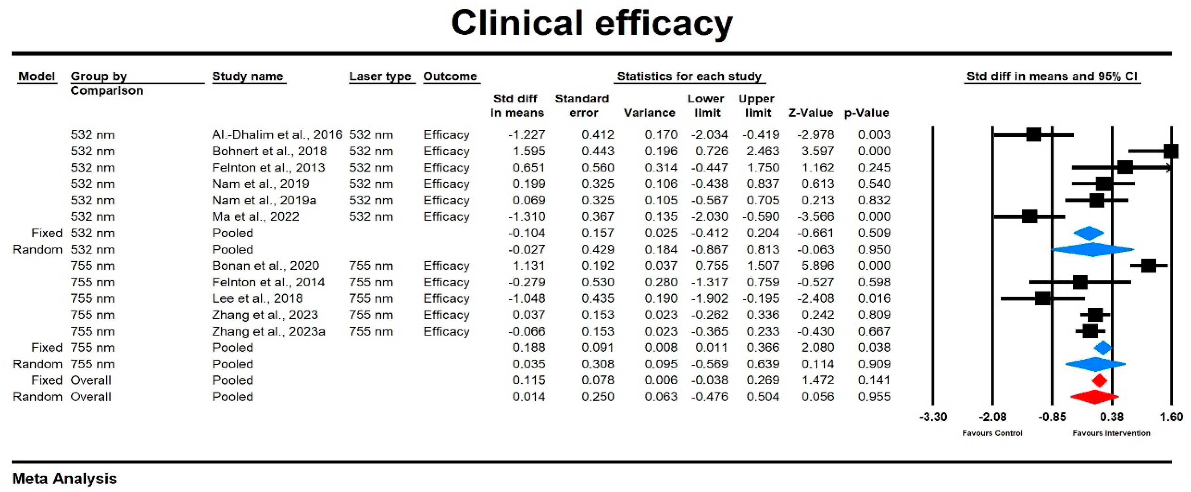
3.3.1. Improvement

Across the studies, laser treatments showed varying degrees of efficacy. This depended on the type of skin lesions and the laser wavelength used, the latter one being the major aim of the present study. Also, the type of tool used to evaluate the efficacy influenced the results. Al-Dhalimi and Al-Janabi (2016) [15], who evaluated port wine stains, demonstrated that the 532 nm laser had a lower improvement score compared to the 1064 nm laser, measured via VAS, however a better (“excellent”) percentage improvement was noted for the 532 nm option (28.5% vs. 0%). Nevertheless, any degree of improvement was noted for all 532 nm treatments, and the outcomes were similar for 13 out of 14 cases. Bohnert et al. (2018) [16] showed better improvement with 532 nm-treatment of the side of face covered with solar lentigines as compared to a 1064 nm approach. In contrast, Felton et al. (2014) [18] reported that the 1064 nm wavelength achieved the highest improvement (97%) for nevus of Ota compared to shorter wavelengths, with rates of 90% and 75% for the 532 nm and 755 nm devices, respectively. Nam et al. (2019) [21] reported comparable improvements in erythema for the 532 nm KTP diode laser and the 595 nm pulsed dye laser (PDL) using both evaluation techniques, i.e., colorimeter analysis and photographic inspection performed by two independent dermatologists. Meanwhile, Uebelhoer et al. (2007) [23] pointed out that the KTP laser achieved 85% clearing for telangiectasia after three treatments compared to 75% for PDL. However, the authors provided no standard deviations with respect to these measurements and thus the results were not included in the synthesis here. West and Alster (1998) [24] observed the long-pulsed dye laser yielding a higher clearance (up to 95% expressed using a 4-point Likert scale) for facial and leg telangiectasias than the KTP laser. As the authors merged data here for low extremities and face, we could not form separate conclusions for facial lesions. As for the 755 nm laser, Bonan et al. (2021) [25] found the MoveoPL mode of the 755 nm alexandrite laser provided superior improvement for benign hyperpigmentation, as compared to the single-pass mode. Levin et al. (2016) [29] noted similar improvement scores for both the picosecond and Q-switched lasers in treating pigmentary disorders, each showing approximately 50% clearance. Ma et al. (2022) [22] found the 755 nm laser achieved higher mean improvement scores (3.78 on a 1–5 scale) compared to the 532 nm picosecond Nd: YAG laser (3.11 on a 1–5 scale) for freckles. Table 2 provides data on the improvement measurements with the name of the tools used.

Improvement Score

Using a mixed-effects model, we found that the overall effect size for the improvement score was low and did not vary significantly by laser type (SMD = 0.014, SE = 0.25, 95% CI [−0.476, 0.504]; p = 0.906). The heterogeneity among studies was high, with I2 = 86.7% and a significant Q-value (Q = 86,05, p = 0.000). Subgroup analysis indicated that the effect size was slightly higher in the case of the 755 nm laser compared to the 532 nm group, though both were not statistically significant (532 nm: SMD = −0.027, SE = 0.429, 95% CI [−0.867, 0.813], p = 0.895; 755 nm: SMD = 0.035, SE = 0.308, 95% CI [−0.569, 0.639], p = 0.909). The results are presented in Figure 2. The findings of the sensitivity analysis indicate that individual studies do not have a differential impact.
Funnel plot inspection revealed no significant publication bias, as indicated by Egger’s test (p = 0.55). The plot was symmetric, further suggesting minimal bias (see Supplemental Figure S1).

Relative Risk for Improvement
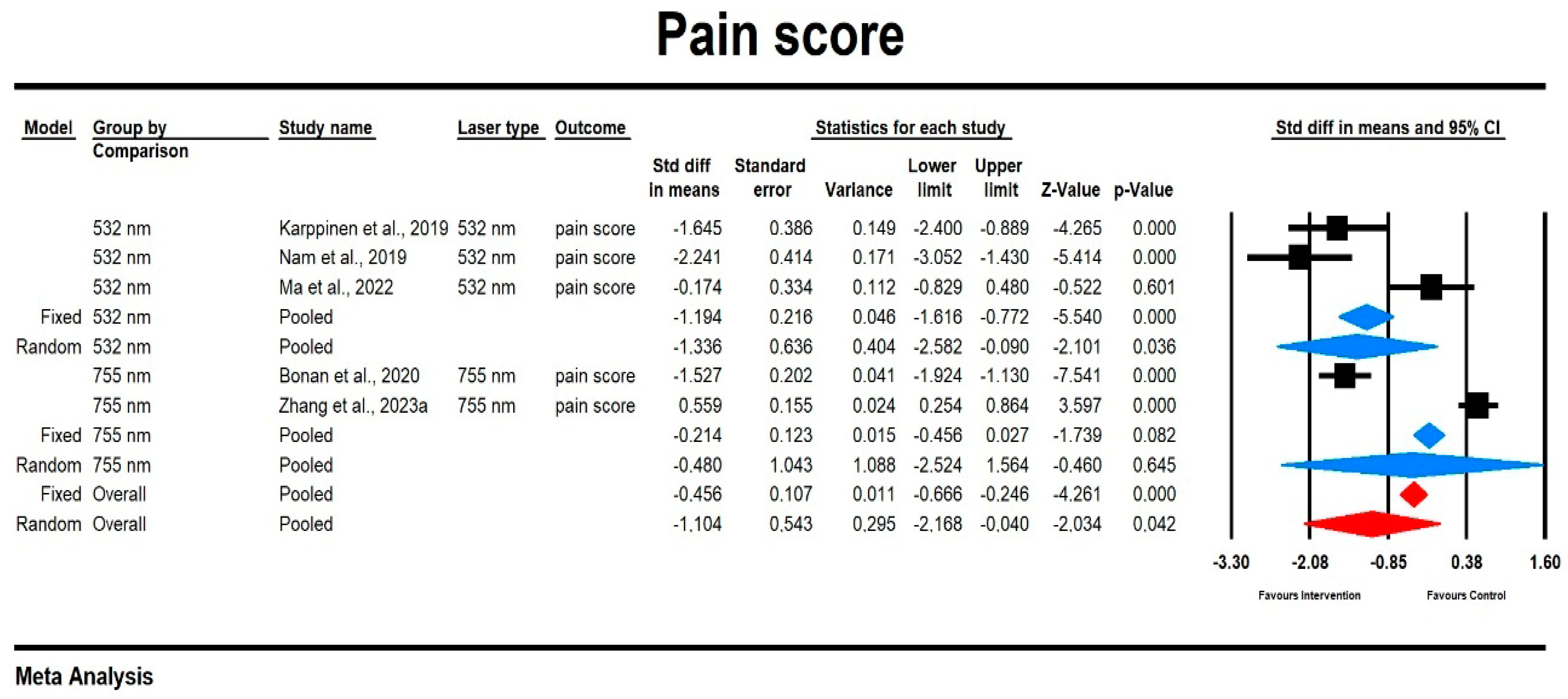
Using a mixed-effects model, we observed that the relative risk for the improvement was small and did not significantly differ between laser types, although there was a trend toward greater improvement with the 755 nm laser (RR = 1.135, 95% CI [0.99, 1.302], p = 0.069). The heterogeneity among studies was low, with I2 = 6.122% and a non-significant Q-value (Q = 5.326, p = 0.327). Subgroup analysis showed that the effect size was slightly higher for the 755 nm laser compared to the 532 nm laser, with the former showing statistical significance (755 nm: RR = 1.512, 95% CI [1.070, 2.136], p = 0.019; 532 nm: RR = 1.077, 95% CI [0.928, 1.249], p = 0.332). The results are presented in Figure 3. The sensitivity analysis findings suggest that individual studies do not exert a differential impact.

3.3.2. Pain

Similarly to improvement indices, pain ratings also varied across studies depending on laser type. Butler et al. (2006) [17] found that patients rated the 532 nm laser as slightly more painful than IPL, with mean pain scores of 5.27 and 4.4 out of 10 Likert scale, respectively. Nevertheless, no standard deviations were reported; thus, these data were not meta-analyzed here. A group studied by Karppinen et al. (2019) [19] rated the 585 nm laser as significantly more painful compared to the KTP laser (532 nm), with scores of 67.7 vs. 34.6 in a 10-point Likert scale evaluation. In contrast, Ma et al. (2022) [22] found only a slight, non-significant difference in pain between QSAL (3.94) and PS532 (3.56), as measured on a numerical visual analog scale. Nam et al. (2019) [21] found that the KTP diode laser was rated less painful (1.49) than the 595 nm PDL (3.73) in treating telangiectatic erythema, similar to Uebelhoer et al. (2007) [23] who reported that more patients found the PDL slightly more painful than KTP for telangiectasia treatments. However, the latter result was not assessed with a pain score.
Bonan et al. (2021) [25] reported that the MoveoPL mode of the 755 nm alexandrite laser was less painful (1.8 on a 0–5 scale) compared to the single-pass mode (3.1 on a 0–5 scale). But Zhang et al. provided data that the 730 nm picosecond laser was less painful than the 755 nm approach (4.69 vs. 5.65, as evidenced with the VAS).

Pain Score

Using a mixed-effects model, we observed that the overall effect size for the pain score was small and did show significant differences between laser types (SMD = −1.104, SE = 0.543, 95% CI [−2.168, 0.064], p = 0.042). There was considerable heterogeneity among the studies, with I2 = 95.977% and a statistically significant Q-value (Q = 99.419, p = 0.000). Subgroup analysis revealed that the effect size was greater in the 532 nm group compared to the 755 nm group, but only the 532 nm result was statistically significant (532 nm: SMD = −1.336, SE = 0.636, 95% CI [−2.582, −0.09], p = 0.036; 755 nm: SMD = −0.480, SE = 1.043, 95% CI [−2.524, 1.564], p = 0.645). The results are displayed in Figure 4. The results of the sensitivity analysis indicate that individual studies do not have a significant influence on the overall effect.
Funnel plot inspection revealed no significant publication bias as indicated by Egger’s test (p = 0.248). The plot was symmetric, further suggesting minimal bias (see Supplemental Figure S3).

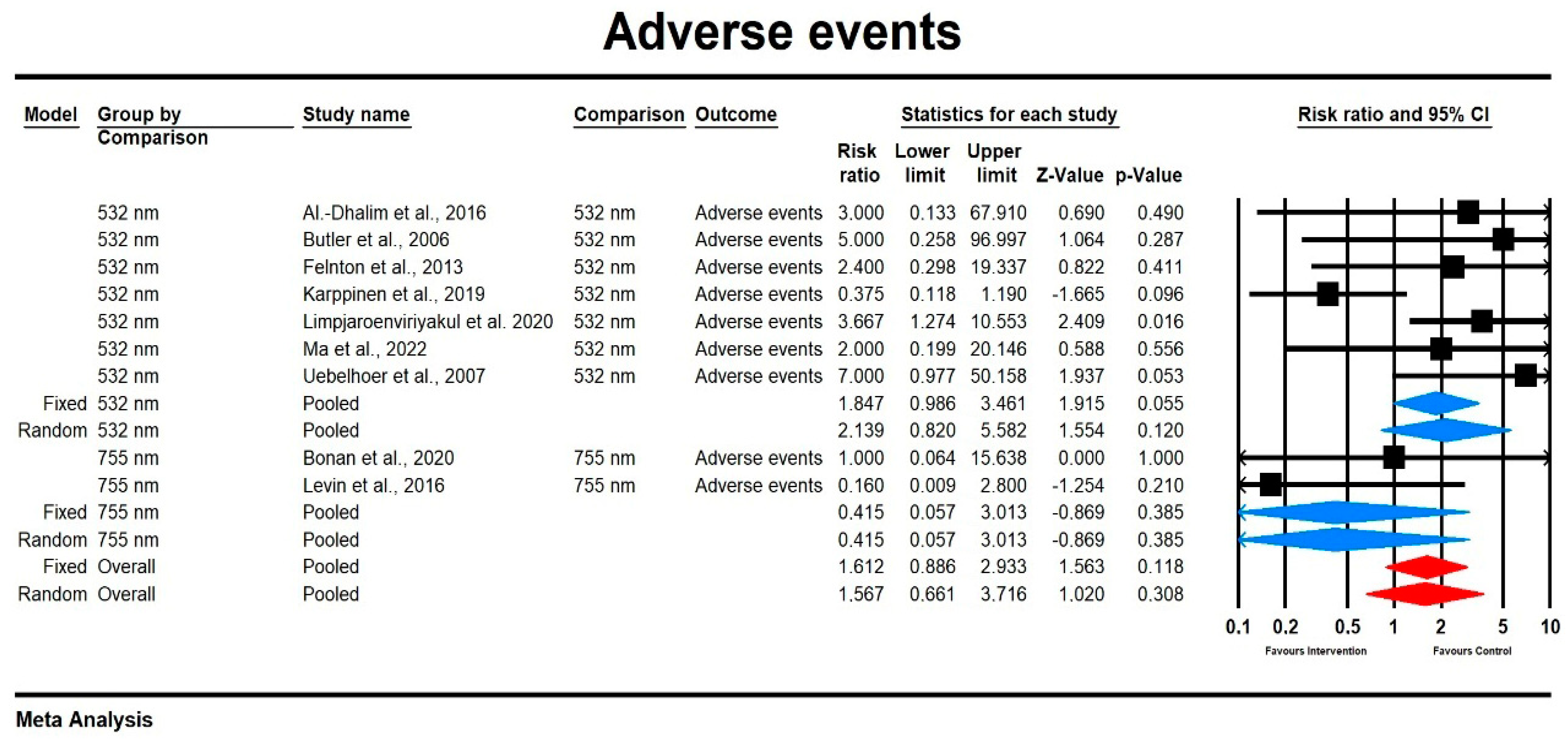
3.3.3. Adverse Events

Safety profiles were generally favourable, though the authors indeed reported some more frequent adverse effects, but predominantly of a mild nature. Al-Dhalimi and Al-Janabi (2016) [15] reported no long-term adverse events, although one case of temporary hyperpigmentation was noted with the 532 nm laser (resolved with hydroquinone cream 4% and no scar). In Bohnert et al.’s [16] study, all patients experienced mild erythema and swelling irrespective of the laser used. Butler et al. [17] noted that erythema and edema, both in the infraorbital area of the KTP-treated side, occurred in two patients. In Karppinen et al.’s study (2019) [19], the PhotoLase yellow laser was associated with more blistering than the KTP laser (8 versus 3 participants), though erythema (17 vs. 16) and crusting (7 vs. 9) rates were similar for both lasers. In the present meta-analysis, data for only blistering were used for calculations.
Ma et al. (2022) [22] reported transient erythema and crusting with both lasers, with no long-term pigmentation changes or scarring. However, two cases developed transient hyperpigmentation on 532 nm-laser side. This was the case in only one man in the 755 nm-treated group. Uebelhoer et al. (2007) [23] noted more prolonged erythema (up to 5 days post treatment) and swelling on the KTP-treated side, though these effects resolved within days. In the present synthesis, data for erythema incidence were used.
In the case of the 755 nm approach, Bonan et al. [25] found a similar incidence of adverse events on the face (2% vs. 2% excluding erythema analyzed as score) between lasers used. Lee et al. [27] demonstrated that mild erythema and swelling without petechiae was present in all patients irrespective of the side of the face. Levin et al. (2016) [29] also reported only temporary side effects for both picosecond and Q-switched lasers, with no long-term complications recorded. However, dyspigmentation only for the Q-switched nanosecond treatment group was noted.

Relative Risk for Adverse Events

Using a mixed-effects model, we found that the relative risk for adverse events was moderate but did not significantly differ across laser types (RR = 1.567, 95% CI [0.661, 3.716], p = 0.383). The heterogeneity among studies was low, with I2 = 31.298% and a non-significant Q-value (Q = 14.084, p = 0.118). Subgroup analysis showed that the effect size was higher for the 532 nm laser compared to the 755 nm laser, although neither was statistically significant (755 nm: RR = 0.415, 95% CI [0.057, 3.013], p = 0.385; 532 nm: RR = 2.139 95% CI [0.820, 5.582], p = 0.12). The results are displayed in Figure 5. The sensitivity analysis results suggest that individual studies do not substantially impact the overall effect. Funnel plot inspection revealed no significant publication bias, as confirmed by Egger’s test (p = 0.602), with a symmetric plot indicating minimal bias (see Supplemental Figure S4).

3.3.4. Patient Satisfaction

Patient satisfaction with laser treatments varied by laser type, modality, and treatment area. Across studies, patient satisfaction generally mirrored the efficacy presented above.
In Al-Dhalimi and Al-Janabi’s study (2016) [15], satisfaction was assessed via patient self-reports, showing high satisfaction levels with the 532 nm Nd: YAG laser over the 1064 nm laser for port wine stain treatment (7.6 vs. 3.3. in a patient 10-point satisfaction score collected via interview). Bohnert et al. (2018) [16] reported higher satisfaction in the arm treated with the 532 nm laser in their study on solar lentigines (82.5% vs. 75%). In Limpjaroenviriyakul et al.’s study [20], a higher satisfaction measured as a changed score was found for the 532 nm device whilst in and Ma et al. [22], higher study scores were noted for the comparative laser (mean score of 8.11) over the 532 nm picosecond Nd: YAG laser (mean score of 6.72). However, in the study by Nam et al. [21], a patient self-assessment using a six-point satisfaction scale produced results which were comparable.
Levin et al. (2016) [29] found 84% of patients were “satisfied” to “completely satisfied” with Q-switched lasers compared to 50% for the picosecond alexandrite, using a 7-point Likert scale, while Uebelhoer et al. (2007) [23] showed greater satisfaction with the 532 nm in terms of the overall effect, downtime, and experience. Bonan et al. (2021) [25] measured satisfaction on a 5-point Likert scale and demonstrated that patients reported greater satisfaction with MoveoPL, scoring it higher (average score of 3.6 for both treatments). However, 93% of the patients reported a preference for MoveoPL, whereas only 7% preferred the standard SP treatment. In the study by Lee et al. (2012) [28], 44% of patients reported “excellent” or “good” satisfaction with the 755 nm long-pulsed alexandrite laser in treating pigmentary lesions, assessed via self-assessment questionnaires. In Lee et al.’s paper (2018) [27], satisfaction for melasma treatment was higher for the 755 nm alexandrite laser than the Q-switched Nd: YAG laser.

Patient Satisfaction Score and Relative Risk

In two studies using the 532 nm laser [15,22], patient satisfaction scores were a reported endpoint, with a pooled SMD of 0.785; SE = 1.692; 95% CI: [−2.532, 4.102]. However, the effect size did not differ significantly by laser type (p = 0.643). The heterogeneity between the studies was high, with I2 = 96.716% and a significant Q-value (Q = 30.449, p = 0.000).
For satisfaction as a qualitative measure, two other studies [16,19] provided data, with a pooled RR of 1.233; 95% CI: [0.814, 1.867]; Z = 0.989. The heterogeneity among these studies was low, with I2 = 0.0% and a non-significant Q-value (Q = 0.249, p = 0.617).

3.4. Meta-Regression Analyses

For endpoint data (SMD) regarding 532 nm laser efficacy, p values for pulse duration (Q = 3.18, df = 2, p = 0.204) and type of lesion (Q = 2.02, df = 2, p = 0.3635) were non-significant, indicating that the predicted effect sizes were not dependent on any of these variables. Compared to millisecond pulse duration, the picosecond pulse duration showed a negative non-significant association with treatment efficacy (coefficient = −1.0263; Z = −1.22; p = 0.22), while the nanosecond pulse duration showed a positive but non-significant association (coefficient = 0.9351; Z = 1.00; p = 0.3195). In the case of lesion type, the coefficients were positive and as follows: pigmented: −1.36, Z = −1.40, p = 0.1601; vascular: −0.5887, Z = −0.74, p = 0.4606. When we conducted meta-regression for RR of improvement, the p values were also non-significant for vascular vs. pigmented lesions (Q = −1.872, Z = −1.28, p = 0.2022).
For the adverse event outcome related to the 532 nm laser (risk ratio), pulse duration was not a significant moderator (Q = 0.01, df = 2, p = 0.9955). However, the overall model examining lesion type was statistically significant (Q = 9.46, df = 2, p = 0.0088), indicating that lesion type significantly moderates the treatment effect. Specifically, when comparing vascular to pigmented lesions, the coefficient for vascular lesions was −1.8687 (p = 0.0085), suggesting a significantly lower effect size—i.e., a reduced risk of adverse events—for vascular lesions. The model explained approx. 71% of the heterogeneity. No additional meta-regression analyses were feasible based on the available data in the present meta-analysis.

3.5. Risk of Bias

Table 3 assesses the risk of bias in each study across seven domains, highlighting key trends, which are as follows:
  • Random Sequence Generation (Selection Bias): All studies demonstrated a “Low Risk” of selection bias, suggesting that the randomization process was robust.
  • Allocation Concealment (Selection Bias): Most studies maintained a low risk by ensuring allocation concealment, though a few studies were rated as “Unclear” due to insufficient reporting.
  • Blinding of Participants and Personnel (Performance Bias): While blinding was achieved in several studies, approximately one-third had a “High Risk” due to lack of blinding of participants and personnel, which could potentially impact performance bias.
  • Blinding of Outcome Assessment (Detection Bias): Most studies ensured low risk for outcome assessment blinding, although some had “Unclear” or “High Risk” ratings due to partial or no blinding of the outcome assessors.
  • Incomplete Outcome Data (Attrition Bias): Nearly all studies were rated as “Low Risk”, indicating good data completeness. Only one study had unclear attrition data, which may affect short-term outcomes.
  • Selective Reporting (Reporting Bias): All studies showed a low risk of reporting bias, suggesting consistency in reporting all specified outcomes.
  • Other Bias: Only one study presented a potential source of other bias due to a device manufacturer affiliation, while all others were free from additional noted biases.
The ROB analysis thus suggests that the studies generally adhered to good research practices, particularly in randomization, allocation concealment, and data completeness. Some areas, notably participant and personnel blinding, could be improved for stronger bias mitigation across all domains.

4. Discussion

This meta-analysis, comprising 16 studies with 509 participants involved, comparatively evaluated the efficacy and safety of 532 nm and 755 nm lasers in treating pigmented and vascular lesions on the face. This meta-analysis demonstrated that overall improvement scores did not significantly differ by laser type (SMD = 0.014, SE = 0.25, 95% CI [−0.476, 0.504], p = 0.906); however, high heterogeneity was present (I2 = 86.7%). Subgroup analysis, however, indicated a notable efficacy advantage for the 755 nm laser (532 nm: SMD = −0.027, SE = 0.429, 95% CI [−0.867, 0.813], p = 0.895; 755 nm: SMD = 0.035, SE = 0.308, 95% CI [−0.569, 0.639], p = 0.909), with a significant relative improvement (755 nm: RR = 1.512, 95% CI [1.070, 2.136], p = 0.019). The observation hereby confirms the greater efficacy of treating pigmentary and vascular lesions on the face with a 755 nm device. Our group previously demonstrated an optimal clearance of melasma-type pigmented lesions and mild hyperpigmented skin lesions with a 755 nm laser, but with a simultaneous reduction in the temperature effect, by using a very short pulse duration in the picosecond range [31]. From our perspective, additionally, 1064 nm lasers with a pulse duration shortened to picoseconds can be effective. A short picosecond pulse duration compared to the widely used millisecond pulse duration is very important in the treatment of pigmented lesions because it minimizes the risk of inflammation associated with temperature PIH (post-inflammatory hyperpigmentation) [32]. However, in the treatment of vascular lesions, it is necessary to achieve an optimal temperature that effectively coagulates and closes the vessel, so it is reasonable to use a pulse duration in the millisecond range [33]. Overall, it seems that the more pronounced efficacy of the 755 nm laser results from effective melanin-targeting, deeper penetration, and reduced thermal damage, making it more versatile for treating both pigmented and vascular lesions compared to the more superficial 532 nm laser.
Pain scores between the two lasers were found to be significantly different in this meta-analysis (SMD = −1.104, SE = 0.543, 95% CI [−2.168, 0.064], p = 0.042) with high heterogeneity (I2 = 95.977%). It turned out that the 532 nm device produces more a painful experience (532 nm: SMD = −1.336, SE = 0.636, 95% CI [−2.582, −0.09], p = 0.036; 755 nm: SMD = −0.480, SE = 1.043, 95% CI [−2.524, 1.564], p = 0.645). Specific studies echoed these findings, as reviewed in the Results section. This variability in pain perception underscores the importance of considering patient comfort and sensitivity to different wavelengths when determining treatment protocols. Unfortunately, the use of anesthetic cream was not consistently reported across all included studies. Some studies explicitly stated that no anesthetic was applied, while others did not provide this detail. Where available, we included such information in the data extraction table. However, due to inconsistent reporting, we could not control for this variable in the meta-analysis—not enough studies provided data with regard to pain scores to be able to conduct subgroup analyses/meta-regression. Other safety outcomes across studies were generally favourable, with minimal adverse events reported. No significant difference in the relative risk of adverse events was observed between the lasers (RR = 1.567, 95% CI [0.661, 3.716], p = 0.383), and heterogeneity was moderate (I2 = 31.289%). However, some studies noted slightly higher incidences of transient adverse events, such as erythema and blistering, with the 532 nm laser, which may be attributable to its higher absorption rate in superficial tissues, as confirmed with subgroup analysis (755 nm: RR = 0.415, 95% CI [0.057, 3.013], p = 0.385; 532 nm: RR = 2.139, 95% CI [0.820, 5.582], p = 0.12). In most studies, the authors suggested that while both lasers maintain acceptable safety profiles, selecting the appropriate wavelength based on the lesion’s depth and patient skin type can optimize safety and efficacy [34,35].
The choice between 532 nm and 755 nm lasers for treating facial pigmented and vascular lesions should be guided by specific clinical considerations, as each wavelength interacts differently with skin chromophores and tissue depths [16,25]. As confirmed earlier, the 532 nm laser is absorbed more by superficial chromophores. Thus, it is primarily effective for epidermal pigmented lesions and superficial vascular conditions but may not reach deeper targets. However, shorter wavelength, higher absorption, and thus greater energy deposition in the epidermis—which contains more melanin and a higher density of nerve endings—can lead to more scattered heat, increasing the risk of surrounding tissue damage and, consequently, pain. This higher absorption rate by melanin can increase the risk of post-inflammatory hyperpigmentation, especially in individuals with darker skin types where melanin content is naturally elevated, making the 532 nm laser more challenging to use in these populations without causing pigmentation changes [22]. Also, typically, the 532 nm laser is used for smaller spot sizes and thus more energy cumulates in the skin as compared to larger 755 nm laser spots [35,36,37,38,39]. Indeed, the meta-regression analysis also demonstrated that 532 nm laser treatment of vascular lesions was associated with significantly fewer adverse events compared to pigmented lesions (coefficient = −1.8687, p = 0.0085). This suggests that vascular lesion treatments using 532 nm lasers may be better tolerated overall. A plausible explanation lies in the underlying tissue characteristics and laser–tissue interaction mechanisms as described above. Vascular lesions primarily involve superficial dermal blood vessels, where hemoglobin serves as the primary chromophore. Hemoglobin absorbs 532 nm light efficiently, allowing for effective treatment at relatively low energy settings and with shorter pulse durations, thereby minimizing collateral thermal damage to surrounding tissues [35]. Conversely, the 755 nm wavelength penetrates deeper into the skin and is less absorbed by melanin, which makes it especially suitable for treating deeper pigmented lesions, such as melasma, and for patients with darker skin types [26,28]. This wavelength bypasses superficial melanin to minimize the risk of hyperpigmentation. Consequently, this provides a safer option for those with higher melanin levels who are at increased risk of pigmentary side effects when treated with shorter wavelengths [21,24]. Studies have demonstrated the efficacy of the 755 nm wavelength in managing various benign hyperpigmentations, with evidence of high patient satisfaction and favorable outcomes, such as higher visual analog scores (VAS) and greater clearance in melasma cases [25,28].
While it is indeed clinically intuitive and already known that factors such as pulse duration and other laser parameters, as well as the type of lesion treated influence treatment outcomes and represent important sources of variability, we performed meta-regression analysis to evaluate whether pulse duration (ps vs. ns vs. ms) and lesion type (pigmented, vascular, or mixed) may significantly influence treatment efficacy and contribute to variability across studies. These findings highlight that variations in laser parameters beyond wavelength—particularly pulse duration—are clinically relevant and should be carefully considered in both research design and clinical practice. For efficacy at 532 nm, although we observed a trend toward smaller effect sizes with picosecond lasers and in the treatment of vascular lesions, this did not reach statistical significance, likely due to the limited number of available studies. Additionally, for the outcome of adverse events at 532 nm, the treatment of vascular lesions was significantly associated (p = 0.02) with a reduced effect size, indicating a lower risk of experiencing adverse events as described above. Overall, accounting for these moderators helps to explain some of the heterogeneity observed in treatment outcomes and reinforces the importance of individualized parameter selection based on lesion characteristics and not only wavelength but other laser parameters. Consequently, clinicians should carefully consider lesion type, lesion depth, skin type, and patient preferences when selecting the appropriate laser wavelength for treatment to maximize efficacy while minimizing adverse effects, as well as pain. Selecting a laser that aligns with the depth and chromophore concentration of the lesion can optimize treatment outcomes and reduce post-treatment complications, especially in diverse skin types where pigmentation risks differ [16,23,26]. Additionally, adjusting laser parameters, such as energy settings and pulse duration, may further tailor treatment to the unique needs of the patient, enhancing safety and satisfaction in laser-based dermatologic procedures [21,30]. It is believed that laser setting parameters such as energy density and pulse duration are fundamental, but they should be expanded to include treatment spot size, which affects the depth of light penetration in the skin, and pulse emission frequency, which affects temperature, i.e., the risk of PIH and pain.
Challenges in data extraction across the studies complicated this analysis, largely due to inconsistencies in outcome reporting. In several studies, data were either missing or presented differently between text and figures, making it challenging to ensure data reliability and comparability. Nam et al. (2019) [21], for example, displayed outcome measurements inconsistently across sections of the study, and subjective measures such as patient satisfaction and improvement ratings varied widely across studies due to differences in scale and assessment methodology. In the study by Butler et al. (2006) [17], patients rated IPL higher for pigment lesions despite KTP’s better performance for vascular lesions, demonstrating how subjective reporting variability impacts outcome comparability. This lack of uniformity underscores the importance of standardized outcome measures in future research to improve reliability and comparability in laser therapy studies.
The limitations of this meta-analysis, including high heterogeneity, small sample sizes, and variability in subjective outcome measures, should be acknowledged. For instance, Fabi et al. (2014) [26] reported wide variability in MMASI scores for melasma, reflecting subjective scoring, while Felton et al. (2014) [18] observed lesion recurrence in some patients after 10 months, underscoring the importance of long-term follow-up data. Moreover, regarding pain severity assessment, inconsistent reporting prevented us from accounting for the use of anesthetic cream prior to the procedure, as too few studies provided such data to allow for subgroup analyses or meta-regression. Lastly, at the screening phase, some of the studies might have been missed. Another important limitation of this meta-analysis is the variability and, in some cases, the lack of long-term follow-up data across the included studies. As the durability of treatment effects is particularly relevant in the management of pigmented lesions—where recurrence can occur over time—this limits our ability to draw conclusions regarding the sustained efficacy of 532 nm and 755 nm lasers. Due to inconsistent reporting and the variability of observation periods following the laser treatment cycle in the majority of trials, we were unable to perform subgroup analyses based on follow-up duration. We therefore recommend that future studies incorporate standardized and sufficiently long follow-up periods to better evaluate the long-term outcomes of laser therapies, especially in the context of pigmented lesion treatment. Finally, while no significant publication bias was detected, the limited number of studies restricts generalizability.
Future research should focus on larger, standardized samples with consistent methodologies and explore long-term outcomes, such as recurrence and quality of life, to strengthen the evidence for clinical applications of 532 nm and 755 nm lasers in facial lesion treatments. Future studies in hyperpigmented lesions should be expanded to include a clear division of efficacy depending on pulse duration in the millisecond, nanosecond, and picosecond ranges, and additionally correlated with the appropriate laser wavelength. Regarding vascular lesions, only pulse duration in the millisecond range seems effective, but an objective comparison of the efficacy of individual laser wavelengths with the broadband effect of pulsed light devices, so-called IPL, seems reasonable. Importantly, we recommend that future trials standardize and clearly report the use of anesthetics, particularly when pain is assessed as a primary outcome. Additionally, they should focus on larger, standardized trials with consistent methodologies and examine long-term outcomes such as disease recurrence and quality of life to strengthen the evidence for the clinical use of 532 nm and 755 nm lasers in the treatment of facial lesions.
In conclusion, this meta-analysis concludes that both 532 nm and 755 nm lasers are effective in treating facial pigmented and vascular lesions, with the 755 nm laser showing a slight advantage and 532 nm producing more painful experiences but less adverse events in the case of vascular lesions. We believe that our work contributes novel insights by providing a structured, evidence-based foundation for optimizing laser therapy and supports the call for individualized treatment based on both clinical and technical factors.

Supplementary Materials

The following supporting information can be downloaded at: https://www.mdpi.com/article/10.3390/jcm14082546/s1, Figure S1: Funnel Plot of Standard Error by Std Diff in Means, Figure S2: Funnel Plot of Standard Error by Log Risk Ratio, Figure S3: Funnel Plot of Standard Error by Std Diff in Means, Figure S4: Funnel Plot of Standard Error by Log Risk Ratio.

Author Contributions

Conceptualization, P.Z. (Piotr Zawodny). methodology, P.Z. (Piotr Zawodny) and K.S.-Ż.; software, K.S.-Ż.; validation, P.Z. (Piotr Zawodny) and K.S.-Ż.; formal analysis, P.Z. (Piotr Zawodny) and K.S.-Ż.; investigation, P.Z. (Paweł Zawodny), A.K.-C., E.S. and M.K.; resources, P.Z. (Piotr Zawodny) and K.S.-Ż.; data curation, P.Z. (Piotr Zawodny) and K.S.-Ż.; writing—original draft preparation, P.Z. (Piotr Zawodny); writing—review and editing, all authors; visualization, K.S.-Ż.; supervision, K.S.-Ż. and J.S.; project administration, P.Z. (Piotr Zawodny) and K.S.-Ż.; funding acquisition, K.S.-Ż. All authors have read and agreed to the published version of the manuscript.

Funding

This research received no external funding.

Institutional Review Board Statement

Not applicable.

Informed Consent Statement

Not applicable.

Data Availability Statement

No new data were created in this manuscript.

Conflicts of Interest

The authors declare no conflicts of interest. Piotr Zawodny is an aesthetic clinic shareholder, but this played no role in producing and presenting the results.

Abbreviations

The following abbreviations are used in this manuscript:
CIConfidence Interval
Cochran Risk-of-Bias ToolCochrane Risk-of-Bias Tool for Randomized Trials
CINAHLCumulative Index to Nursing and Allied Health Literature
EmbaseExcerpta Medica Database
IPLIntense Pulsed Light
I2I-Squared Statistic
KTP laserPotassium Titanyl Phosphate Laser
MMASIMelasma Area and Severity Index
Nd: YAG laserNeodymium-Doped Yttrium Aluminum Garnet Laser
Nd laserNeodymium Laser
OCObserved Cases
PIHPost-inflammatory Hyperpigmentation
PDLPulsed Dye Laser
PLPhotolysis
PRISMAPreferred Reporting Items for Systematic Reviews and Meta-Analyses
Q Statistic, Q-valueQ Statistic
Q-switched laserQ-switched Laser
RCTsRandomized Controlled Trials
RRRisk Ratio
ROBRisk of Bias
SEStandard Error
SMDStandardized Mean Difference
SPStandard Pulsed
τ2Between-Study Variance
USAUnited States of America
VASVisual Analog Scale
WHOWorld Health Organization
nNumber
nmNanometer
pp-value
MoveoPL modeMoveo Photo Light Mode
PubMedPublic/Publisher MEDLINE

References

  1. Barone, M.; De Bernardis, R.; Persichetti, P. Aesthetic Medicine Across Generations: Evolving Trends and Influences. Aesthetic Plast. Surg. 2024, 1–3. [Google Scholar] [CrossRef]
  2. Khavkin, J.; Ellis, D.A.F. Aging Skin: Histology, Physiology, and Pathology. Facial Plast. Surg. Clin. N. Am. 2011, 19, 229–234. [Google Scholar] [CrossRef]
  3. Cestari, T.F.; Dantas, L.P.; Boza, J.C. Acquired Hyperpigmentations. An. Bras. Dermatol. 2014, 89, 11–25. [Google Scholar] [CrossRef] [PubMed]
  4. Ratajczak-Stefańska, V.; Maleszka, R.; Boer, M.; Kiedrowicz, M. Erythematotelangiectatic skin–diagnostic difficulties. Ann. Acad. Med. Stetin. 2009, 55, 58–65, discussion 65. [Google Scholar] [PubMed]
  5. Zhou, Y.; Hamblin, M.R.; Wen, X. An Update on Fractional Picosecond Laser Treatment: Histology and Clinical Applications. Lasers Med. Sci. 2023, 38, 45. [Google Scholar] [CrossRef] [PubMed]
  6. Freedman, J.R.; Kaufman, J.; Metelitsa, A.I.; Green, J.B. Picosecond Lasers: The next Generation of Short-Pulsed Lasers. Semin. Cutan. Med. Surg. 2014, 33, 164–168. [Google Scholar] [CrossRef]
  7. Haykal, D.; Cartier, H.; Maire, C.; Mordon, S. Picosecond Lasers in Cosmetic Dermatology: Where Are We Now? An Overview of Types and Indications. Lasers Med. Sci. 2023, 39, 8. [Google Scholar] [CrossRef]
  8. Zawodny, P.; Stój, E.; Kulig, P.; Skonieczna-Żydecka, K.; Sieńko, J. VISIA Skin Analysis System as a Tool to Evaluate the Reduction of Pigmented Skin and Vascular Lesions Using the 532 Nm Laser. CCID 2022, 15, 2187–2195. [Google Scholar] [CrossRef]
  9. Zawodny, P.; Malec, W.; Gill, K.; Skonieczna-Żydecka, K.; Sieńko, J. Assessment of the Effectiveness of Treatment of Vascular Lesions within the Facial Skin with a Laser with a Wavelength of 532 Nm Based on Photographic Diagnostics with the Use of Polarized Light. Sensors 2023, 23, 1010. [Google Scholar] [CrossRef]
  10. DerSimonian, R.; Laird, N. Meta-Analysis in Clinical Trials. Control Clin. Trials 1986, 7, 177–188. [Google Scholar]
  11. Higgins, J.P.T.; Thompson, S.G.; Deeks, J.J.; Altman, D.G. Measuring Inconsistency in Meta-Analyses. BMJ 2003, 327, 557–560. [Google Scholar]
  12. Egger, M.; Davey Smith, G.; Schneider, M.; Minder, C. Bias in Meta-Analysis Detected by a Simple, Graphical Test. BMJ 1997, 315, 629–634. [Google Scholar] [CrossRef]
  13. Duval, S.; Tweedie, R. Trim and Fill: A Simple Funnel-Plot-Based Method of Testing and Adjusting for Publication Bias in Meta-Analysis. Biometrics 2000, 56, 455–463. [Google Scholar] [CrossRef]
  14. RoB 2: A Revised Cochrane Risk-of-Bias Tool for Randomized Trials|Cochrane Bias. Available online: https://methods.cochrane.org/bias/resources/rob-2-revised-cochrane-risk-bias-tool-randomized-trials (accessed on 1 November 2024).
  15. Al-Dhalimi, M.A.; Al-Janabi, M.H. Split Lesion Randomized Comparative Study between Long Pulsed Nd:YAG Laser 532 and 1064 Nm in Treatment of Facial Port-Wine Stain. Lasers Surg. Med. 2016, 48, 852–858. [Google Scholar] [CrossRef]
  16. Bohnert, K.; Dorizas, A.; Sadick, N. A Prospective, Randomized, Double-Blinded, Split-Face Pilot Study Comparing Q-Switched 1064-Nm Nd:YAG versus 532-Nm Nd:YAG Laser for the Treatment of Solar Lentigines. J. Cosmet. Laser Ther. 2018, 20, 395–397. [Google Scholar] [CrossRef] [PubMed]
  17. Butler, E.G.; McClellan, S.D.; Ross, E.V. Split Treatment of Photodamaged Skin with KTP 532 Nm Laser with 10 Mm Handpiece versus IPL: A Cheek-to-Cheek Comparison. Lasers Surg. Med. 2006, 38, 124–128. [Google Scholar] [CrossRef] [PubMed]
  18. Felton, S.J.; Al-Niaimi, F.; Ferguson, J.E.; Madan, V. Our Perspective of the Treatment of Naevus of Ota with 1,064-, 755- and 532-Nm Wavelength Lasers. Lasers Med. Sci. 2014, 29, 1745–1749. [Google Scholar] [CrossRef] [PubMed]
  19. Karppinen, T.; Kantola, E.; Karppinen, A.; Rantamäki, A.; Kautiainen, H.; Mordon, S.; Guina, M. Treatment of Telangiectasia on the Cheeks with a Compact Yellow (585 Nm) Semiconductor Laser and a Green (532 Nm) KTP Laser: A Randomized Double-Blinded Split-Face Trial. Lasers Surg. Med. 2019, 51, 223–229. [Google Scholar] [CrossRef]
  20. Limpjaroenviriyakul, N.; Jurairattanaporn, N.; Kamanamool, N.; Rojhirunsakool, S.; Kanokrungsee, S.; Udompataikul, M. Low-Fluence Q-Switched Nd:YAG 1064-Nm Laser versus Q-Switched Nd:YAG 532-Nm Laser in the Treatment of Hyperpigmented Lips: A Prospective, Randomized, Controlled, Evaluator-Blinded Trial. Lasers Med. Sci 2020, 35, 165–171. [Google Scholar] [CrossRef]
  21. Nam, C.H.; Kim, M.H.; Hong, S.P.; Park, B.C. Fractional 532-Nm KTP Diode Laser and 595-Nm Pulsed Dye Laser in Treatment of Facial Telangiectatic Erythema. J. Cosmet. Dermatol. 2019, 18, 783–787. [Google Scholar] [CrossRef]
  22. Ma, S.Y.; Gong, Y.Q.; Zhang, W.J.; Liang, B.H.; Li, Y.M.; Xie, Z.M.; Zhu, H.L. Split-Face Comparison of the Efficacy of Picosecond 532 Nm Nd:YAG Laser and Q-Switched 755 Nm Alexandrite Laser for Treatment of Freckles. J. Cosmet. Laser Ther. 2022, 24, 22–27. [Google Scholar] [CrossRef]
  23. Uebelhoer, N.S.; Bogle, M.A.; Stewart, B.; Arndt, K.A.; Dover, J.S. A Split-Face Comparison Study of Pulsed 532-Nm KTP Laser and 595-Nm Pulsed Dye Laser in the Treatment of Facial Telangiectasias and Diffuse Telangiectatic Facial Erythema. Dermatol. Surg. 2007, 33, 441. [Google Scholar] [PubMed]
  24. West, T.B.; Alster, T.S. Comparison of the Long-Pulse Dye (590–595 Nm) and KTP (532 Nm) Lasers in the Treatment of Facial and Leg Telangiectasias. Dermatol. Surg. 1998, 24, 221. [Google Scholar] [CrossRef] [PubMed]
  25. Bonan, P.; Troiano, M.; Bruscino, N.; Verdelli, A. Treatment of Benign Hyperpigmentations and Pigmented Scars by 755 Alexandrite Laser Comparing the Single Pass versus MultiPass (MoveoPL) Emission in Skin Types I-IV. Dermatol. Ther. 2021, 34, e14819. [Google Scholar] [CrossRef]
  26. Fabi, S.G.; Friedmann, D.P.; Niwa Massaki, A.B.; Goldman, M.P. A Randomized, Split-Face Clinical Trial of Low-Fluence Q-Switched Neodymium-Doped Yttrium Aluminum Garnet (1064 Nm) Laser versus Low-Fluence Q-Switched Alexandrite Laser (755 Nm) for the Treatment of Facial Melasma. Lasers Surg. Med. 2014, 46, 531–537. [Google Scholar] [CrossRef] [PubMed]
  27. Lee, M.-C.; Lin, Y.-F.; Hu, S.; Huang, Y.-L.; Chang, S.-L.; Cheng, C.-Y.; Chang, C.-S. A Split-Face Study: Comparison of Picosecond Alexandrite Laser and Q-Switched Nd:YAG Laser in the Treatment of Melasma in Asians. Lasers Med. Sci. 2018, 33, 1733–1738. [Google Scholar] [CrossRef]
  28. Lee, Y.B.; Shin, J.Y.; Cheon, M.S.; Oh, S.T.; Cho, B.K.; Park, H.J. Photorejuvenation Using Long-Pulsed Alexandrite and Long-Pulsed Neodymium:Yttrium-Aluminum-Garnet Lasers: A Pilot Study of Clinical Outcome and Patients’ Satisfaction in Koreans. J. Dermatol. 2012, 39, 425–429. [Google Scholar] [CrossRef]
  29. Levin, M.K.; Ng, E.; Bae, Y.C.; Brauer, J.A.; Geronemus, R.G. Treatment of Pigmentary Disorders in Patients with Skin of Color with a Novel 755 Nm Picosecond, Q-Switched Ruby, and Q-Switched Nd:YAG Nanosecond Lasers: A Retrospective Photographic Review. Lasers Surg. Med. 2016, 48, 181–187. [Google Scholar] [CrossRef]
  30. Zhang, M.; Zheng, H.; Ge, Y.; Yang, Y.; Guo, L.; Wu, Q.; Zeng, R.; Shi, H.; Huang, Y.; Zhao, W.; et al. Comparison of the Efficacy and Safety of a 730 Nm Picosecond Titanium Sapphire Laser and a 755 Nm Picosecond Alexandrite Laser for the Treatment of Freckles in Asian Patients: A Two-Center Randomized, Split-Face, Controlled Trial. Lasers Surg. Med. 2023, 55, 636–641. [Google Scholar] [CrossRef]
  31. Zawodny, P.; Wahidi, N.; Zawodny, P.; Duchnik, E.; Stój, E.; Malec, W.R.; Kulaszyńska, M.; Skonieczna-Żydecka, K.; Sieńko, J. Evaluation of the Efficacy of the 755 Nm Picosecond Laser in Eliminating Pigmented Skin Lesions after a Single Treatment Based on Photographic Analysis with Polarised Light. J. Clin. Med. 2024, 13, 304. [Google Scholar] [CrossRef]
  32. Pigment—Detailed Explanation. Available online: https://cambridgelaserclinic.com/laser-treatments/pigment/detailed-explanation/ (accessed on 3 January 2025).
  33. Laser and Light-Based Treatments for Pigmented Lesions. Available online: https://www.thepmfajournal.com/features/features/post/laser-and-light-based-treatments-for-pigmented-lesions (accessed on 3 January 2025).
  34. Alster, T.S.; Tanzi, E.L. Laser Surgery in Dark Skin. Skinmed 2003, 2, 80–85. [Google Scholar] [CrossRef] [PubMed]
  35. Anderson, R.R.; Parrish, J.A. Selective Photothermolysis: Precise Microsurgery by Selective Absorption of Pulsed Radiation. Science 1983, 220, 524–527. [Google Scholar] [CrossRef] [PubMed]
  36. Tanghetti Md, E.; Jennings, J. A Comparative Study with a 755 Nm Picosecond Alexandrite Laser with a Diffractive Lens Array and a 532 Nm/1064 Nm Nd:YAG with a Holographic Optic. Lasers Surg. Med. 2018, 50, 37–44. [Google Scholar] [CrossRef] [PubMed]
  37. Ross, E.V. Light-Based Treatment of Pigmented Lesions. Cosmet. Dermatol. 2011, 24, 515–522. [Google Scholar]
  38. Tanghetti, E.A. The Histology of Skin Treated with a Picosecond Alexandrite Laser and a Fractional Lens Array. Lasers Surg. Med. 2016, 48, 646–652. [Google Scholar] [CrossRef]
  39. Chan, H.H.L.; Kono, T. The Use of Lasers and Intense Pulsed Light Sources for the Treatment of Pigmentary Lesions. Skin. Therapy Lett. 2004, 9, 5–7. [Google Scholar]
Figure 1. Study flow chart.
Jcm 14 02546 g001
Figure 2. The effect size standardized mean differences (SDM) for the improvement score in patients subjected to the 532/755 nm (intervention) vs. the comparator (control) [15,16,18,21,22,25,27,30].
Jcm 14 02546 g002
Figure 3. The effect size risk ratios (RR) for the improvement in patients subjected to 532/755 nm (intervention) vs. comparator (control). Examination of the funnel plot revealed no significant publication bias, as confirmed by Egger’s test (p = 0.17) (see Supplemental Figure S2) [15,19,20,23,26,28].
Jcm 14 02546 g003
Figure 4. The effect size (SDM) for the pain score pain in patients subjected to 532/755 nm (intervention) vs. comparator (control) [19,21,22,25,30].
Jcm 14 02546 g004
Figure 5. The effect size (RR) for the adverse events in patients subjected to 532/755 nm (intervention) vs. comparator (control) [15,17,18,19,20,22,23,25,29].
Jcm 14 02546 g005
Table 1. Study characteristics.
Study CharacteristicsInterventionComparator Sample Characteristics
Publication Year/CountrySponsorship/Blinding/
Study Type
N Total Randomized/AnalyzedLaser 1 NameLength of Wave [nm]Fluence [J/cm2]Spot Size [mm]Frequency [Hz]Pulse Duration [ps]Anesthetic Cream Applied (Y/N)Laser 2 NameLength of Wave [nm]Fluence [J/cm2]Spot Size [mm]Frequency [Hz]Pulse Duration [ps}Intervention DurationAge: Mean/SDN MalesType of LesionFitzpatrick Type (List Type with n)
Al-Dhalim et al., 2016/Iraq [15]A/None/SF19/14Nd: YAG532303125 [ms]Y Nd: YAG10643003220 [ms]6 every 4 weeks22.07/9.0031Facial port wine stainIII-3, IV-11
Bohnert et al., 2018/USA [16]A/DB/SF10/10KTP/Nd: YAG532/10644; 4.53; 610NDYNd: YAG10644610ND6 every 2–3 weeks55/(40–63) range0Mild-to-severe facial mottled pigmentationI-1, II-7, III-2
Butler et al., 2006/USA [17]Ind/DB/SF19/19KTP5327–1010ND15–30 [ms]NIPL (Starlux)IPL25–3610 × 15ND20 [ms]NDOver 18 years old/NDNDPigmented and/or vascular dyschromiasND
Felton et al., 2014/England [18]A/None/Other participants21/15 (11 for 532 and 755)Nd: YAG-532 nm5320.5–52–5ND50 [ns]YQS alexandrite (Alex)—755 nm7554.2–182–4ND100 [ns]Median 8 (4–13)24 (median)/15–61 (range)5Nevus of Ota (NO)II-2, IV-5, V-8
Karppinen et al., 2019/Multicenter [19]A/DB/SF24/18KTP53220–301ND10 [ms]NYellow laser—PhotoLase (585 nm)5855.6–8.11.4ND25 [ms]1–2 every 1–2 months48/27–62 (range)2TelangiectasiaI-6, II-8, III-4
Limpjaroenviriyakul et al., 2020/England [20]Ind/SB/Other participants30/30Q-switched Nd: YAG 532 nm laser (QS 532-nm)532123NDYLow-fluence Q-switched Nd: YAG 1064 nm laser (LFQS 1064 nm)10642.465ND1064 nm: 5 every 2 weeks, 532 nm: once27.73/7.918Idiopathic hyperpigmented lipND
Nam et al., 2019/Korea [21]A/DB/SF20/19Fractional 532 nm KTP laser5323.8620 × 20 [mm2]ND5 [ms]ND595 nm pulsed dye laser (PDL)5957.510ND6 [ms]3 every 4 weeks41.5/21–59 (range)5Facial erythema and telangiectasiaND
Ma et al., 2022/China [22]A/DB/SF18/18QSAL-treated group or picosecond 532 nm Nd: YAG5320.5–0.632 [MHz]375YQ-switched alexandrite laser7555–632 [MHz]100 ± 10 [ns]127.11/4.813FrecklesIII-14, IV-4
Uebelhoer et al., 2007/USA [23]Ind/SB/SF15/14532 nm KTP laser (Gemini, Laserscope)53210; 95; 10ND23 [ms]ND595 nm flashlamp-pumped, long-pulsed PDL (V-Beam, Candela)5957.510110 [ms]3 every 3 weeks52.4/35–70 (range)8Facial telangiectasias, diffuse telangiectatic facial erythemaI-6, II-6, III-2, IV-1
West and Alster 1998/USA [24]A/None/Other participants20/17KTP (532 nm) laser (Aura; Laserscope, San Jose, CA, USA)532151ND10 [ms]ND590 or 595 nm long-pulse dye laser (ScleroPlus Laser; Candela Corporation, Wayland, MA, USA)590, 59515; 202 × 7, 2 × 7ND1.5 [ms]1 or 2 (8-week interval)40/23–690Facial or leg telangiectasiaI-Nd, II-Nd, III-Nd
Bonan et al., 2021/Italy [25]Ind/SB/SF63/63MoveoPL 755 alexandrite laser75518–25ND2.5–3.5NDNDStandard SP emissionSP18–255–101–1.5 ND2 every 50 days57/129Benign hyperpigmentation, pigmentation scarsI-16, II-12, III-19, IV-16
Fabi et al., 2014/USA [26]Ind/DB/SF20/16Low-fluence QSAL7552.0, 1.8, 1.2, 1.185NDNDLow-fluence Q-switched Nd: YAG10642.085ND6 every 1 week43.4/32–64 (range)1MelasmaII-2, III-7, IV-11
Lee et al., 2018/England [27]Ind/None/SF12/12755 nm picosecond alexandrite laser7550.88–1.184.4–5.1ND650ND1064 nm QS-Nd: YAG10642.0, 3.5, 3.28, 6, 4NDND4 every 1 monthND/32–52 (range)1MelasmaIII-Nd, IV-Nd
Lee et al., 2012/Korea [28]A/None/Two groups based on type of lesions on the face ((i) those with pigmentation; and (ii) those with facial flushing, skin laxity or some pigmentation.)116/116Long-pulsed 755 nm alexandrite7551251.5–18ND0.25–0.5 [ms]YLong-pulsed 1064 nm Nd: YAG106440–5010ND0.25–300 [ms]140.73/2.356Facial flushing and telangiectasiaIII-Nd, IV-Nd
Levin et al., 2016/USA [29]Ind/TB/Side-by-side42/42755 nm alexandrite picosecond laser7550.71–4.072.5–6ND750–900NDQ-switched frequency-doubled 532 nm Nd: YAG nanosecond, Q-switched 694 nm ruby nanosecond532, 694, 1064NDNDNDNDpicosecond: 4.12; nanosecond: 5.46 treatments37.1/1–71 (range)6Nevus of OtaIII-16, IV-11, V-14, VI-1
Zhang et al., 2023/China [30]A/SB/SF86/86Picosecond alexandrite laser (PicoSure; Cynosure)7553.77–4.802.3–2.61550–750NDTi:sapphire laser (PicoWay; Syneron-Candela)7303.75 2-31250130.7/21–62 (range)7FrecklesIII-16, IV-70
A—academia; DB—double-blind; SF—split face; ND—no data; Ind—industry; SB—single-blind.
Table 2. Improvement scores in meta-analyzed studies.
Improvement/Clinical Efficacy Endpoint
Publication YearName of ToolLaser 1 ØLaser 1 ±Laser 1 nLaser 2 ØLaser 2 ±Laser 2 n
532 nm
Al-Dhalim et al., 2016 [15]VAS 5-point visual analog scale assessment for halves of PWS with photos2.281.43143.710.8214
Bohnert et al., 2018 [16]Global Aesthetic Improvement scale (1–5) with CR VISIA1.350.37190.760.3710
Felton et al., 2014 [18]ND906.32574.2828.710
Nam et al., 2019 [21]Colorimetric Endpoint Analysis: CR-400 device (Minolta, Tokyo, Japan)12.772.771912.222.7519
Nam et al., 2019 [21]Clinical photography endpoint analysis: 10-point Percent Change Scale2.891.71192.791.1319
Ma et al., 2022 [22]Photographic Endpoint Analysis: Five-grade Percent Change Scale3.110.583183.780.42818
Uebelhoer et al., 2007 [23]Photographic Endpoint Analysis: Five-grade Percent Change Scale85nd875nd8
West and Alster 1998 [24]Photographic Evaluation: assessed by a physician and nurse.1.45nd203nd20
755 nm
Bonan et al., 2020 [25]Grading score (0–4) using LifeViz digital imaging system3.60.6632.80.863
Lee et al., 2018 [27]VAS-5-point visual analog scale assessment1.380.48122.040.7512
Levin et al., 2016 [29]VAS 5-point visual analog scale assessment2.44nd82.57nd22
Zhang et al., 2023 [30]Photographic Evaluation via VISIA-CR camera (Canfield Scientific)69.277.758668.997.4286
Zhang et al., 2023 [30]Global Aesthetic Improvement Scale (GAIS) score (5-point scale)4.020.3864.040.3186
Felton et al., 2014 [18]Endpoint Analysis of Improvement: percentage-based evaluation7518.02682.2229.739
Laser 1—intervention laser; Laser 2—control device.
Table 3. The risk of bias in the studies in the present meta-analysis.
AuthorRandom Sequence Generation (Selection Bias)Allocation Concealment (Selection Bias)Blinding of Participants and Personnel (Performance Bias)Blinding of Outcome Assessment (Detection Bias) (Mortality)Incomplete Outcome Data Addressed (Attrition Bias) (Short-Term Outcomes)Selective Reporting (Reporting Bias)Other Bias
Al-Dhalimi and Al-Janabi (2016) [15]Low Risk—Randomization by simple drawLow Risk—Half-lesion treatmentHigh Risk—Not blindedHigh Risk—Outcome assessors not blindedUnclear—Attrition not fully explained (5 dropouts)Low RiskNone noted
Bohnert et al. (2018) [16]Low Risk—Randomized split-face studyLow Risk—Side randomizationLow Risk—Double-blindedLow Risk—Blinded investigator assessmentsLow Risk—All participants completedLow RiskNone noted
Bonan et al. (2021) [25]Low Risk—Randomization conductedUnclear Risk—Allocation details unclearLow Risk—Controlled and blindedLow Risk—Outcome assessments were blindedLow Risk—High completion rateLow RiskNone noted
Butler et al. (2006) [17]Low Risk—Randomized, split-face designLow Risk—Clear allocationLow Risk—Blinding maintainedLow Risk—Blinded assessorsLow Risk—All participants completedLow RiskNone noted
Fabi et al. (2014) [26]Low Risk—Randomization appliedLow Risk—Allocation well managedLow Risk—Double-blind protocolLow Risk—Independent evaluationsLow Risk—High follow-up rate for each groupLow RiskNone noted
Felton et al. (2014) [18]Low Risk—Randomization via pre-test patchesLow Risk—Allocated by laser responseUnclear Risk—No personnel blinding detailsLow Risk—Independent evaluationsLow Risk—High follow-up completionLow RiskNone noted
Karppinen et al. (2019) [19]Low Risk—Randomized, double-blindedLow Risk—Blinded side allocationLow Risk—Double-blinded designLow Risk—Blinded assessorsLow Risk—All participants completedLow RiskDevice bias (manufacturer affiliation)
Limpjaroenviriyakul et al. (2020) [20]Low Risk—Randomized controlled studyLow Risk—Allocation by blockHigh Risk—No blinding reportedUnclear Risk—No specific blinding for outcome assessorsLow Risk—All patients completed studyLow RiskNone noted
Zhang et al. (2023) [30]Low Risk—Randomized, split-face controlledLow Risk—Controlled side allocationLow Risk—Blinded split-face trialLow Risk—Blinded outcome assessmentsLow Risk—Complete datasetLow RiskNone noted
Lee et al. (2012) [28]Low Risk—Randomized split treatmentUnclear Risk—Allocation not specifiedHigh Risk—No blindingLow Risk—Blinded assessorsLow Risk—Full data presentedLow RiskNone noted
Lee et al. (2018) [27]Low Risk—Randomized, split-face designUnclear Risk—Random allocation unclearHigh Risk—Blinding not reportedUnclear-No blinding for outcome assessmentLow Risk—High retention rateLow RiskNone noted
Levin et al. (2016) [29]Low Risk—Retrospective analysisUnclear Risk—Allocation not describedHigh Risk—No blinding for personnelLow Risk—Blinded evaluation of outcomesLow Risk—Complete data presentedLow RiskRetrospective design
Ma et al. (2022) [22]Low Risk—Randomized split-face studyLow Risk—Clear allocationHigh Risk—No blindingLow Risk—Blinded outcome assessorsLow Risk—All participants completedLow RiskNone noted
Nam et al. (2019) [21]Low Risk—Randomized controlled studyUnclear Risk—Allocation methods unclearHigh Risk—No blinding for personnelLow Risk—Outcome assessment blindedLow Risk—High completion rateLow RiskNone noted
Uebelhoer et al. (2007) [23]Low Risk—Randomized, split-face designLow Risk—Clear allocationLow Risk—Single-blinded for participantsLow Risk—Blinded outcome assessmentsLow Risk—High retention rateLow RiskNone noted
West and Alster (1998) [24]Low Risk—Randomized, split-face comparisonLow Risk—Clear allocationLow Risk—Single-blinding for patientsLow Risk—Independent blinded evaluationLow Risk—All data completeLow RiskNone noted
Disclaimer/Publisher’s Note: The statements, opinions and data contained in all publications are solely those of the individual author(s) and contributor(s) and not of MDPI and/or the editor(s). MDPI and/or the editor(s) disclaim responsibility for any injury to people or property resulting from any ideas, methods, instructions or products referred to in the content.

