
Thermal and Non-Thermal Energies for Atrial Fibrillation Ablation

Abstract
1. Introduction
2. Available Ablation Technologies
2.1. Radiofrequency
2.2. Cryoablation
2.3. Pulsed Field Ablation
- The extent and characteristics of the lesion obtained depend on factors such as voltage, pulse amplitude, waveform, polarity, and physical shape of the catheter, which cannot be subject to adjustment by the end user and are strictly dependent on the system used. Efficacy and lesion geometry are therefore specific, and results obtained with individual systems are not generalizable.
- PFA results in an increase in tissue temperature due to the Joule effect (resistive heating), although it does not contribute to lesion formation [17]. Verma et al. report muscle temperature changes of less than 2.8 °C at a depth of 3 mm [18]. Although small, this temperature increase at the catheter tip can be used to monitor energy transfer to the tissue during energy delivery for optimal lesion formation. Indeed, the use of an esophageal probe [19] or techniques such as endoscopy, endoscopic ultrasound, and electrogastrography before and after PVI have confirmed insignificant rises in temperature and no tissue damage [20].
- Thermal methods act nonspecifically on tissues. In contrast, sensitivity to PFA is extremely different for different tissues. Heart muscle cells are particularly vulnerable to PFA, three times more so than the esophageal wall and four times more so than the phrenic nerve [21]. In addition, even thin layers of adipose tissue, such as those separating the posterior wall of the atrium from the esophagus, act as insulators against PFA. For these reasons, the introduction of PFA has been accompanied by positive expectations regarding safety [22]. Indeed, in clinical and preclinical trials, the use of PFA has not been associated with long-term esophageal injury [23,24].
- PFA is associated with the formation of microbubbles, likely due to the release of nitrogen in the gaseous state. The small size of the bubbles (<40µ) and their composition would allow for their rapid reabsorption and explain the low incidence of silent cardioembolic events on MR [29].
- PFA can induce cough, even in patients under general anesthesia, by direct stimulation of pulmonary J receptors.
3. Technology Application to Ablation Strategies
- PVI;
- Atrial lines (CT isthmus, mitral isthmus, anterior mitral line, left atrial roof, posterior wall isolation);
- Debulking of the posterior wall;
- Substrate ablation, targeting low voltages, areas of slow conduction, fragmented potentials.
3.1. Pulmonary Vein Isolation
3.2. Linear Lesions
3.3. Debulking or Isolation of the Posterior Wall
3.4. Substrate-Guided Ablation
4. Limitations
5. Future Directions
6. Conclusions
Funding
Conflicts of Interest
References
- Tzeis, S.; Gerstenfeld, E.; Kalman, J.; Saad, E.; Shamloo, A.; Andrade, J.; Barbhaiya, C.; Baykaner, T.; Boveda, S.; Calkins, H.; et al. 2024 European Heart Rhythm Association/Heart Rhythm Society/Asia Pacific Heart Rhythm Society/Latin American Heart Rhythm Society expert consensus statement on catheter and surgical ablation of atrial fibrillation. Europace 2024, 26, euae043. [Google Scholar] [PubMed]
- Kirchhof, P.; Camm, A.J.; Goette, A.; Brandes, A.; Eckardt, L.; Elvan, A.; Fetsch, T.; van Gelder, I.C.; Haase, D.; Haegeli, L.M.; et al. Early Rhythm-Control Therapy in Patients with Atrial Fibrillation. N. Engl. J. Med. 2020, 383, 1305–1316. [Google Scholar] [CrossRef]
- Kuck, K.H.; Lebedev, D.S.; Mikhaylov, E.N.; Romanov, A.; Gellér, L.; Kalējs, O.; Neumann, T.; Davtyan, K.; On, Y.K.; Popov, S.; et al. Catheter ablation or medical therapy to delay progression of atrial fibrillation: The randomized controlled atrial fibrillation progression trial (ATTEST). Europace 2021, 23, 362–369. [Google Scholar] [CrossRef]
- Nath, S.; Haines, D.E. Biophysics and pathology of catheter energy delivery systems. Prog. Cardiovasc. Dis. 1995, 37, 185–204. [Google Scholar] [CrossRef] [PubMed]
- Michaud, G.F.; Kumar, S. Eliminating Coagulum Formation with Charge Delivery During Radiofrequency Ablation Negative May Be a Positive! JACC Clin. Electrophysiol. 2016, 2, 242–245. [Google Scholar] [CrossRef]
- La Fazia, V.M.; Pierucci, N.; Schiavone, M.; Compagnucci, P.; Mohanty, S.; Gianni, C.; Della Rocca, D.G.; Horton, R.; Al-Ahmad, A.; Di Biase, L.; et al. Comparative effects of different power settings for achieving transmural isolation of the left atrial posterior wall with radiofrequency energy. Europace 2024, 26, euae265. [Google Scholar] [CrossRef] [PubMed]
- Lee, A.C.; Voskoboinik, A.; Cheung, C.C.; Yogi, S.; Tseng, Z.H.; Moss, J.D.; Dewland, T.A.; Lee, B.K.; Lee, R.J.; Hsia, H.H.; et al. A Randomized Trial of High vs Standard Power Radiofrequency Ablation for Pulmonary Vein Isolation: SHORT-AF. JACC Clin. Electrophysiol. 2023, 9, 1038–1047. [Google Scholar] [CrossRef]
- Reddy, V.Y.; Schilling, R.; Grimaldi, M.; Horton, R.; Natale, A.; Riva, S.; Tondo, C.; Kuck, K.H.; Neuzil, P.; McInnis, K.; et al. Pulmonary Vein Isolation with a Novel Multielectrode Radiofrequency Balloon Catheter That Allows Directionally Tailored Energy Delivery: Short-Term Outcomes from a Multicenter First-in-Human Study (RADIANCE). Circ. Arrhythm. Electrophysiol. 2019, 12, e007541. [Google Scholar] [CrossRef]
- Reddy, V.Y.; Peichl, P.; Anter, E.; Rackauskas, G.; Petru, J.; Funasako, M.; Minami, K.; Koruth, J.S.; Natale, A.; Jais, P.; et al. A Focal Ablation Catheter Toggling Between Radiofrequency and Pulsed Field Energy to Treat Atrial Fibrillation. JACC Clin. Electrophysiol. 2023, 9, 1786–1801. [Google Scholar] [CrossRef]
- Schoene, K.; Arya, A.; Jahnke, C.; Paetsch, I.; Nedios, S.; Hilbert, S.; Bollmann, A.; Hindricks, G.; Sommer, P. Acquired Pulmonary Vein Stenosis After Radiofrequency Ablation for Atrial Fibrillation: Single-Center Experience in Catheter Interventional Treatment. JACC Cardiovasc. Interv. 2018, 11, 1626–1632. [Google Scholar] [CrossRef]
- Nair, K.K.; Shurrab, M.; Skanes, A.; Danon, A.; Birnie, D.; Morillo, C.; Chauhan, V.; Mangat, I.; Ayala-Paredes, F. The prevalence and risk factors for atrioesophageal fistula after percutaneous radiofrequency catheter ablation for atrial fibrillation: The Canadian experience. J. Interv. Card. Electrophysiol. 2014, 39, 139–144. [Google Scholar] [CrossRef] [PubMed]
- Khairy, P.; Chauvet, P.; Lehmann, J.; Lambert, J.; Macle, L.; Tanguay, J.F.; Sirois, M.G.; Santoianni, D.; Dubuc, M. Lower incidence of thrombus formation with cryoenergy versus radiofrequency catheter ablation. Circulation 2003, 107, 2045–2050. [Google Scholar] [CrossRef]
- Mazur, P. Freezing of Living Cells: Mechanisms and Implications. Am. J. Physiol.-Cell Physiol. 1984, 247, C125–C142. [Google Scholar] [CrossRef] [PubMed]
- Guhl, E.N.; Siddoway, D.; Adelstein, E.; Bazaz, R.; Mendenhall, G.S.; Nemec, J.; Saba, S.; Schwartzman, D.; Voigt, A.; Wang, N.C.; et al. Incidence and Predictors of Complications During Cryoballoon Pulmonary Vein Isolation for Atrial Fibrillation. J. Am. Heart Assoc. 2016, 5, e003724. [Google Scholar] [CrossRef]
- Scheinman, M.M.; Scheinman, M.M. Catheter-induced ablation of the atrioventricular junction to control refractory supraventricular arrhythmias. JAMA J. Am. Med. Assoc. 1982, 248, 851–855. [Google Scholar] [CrossRef]
- Xie, F.; Varghese, F.; Pakhomov, A.G.; Semenov, I.; Xiao, S.; Philpott, J.; Zemlin, C. Ablation of myocardial tissue with nanosecond pulsed electric fields. PLoS ONE 2015, 10, e0144833. [Google Scholar] [CrossRef] [PubMed]
- Sugrue, A.; Maor, E.; Del-Carpio Munoz, F.; Killu, A.M.; Asirvatham, S.J. Cardiac ablation with pulsed electric fields: Principles and biophysics. Europace 2022, 24, 1213–1222. [Google Scholar] [CrossRef]
- Verma, A.; Zhong, P.; Castellvi, Q.; Girouard, S.; Mediratta, V.; Neal, R.E. Thermal Profiles for Focal Pulsed Electric Field Ablation. JACC Clin. Electrophysiol. 2023, 9, 1854–1863. [Google Scholar] [CrossRef]
- Kirstein, B.; Heeger, C.H.; Vogler, J.; Eitel, C.; Feher, M.; Phan, H.L.; Mushfiq, I.; Traub, A.; Hatahet, S.; Samara, O.; et al. Impact of pulsed field ablation on intraluminal esophageal temperature. J. Cardiovasc. Electrophysiol. 2024, 35, 78–85. [Google Scholar] [CrossRef]
- Grosse Meininghaus, D.; Freund, R.; Koerber, B.; Kleemann, T.; Matthes, H.; Geller, J.C. Pulsed-field ablation does not induce esophageal and periesophageal injury—A new esophageal safety paradigm in catheter ablation of atrial fibrillation. J. Cardiovasc. Electrophysiol. 2024, 35, 86–93. [Google Scholar] [CrossRef]
- Tabaja, C.; Younis, A.; Hussein, A.A.; Taigen, T.L.; Nakagawa, H.; Saliba, W.I.; Sroubek, J.; Santangeli, P.; Wazni, O.M. Catheter-Based Electroporation: A Novel Technique for Catheter Ablation of Cardiac Arrhythmias. JACC Clin. Electrophysiol. 2023, 9, 2008–2023. [Google Scholar] [CrossRef] [PubMed]
- Maor, E.; Ivorra, A.; Rubinsky, B. Non thermal irreversible electroporation: Novel technology for vascular smooth muscle cells ablation. PLoS ONE 2009, 4, e4757. [Google Scholar] [CrossRef]
- Stewart, M.T.; Haines, D.E.; Miklavčič, D.; Kos, B.; Kirchhof, N.; Barka, N.; Mattison, L.; Martien, M.; Onal, B.; Howard, B.; et al. Safety and chronic lesion characterization of pulsed field ablation in a Porcine model. J. Cardiovasc. Electrophysiol. 2021, 32, 958–969. [Google Scholar] [CrossRef] [PubMed]
- Koruth, J.S.; Kuroki, K.; Kawamura, I.; Brose, R.; Viswanathan, R.; Buck, E.D.; Donskoy, E.; Neuzil, P.; Dukkipati, S.R.; Reddy, V.Y. Pulsed Field Ablation Versus Radiofrequency Ablation: Esophageal Injury in a Novel Porcine Model. Circ. Arrhythm. Electrophysiol. 2020, 13, E008303. [Google Scholar] [CrossRef]
- Reddy, V.Y.; Neuzil, P.; Koruth, J.S.; Petru, J.; Funosako, M.; Cochet, H.; Sediva, L.; Chovanec, M.; Dukkipati, S.R.; Jais, P. Pulsed Field Ablation for Pulmonary Vein Isolation in Atrial Fibrillation. J. Am. Coll. Cardiol. 2019, 74, 315–326. [Google Scholar] [CrossRef] [PubMed]
- Verma, A.; Boersma, L.; Haines, D.E.; Natale, A.; Marchlinski, F.E.; Sanders, P.; Calkins, H.; Packer, D.L.; Hummel, J.; Onal, B.; et al. First-in-Human Experience and Acute Procedural Outcomes Using a Novel Pulsed Field Ablation System: The PULSED AF Pilot Trial. Circ. Arrhythm. Electrophysiol. 2020, 15, E010168. [Google Scholar] [CrossRef]
- Reddy, V.Y.; Petru, J.; Funasako, M.; Kopriva, K.; Hala, P.; Chovanec, M.; Janotka, M.; Kralovec, S.; Neuzil, P. Coronary Arterial Spasm during Pulsed Field Ablation to Treat Atrial Fibrillation. Circulation 2022, 146, 1808–1819. [Google Scholar] [CrossRef]
- Zhang, C.; Neuzil, P.; Petru, J.; Funasako, M.; Hala, P.; Kopriva, K.; Koruth, J.S.; Dukkipati, S.R.; Reddy, V.Y. Coronary Artery Spasm during Pulsed Field vs. Radiofrequency Catheter Ablation of the Mitral Isthmus. JAMA Cardiol. 2024, 9, 72–77. [Google Scholar] [CrossRef]
- Reinsch, N.; Füting, A.; Höwel, D.; Bell, J.; Lin, Y.; Neven, K. Cerebral safety after pulsed field ablation for paroxysmal atrial fibrillation. Heart Rhythm. 2022, 19, 1813–1818. [Google Scholar] [CrossRef]
- Serban, T.; Mannhart, D.; Abid, Q.U.; Höchli, A.; Lazar, S.; Krisai, P.; Bettelini, A.S.; Knecht, S.; Kühne, M.; Sticherling, C.; et al. Durability of pulmonary vein isolation for atrial fibrillation: A meta-Analysis and systematic review. Europace 2023, 25, euad335. [Google Scholar] [CrossRef]
- Castrejón-Castrejón, S.; Martínez Cossiani, M.; Jáuregui-Abularach, M.; Basterra Sola, N.; Ibáñez Criado, J.L.; Osca Asensi, J.; Roca Luque, I.; Moya Mitjans, A.; Quesada Dorador, A.; Hidalgo Olivares, V.M.; et al. Multicenter prospective comparison of conventional and high-power short duration radiofrequency application for pulmonary vein isolation: The high-power short-duration radiofrequency application for faster and safer pulmonary vein ablation (POWER FAST III) trial. J. Interv. Card. Electrophysiol. 2023, 66, 1889–1899. [Google Scholar] [PubMed]
- Kottmaier, M.; Popa, M.; Bourier, F.; Reents, T.; Cifuentes, J.; Semmler, V.; Telishevska, M.; Otgonbayar, U.; Koch-Büttner, K.; Lennerz, C.; et al. Safety and outcome of very high-power short-duration ablation using 70 W for pulmonary vein isolation in patients with paroxysmal atrial fibrillation. Europace 2020, 22, 388–393. [Google Scholar] [CrossRef] [PubMed]
- Kuck, K.H.; Brugada, J.; Fürnkranz, A.; Metzner, A.; Ouyang, F.; Chun, K.R.; Elvan, A.; Arentz, T.; Bestehorn, K.; Pocock, S.J.; et al. Cryoballoon or radiofrequency ablation for paroxysmal atrial fibrillation. J. Cardiopulm. Rehabil. Prev. 2016, 36, 393–394. [Google Scholar] [CrossRef]
- Andrade, J.G.; Champagne, J.; Dubuc, M.; Deyell, M.W.; Verma, A.; Macle, L.; Leong-Sit, P.; Novak, P.; Badra-Verdu, M.; Sapp, J.; et al. Cryoballoon or Radiofrequency Ablation for Atrial Fibrillation Assessed by Continuous Monitoring: A Randomized Clinical Trial. Circulation 2019, 140, 1779–1788. [Google Scholar] [CrossRef]
- Turagam, M.K.; Neuzil, P.; Schmidt, B.; Reichlin, T.; Neven, K.; Metzner, A.; Hansen, J.; Blaauw, Y.; Maury, P.; Arentz, T.; et al. Safety and Effectiveness of Pulsed Field Ablation to Treat Atrial Fibrillation: One-Year Outcomes from the MANIFEST-PF Registry. Circulation 2023, 148, 35–46. [Google Scholar] [CrossRef]
- Reddy, V.Y.; Gerstenfeld, E.P.; Natale, A.; Whang, W.; Cuoco, F.A.; Patel, C.; Mountantonakis, S.E.; Gibson, D.N.; Harding, J.D.; Ellis, C.R.; et al. Pulsed Field or Conventional Thermal Ablation for Paroxysmal Atrial Fibrillation. N. Engl. J. Med. 2023, 389, 1660–1671. [Google Scholar] [CrossRef]
- Verma, A.; Haines, D.E.; Boersma, L.V.; Sood, N.; Natale, A.; Marchlinski, F.E.; Calkins, H.; Sanders, P.; Packer, D.L.; Kuck, K.H.; et al. Pulsed Field Ablation for the Treatment of Atrial Fibrillation: PULSED AF Pivotal Trial. Circulation 2023, 147, 1422–1432. [Google Scholar] [CrossRef]
- Duytschaever, M.; De Potter, T.; Grimaldi, M.; Anic, A.; Vijgen, J.; Neuzil, P.; Van Herendael, H.; Verma, A.; Skanes, A.; Scherr, D.; et al. Paroxysmal Atrial Fibrillation Ablation Using a Novel Variable-Loop Biphasic Pulsed Field Ablation Catheter Integrated with a 3-Dimensional Mapping System: 1-Year Outcomes of the Multicenter inspIRE Study. Circ. Arrhythm. Electrophysiol. 2023, 16, E011780. [Google Scholar] [CrossRef] [PubMed]
- Reddy, V.Y.; Anter, E.; Peichl, P.; Rackauskas, G.; Petru, J.; Funasako, M.; Koruth, J.S.; Marinskis, G.; Turagam, M.; Aidietis, A.; et al. First-in-human clinical series of a novel conformable large-lattice pulsed field ablation catheter for pulmonary vein isolation. Europace 2024, 26, euae090. [Google Scholar] [CrossRef]
- Della Rocca, D.G.; Marcon, L.; Magnocavallo, M.; Menè, R.; Pannone, L.; Mohanty, S.; Sousonis, V.; Sorgente, A.; Almorad, A.; Bisignani, A.; et al. Pulsed electric field, cryoballoon, and radiofrequency for paroxysmal atrial fibrillation ablation: A propensity score-matched comparison. Europace 2024, 26, euae016. [Google Scholar] [CrossRef]
- Maurhofer, J.; Kueffer, T.; Madaffari, A.; Stettler, R.; Stefanova, A.; Seiler, J.; Thalmann, G.; Kozhuharov, N.; Galuszka, O.; Servatius, H.; et al. Pulsed-field vs. cryoballoon vs. radiofrequency ablation: A propensity score matched comparison of one-year outcomes after pulmonary vein isolation in patients with paroxysmal atrial fibrillation. J. Interv. Card. Electrophysiol. 2024, 67, 389–397. [Google Scholar] [CrossRef] [PubMed]
- Badertscher, P.; Weidlich, S.; Knecht, S.; Stauffer, N.; Krisai, P.; Voellmin, G.; Osswald, S.; Sticherling, C.; Kühne, M. Efficacy and safety of pulmonary vein isolation with pulsed field ablation vs. novel cryoballoon ablation system for atrial fibrillation. Europace 2023, 25, euad329. [Google Scholar] [CrossRef]
- Rattka, M.; Mavrakis, E.; Vlachopoulou, D.; Rudolph, I.; Kohn, C.; Bohnen, J.; Yahsaly, L.; Siebermair, J.; Wakili, R.; Jungen, C.; et al. Pulsed field ablation and cryoballoon ablation for pulmonary vein isolation: Insights on efficacy, safety and cardiac function. J. Interv. Card. Electrophysiol. 2024, 67, 1191–1198. [Google Scholar] [CrossRef]
- Schipper, J.H.; Steven, D.; Lüker, J.; Wörmann, J.; van den Bruck, J.H.; Filipovic, K.; Dittrich, S.; Scheurlen, C.; Erlhöfer, S.; Pavel, F.; et al. Comparison of pulsed field ablation and cryoballoon ablation for pulmonary vein isolation. J. Cardiovasc. Electrophysiol. 2023, 34, 2019–2026. [Google Scholar] [CrossRef] [PubMed]
- Urbanek, L.; Bordignon, S.; Schaack, D.; Chen, S.; Tohoku, S.; Efe, T.H.; Ebrahimi, R.; Pansera, F.; Hirokami, J.; Plank, K.; et al. Pulsed Field Versus Cryoballoon Pulmonary Vein Isolation for Atrial Fibrillation: Efficacy, Safety, and Long-Term Follow-up in a 400-Patient Cohort. Circ. Arrhythm. Electrophysiol. 2023, 16, 389–398. [Google Scholar] [CrossRef]
- Reinsch, N.; Füting, A.; Hartl, S.; Höwel, D.; Rausch, E.; Lin, Y.; Kasparian, K.; Neven, K. Pulmonary Vein Isolation Using Pulsed Field Ablation versus High-Power-Short-Duration Radiofrequency Ablation for Paroxysmal Atrial Fibrillation: Efficacy, Safety, and Long-Term Follow-up. Europace 2024, 26, euae194. [Google Scholar] [CrossRef] [PubMed]
- Takagi, T.; Derval, N.; Duchateau, J.; Chauvel, R.; Tixier, R.; Marchand, H.; Bouyer, B.; André, C.; Kamakura, T.; Krisai, P.; et al. Gaps after linear ablation of persistent atrial fibrillation (Marshall-PLAN): Clinical implication. Heart Rhythm. 2023, 20, 14–21. [Google Scholar] [CrossRef]
- Chae, S.; Oral, H.; Good, E.; Dey, S.; Wimmer, A.; Crawford, T.; Wells, D.; Sarrazin, J.F.; Chalfoun, N.; Kuhne, M.; et al. Atrial Tachycardia After Circumferential Pulmonary Vein Ablation of Atrial Fibrillation. Mechanistic Insights, Results of Catheter Ablation, and Risk Factors for Recurrence. J. Am. Coll. Cardiol. 2007, 50, 1781–1787. [Google Scholar] [CrossRef]
- Ouyang, F.; Ernst, S.; Vogtmann, T.; Goya, M.; Volkmer, M.; Schaumann, A.; Bänsch, D.; Antz, M.; Kuck, K.H. Characterization of reentrant circuits in left atrial macroreentrant tachycardia: Critical isthmus block can prevent atrial tachycardia recurrence. Circulation 2002, 105, 1934–1942. [Google Scholar] [CrossRef]
- Sawhney, N.; Anousheh, R.; Chen, W.; Feld, G.K. Circumferential pulmonary vein ablation with additional linear ablation results in an increased incidence of left atrial flutter compared with segmental pulmonary vein isolation as an initial approach to ablation of paroxysmal atrial fibrillation. Circ. Arrhythm. Electrophysiol. 2010, 3, 243–248. [Google Scholar] [CrossRef]
- Reddy, V.Y.; Anter, E.; Rackauskas, G.; Peichl, P.; Koruth, J.S.; Petru, J.; Funasako, M.; Minami, K.; Natale, A.; Jais, P.; et al. Lattice-Tip Focal Ablation Catheter That Toggles Between Radiofrequency and Pulsed Field Energy to Treat Atrial Fibrillation: A First-in-Human Trial. Circ. Arrhythm. Electrophysiol. 2020, 13, E008718. [Google Scholar] [CrossRef] [PubMed]
- Anter, E.; Mansour, M.; Nair, D.G.; Sharma, D.; Taigen, T.L.; Neuzil, P.; Kiehl, E.L.; Kautzner, J.; Osorio, J.; Mountantonakis, S.; et al. Dual-energy lattice-tip ablation system for persistent atrial fibrillation: A randomized trial. Nat. Med. 2024, 30, 2303–2310. [Google Scholar] [CrossRef] [PubMed]
- Kim, M.Y.; Sandler, B.C.; Sikkel, M.B.; Cantwell, C.D.; Leong, K.M.; Luther, V.; Malcolme-Lawes, L.; Koa-Wing, M.; Ng, F.S.; Qureshi, N.; et al. Anatomical Distribution of Ectopy-Triggering Plexuses in Patients with Atrial Fibrillation. Circ. Arrhythmia Electrophysiol. 2020, 13, E008715. [Google Scholar] [CrossRef]
- Markides, V.; Schilling, R.J.; Ho, S.Y.; Chow, A.W.; Davies, D.W.; Peters, N.S. Characterization of left atrial activation in the intact human heart. Circulation 2003, 107, 733–739. [Google Scholar] [CrossRef]
- Bisignani, A.; Pannone, L.; Miraglia, V.; Sieira, J.; Iacopino, S.; Bala, G.; Ströker, E.; Overeinder, I.; Almorad, A.; Gauthey, A.; et al. Feasibility and safety of left atrial posterior wall isolation with a new Cryoballoon technology in patients with persistent atrial fibrillation. PACE—Pacing Clin. Electrophysiol. 2022, 45, 605–611. [Google Scholar] [CrossRef]
- Reddy, V.Y.; Anic, A.; Koruth, J.; Petru, J.; Funasako, M.; Minami, K.; Breskovic, T.; Sikiric, I.; Dukkipati, S.R.; Kawamura, I.; et al. Pulsed Field Ablation in Patients with Persistent Atrial Fibrillation. J. Am. Coll. Cardiol. 2020, 76, 1068–1080. [Google Scholar] [CrossRef] [PubMed]
- Karakasis, P.; Theofilis, P.; Vlachakis, P.K.; Korantzopoulos, P.; Patoulias, D.; Antoniadis, A.P.; Fragakis, N. Atrial Fibrosis in Atrial Fibrillation: Mechanistic Insights, Diagnostic Challenges, and Emerging Therapeutic Targets. Int. J. Mol. Sci. 2025, 26, 209. [Google Scholar] [CrossRef]
- Chelu, M.G.; King, J.B.; Kholmovski, E.G.; Ma, J.; Gal, P.; Marashly, Q.; AlJuaid, M.A.; Kaur, G.; Silver, M.A.; Johnson, K.A.; et al. Atrial fibrosis by late gadolinium enhancement magnetic resonance imaging and catheter ablation of atrial fibrillation: 5-year follow-up data. J. Am. Heart Assoc. 2018, 7, e006313. [Google Scholar] [CrossRef]
- Marrouche, N.F.; Wazni, O.; McGann, C.; Greene, T.; Dean, J.M.; Dagher, L.; Kholmovski, E.; Mansour, M.; Marchlinski, F.; Wilber, D.; et al. Effect of MRI-Guided Fibrosis Ablation vs. Conventional Catheter Ablation on Atrial Arrhythmia Recurrence in Patients with Persistent Atrial Fibrillation: The DECAAF II Randomized Clinical Trial. JAMA 2022, 327, 2296–2305. [Google Scholar] [CrossRef]
- Chen, H.; Li, C.; Han, B.; Xiao, F.; Yi, F.; Wei, Y.; Jiang, C.; Zou, C.; Shi, L.; Ma, W.; et al. Circumferential Pulmonary Vein Isolation with vs. Without Additional Low-Voltage-Area Ablation in Older Patients with Paroxysmal Atrial Fibrillation: A Randomized Clinical Trial. JAMA Cardiol. 2023, 8, 765–772. [Google Scholar] [CrossRef]
- Huo, Y.; Gaspar, T.; Schönbauer, R.; Wójcik, M.; Fiedler, L.; Roithinger, F.X.; Martinek, M.; Pürerfellner, H.; Kirstein, B.; Richter, U.; et al. Low-Voltage Myocardium-Guided Ablation Trial of Persistent Atrial Fibrillation. NEJM Evidence 2022, 1, EVIDoa2200141. [Google Scholar] [CrossRef] [PubMed]
- Ahn, H.J.; Lee, S.R.; Lee, K.Y.; Choi, J.M.; Kwon, S.; Choi, E.K.; Oh, S. Left atrial fibrosis-guided ablation in patients with atrial fibrillation: A systematic review and meta-analysis of randomized trials. Eur. Heart J. 2023, 44, 44. [Google Scholar] [CrossRef]
- Rivera, A.; Gewehr, D.M.; Braga, M.A.P.; Carvalho, P.E.P.; Ternes, C.M.P.; Pantaleao, A.N.; Hincapie, D.; Serpa, F.; Romero, J.E.; d’Avila, A. Adjunctive low-voltage area ablation for patients with atrial fibrillation: An updated meta-analysis of randomized controlled trials. J. Cardiovasc. Electrophysiol. 2024, 35, 1329–1339. [Google Scholar] [CrossRef] [PubMed]
- Verma, A.; Wazni, O.M.; Marrouche, N.F.; Martin, D.O.; Kilicaslan, F.; Minor, S.; Schweikert, R.A.; Saliba, W.; Cummings, J.; Burkhardt, J.D.; et al. Pre-existent left atrial scarring in patients undergoing pulmonary vein antrum isolation: An independent predictor of procedural failure. J. Am. Coll. Cardiol. 2005, 45, 285–292. [Google Scholar] [CrossRef] [PubMed]
- Younis, A.; Zilberman, I.; Krywanczyk, A.; Higuchi, K.; Yavin, H.D.; Sroubek, J.; Anter, E. Effect of Pulsed-Field and Radiofrequency Ablation on Heterogeneous Ventricular Scar in a Swine Model of Healed Myocardial Infarction. Circ. Arrhythm. Electrophysiol. 2022, 15, 683–692. [Google Scholar] [CrossRef]
- Kawamura, I.; Reddy, V.Y.; Santos-Gallego, C.G.; Wang, B.J.; Chaudhry, H.W.; Buck, E.D.; Mavroudis, G.; Jerrell, S.; Schneider, C.W.; Speltz, M.; et al. Electrophysiology, Pathology, and Imaging of Pulsed Field Ablation of Scarred and Healthy Ventricles in Swine. Circ. Arrhythm. Electrophysiol. 2023, 16, E011369. [Google Scholar] [CrossRef]
- Gardziejczyk, P.; Wlazłowska-Struzik, E.; Skowrońska, M.; Baran, J. Pulsed-field ablation using pentaspline catheter as a bail-out strategy for perimitral flutter related to the left atrium anterior wall scar. Hear. Case Rep. 2023, 9, 906–909. [Google Scholar] [CrossRef]
- Mohanty, S.; Torlapati, P.G.; Casella, M.; Della Rocca, D.G.; Schiavone, M.; Doty, B.; La Fazia, V.M.; Pahi, S.; Pierucci, N.; Valeri, Y.; et al. Redefining the blanking period after pulsed-field ablation in patients with atrial fibrillation. Heart Rhythm, 2024; in press. [Google Scholar] [CrossRef]


| Farapulse™ (by Boston Scientific, Natick, MA, USA) 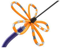 Bipolar penta spline catheter | It proved effective in terms of lesion persistence 641, with a 1-year arrhythmia-free survival of 78% in PAF. Two multicenter registries (MANIFEST-PV and EU-PORIA) gave similar results in a mixed population of PAF and PerAF [35]. The same device was evaluated in the randomized ADVENT trial, which indicated the noninferiority of PFA to both contact force RF and latest-generation cryoballoon ablation [36]. |
| PulseSelectTM (by Medtronic Inc., Minneapolis, MN, USA) 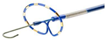 Circular over-the-wire | In the PULSED AF study, the PulseSelectTM System achieved a 100% acute success rate for PVI with essentially no acute complications and a 1-year recurrence rate comparable to that of RF [37]. |
| Varipulse VLCC™ (by Biosense Webster Inc., Irvine, CA, USA) 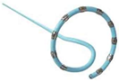 Variable loop biphasic circular catheter | VLCCTM obtained 71% freedom from atrial arrhythmias at 1 year in PAF, with no procedure-related adverse events [38]. |
| Sphere9TM (by Medtronic Inc., Minneapolis, MN, USA)  Large-footprint lattice catheter | Sphere9TM, delivering both radiofrequency and PFA, achieved 78% freedom from atrial arrhythmias at 1 year in a mixed population of PAF and PerAF [9]. |
| Sphere 360TM (by Medtronic Inc., Minneapolis, MN, USA)  Lattice PVI-only single-shot | Currently under development. Preliminary data suggest an efficacy of PVI, with a 45-day isolation durability, of up to 99% [39]. |
| Study | n | Endpoint | Freedom from Endpoint (at 12 mos.) | p | Procedure Duration (min) | p | Fluoroscopy Time (min) | p | Complications | p |
|---|---|---|---|---|---|---|---|---|---|---|
| PFA vs. Thermal (Cryo and RF) | ||||||||||
| Reddy et al. [39] ADVENT | 305 PFA 302 thermal | Recurrence of AT/AF or AADs or repeat TCA | 73.1% 71.3% | n.s. | 105.8 ± 29.4 123.1 ± 42.1 | <0.05 | 21.1 ± 11.0 13.9 ± 12.8 | <0.05 | 2.0% 1.3% | n.s. |
| Della Rocca et al. [40] (HRMC trial) | 174 PFA 348 Cryo 348 RF | AF/AT Recurrence | 79.3% 74.7% 72.4% | n.s. | 52.1 ± 14.6 64.5 ± 21.8 84.8 ± 24.8 | <0.001 | 14.8 ± 3.4 17.6 ± 8.1 12.9 ± 6.9 | <0.001 | 1.1% (*) 1.1% (*) 0.9% (*) | n.s. |
| Maurhofer et al. [41] | 40 PFA 80 Cryo 80 RF | AF/AT Recurrence | 85.0% 76.8% 66.2% | n.s. | 93 (79–116) 75 (60–97) 182 (134–23) | <0.001 | 26 (21–31) 17 (13–24) 7 (3–13) | <0.001 | 1 (**) 0% 0% | <0.04 |
| PFA vs. cryo | ||||||||||
| Badertscher et al. [42] | 106 PFA 75 Cryo | AF/AT Recurrence | 76% 70% | n.s. | 55 (43–64) 58 (48–69) | 0.09 | 11 (9.3–14) 11 (8.7–16) | n.s. | 2.8% 4% | n.s. |
| Rattka et al. [43] | 94 PFA 47 Cryo | AF/AT Recurrence | 70% 61% | n.s. | 162 ± 64 163 ± 62 | n.s. | 26 ± 9 23 ± 9 | 0.06 | 4.2% 2.1% | n.s. |
| Schipper et al. [44] | 54 PFA 54 Cryo | AF/AT Recurrence | 74% 72% | n.s. | 64.5 ± 17.5 73.0 ± 24.8 | 0.07 | 15.3 ± 4.7 12.3 ± 5.3 | n.s. | 3.7% 11% | n.s. |
| Urbanek et al. [45] | 200 PFA 200 Cryo | AF/AT Recurrence | 74% 78% | n.s. | 34.5 (29–40) 50 (45–60) | <0.001 | 7.1 (5.5–8.9) 6.9 (5.5–8.8) | n.s. | 6 13 | n.s. |
| PFA vs. RF HPSD | ||||||||||
| Reinsch et al. [46] (PRIORI study) | 201 PFA 210 RF | AF/AT Recurrence | 85% 79% | n.s. | 61 (44–103) 125 (105–143) | <0.001 | 16 (13–20) 4 (2–5) | <0.001 | 3% 6.2% | n.s. |
Disclaimer/Publisher’s Note: The statements, opinions and data contained in all publications are solely those of the individual author(s) and contributor(s) and not of MDPI and/or the editor(s). MDPI and/or the editor(s) disclaim responsibility for any injury to people or property resulting from any ideas, methods, instructions or products referred to in the content. |
© 2025 by the authors. Licensee MDPI, Basel, Switzerland. This article is an open access article distributed under the terms and conditions of the Creative Commons Attribution (CC BY) license (https://creativecommons.org/licenses/by/4.0/).
Share and Cite
Brasca, F.M.; Curti, E.; Perego, G.B. Thermal and Non-Thermal Energies for Atrial Fibrillation Ablation. J. Clin. Med. 2025, 14, 2071. https://doi.org/10.3390/jcm14062071
Brasca FM, Curti E, Perego GB. Thermal and Non-Thermal Energies for Atrial Fibrillation Ablation. Journal of Clinical Medicine. 2025; 14(6):2071. https://doi.org/10.3390/jcm14062071
Chicago/Turabian StyleBrasca, Francesco M., Emanuele Curti, and Giovanni B. Perego. 2025. "Thermal and Non-Thermal Energies for Atrial Fibrillation Ablation" Journal of Clinical Medicine 14, no. 6: 2071. https://doi.org/10.3390/jcm14062071
APA StyleBrasca, F. M., Curti, E., & Perego, G. B. (2025). Thermal and Non-Thermal Energies for Atrial Fibrillation Ablation. Journal of Clinical Medicine, 14(6), 2071. https://doi.org/10.3390/jcm14062071