Article

Diagnostic Role of Immunofluorescence Analysis in Primary Ciliary Dyskinesia-Suspected Individuals

1
Department of Histology and Embryology, Faculty of Medicine, Hacettepe University, 06230 Ankara, Turkey
2
Department of General Pediatrics, University Hospital Muenster, 48149 Muenster, Germany
3
Department of Neuroscience, Mayo Clinic, Jacksonville, FL 32224, USA
4
Department of Pediatric Pulmonology, Faculty of Medicine, Hacettepe University, 06230 Ankara, Turkey
*
Authors to whom correspondence should be addressed.
These authors contributed equally to the work.
J. Clin. Med. 2025, 14(6), 1941; https://doi.org/10.3390/jcm14061941
Submission received: 23 January 2025 / Revised: 9 March 2025 / Accepted: 10 March 2025 / Published: 13 March 2025
(This article belongs to the Special Issue Pediatric Pulmonology: Recent Developments and Emerging Trends)

Abstract

:
Background/Objectives: Primary ciliary dyskinesia (PCD) (OMIM: 244400) is a hereditary, rare disorder with a high prevalence in Turkey due to a high rate of consanguinity. The disorder is caused by malfunctioning motile cilia and is characterized by a variety of clinical symptoms including sinusitis, otitis media and chronic obstructive pulmonary disease. This study presents the first assessment of the efficacy of immunofluorescence (IF) labeling for diagnosing PCD in Turkey by correlating IF with clinical observations when genetic data are scarce. Methods: We have a cohort of 54 PCD-suspected individuals with an age range of 5–27 years classified into two groups: group A with available genomic data (8 individuals) and group B with no available genomic data (46 individuals). We performed immunofluorescence analysis to confirm the pathogenicity of the variants in individuals with a prior genetic diagnosis and to confirm a PCD diagnosis in individuals with typical PCD symptoms and no genetic diagnosis. Results: All individuals had airway infections and displayed clinical symptoms of PCD. Our data revealed an absence of outer dynein arm dynein heavy chain DNAH5 in individuals with pathogenic variants in DNAH5 and DNAAF1 and in 17 other PCD-suspected individuals, an absence of nexin–dynein regulatory complex component GAS8 in 8 PCD-suspected individuals, an absence of outer dynein arm dynein heavy chain DNAH11 in 6 PCD-suspected individuals and an absence of radial spoke head component RSPH9 in 2 PCD-suspected individuals. Furthermore, the pathogenicity of ARMC4 variants was confirmed by the absence of the outer dynein arm docking complex component ARMC4 and the proximal localization of DNAH5. Conclusions: Immunofluorescence analysis, owing to its lower cost and quicker turnaround time, proves to be a powerful tool for diagnosing PCD even in the absence of genetic data or electron microscopy results.

1. Introduction

Primary ciliary dyskinesia (PCD) (OMIM: 244,400) is an inherited rare disorder with a prevalence of approximately 1:10,000 to 1:20,000 and a mortality rate of around 5% among adults [1]. PCD is caused by dysfunction of motile cilia and is characterized by various clinical symptoms such as chronic airway illness, otitis media, sinusitis, hydrocephalus, hearing deficits and male infertility [2,3,4]. Additionally, 50% of individuals with PCD exhibit laterality defects such as situs inversus, and 6% are affected with congenital heart disease [3,5].
Cilia are hair-like organelles composed of microtubules capable of movement, driven by their distinct motor protein complexes [6]. The classical 9+2 axonemal structure of cilia consists of two microtubules located centrally (central pair apparatus) and nine doublets peripherally [7]. Outer dynein arms (ODAs) and inner dynein arms (IDAs) regulate ciliary motility [6]. The nexin–dynein regulatory complex (N-DRC) connects the peripheral microtubules, and, together with ruler proteins CCDC39 and CCDC40, maintains the integrity of the microtubules [8,9]. The outer doublets are connected to the central pair by radial spokes (RS) [6].
Axonemal structures are preassembled in the cytoplasm with the help of chaperones and preassembly factors such as DNAAF1, DNAAF2, DNAAF3, DNAAF4, DNAAF5, DNAAF6, DNAAF7 and DNAAF11 [10,11,12,13,14,15,16]. These structures interact together to form the 9 + 2 axonemal structure, creating a dependency on each other for the assembly of ciliary proteins. In fact, studies over the last 15 years have confirmed this, showing that a deficiency in any of the DNAAFs or outer dynein arm docking components (such as ODAD1, ODAD2, ODAD3, ODAD4 and ODAD5) leads to a deficiency in the assembly of ODAs to the axonemes [17,18,19,20,21]. Consequently, the outer dynein arm heavy chain DNAH5 is partially or completely absent from the axonemes in individuals carrying pathogenic variants not only in DNAH5 but also in ODAD and DNAAF genes.
To date, pathogenic variants in more than 50 genes have been identified to cause PCD [22,23]. The diagnosis of PCD relies on several tools, both clinical and pre-clinical [24,25,26,27]. After a clinical examination, measurements of nasal nitric oxide (nNO) are performed, as low nNO levels correlate with PCD. Nasal brushing samples are then examined by high-speed video microscopy analysis (HSVMA) to evaluate ciliary beat frequency and by transmission electron microscopy (TEM) to investigate ultrastructural defects [6,20,24,26]. Nowadays, genetic analysis has become more accessible and affordable. To confirm the genetic analysis and address potential secondary defects and artifacts from TEM, immunofluorescence analysis (IF) of samples is also recommended [24,28].
Despite PCD being a common hereditary disease in consanguineous marriages, more investigations are needed in Turkey. Not only clinical, but also preclinical approaches for diagnosis should be carried out according to global algorithms. The exact incidence of PCD is unknown, but an increasing number of cases predicted as PCD remain underdiagnosed. Since 2013, nasal brushing samples for IF and TEM have been prepared from individuals suspected of having PCD. Although no single diagnostic tool is sufficient to detect all PCD cases, IF analysis, being less expensive and faster, has a strong chance of becoming a more popular diagnostic tool [29,30]. This study aims to evaluate IF analysis for PCD-suspected individuals and to correlate clinical and genetic findings (when available) with IF results. As there are no clinical and IF-correlated studies reported from Turkey, this study represents the first IF results from PCD-affected individuals in Turkey.

2. Materials and Methods

2.1. Study Design

All individuals suspected of having PCD who originated from Turkey were included in this cross-sectional study. A highly likely diagnosis of PCD in these individuals was determined according to the following criteria: (1) a clinical history of symptoms suggestive of PCD in combination with (2) low nasal nitric oxide, (3) abnormal ciliary beat frequency and pattern in high-speed video-microscopy analysis and/or (4) pathogenic biallelic mutations. Individuals with other chronic lung diseases with less likely diagnosis of PCD according to ERS guidelines were excluded. All participants provided informed consent.
Physical examinations of 54 individuals (31 females and 23 males) were conducted at the Hacettepe University Faculty of Medicine in the department of Pediatric Pulmonology. Infants with typical symptoms, such as nasal congestion, recurrent airway infections, and bronchiectasis, were examined using X-ray imaging. For all medical tests, including physical examinations, blood tests and further PCD diagnostic protocols, mandatory approval was obtained from the Institutional Ethics Review Board of the Hacettepe University Faculty of Medicine. Cases with low nNO were considered candidate PCD individuals and were subjected to ciliary beat frequency analysis by High-Speed Video Microscopy (HSVM). All PCD-suspected cases were diagnosed based on the ERS clinical guidelines [9].

2.2. Public and Patient Involvement Statement

In this research, the study was designed entirely by the scientists and the participants only provided consent without having a role in the study’s design, analysis, or dissemination. Because of this, the study would not be considered as Public and Patient Involvement research.

2.3. X-Ray Imaging

Routine X-ray imaging for candidate patients was performed at Hacettepe University.

2.4. Nasal Nitric Oxide (nNO)

Nasal NO was measured by an experienced physician at Pediatric Pulmonology Department through a NIOX-MINO (Circassia, Oxford, UK) device. All tests were made based on a flow ratio as previously described [31].

2.5. High-Speed Video Microscopy (HSVM)

High-speed video microscopy analysis was conducted according to previous studies [27]. Nasal brushing samples of the inferior turbinate were obtained by physicians at the Department of Pediatric Pulmonology of Hacettepe University. The nasal brushing samples were immediately immersed in RPMI medium, and a small droplet was then transferred onto a glass slide. A high-speed camera attached to an inverted microscope Leica DMI3000B (Leica Microsystems, Wetzlar, Germany) was used to capture images by using a 100× objective. For each individual, the frequency of beating and different beat patterns were recorded and noted in at least three or four non-overlapping areas. Matlab software (R2018b, MATLAB 9.5) was used to calculate the CBF (ciliary beating frequency) value (reference values were CBF 12 Hz, SD 0.8 at 37 °C).

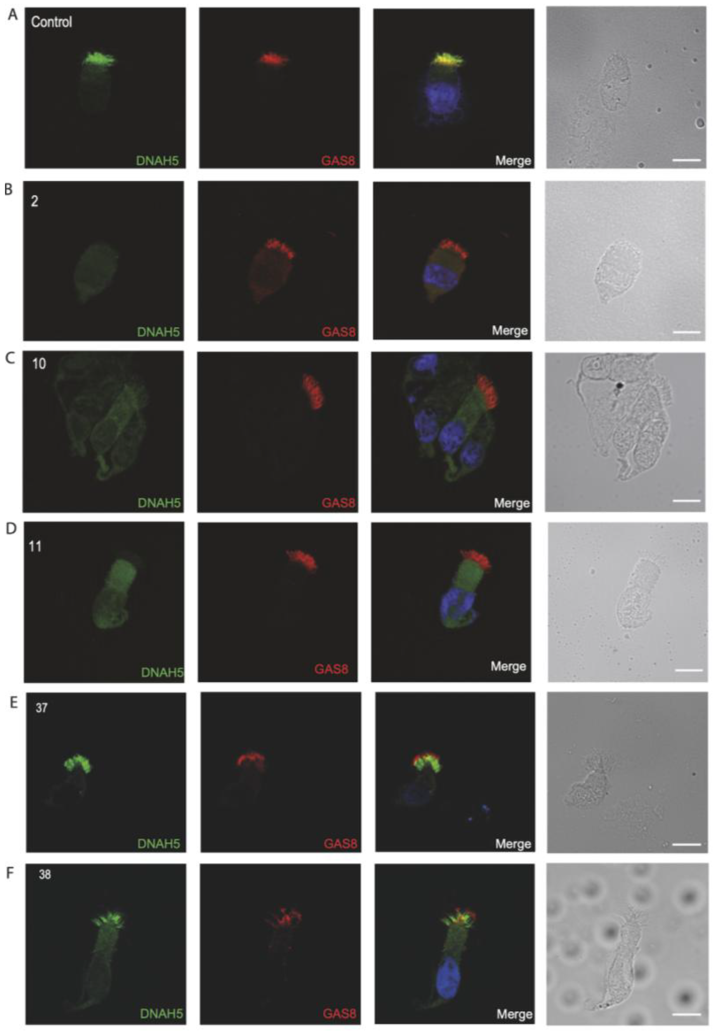
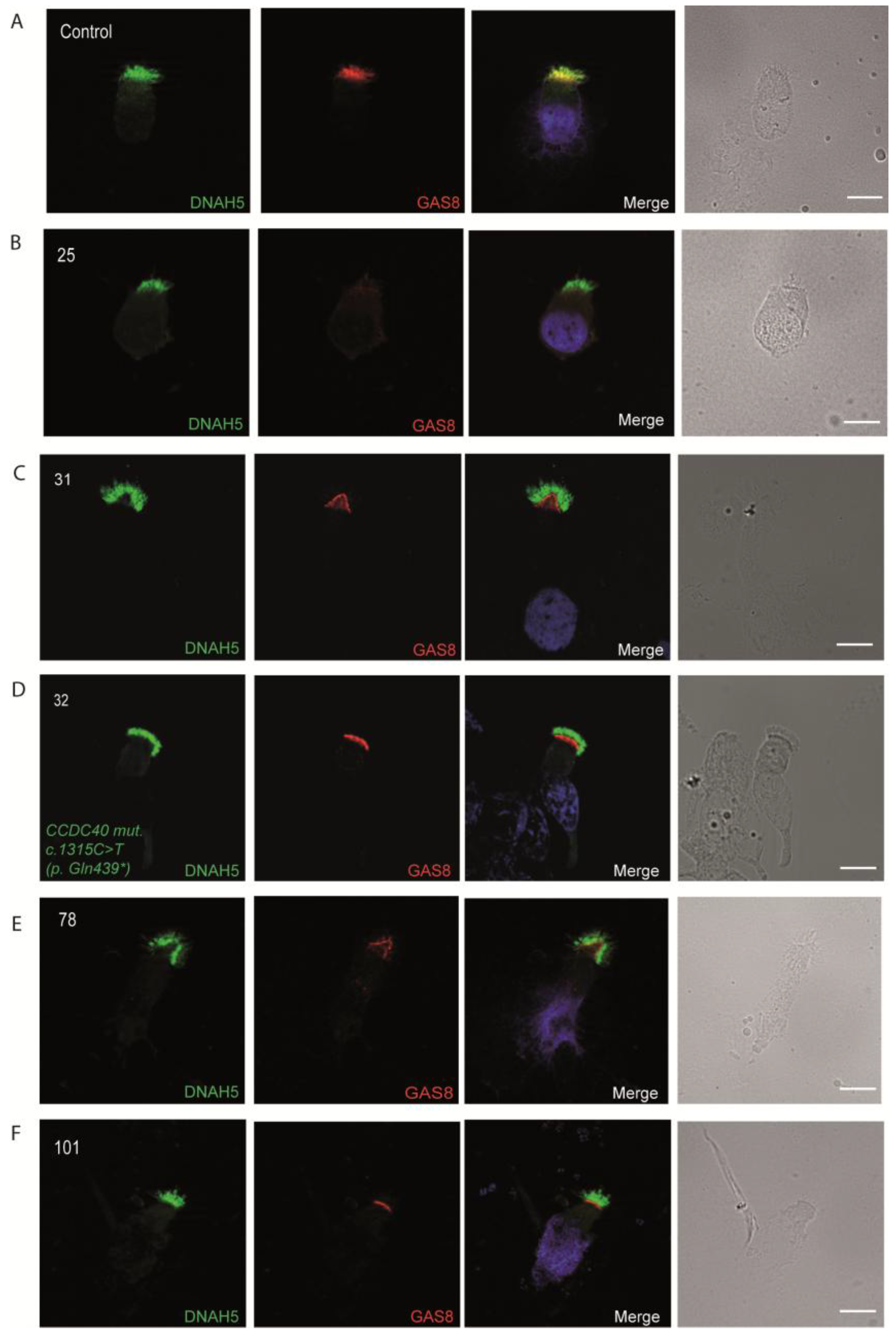
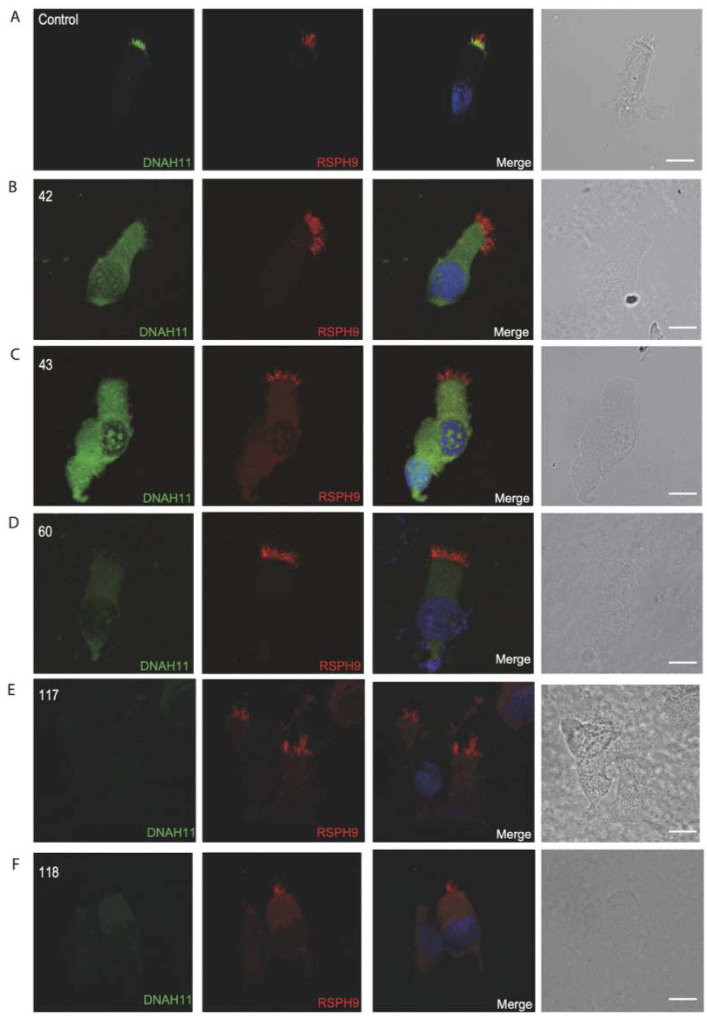
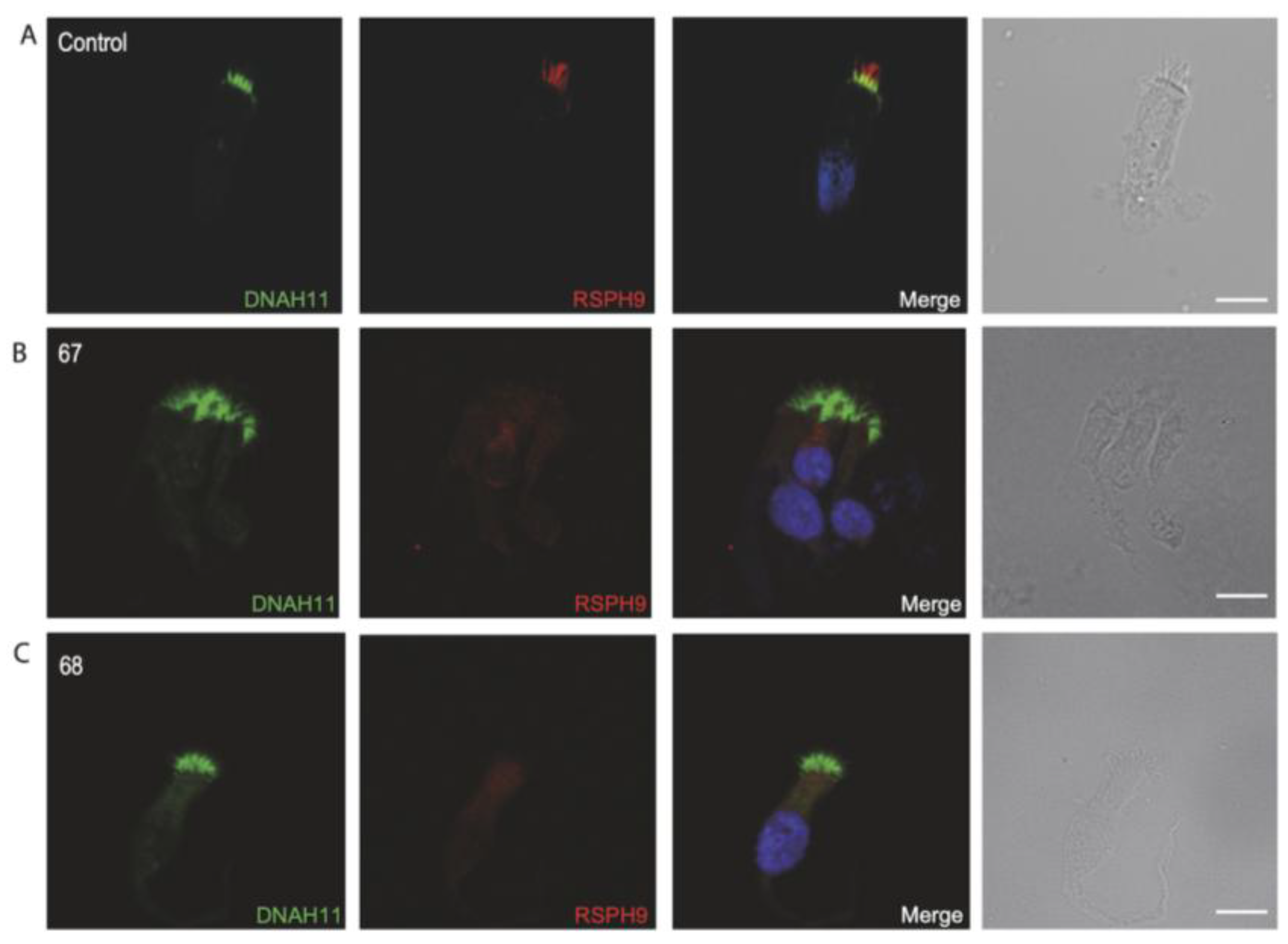
2.6. Immunofluorescence Analysis

After obtaining respiratory epithelial cells by brushing the nasal epithelial area of patients (#21103, Medbar, İzmir, Turkey), the cells were suspended in RPMI 1640 medium (#01-100-1A, Biological Industries, Cromwell, CT, USA). To make a fresh, well-preserved ciliated epithelial cell smear, samples were immediately spread onto glass slides. For physical fixation, the slides were air-dried and then stored at −80 °C. For the IF procedure, samples were fixed with 4% paraformaldehyde for 15 min (RT). Then, they were permeabilized with 0.2% Triton-X for 10 min (RT) and incubated with 1% skim milk for non-specific blockage at RT overnight. These steps were followed by primary antibody incubation (3–4 h at RT) and secondary antibody incubation (30 min RT). Slides from healthy control individuals and PCD-suspected individuals were tested simultaneously with primary antibodies. Monoclonal mouse anti-DNAH5 and polyclonal rabbit anti-GAS8 (HPA041311) primary antibodies were used for double labeling at 1:500 dilution. Monoclonal mouse anti-DNAH11 and polyclonal rabbit anti-RSPH9 (HPA031703) primary antibodies were double-labeled at 1:300 dilution. Polyclonal ARMC4 antibodies were obtained from Atlas Antibodies (HPA037829; 1:100 dilution). Goat anti-mouse Alexa Fluor 488 and anti-rabbit Alexa Fluor 546 secondary antibodies were used as 1:1000 dilution. To stain the nuclei, we used Hoechst 33342 (Sigma, St. Louis, MO, USA). After mounting the slides, IF analyses were performed using a Zeiss Apotome Axiovert 200 microscope manufactured by Carl Zeiss AG (Jena, Germany) (AxioVision v.4.8 software). Presentative figures were created accordingly [20].

2.7. Statistical Analysis

Descriptive statistics were used to summarize clinical (e.g., age, gender, lobectomy, etc.) and laboratory findings (e.g., nasal NO levels, immunofluorescence results, high-speed video microscopy patterns). Continuous variables are presented as median (range), while categorical variables are expressed as valid percentages. ANOVA was used to compare continuous variables across motility categories, and Chi-square tests were used to assess associations between categorical variables. Crosstab analysis was conducted to evaluate relationships between motility patterns and immunofluorescence results. A significance level of p < 0.05 was applied to all tests, with statistical analyses performed using the Statistical Package for the Social Sciences (SPSS) version 23.0 (SPSS Inc, Chicago, IL, USA).

3. Results

3.1. Clinical Features

The study involved patients who had been clinically diagnosed as PCD-suspected individuals. Among the participants, 44.4% were male, ranging in age from 5 to 24 years. Females composed 55.6% of the population, with an age range between 12 and 27 years. All individuals included in this study, with a consanguinity rate of 79.2%, exhibited clinical symptoms of PCD, suffering from recurrent symptoms of upper and lower airway infections and chronic wet cough. Clinical findings varied among individuals. Among them, 61% suffered from sinusitis, 67.9% had a history of bronchiectasis, 40.7% had hearing defects, and 76% had a low nNO ratio. Additionally, X-ray imaging revealed that 37.7% had situs inversus. Clinical and diagnostic findings are summarized in Table 1, while clinical, laboratory and immunofluorescence labeling statistics are presented in Table 2 and Table 3.
The study included 17 individuals presenting with hearing defects, of which 14 are descendants from consanguineous marriages, 11 are associated with bronchiectasis, and 8 with situs inversus. Of the 17, 8 presented abnormal labeling of DNAH5 and only 1 individual showed abnormal labeling of DNAH11.
Five individuals had lobectomy history. One individual presented with absent labeling of DNAH5, another one with absent labeling of GAS8 and the last one with absent labeling of DNAH11.
Individual 60 (female) was a distinct case that was observed at Hacettepe Pediatric Pulmonology Clinic since infancy. She exhibited recurrent lung infections, low nNO levels and a stiff ciliary beat pattern. Nasal epithelial samples were collected in 2015. Having become an adult recently, she was diagnosed with infertility and, in current study respiratory epithelial cells, displayed DNAH11 negative staining.

3.2. HSVM Analysis

HSVM analysis determined that 39.6% of the individuals showed minimal residual ciliary beating, 22.9% had almost immotile ciliary movement, 12.5% had reduced amplitude, and 14.6% had stiff beating pattern. Only four samples were completely immotile, and one was recorded as hyperkinetic. Ciliary beating patterns are presented in Table 3 (the Supplementary Videos S1–S10 represent the ciliary beating patterns from unaffected control and individuals with DNAH5, ARMC4 mutations and suspected PCD).

3.3. Genetic Results

Genetic results were available for eight individuals. We identified individuals 33, 119 and 30 with homozygous variants in DNAH5, individuals 9, 107 and 109 with homozygous variants in DNAAF1, individual 19 with homozygous ARMC4 missense variants and individual 32 with homozygous variants in CCDC40.
Individual 33, a descendant of a consanguineous family, presenting with situs inversus totalis and bronchiectasis, and carried biallelic missense variants in DNAH5 (c.7615T>C; p.Trp2539Arg). Individuals 119 and 30 carried biallelic nonsense variants in DNAH5, respectively: c.5747G>A; p.Trp1916Ter and c.2710G>T; p.Glu904Ter. Individual 119 is also a descendant of a consanguineous family, presenting with bronchiectasis, while individual 30 did not present any consanguinity nor laterality defects. Respiratory cilia of individual 30 were almost immotile, whereas respiratory cilia of individuals 33 and 119 displayed minimal residual ciliary movements.
Individual 9, a descendant of a consanguineous family, presenting with situs inversus totalis, carried biallelic missense variants in DNAAF1 (c.1385A>C; p.Gln462Pro) and had respiratory epithelial cells displaying a minimal residual ciliary movement. Individual 107, a descendant of a consanguineous family, presented with situs inversus totalis, carried biallelic frameshift variants in DNAAF1 (c.1349dupC; p.Pro451fs*6) and had respiratory epithelial cells displaying hypokinetic ciliary movement. Individual 109, a descendant of a consanguineous family, presented with situs inversus totalis, carried biallelic frameshift variants in DNAAF1 (c.1228_1232delCCAGA; p.Pro410fs*8) and had respiratory epithelial cells that were were almost immotile.
Individual 19 carried biallelic ARMC4 missense variants (c. 2780T>G, p. Leu927Trp) and had respiratory epithelial cells displaying minimal residual ciliary movement with a reduced beating pattern.
Individual 32 carried biallelic CCDC40 nonsense variants (c.1315C>T; p.Gln439*), is a descendant of consanguineous family, and had hypokinetic ciliary movements.

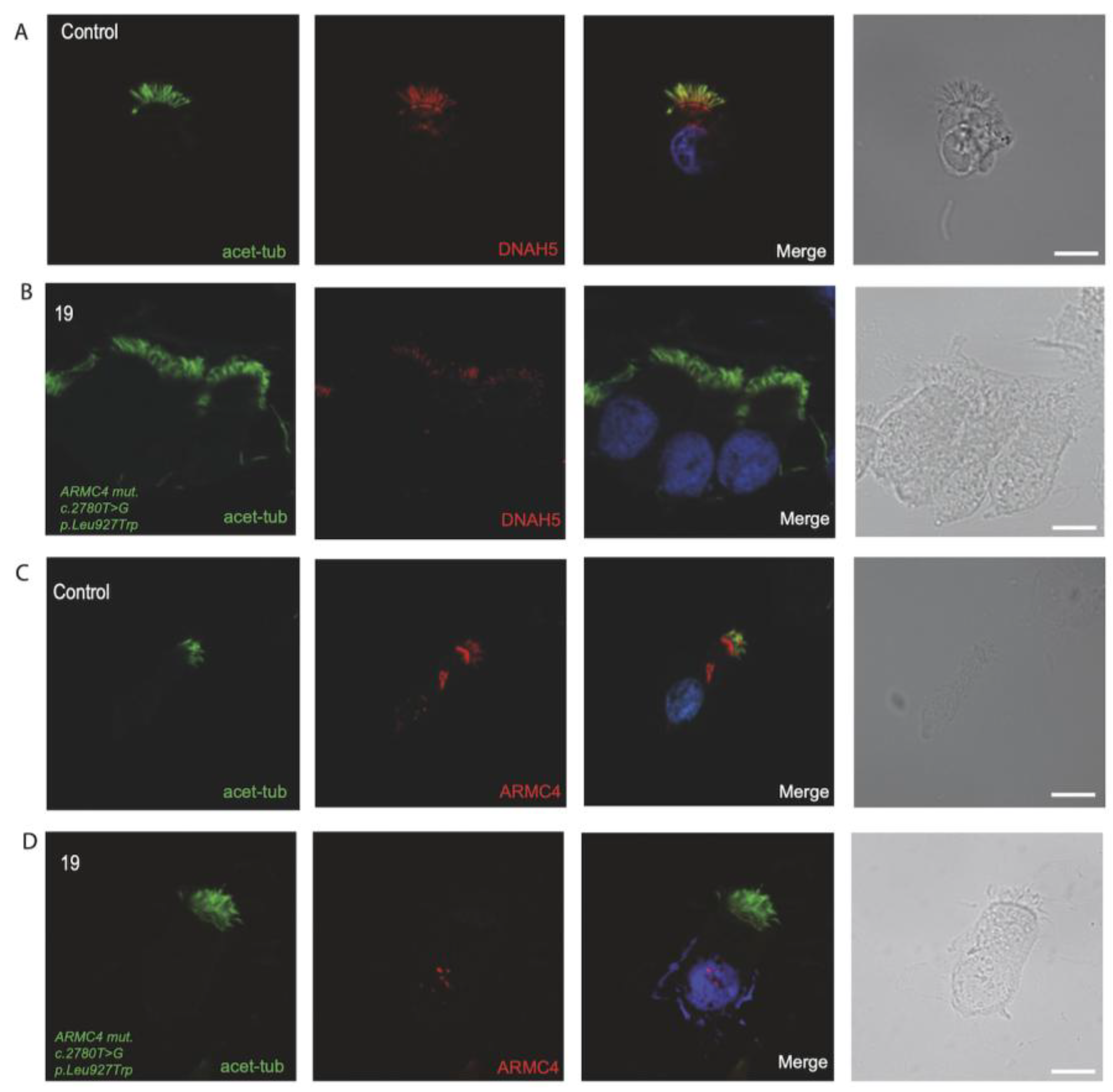
3.4. Immunofluorescence Analysis for Genetically Analyzed Individuals

To confirm the pathogenicity of the identified variants, we analyzed the localization of DNAH5 in the respiratory epithelial cells of the seven individuals identified by IF microscopy. DNAH5 was undetectable in individuals 33, 119, 30, 9, 107 and 109 (shown in Figure 1) and localized only to the proximal axonemes in individual 19 (shown in Figure 2). We also analyzed the localization of ARMC4 in the respiratory epithelial cells of individual 19, and we could not detect any signal, confirming the absence of the protein from the ciliary axonemes (shown in Figure 2). DNAH5 was also abnormal in individuals without genetic analysis (shown in Figure 3). In individual 32, we also did not detect GAS8 signaling by IF (shown in Figure 4). The staining results correlated with the genetic findings.

3.5. Diagnostic IF Analysis for Non-Genetically Analyzed Individuals

As the genetic analysis of the remaining individuals was not available and given the efficiency of IF analysis as a diagnostic tool, we performed IF analysis with different antibodies to reveal the ciliary defect at the molecular level. We used antibodies targeting DNAH5 and DNAH11 (dynein heavy chains of the outer dynein arm complex), GAS8 (component of the nexin–dynein regulatory complex n-DRC) and RSPH9 (component of the radial spoke complex).
As mentioned above, the localization of DNAH5 is affected in individuals with pathogenic variants in DNAH5, other ODA structural components, ODA docking-associated genes (ODADs) and preassembly factors (DNAAFs). We also targeted the localization of DNAH11 as well, as this component of the outer dynein arm is affected only in individuals with pathogenic variants in DNAH11, CFAP45 and CFAP52 [32]. Furthermore, we analyzed the localization of GAS8, which not only indicates a deficiency in GAS8 but also in the ruler proteins CCDC39 and CCDC40 [9]. Additionally, we targeted the localization of RSPH9, as this radial spoke component is absent in individuals with pathogenic variants in RSPH9, RSPH4A and other radial spoke genes. Although the four antibodies cover a wide range of genetic defects related to PCD, they do not cover CP, isolated IDA and other N-DRC and RSP defects, for example. Therefore, a normal IF does not exclude PCD in these cases.
Repeated IF analysis revealed an absent localization of DNAH5 in 16 individuals, an absent localization of GAS8 in 7 individuals, an absent localization of DNAH11 in 5 individuals and an absent localization of RSPH9 in 1 individual, pointing to the possible PCD defect in these cases (shown in Figure 3, Figure 4, Figure 5 and Figure 6). To corroborate the negative staining observed in some individuals, we analyzed affected siblings whenever available. Consequently, siblings 37 and 38 were DNAH5-negative (proximal), siblings 31 and 32 were negative for GAS8, siblings 117 and 118 were DNAH11-negative, and siblings 67 and 68 were negative for RSPH9 (shown in Figure 3, Figure 4, Figure 5 and Figure 6) (See also Supplementary Figures S1 and S2, providing additional information for the remaining DNAH5 and GAS8 abnormal immunofluorescence analysis).
Figure 3. Immunofluorescence analysis of PCD-suspected individuals reveals the absence or abnormal localization of DNAH5 in the respiratory ciliary axonemes. (A) Respiratory cilia double-labeled with antibodies directed against DNAH5 (green) and GAS8 (red) show colocalization of DNAH5 with GAS8 along the cilia from healthy controls (yellow color). (BD) In contrast, DNAH5 is absent in respiratory axonemes of PCD-suspected individuals. (E,F) Two siblings, showing proximal staining pattern of DNAH5 in the ciliary axonemes of respiratory epithelium. Nuclei were stained with Hoechst 33342 (blue). Scale bars represent 10 mm.
Jcm 14 01941 g003
Figure 4. Immunofluorescence analysis of PCD-suspected individuals reveals an absence of GAS8 from the respiratory ciliary axonemes. (A) Respiratory cilia double-labeled with antibodies directed against DNAH5 (green) and GAS8 (red) show colocalization of DNAH5 with GAS8 along the cilia from unaffected controls (yellow color). (B,E,F) In contrast, GAS8 is absent in respiratory axonemes of PCD-suspected individuals. (C,D) Two siblings, showing absence of GAS8 in the ciliary axonemes of respiratory epithelium. (D) CCDC40 mutant respiratory cells were negative for GAS8. Nuclei were stained with Hoechst 33342 (blue). Scale bars represent 10 mm. * stands for termination of translation of the protein.
Jcm 14 01941 g004
Figure 5. Immunofluorescence analysis of PCD-suspected individuals reveals the absence of DNAH11 from the respiratory ciliary axonemes. (A) Respiratory cilia double-labeled with antibodies directed against DNAH11 (green) and RSPH9 (red) show colocalization of DNAH11 with RSPH9 in the proximal ciliary axonemes from unaffected controls (yellow color). (BD) In contrast, DNAH11 is absent in respiratory axonemes of PCD-suspected individuals. (E,F) Two siblings, showing absence of DNAH11 in the ciliary axonemes of respiratory epithelium. Nuclei were stained with Hoechst 33342 (blue). Scale bars represent 10 mm.
Jcm 14 01941 g005
Figure 6. Immunofluorescence analysis of PCD-suspected individuals reveals the absence of RSPH9 from the respiratory ciliary axonemes. (A) Respiratory cilia double-labeled with antibodies directed against DNAH11 (green) and RSPH9 (red) show a proximal localization of DNAH11 with RSPH9 along the cilia from unaffected controls (yellow color). (B,C) Siblings, showing RSPH9 is absent in respiratory axonemes of PCD-suspected individuals. Nuclei were stained with Hoechst 33342 (blue). Scale bars represent 10 mm.
Jcm 14 01941 g006

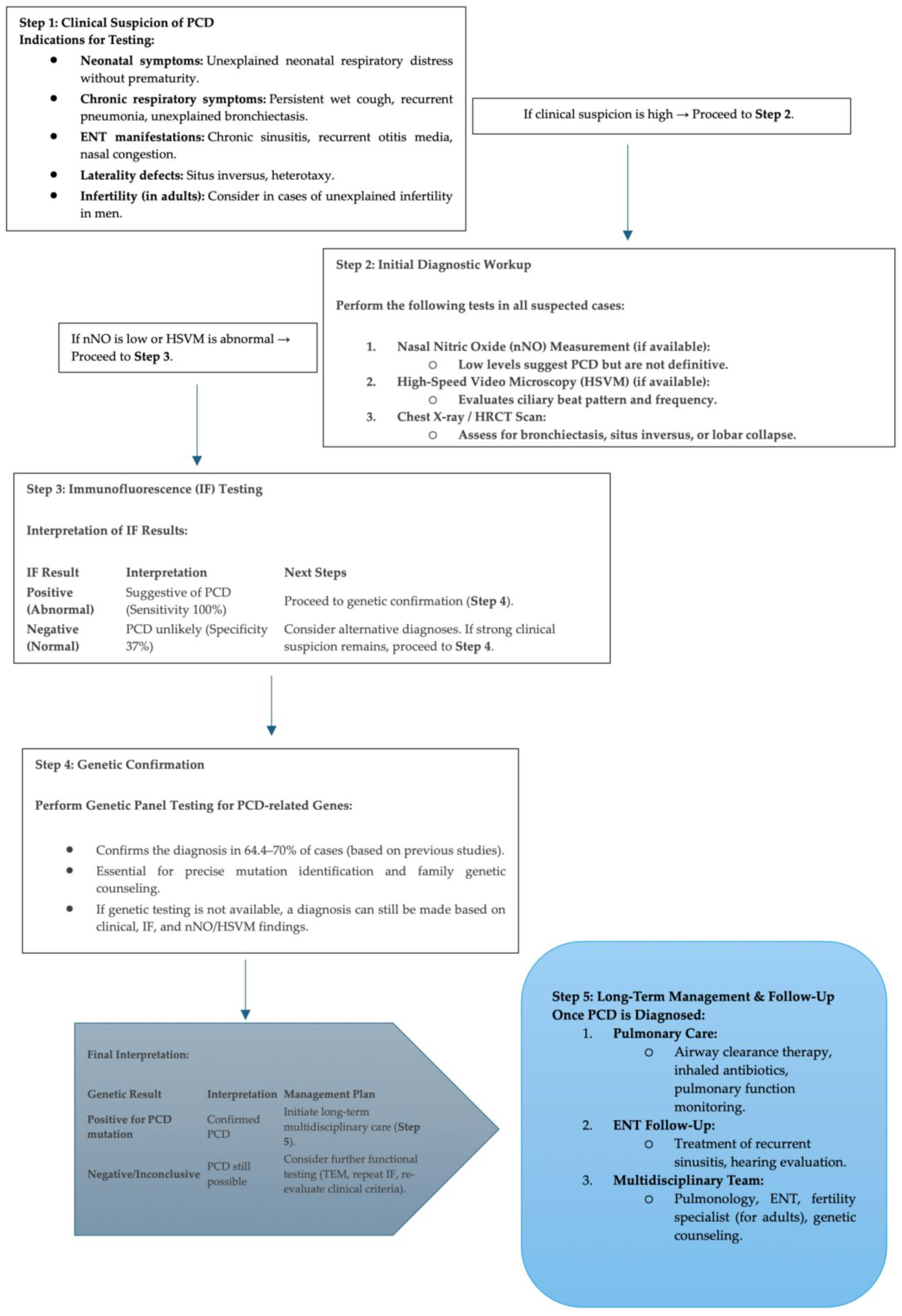
3.6. Proposed Diagnostic Algorithm for PCD

To evaluate the performance of immunofluorescence (IF) as a diagnostic tool for PCD, we analyzed individuals in two groups: Group A: Individuals with available genetic analysis and Group B: Individuals without genetic analysis. We calculated the sensitivity and specificity of IF compared to genetic analysis using a 2 × 2 contingency table (Table 4):
In group A (n = 8, available genetic analysis), IF correlated with genetic results in 8 cases (True Positives, TP = 8). IF was absent in 0 genetically confirmed cases (False Negative, FN = 0). In group B (n = 46, without genetic analysis), IF was absent in 17 of 46 individuals (True Negatives, TN = 17), and IF was present in 29 of 46 individuals (False Positives, FP = 29). Sensitivity and specificity calculations were made depending on the formula: Sensitivity = TP/(TP + FN) × 100 = 8/(8 + 0) × 100 = 100% and Specificity = TN/(TN + FP) × 100 = 17/(29 + 17) × 100 = 17/46 ≈ 37%.
IF correctly identified 100% of PCD cases when genetic analysis confirmed the diagnosis and correctly excluded 37% of non-PCD cases. An algorithm for PCD diagnosis is formed (shown in Figure 7).

4. Discussion

Despite the identification of more than 50 genes associated with PCD, more than 30% of PCD cases remain genetically unresolved [29,33]. Recently, our colleagues published a survey on PCD cases identified in Turkey, both clinically and genetically [31]. However, the approach to these cases is complex and includes not only clinical diagnostics but also laboratory techniques like IF and TEM analyses [17,34]. In our study, we aimed to provide insights using clinical approaches guided by global algorithms, employing IF on Turkey-based cases.
Here, we present the first clinical PCD cases from Turkey with IF correlation in a large patient group. As a result, PCD candidate individuals with symptoms such as persistent cough, purulent expectoration, and rhinorrhea were identified [6,24]. Although results varied, bronchiectasis was the most prevalent clinical finding in our patients, which was consistent with the meta-analysis performed by Goutaki et al. [35].
A global perspective on PCD diagnosis has emerged through cohort studies conducted across diverse populations. In 2022, Hannah et al. conducted a large cohort study, comparing the prevalence of PCD across different ethnic groups. Since most PCD genetic studies have been performed in European and North American populations, their research aimed to provide a more global perspective. Their study suggested that including a worldwide cohort study could highlight the genetic variations between different ethnic groups [36]. Another cohort study performed in China included 26 PCD patients and identified DNAH5 as the most prevalent mutation among genetically diagnosed individuals. Through clinical assessments and whole-genome sequencing (WGS), they demonstrated that clinical features aligned with genetic findings [37]. In the North American population, PCD studies have been relatively limited. However, Poplawska et al. investigated PCD cases and highlighted the significance of genetic testing in the PCD area, particularly while socioeconomic factors were thought to be the cause of recurrent respiratory infections [38].
Our study adds to this growing body of evidence by providing the first clinical and IF-based insights into PCD cases from Turkey. The high prevalence of DNAH5 mutations in our cohort aligns with findings from European populations and a recent Chinese study, suggesting a shared genetic basis. However, the identification of unique clinical presentations and genetic variants in our patients highlights the importance of expanding PCD research to diverse populations.
Furthermore, an international survey, as a part of the COVID-PCD cohort, including participants from Europe, North America and other non-European countries, was conducted [39]. This questionnaire study revealed significant variations in diagnostic testing for PCD between countries. While most of participants were tested, not all underwent all three major tests (nNO, biopsy and genetics). The study underlined that genetic testing is more common now than in previous surveys, but also highlighted the need for more complete diagnostics, especially for people diagnosed long ago or with situs abnormalities. Similarly, Pedersen et al. emphasized the importance of correlating genetic findings with clinical outcomes to improve diagnostic accuracy [40]. Our study reinforces these conclusions, demonstrating that IF serves as a reliable initial screening tool, particularly in resource-limited settings where genetic testing is not always accessible.
This study was limited by having only genomic findings from eight individuals with genetic variants. One of the key strengths of our study is its demonstration of high sensitivity (100%) of IF for detecting PCD in genetically confirmed cases. However, its moderate specificity (37%) highlights the need for confirmatory genetic testing in IF-positive individuals to reduce false positives.
Despite this limitation, IF remains a valuable diagnostic tool, particularly in clinically suspected individuals. It is widely accessible and cost-effective, and provides rapid results compared to genetic testing and transmission electron microscopy.
The relatively lower specificity observed in this study may reflect the complexity of diagnosing PCD, as multiple ciliary protein abnormalities contribute to the disease, and secondary ciliary defects can influence the IF pattern. Although this study’s IF panel covered key PCD-related proteins, more than 50 genes and their related proteins are linked to PCD [22]. Even so, previous studies have shown that when interpreted within the appropriate clinical context, IF remains a reliable and feasible method for initial screening and diagnosis [25,28,41].
Nevertheless, IF analysis in individuals with DNAH5 and DNAAF1 variants (patients 33, 119, 30 and 9, 107, 109) correlated with the genetic results. Moreover, HSVM analysis showed that these individuals displayed a range from minimal residual to almost immotile ciliary beating, as previously reported in individuals with pathogenic variants in DNAH5 and DNAAF1 [10,13].
Consanguinity in individuals with DNAH5 pathogenic variants and negative immunofluorescence labeling was consistent with the literature [42].
Respiratory cells from individual 19 with homozygous ARMC4 missense variants (c. 2780T>G, p. Leu927Trp) displayed a proximal staining pattern of DNAH5 and complete absence of ARMC4, consistent with previous publications [18]. HSVM analysis in the cilia of this individual revealed a minimal residual beating pattern with reduced amplitude, consistent with the findings of Raidt et al. [27].
Respiratory cells from siblings 37 and 38 showed a proximal localization of DNAH5 by IF, and both had immotile cilia. The male sibling did not have bronchiectasis or situs inversus, but the female sibling did, indicating that DNAH5 deficiency had different effects on the two siblings [41,43]. Both siblings showed an almost immotile cilia beat pattern via HSVM, consistent with previous studies [27,41].
Respiratory cells from siblings 31 and 32 showed an absence of GAS8 from the ciliary axonemes, consistent with CCDC40 deficiency. They shared similar clinical symptoms, and their parents were consanguineous. The remaining GAS8-absent individuals had mostly consanguinity-positive backgrounds (with no genetic data), supporting the literature [44]. We observed that the cilia of these siblings had reduced amplitude, in line with the fact that GAS8 abnormalities cause reduced or disorganized ciliary beating [45]. The remaining individuals with abnormal GAS8 staining exhibited a minimal residual and reduced beating pattern, except for one individual recorded as having minimal residual and almost immotile beating pattern.
Respiratory cells from siblings 117 and 118 showed an absence of DNAH11. The male sibling had a history of lobectomy. Both siblings exhibited consanguinity and bronchiectasis but showed no concerns with heterotaxy. The beating pattern of their cilia was stiff, correlating with the previous studies [27,46] that reported increased ciliary beating frequency in patients with known DNAH11 mutations. Other individuals with DNAH11-negative staining included two individuals with hearing loss; HSVM analysis in one of them showed a stiff beating pattern, aligning with the previous studies [27,46,47]. Another female patient with DNAH11 negative staining had fertility issues. Although DNAH11 abnormality was associated with male infertility recently [48], a woman with primary infertility has been reported to carry homozygous pathogenic variants in DNAH11 [49]. Additionally, HSVM revealed that the stiff beating pattern was present at the distal part of the cilia, and the cilia were likely shorter in this individual. These findings aligned with the study by Nussbaumer et al. [47]. The remaining individuals with DNAH11 abnormality exhibited stiff ciliary beating patterns. Schreck et al. examined fertility outcomes in male and female PCD diagnosed patients from the U.S.A., Canada, European countries, the U.K. and some other countries. The study included clinical features and some available genetic results. Among women with known genetic results, those with infertility had DNAH5, DNAAF3, ZMYND10, CCDC40, RSPH9 and HYDIN mutations [50]. However, no immunofluorescence labeling was presented. In our cohort, the female patient with DNAH11-negative staining with infertility issues, consistent with previous reports of primary infertility in individuals with DNAH11 mutations [49]. These findings highlight the broader clinical implications of PCD and the need for comprehensive genetic, clinical and IF evaluations, particularly when TEM or WES is not feasible.
Respiratory cells from siblings 67 and 68, descendants of a consanguineous marriage and born deaf, tested negative for RSPH9 by IF analyses. Interestingly, these individuals did not display any laterality defects, consistent with the observation that individuals with radial spoke defects typically present with situs solitus [51]. Although the genetic background of these siblings remained unknown throughout our investigation, PCD could be indicated due to the IF results and clinical phenotype [52]. It has been noted that some rotational or metronome-like (stiff) movements can be seen in patients with radial spoke abnormalities on HSVM; our study revealed that these siblings had stiff ciliary beating [53].
Moreover, the majority of the remaining individuals had impaired mucociliary clearance and bronchiectasis due to minimal residual or immotile cilia, and most had abnormal DNAH5 immunostaining (either negative or proximal staining pattern). Consistently, aberrant DNAH5 localization found by IF in individuals correlated with immotile or minimal residual ciliary beating found by HSVM, indicating that our findings were in line with previous studies [41].
DNAH5 is recognized as the gene with the most prevalent mutations in European families, as previously reported [54]. We discovered that 50% of the patients had abnormal DNAH5 staining results and, consequently, defects in the outer dynein arms—the ciliary stroke power generators. ODA defects can be easily detected under an electron microscope [55]. Indeed, we demonstrated here that individuals with homozygous DNAH5 pathogenic variants could be identified by IF labeling, verifying their clinical PCD symptoms.
A key limitation of this study is the absence of ultrastructural analysis using transmission electron microscopy (TEM), which is traditionally considered a gold standard for diagnosing PCD-related ciliary defects. While our study effectively utilized IF and genetic testing, TEM provides direct visualization of ciliary ultrastructural defects, aiding in the confirmation of PCD cases, particularly in genetically unresolved individuals. TEM was not included in this study due to lack of availability, cost constraints, and limited accessibility in our clinical setting. Given the requirement for specialized expertise and sample preparation challenges, TEM is often not feasible in all diagnostic centers. Despite the absence of TEM, IF demonstrated high sensitivity (100%) and provided molecular insights into ciliary defects, complementing genetic diagnostics.

5. Conclusions

The study identified a correlation between IF and genetics in genetically analyzed individuals and a correlation between IF and clinically suspected PCD individuals. According to our data, the most frequent defect identified in clinically suspected PCD candidates in our group is related to the outer dynein arms (via the absence of DNAH5). We showed that immunofluorescence, compared to TEM, has significant potential to become a more common diagnostic strategy by being less expensive and less time-consuming. Overall, even in the absence of genetic data or electron microscopy, immunofluorescence remains a potent tool for PCD diagnosis.

Supplementary Materials

The following supporting information can be downloaded at: https://www.mdpi.com/article/10.3390/jcm14061941/s1, Figure S1. Immunofluorescence analysis of PCD-suspected individuals reveals the absence or abnormal localization of DNAH5 in the respiratory ciliary axonemes. (A) Respiratory cilia double-labeled with antibodies directed against DNAH5 (green) and GAS8 (red) show colocalization of DNAH5 with GAS8 along the cilia from healthy controls. (B–G,J,K) In contrast, DNAH5 is absent in respiratory axonemes of PCD-suspected individuals. (H,I) Two PCD-suspected individuals, showing proximal staining pattern of DNAH5 in the ciliary axonemes of respiratory epithelium. Nuclei were stained with Hoechst 33342 (blue). Scale bars represent 10 mm.; Figure S2. Immunofluorescence analysis of PCD-suspected individuals reveals an absence of GAS8 from the respiratory ciliary axonemes. (A) Respiratory cilia double-labeled with antibodies directed against DNAH5 (green) and GAS8 (red) show colocalization of DNAH5 with GAS8 along the cilia from unaffected controls. (B–D) In contrast, GAS8 is absent in respiratory axonemes of PCD-suspected individuals. Nuclei were stained with Hoechst 33342 (blue). Scale bars represent 10 mm.; Video S1. Control HSVM video of unaffected control; Video S2. DNAH5 mutant (c.2710G>T; p.Glu904Ter) Almost immotile ciliary beat of individual 30.; Video S3. DNAH5 mutant (c.7615T>C;p.Trp2539Arg) Minimal residual ciliary beat of individual 33.; Video S4. DNAH5 mutant (c.5747G>A; p.Trp1916Ter) Minimal residual ciliary beat of individual 119.; Video S5. ARMC4 mutant (c.2780T>G; p.Leu927Trp) Minimal residual and reduced amplitude ciliary beat of individual 19.; Video S6. Almost immotile cilia of PCD-suspected individual 38.; Video S7. Almost immotile cilia of individual 24.; Video S8. Hyperkinetic ciliary beat pattern of individual 36.; Video S9. Minimal residual and almost immotile cilia of individual 25.; Video S10. Stiff ciliary beat of individual 63.

Author Contributions

Conceptualization, E.K., R.H. and H.O.; methodology, E.K., R.H., U.Ö. and N.E.; software, E.K. and R.H.; validation, E.K. and R.H.; formal analysis, E.K. and R.H.; investigation, E.K., R.H., U.Ö. and N.E.; resources, E.K., R.H., Z.B.K., D.A.T., N.E. and P.A.; data curation, E.K., R.H., Z.B.K. and N.E.; writing—original draft preparation, E.K. and R.H.; writing—review and editing, E.K., R.H. and H.O.; visualization, E.K. and R.H.; supervision, H.O.; project administration, E.K., R.H. and H.O.; funding acquisition, E.K., R.H. and H.O. All authors have read and agreed to the published version of the manuscript.

Funding

This work was supported by the Hacettepe University Scientific Research Council (#TBI-2018-17641)—the grant holders were U.E. and N.E.—and COST STSM Applications (#42518-BM1407)—the grant holder was E.K.; grants from the Deutsche Forschungsgemeinschaft DFG OM6/7, OM6/8, OM6/10 and OM6/11 to H. Omran and HJ 7/1-1, HJ 7/1-3 to R. Hjeij. This work is generated within the European Reference Network for rare respiratory diseases ERN-LUNG. The funders had no role in the design, data collection, data analysis, and reporting of this study.

Institutional Review Board Statement

This study was performed in accordance with the Declaration of Helsinki. Ethical approval for this project was given by Hacettepe University Clinical Ethics Board (#GO-15/638-14; 21 October 2015) and the ethics committee from the Medical Association Westphalia Lippe and the Westphalian University Muenster (Ärztekammer Westfalen Lippe und Westfälische Universität), (reference number: 2015-104-f-S; 21 April 2015). All participating individuals meeting diagnostic criteria, or a responsible parent in the case of children, as well as participating family members, provided written informed consent to participate in the study according to protocols approved by the institutional ethics review board at Hacettepe University.

Informed Consent Statement

Informed consent was obtained from all subjects involved in the study.

Data Availability Statement

The data generated and/or analyzed during the current study are available from the corresponding author upon reasonable request.

Acknowledgments

We thank all PCD-affected individuals and their families who participated in the study. We also acknowledge the Hacettepe University Faculty of Medicine Department of Genetics for providing the genetic information. We appreciate the technical assistance provided by the members of Heymut Omran’s laboratory.

Conflicts of Interest

The authors declare no conflicts of interest.

References

  1. Mirra, V.; Werner, C.; Santamaria, F. Primary Ciliary Dyskinesia: An Update on Clinical Aspects, Genetics, Diagnosis, and Future Treatment Strategies. Front. Pediatr. 2017, 5, 135. [Google Scholar] [CrossRef] [PubMed]
  2. Horani, A.; Ferkol, T.W. Advances in the Genetics of Primary Ciliary Dyskinesia. Chest 2018, 154, 645–652. [Google Scholar] [CrossRef] [PubMed]
  3. Reilly, M.L.; Benmerah, A. Ciliary kinesins beyond IFT: Cilium length, disassembly, cargo transport and signalling. Biol. Cell 2019, 111, 79–94. [Google Scholar] [CrossRef] [PubMed]
  4. Dong, M.; Shi, X.; Zhou, Y.; Duan, J.; He, L.; Song, X.; Huang, Z.; Chen, R.; Li, J.; Jia, N. Genetic spectrum and genotype–phenotype correlations in DNAH5-mutated primary ciliary dyskinesia: A systematic review. Orphanet J. Rare Dis. 2025, 20, 97. [Google Scholar] [CrossRef]
  5. Olbrich, H.; Schmidts, M.; Werner, C.; Onoufriadis, A.; Loges, N.T.; Raidt, J.; Banki, N.F.; Shoemark, A.; Burgoyne, T.; Al Turki, S.; et al. Recessive HYDIN mutations cause primary ciliary dyskinesia without randomization of left-right body asymmetry. Am. J. Hum. Genet. 2012, 91, 672–684. [Google Scholar] [CrossRef]
  6. Loges, N.T.; Antony, D.; Maver, A.; Deardorff, M.A.; Gulec, E.Y.; Gezdirici, A.; Nothe-Menchen, T.; Hoben, I.M.; Jelten, L.; Frank, D.; et al. Recessive DNAH9 Loss-of-Function Mutations Cause Laterality Defects and Subtle Respiratory Ciliary-Beating Defects. Am. J. Hum. Genet. 2018, 103, 995–1008. [Google Scholar] [CrossRef]
  7. Zhao, L.; Hou, Y.; Picariello, T.; Craige, B.; Witman, G.B. Proteome of the central apparatus of a ciliary axoneme. J. Cell Biol. 2019, 218, 2051–2070. [Google Scholar] [CrossRef]
  8. Merveille, A.C.; Davis, E.E.; Becker-Heck, A.; Legendre, M.; Amirav, I.; Bataille, G.; Belmont, J.; Beydon, N.; Billen, F.; Clement, A.; et al. CCDC39 is required for assembly of inner dynein arms and the dynein regulatory complex and for normal ciliary motility in humans and dogs. Nat. Genet. 2011, 43, 72–78. [Google Scholar] [CrossRef]
  9. Becker-Heck, A.; Zohn, I.E.; Okabe, N.; Pollock, A.; Lenhart, K.B.; Sullivan-Brown, J.; McSheene, J.; Loges, N.T.; Olbrich, H.; Haeffner, K.; et al. The coiled-coil domain containing protein CCDC40 is essential for motile cilia function and left-right axis formation. Nat. Genet. 2011, 43, 79–84. [Google Scholar] [CrossRef]
  10. Loges, N.T.; Olbrich, H.; Becker-Heck, A.; Häffner, K.; Heer, A.; Reinhard, C.; Schmidts, M.; Kispert, A.; Zariwal, M.A.; Leigh, M.W.; et al. Deletions and Point Mutations of Cause Primary Ciliary Dyskinesia Due to Dynein Arm Defects. Am. J. Hum. Genet. 2009, 85, 883–889. [Google Scholar] [CrossRef]
  11. Mitchison, H.M.; Schmidts, M.; Loges, N.T.; Freshour, J.; Dritsoula, A.; Hirst, R.A.; O’Callaghan, C.; Blau, H.; Al Dabbagh, M.; Olbrich, H.; et al. Mutations in axonemal dynein assembly factor DNAAF3 cause primary ciliary dyskinesia. Nat. Genet. 2012, 44, 381–389. [Google Scholar] [CrossRef] [PubMed]
  12. Tarkar, A.; Loges, N.T.; Slagle, C.E.; Francis, R.; Dougherty, G.W.; Tamayo, J.V.; Shook, B.; Cantino, M.; Schwartz, D.; Jahnke, C.; et al. DYX1C1 is required for axonemal dynein assembly and ciliary motility. Nat. Genet. 2013, 45, 995–1003. [Google Scholar] [CrossRef] [PubMed]
  13. Horani, A.; Druley, T.E.; Zariwala, M.A.; Pate, A.C.; Levinson, B.T.; Van Arendonk, L.G.; Thornton, K.C.; Giacalone, J.C.; Albee, A.J.; Wilson, K.S.; et al. Whole-Exome Capture and Sequencing Identifies Mutation as a Cause of Primary Ciliary Dyskinesia. Am. J. Hum. Genet. 2012, 91, 685–693. [Google Scholar] [CrossRef]
  14. Paff, T.; Loges, N.T.; Aprea, I.; Wu, K.; Bakey, Z.; Haarman, E.G.; Daniels, J.M.A.; Sistermans, E.A.; Bogunovic, N.; Dougherty, G.W.; et al. Mutations in PIH1D3 Cause X-Linked Primary Ciliary Dyskinesia with Outer and Inner Dynein Arm Defects. Am. J. Hum. Genet. 2017, 100, 160–168. [Google Scholar] [CrossRef]
  15. Zariwala, M.A.; Gee, H.Y.; Kurkowiak, M.; Al-Mutairi, D.A.; Leigh, M.W.; Hurd, T.W.; Hjeij, R.; Dell, S.D.; Chaki, M.; Dougherty, G.W.; et al. Is Mutated in Primary Ciliary Dyskinesia and Interacts with LRRC6. Am. J. Hum. Genet. 2013, 93, 336–345. [Google Scholar] [CrossRef] [PubMed]
  16. Kott, E.; Duquesnoy, P.; Copin, B.; Legendre, M.; Dastot-Le Moal, F.; Montantin, G.; Jeanson, L.; Tamalet, A.; Papon, J.F.; Siffroi, J.P.; et al. Loss-of-function mutations in LRRC6, a gene essential for proper axonemal assembly of inner and outer dynein arms, cause primary ciliary dyskinesia. Am. J. Hum. Genet. 2012, 91, 958–964. [Google Scholar] [CrossRef]
  17. Hjeij, R.; Aprea, I.; Poeta, M.; Nothe-Menchen, T.; Bracht, D.; Raidt, J.; Honecker, B.I.; Dougherty, G.W.; Olbrich, H.; Schwartz, O.; et al. Pathogenic variants in CLXN encoding the outer dynein arm docking-associated calcium-binding protein calaxin cause primary ciliary dyskinesia. Genet. Med. 2023, 25, 100798. [Google Scholar] [CrossRef]
  18. Hjeij, R.; Lindstrand, A.; Francis, R.; Zariwala, M.A.; Liu, X.; Li, Y.; Damerla, R.; Dougherty, G.W.; Abouhamed, M.; Olbrich, H.; et al. ARMC4 mutations cause primary ciliary dyskinesia with randomization of left/right body asymmetry. Am. J. Hum. Genet. 2013, 93, 357–367. [Google Scholar] [CrossRef]
  19. Onoufriadis, A.; Paff, T.; Antony, D.; Shoemark, A.; Micha, D.; Kuyt, B.; Schmidts, M.; Petridi, S.; Dankert-Roelse, J.E.; Haarman, E.G.; et al. Splice-site mutations in the axonemal outer dynein arm docking complex gene CCDC114 cause primary ciliary dyskinesia. Am. J. Hum. Genet. 2013, 92, 88–98. [Google Scholar] [CrossRef]
  20. Hjeij, R.; Onoufriadis, A.; Watson, C.M.; Slagle, C.E.; Klena, N.T.; Dougherty, G.W.; Kurkowiak, M.; Loges, N.T.; Diggle, C.P.; Morante, N.F.; et al. CCDC151 mutations cause primary ciliary dyskinesia by disruption of the outer dynein arm docking complex formation. Am. J. Hum. Genet. 2014, 95, 257–274. [Google Scholar] [CrossRef]
  21. Wallmeier, J.; Shiratori, H.; Dougherty, G.W.; Edelbusch, C.; Hjeij, R.; Loges, N.T.; Menchen, T.; Olbrich, H.; Pennekamp, P.; Raidt, J.; et al. TTC25 Deficiency Results in Defects of the Outer Dynein Arm Docking Machinery and Primary Ciliary Dyskinesia with Left-Right Body Asymmetry Randomization. Am. J. Hum. Genet. 2016, 99, 460–469. [Google Scholar] [CrossRef] [PubMed]
  22. Schultz, R.; Elenius, V.; Fassad, M.R.; Freke, G.; Rogers, A.; Shoemark, A.; Koistinen, T.; Mohamed, M.A.; Lim, J.S.Y.; Mitchison, H.M.; et al. CFAP300 mutation causing primary ciliary dyskinesia in Finland. Front. Genet. 2022, 13, 985227. [Google Scholar] [CrossRef] [PubMed]
  23. Wu, R.; Li, H.; Wu, P.; Yang, Q.; Wan, X.; Wu, Y. LRRC56 deletion causes primary ciliary dyskinesia in mice characterized by dynein arms defects. Biol. Open 2025, 14, bio061846. [Google Scholar] [CrossRef] [PubMed]
  24. Lucas, J.S.; Barbato, A.; Collins, S.A.; Goutaki, M.; Behan, L.; Caudri, D.; Dell, S.; Eber, E.; Escudier, E.; Hirst, R.A.; et al. European Respiratory Society guidelines for the diagnosis of primary ciliary dyskinesia. Eur. Respir. J. 2017, 49, 1601090. [Google Scholar] [CrossRef]
  25. Shoemark, A.; Frost, E.; Dixon, M.; Ollosson, S.; Kilpin, K.; Patel, M.; Scully, J.; Rogers, A.V.; Mitchison, H.M.; Bush, A.; et al. Accuracy of Immunofluorescence in the Diagnosis of Primary Ciliary Dyskinesia. Am. J. Respir. Crit. Care Med. 2017, 196, 94–101. [Google Scholar] [CrossRef]
  26. Shoemark, A. Applications of emerging transmission electron microscopy technology in PCD research and diagnosis. Ultrastruct. Pathol. 2017, 41, 408–414. [Google Scholar] [CrossRef]
  27. Raidt, J.; Wallmeier, J.; Hjeij, R.; Onnebrink, J.G.; Pennekamp, P.; Loges, N.T.; Olbrich, H.; Haffner, K.; Dougherty, G.W.; Omran, H.; et al. Ciliary beat pattern and frequency in genetic variants of primary ciliary dyskinesia. Eur. Respir. J. 2014, 44, 1579–1588. [Google Scholar] [CrossRef]
  28. Farley, H.; Rubbo, B.; Bukowy-Bieryllo, Z.; Fassad, M.; Goutaki, M.; Harman, K.; Hogg, C.; Kuehni, C.E.; Lopes, S.; Nielsen, K.G.; et al. Proceedings of the 3rd BEAT-PCD Conference and 4th PCD Training School. BMC Proc. 2018, 12, 64. [Google Scholar] [CrossRef]
  29. Wallmeier, J.; Nielsen, K.G.; Kuehni, C.E.; Lucas, J.S.; Leigh, M.W.; Zariwala, M.A.; Omran, H. Motile ciliopathies. Nat. Rev. Dis. Primers 2020, 6, 77. [Google Scholar] [CrossRef]
  30. Chen, W.; Guo, Z.; Li, M.; Sheng, W.; Huang, G. Next-Generation Sequencing-Based Copy Number Variation Analysis in Chinese Patients with Primary Ciliary Dyskinesia Revealed Novel DNAH5 Copy Number Variations. Phenomics 2024, 4, 24–33. [Google Scholar] [CrossRef]
  31. Emiralioglu, N.; Taskiran, E.Z.; Kosukcu, C.; Bilgic, E.; Atilla, P.; Kaya, B.; Gunaydin, O.; Yuzbasioglu, A.; Tugcu, G.D.; Ademhan, D.; et al. Genotype and phenotype evaluation of patients with primary ciliary dyskinesia: First results from Turkey. Pediatr. Pulmonol. 2020, 55, 383–393. [Google Scholar] [CrossRef] [PubMed]
  32. Dougherty, G.W.; Mizuno, K.; Nothe-Menchen, T.; Ikawa, Y.; Boldt, K.; Ta-Shma, A.; Aprea, I.; Minegishi, K.; Pang, Y.P.; Pennekamp, P.; et al. CFAP45 deficiency causes situs abnormalities and asthenospermia by disrupting an axonemal adenine nucleotide homeostasis module. Nat. Commun. 2020, 11, 5520. [Google Scholar] [CrossRef] [PubMed]
  33. Al-Mutairi, D.A.; Alsabah, B.H.; Alkhaledi, B.A.; Pennekamp, P.; Omran, H. Identification of a novel founder variant in DNAI2 cause primary ciliary dyskinesia in five consanguineous families derived from a single tribe descendant of Arabian Peninsula. Front. Genet. 2022, 13, 1017280. [Google Scholar] [CrossRef]
  34. Guo, T.; Lu, C.; Yang, D.; Lei, C.; Liu, Y.; Xu, Y.; Yang, B.; Wang, R.; Luo, H. Case Report: DNAAF4 Variants Cause Primary Ciliary Dyskinesia and Infertility in Two Han Chinese Families. Front. Genet. 2022, 13, 934920. [Google Scholar] [CrossRef]
  35. Goutaki, M.; Meier, A.B.; Halbeisen, F.S.; Lucas, J.S.; Dell, S.D.; Maurer, E.; Casaulta, C.; Jurca, M.; Spycher, B.D.; Kuehni, C.E. Clinical manifestations in primary ciliary dyskinesia: Systematic review and meta-analysis. Eur. Respir. J. 2016, 48, 1081–1095. [Google Scholar] [CrossRef]
  36. Hannah, W.B.; Seifert, B.A.; Truty, R.; Zariwala, M.A.; Ameel, K.; Zhao, Y.; Nykamp, K.; Gaston, B. The global prevalence and ethnic heterogeneity of primary ciliary dyskinesia gene variants: A genetic database analysis. Lancet Respir. Med. 2022, 10, 459–468. [Google Scholar] [CrossRef]
  37. Zhao, X.; Bian, C.; Liu, K.; Xu, W.; Liu, Y.; Tian, X.; Bai, J.; Xu, K.-F.; Zhang, X. Clinical characteristics and genetic spectrum of 26 individuals of Chinese origin with primary ciliary dyskinesia. Orphanet J. Rare Dis. 2021, 16, 293. [Google Scholar] [CrossRef]
  38. Poplawska, K.; Griffiths, A.; Temme, R.; Adamko, D.J.; Nykamp, K.; Shapiro, A.J. Deletions in DNAL1 Cause Primary Ciliary Dyskinesia Across North American Indigenous Populations. J. Pediatr. 2023, 261, 113362. [Google Scholar] [CrossRef]
  39. Rodriguez, N.M.; Schreck, L.D.; Pedersen, E.S.L.; Cizeau, I.; Müller, L.; Kruljac, C.; Lucas, J.S.; Goutaki, M.; Kuehni, C.E. Diagnostic testing in people with primary ciliary dyskinesia: An international participatory study. PLoS Glob. Public Health 2023, 3, e0001522. [Google Scholar] [CrossRef]
  40. Pedersen, E.S.L.; Goutaki, M.; Schreck, L.D.; Rindlisbacher, B.; Dixon, L.; Lucas, J.S.; Kuehni, C.E. Questionnaire-assessed genotypes and associations with symptoms in primary ciliary dyskinesia. ERJ Open Res. 2024, 10, 00288-2024. [Google Scholar] [CrossRef]
  41. Baz-Redon, N.; Rovira-Amigo, S.; Fernandez-Cancio, M.; Castillo-Corullon, S.; Cols, M.; Caballero-Rabasco, M.A.; Asensio, O.; Martin de Vicente, C.; Martinez-Colls, M.D.M.; Torrent-Vernetta, A.; et al. Immunofluorescence Analysis as a Diagnostic Tool in a Spanish Cohort of Patients with Suspected Primary Ciliary Dyskinesia. J. Clin. Med. 2020, 9, 3603. [Google Scholar] [CrossRef] [PubMed]
  42. Ta-Shma, A.; Hjeij, R.; Perles, Z.; Dougherty, G.W.; Abu Zahira, I.; Letteboer, S.J.F.; Antony, D.; Darwish, A.; Mans, D.A.; Spittler, S.; et al. Homozygous loss-of-function mutations in MNS1 cause laterality defects and likely male infertility. PLoS Genet. 2018, 14, e1007602. [Google Scholar] [CrossRef]
  43. Yue, Y.; Huang, Q.; Zhu, P.; Zhao, P.; Tan, X.; Liu, S.; Li, S.; Han, X.; Cheng, L.; Li, B.; et al. Identification of Pathogenic Mutations and Investigation of the NOTCH Pathway Activation in Kartagener Syndrome. Front. Genet. 2019, 10, 749. [Google Scholar] [CrossRef]
  44. Olbrich, H.; Cremers, C.; Loges, N.T.; Werner, C.; Nielsen, K.G.; Marthin, J.K.; Philipsen, M.; Wallmeier, J.; Pennekamp, P.; Menchen, T.; et al. Loss-of-Function GAS8 Mutations Cause Primary Ciliary Dyskinesia and Disrupt the Nexin-Dynein Regulatory Complex. Am. J. Hum. Genet. 2015, 97, 546–554. [Google Scholar] [CrossRef]
  45. Jeanson, L.; Thomas, L.; Copin, B.; Coste, A.; Sermet-Gaudelus, I.; Dastot-Le Moal, F.; Duquesnoy, P.; Montantin, G.; Collot, N.; Tissier, S.; et al. Mutations in GAS8, a Gene Encoding a Nexin-Dynein Regulatory Complex Subunit, Cause Primary Ciliary Dyskinesia with Axonemal Disorganization. Hum. Mutat. 2016, 37, 776–785. [Google Scholar] [CrossRef] [PubMed]
  46. Zlotina, A.; Barashkova, S.; Zhuk, S.; Skitchenko, R.; Usoltsev, D.; Sokolnikova, P.; Artomov, M.; Alekseenko, S.; Simanova, T.; Goloborodko, M.; et al. Characterization of pathogenic genetic variants in Russian patients with primary ciliary dyskinesia using gene panel sequencing and transcript analysis. Orphanet J. Rare Dis. 2024, 19, 310. [Google Scholar] [CrossRef]
  47. Nussbaumer, M.; Kieninger, E.; Tschanz, S.A.; Savas, S.T.; Casaulta, C.; Goutaki, M.; Blanchon, S.; Jung, A.; Regamey, N.; Kuehni, C.E.; et al. Diagnosis of primary ciliary dyskinesia: Discrepancy according to different algorithms. ERJ Open Res. 2021, 7, 00353-2021. [Google Scholar] [CrossRef]
  48. Guo, S.; Tang, D.; Chen, Y.; Yu, H.; Gu, M.; Geng, H.; Fang, J.; Wu, B.; Ruan, L.; Li, K.; et al. Association of novel DNAH11 variants with asthenoteratozoospermia lead to male infertility. Hum. Genom. 2024, 18, 97. [Google Scholar] [CrossRef]
  49. Maddirevula, S.; Awartani, K.; Coskun, S.; AlNaim, L.F.; Ibrahim, N.; Abdulwahab, F.; Hashem, M.; Alhassan, S.; Alkuraya, F.S. A genomics approach to females with infertility and recurrent pregnancy loss. Hum. Genet. 2020, 139, 605–613. [Google Scholar] [CrossRef]
  50. Schreck, L.D.; Pedersen, E.S.L.; Dexter, K.; Manion, M.; Bellu, S.; Cizeau, I.; Dexter, K.; Dixon, L.; Fernández, T.L.; Grieder, S.; et al. Infertility and pregnancy outcomes among adults with primary ciliary dyskinesia. Hum. Reprod. Open 2024, 2024, hoae039. [Google Scholar] [CrossRef]
  51. Frommer, A.; Hjeij, R.; Loges, N.T.; Edelbusch, C.; Jahnke, C.; Raidt, J.; Werner, C.; Wallmeier, J.; Grosse-Onnebrink, J.; Olbrich, H.; et al. Immunofluorescence Analysis and Diagnosis of Primary Ciliary Dyskinesia with Radial Spoke Defects. Am. J. Respir. Cell Mol. Biol. 2015, 53, 563–573. [Google Scholar] [CrossRef] [PubMed]
  52. Mabrouk, I.; Al-Harthi, N.; Mani, R.; Montantin, G.; Tissier, S.; Lagha, R.; Ben Abdallah, F.; Hassan, M.M.; Alhomrani, M.; Gaber, A.; et al. Combining RSPH9 founder mutation screening and next-generation sequencing analysis is efficient for primary ciliary dyskinesia diagnosis in Saudi patients. J. Hum. Genet. 2022, 67, 381–386. [Google Scholar] [CrossRef]
  53. Shoemark, A.; Boon, M.; Brochhausen, C.; Bukowy-Bieryllo, Z.; De Santi, M.M.; Goggin, P.; Griffin, P.; Hegele, R.G.; Hirst, R.A.; Leigh, M.W.; et al. International consensus guideline for reporting transmission electron microscopy results in the diagnosis of primary ciliary dyskinesia (BEAT PCD TEM Criteria). Eur. Respir. J. 2020, 55, 1900725. [Google Scholar] [CrossRef] [PubMed]
  54. Fassad, M.R.; Patel, M.P.; Shoemark, A.; Cullup, T.; Hayward, J.; Dixon, M.; Rogers, A.V.; Ollosson, S.; Jackson, C.; Goggin, P.; et al. Clinical utility of NGS diagnosis and disease stratification in a multiethnic primary ciliary dyskinesia cohort. J. Med. Genet. 2020, 57, 322–330. [Google Scholar] [CrossRef]
  55. Horani, A.; Ferkol, T.W.; Dutcher, S.K.; Brody, S.L. Genetics and biology of primary ciliary dyskinesia. Paediatr. Respir. Rev. 2016, 18, 18–24. [Google Scholar] [CrossRef]
Figure 1. DNAH5 is undetectable in respiratory ciliary axonemes of PCD-affected individuals with pathogenic variants in DNAH5 and DNAAF1. (A) Respiratory cilia double-labeled with antibodies directed against DNAH5 (green) and GAS8 (red) show colocalization of DNAH5 with GAS8 along the cilia from healthy controls (yellow color). (BG) In DNAH5- and DNAAF1-mutant cells, DNAH5 is absent from the ciliary axonemes. Nuclei were stained with Hoechst 33342 (blue). Scale bars represent 10 mm.
Jcm 14 01941 g001
Figure 2. Defects in the assembly of ARMC4 and DNAH5 in respiratory ciliary axonemes of PCD-affected individual with pathogenic variants in ARMC4. (A) Respiratory cilia double-labeled with antibodies directed against acetylated tubulin alpha (green) and DNAH5 (red) show colocalization of acetylated tubulin with DNAH5 along the cilia from healthy controls (yellow color). (B) In contrast, proximal localization of DNAH5 is shown in ARMC4 mutant axonemes (C). Respiratory cilia double-labeled with antibodies directed against acetylated alpha tubulin (green) and ARMC4 (red) show colocalization of acetylated tubulin with ARMC4 on the cilia from unaffected controls. (D) In ARMC4-mutant cells, ARMC4 is absent from the ciliary axonemes. Nuclei were stained with Hoechst 33342 (blue). Scale bars represent 10 mm.
Jcm 14 01941 g002
Figure 7. PCD diagnostic algorithm flow.
Jcm 14 01941 g007
Table 1. Clinical, genetic and demographic findings of PCD-affected individuals.
Individual IDAgeSexSymptomsBronchiectasisSitus InversusConsanguinityGenetic VariantHSVMDNAH5GAS8DNAH11RSPH9
219 yFRecurrent Lung Infection, Hearing Defectyesyesyes NAnegativenormalnormalnormal
611 yMRecurrent Lung Infection, Low Nasal NO, Sinusitis, Hearing Defectnonono Minimal residue movementnormalnormalnormalnormal
919 yMRecurrent Lung Infection, Low Nasal NO, SinusitisnoyesyesDNAAF1 hom:
c.1385A>C;
p.Gln462Pro
Minimal residue movementnegativenormalnormalnormal
1015 yFRecurrent Lung Infection, Low Nasal NO, Sinusitis, Hearing Defectyesnoyes Minimal residue movementnegativenormalnormalnormal
1124 yFRecurrent Lung Infection, Low Nasal NO, Sinusitis, Lobectomyyesnoyes Immotile cilianegativenormalnormalnormal
1224 yMRecurrent Lung Infectionyesnono Minimal residue movementnormalnormalnormalnormal
1415 yFRecurrent Lung Infection, Hearing Defectnoyesyes Almost immotile cilianegativenormalnormalnormal
1512 yFRecurrent Lung Infection, Low Nasal NO, Sinusitisnonoyes Minimal residue movementnegativenormalnormalnormal
1713 yFRecurrent Lung Infection, Low Nasal NOyesnono Immotile cilianegativenormalnormalnormal
1817 yFRecurrent Lung Infectionnoyesyes Almost immotile cilianormalnormalnormalnormal
1922 yFRecurrent Lung Infection, Hearing DefectnoyesyesARMC4 hom: c.2780T>; p.Leu927TrpMinimal residue movement and reduced amplitudeproximalnormalnormalnormal
2020 yFRecurrent Lung Infectionnonoyes Almost immotile cilianormalnormalnormalnormal
2122 yFRecurrent Lung Infection, Low Nasal NO, Sinusitis, Hearing Defectyesnoyes Minimal residue movementnormalnormalnormalnormal
2420 yFRecurrent Lung Infection, Low Nasal NOyesnoyes Almost immotile cilianegativenormalnormalnormal
2510 yMRecurrent Lung Infection, Low Nasal NO, Sinusitisyesyesyes Minimal residue movementnormalnegativenormalnormal
2813 yMRecurrent Lung Infection, Low Nasal NOyesnoyes Reduced amplitudenormalnegativenormalnormal
2920 yMRecurrent Lung Infection, Low Nasal NO, Hearing Defectyesyesno Minimal residue movementnormalnegativenormalnormal
3015 yMRecurrent Lung Infection, Low Nasal NOyesnonoDNAH5 hom: c.2710G>; p.Glu904TerAlmost immotile cilianegativenormalnormalnormal
3122 yFRecurrent Lung Infection, Low Nasal NO, Sinusitisyesnoyes Reduced amplitudenormalnegativenormalnormal
328 yMRecurrent Lung InfectionnonoyesCCDC40 hom:
c.1315C>T; p. Gln439Ter
Reduced amplitudenormalnegativenormalnormal
3321 yFRecurrent Lung Infection, Low Nasal NO, SinusitisyesyesyesDNAH5 hom: c.7615T>; p.Trp2539ArgMinimal residue movementnegativenormalnormalnormal
3615 yFRecurrent Lung Infection, Low Nasal NOyesnono Hyperkinetic cilianormalnormalnormalnormal
3722 yMRecurrent Lung Infection, Low Nasal NO, Sinusitisnonoyes Almost immotile ciliaproximalnormalnormalnormal
3816 yFRecurrent Lung Infection, Low Nasal NO, Sinusitisyesyesyes Almost immotile ciliaproximalnormalnormalnormal
4214 yFLow Nasal NOnoyesyes Stiff patternnormalnormalnegativenormal
4324 yFLow Nasal NO,
Recurrent Lung Infection, Hearing Defect
nonono NAnormalnormalnegativenormal
4416 yFRecurrent Lung Infection, Low Nasal NO, Sinusitis, Hearing Defectyesnoyes Minimal residue movementnegativenormalnormalnormal
4616 yMRecurrent Lung Infection, Low Nasal NO, Sinusitis, Hearing Defectyesnoyes Minimal residue movementproximalnormalnormalnormal
4814 yMRecurrent Lung Infection, Low Nasal NO, Sinusitisyesnoyes Reduced amplitudenormalnegativenormalnormal
5113 yMRecurrent Lung Infection, Sinusitis, Hearing Defectyesnoyes Almost immotile cilianormalnormalnormalnormal
5320 yFRecurrent Lung Infection, Low Nasal NO, Sinusitis, Lobectomyyesnono NAnormalnormalnormalnormal
6024 yFRecurrent lung infection, Low Nasal NO, Infertilitynonono Stiff patternnormalnormalnegativenormal
635 yMRecurrent Lung Infection, Hearing Loss, Low Nasal NOnonoyes Stiff patternnormalnormalnormalnormal
6621 yMRecurrent Lung Infection, Low Nasal NO, Sinusitisyesnoyes Minimal residue movementproximalnormalnormalnormal
6720 yFRecurrent Lung Infection, Low Nasal NO, Sinusitis, Hearing Defectyesnoyes Stiff patternnormalnormalnormalnegative
6817 yMRecurrent Lung Infection, Low Nasal NO, Sinusitis, Hearing Defect, Lobectomyyesnoyes Stiff patternnormalnormalnormalnegative
7214 yMRecurrent Lung Infection, Low Nasal NOyesyesno Minimal residue movementnormalnormalnormalnormal
7321 yMRecurrent Lung Infection, Low Nasal NO, Sinusitis, Hearing Defectyesnoyes Minimal residue movementnormalnormalnormalnormal
7616 yMRecurrent Lung Infection, Sinusitis, Hearing Defectyesnoyes Minimal residue movementnegativenormalnormalnormal
7719 yMRecurrent Lung Infection, Low Nasal NOyesnoyes NAnormalnormalnormalnormal
7812 yMRecurrent Lung Infection, Low Nasal NO, Sinusitisnoyesyes Minimal residue movementnormalnegativenormalnormal
8315 yFRecurrent Lung Infection, Low Nasal NO, Sinusitis, Hearing Defectnoyesyes NAnormalnormalnormalnormal
9622 yMRecurrent Lung Infection, Low Nasal NO, Sinusitis, Hearing Defectyesnoyes Minimal residue movementnegativenormalnormalnormal
10012 yFRecurrent Lung Infection, Low Nasal NOnoyesyes Almost immotile cilianormalnormalnormalnormal
10123 yFRecurrent Lung Infection, Low Nasal NO, Sinusitis, Lobectomyyesnoyes Reduced amplitudenormalnegativenormalnormal
10224 yFRecurrent Lung Infection, Hearing Defectyesyesyes NAnormalnormalnormalnormal
10717 yFRecurrent Lung Infection, Hearing Defect, SinusitisyesyesyesDNAAF1 hom:
c.1349dupC; p.Pro451fs Ter6
Minimal residue movementnegativenormalnormalnormal
10927 yFRecurrent Lung Infection, Hearing Defect, SinusitisnoyesyesDNAAF1 hom:
c.1228_1232delCCAGA; p.Pro410fs Ter8
Almost immotile cilianegativenormalnormalnormal
11022 yFRecurrent Lung Infection, Sinusitisyesyesyes Almost immotile cilianegativenormalnormalnormal
11714 yFRecurrent Lung Infection, Low Nasal NO, Sinusitisyesnoyes Stiff patternnormalnormalnegativenormal
11817 yMRecurrent Lung Infection, Low Nasal NO, Sinusitis, Lobectomyyesnoyes Stiff patternnormalnormalnegativenormal
11918 yFRecurrent Lung Infection, Low Nasal NO, Sinusitis, Hearing DefectyesyesnoDNAH5 hom: c.5747G>A; p.Trp1916TerMinimal residue movementnegativenormalnormalnormal
#PH 17 yMRecurrent Lung Infection, Low Nasal NO, SinusitisNANANA Minimal residue movementnormalnormalnormalnormal
#PH 212 yFRecurrent Lung Infection, Low Nasal NO, Sinusitisyesyesyes Minimal residue movementnormalnormalnormalnormal
y: years; M: male; F: female; NO: nitric oxide; hom: homozygous; NA: not available.
Table 2. Clinical and immunofluorescence labeling statistics 1.
Clinical Statistics GenderN MedianValid PercentDNAH5 Abnormality
n (% Within Group)
GAS8
Abnormality
n (% Within Group)
DNAH11 Abnormality
n (% Within Group)
RSPH9 Abnormality
n (% Within Group)
p Value
Female231.444455.6
44.4
14 (63.6%)
8 (36.4%)
2 (25.0%)
6 (75.0%)
3 (60.0%)
2 (40.0%)
1 (50.0%)
1 (50.0%)
0.432
Male31
Age, median (min-max)17 (5–32)
Consanguinity, n4179.286.4%87.5%60%100%0.409
Sinusitis, n3361.172.7%62.5%20%100%0.121
Bronchiectasis, n3667.972.7%75%20%100%0.144
Recurrent lung infection, n5398.1100%100%80%100%-
Hearing defect, n2240.750%12.5%40%100%0.105
Situs inversus
totalis, n
2037.745.5%37.5%20%0%0.529
Lobectomy, n59.34.5%12.5%20%50%0.438
Infertility, n11.90%0%20%0%-
N (n): Number of patients; DNAH5: Dynein Axonemal Heavy Chain 5; GAS8: Nexin–Dynein Regulatory Complex Component; DNAH11: Dynein Axonemal Heavy Chain 11; RSPH9: Radial Spoke Head Component 9. 1 p-Value < 0.05 is considered statistically significant.
Table 3. Laboratory statistics.
Lab StatisticsnMedian
(Min–Max)
Valid Percent
Nasal NO (ppm)439 (5–16)
High-speed video microscopyminimal residual22 39.6
hyperkinetic1 2.1
immotile4 8.3
almost immotile10 22.9
reduced6 12.5
stiff7 14.6
Immunofluorescence labelingNormal17 9.2
DNAH5 abnormal22 * 11.8
GAS8 abnormal8 4.3
DNAH11 abnormal5 2.7
RSPH9 abnormal2 1
ARMC4 abnormal1 * 0.5
ARMC4: outer dynein arm docking complex component. * Individual 19 had both DNAH5 and ARMC4 negative immunofluorescence labeling. Valid percentages represent the proportion of individuals with each ciliary motion pattern among those assessed, excluding missing data.
Table 4. Sensitivity and Specificity Contingency table.
Genetic PCD (+)Genetic PCD (−)Total
IF-Positive (TP)817 (FP)25
IF-Negative (FN)029 (TN)29
Total84654
Disclaimer/Publisher’s Note: The statements, opinions and data contained in all publications are solely those of the individual author(s) and contributor(s) and not of MDPI and/or the editor(s). MDPI and/or the editor(s) disclaim responsibility for any injury to people or property resulting from any ideas, methods, instructions or products referred to in the content.

