
Technical Notes on Liver Elastography: A Guide for Use in Neonates in Intensive Care Units

 , ,
, ,  and
and 
Abstract
1. Introduction
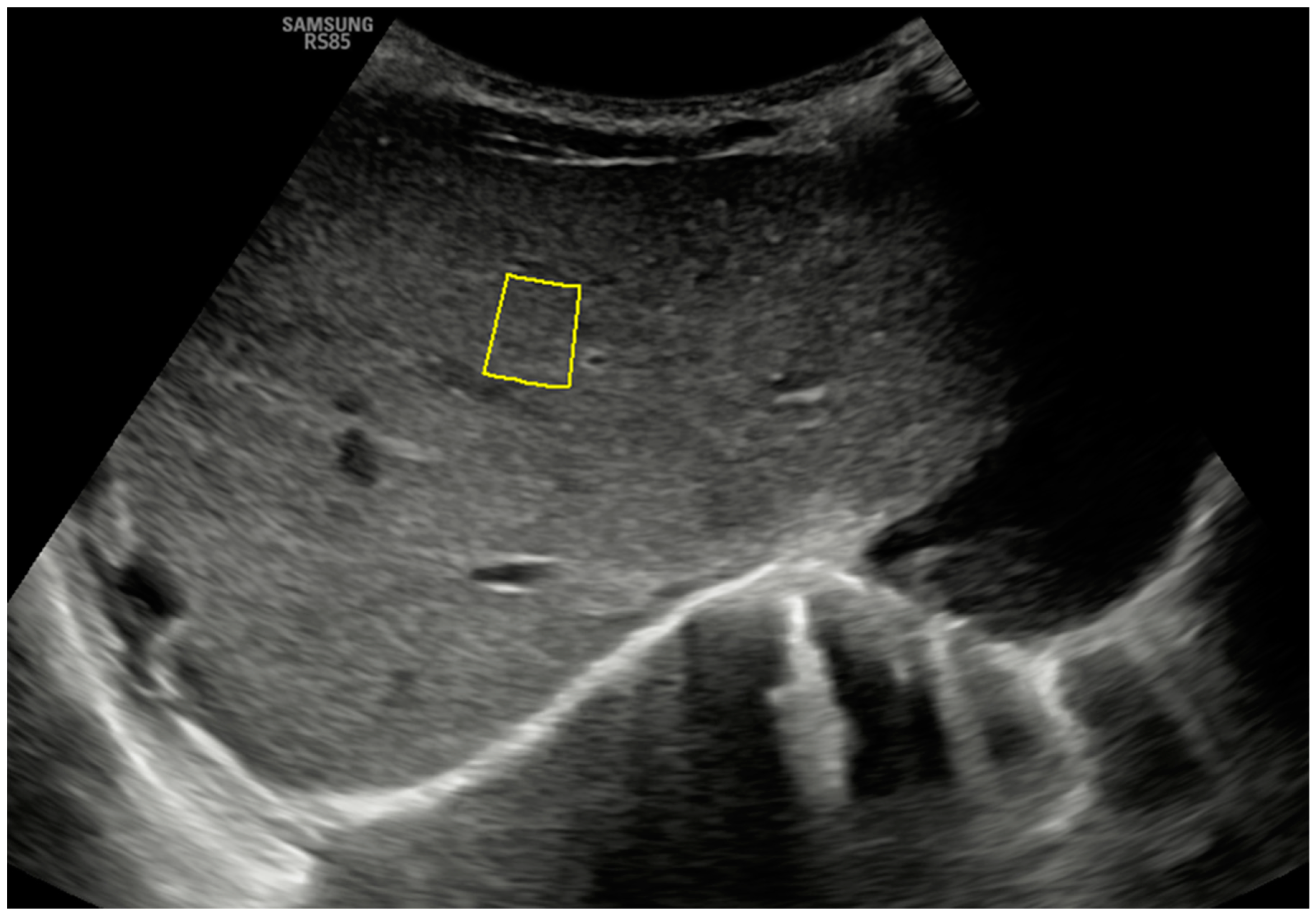
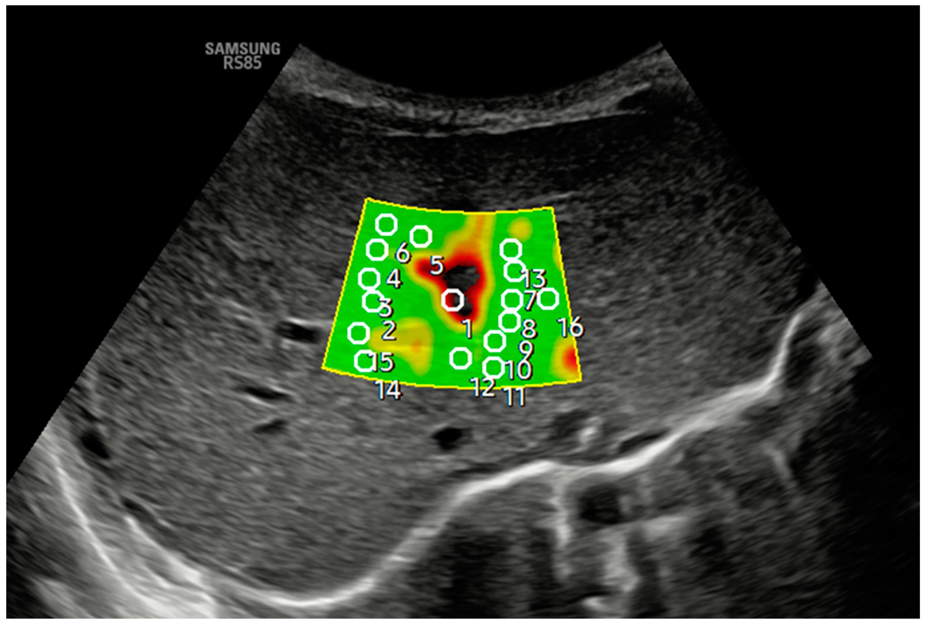
2. Technique
3. Peculiarities in ICUs and Neonatal Care Units and Discussion
- It is crucial to minimize patient contact time to maintain their overall stability and an aseptic environment. Elastography should take no longer than 1 to 2 min, with the mechanical index (MI) and thermal index (TI) values set to the lowest possible settings from the beginning (ideally below 0.4 MI) for biosecurity reasons [10], ensuring that the total scan time, including elastography, does not exceed 5 to 6 min.
- The patient must remain as comfortable as possible during the exam. Nurses play a vital role in improving patient comfort and ensuring the proper functioning of any respiratory, digestive, and vascular devices the patient may have. This is especially important in intensive care units, where at least one staff member must always be at the patient’s bedside.
- The patient should be examined supine, ideally without removing the kangaroo from its crib or incubator. The patient should remain calm and stable, avoiding interruptions of kangaroo care or feeding hours.
- Unlike older children or adolescents, neonates are not allowed to fast for extended periods, as irritability and crying would make the examination impossible. Therefore, elastography should be performed at the most convenient time, regardless of the fasting state. The patient should be in a postprandial state to ensure peace of mind, thus minimizing the time of exploration. There is ample literature on other age groups that shows fasting does not significantly affect the stiffness values obtained through elastography [11,12].
- In more minor patients (<1000 g), obtaining sufficient measurements is often more difficult, as the available depth cannot exceed 3 cm. To address this, we recommend using contact surface expansion devices, such as ultrasound gel pads, to increase the depth or, more simply, take measurements of the left hepatic lobe. This measurement has not significantly altered the values obtained in other age groups [11,12].
- Elastography can be challenging in patients undergoing high-frequency oscillatory ventilation (HFO), a treatment for respiratory failure in preterm infants. In some cases, the exam length can increase significantly, from less than a minute to 14 minutes. In other situations, obtaining a valid measurement may not be possible at all. Therefore, especially if the patient is hemodynamically unstable, the examination should only be performed if it is essential for their management.
4. Conclusions
Author Contributions
Funding
Institutional Review Board Statement
Informed Consent Statement
Data Availability Statement
Conflicts of Interest
References
- Sandberg, J.K.; Sun, Y.; Ju, Z.; Liu, S.; Jiang, J.; Koci, M.; Rosenberg, J.; Rubesova, E.; Barth, R.A. Ultrasound elastography with shear waves: Does grayscale ultrasound add value to differentiate biliary atresia from other causes of neonatal jaundice? Pediatr. Radiol. 2021, 51, 1654–1666. [Google Scholar] [CrossRef]
- Kollmann, C.; Jenderka, K.V.; Moran, C.M.; Draghi, F.; Jiménez Díaz, J.F.; Sande, R. EFSUMB Clinical Safety Statement for Diagnostic Ultrasound—(2019 review). Ultrasound Medizin. 2020, 41, 387–389. [Google Scholar]
- British Society of Medical Ultrasound Committee on Physics and Safety. Guidance Document on Ultrasound Safety Issues During Newborn Examination; The British Medical Ultrasound Society: London, UK, 2021. [Google Scholar]
- Martínez, S.M.; Crespo, G.; Navasa, M.; Forns, X. Non-invasive assessment of liver fibrosis. Hepatology 2011, 53, 325–335. [Google Scholar] [CrossRef] [PubMed]
- Barr, R.G.; Ferraioli, G.; Palmeri, M.L.; Goodman, Z.D.; Garcia-Tsao, G.; Rubin, J.; Garra, B.; Myers, R.P.; Wilson, S.R.; Rubens, D.; et al. Elastographic evaluation of liver fibrosis: Statement of the Society of Ultrasound Radiologists consensus conference. Radiology 2015, 276, 845–861. [Google Scholar] [CrossRef] [PubMed]
- Cast́era, L.; Foucher, J.; Bernard, P.H.; Carvalho, F.; Allaix, D.; Merrouche, W.; Couzigou, P.; De Ĺedinghen, V. Pitfalls of liver stiffness measurement: A 5-year prospective study of 13,369 examinations. Hepatology 2010, 51, 828–835. [Google Scholar] [CrossRef] [PubMed]
- Bamber, J.; Cosgrove, D.; Dietrich, C.F.; Fromageau, J.; Bojunga, J.; Calliada, F.; Cantisani, V.; Correas, J.-M.; D’Onofrio, M.; Drakonaki, E.E.; et al. EFSUMB Guidelines and Recommendations on the Clinical Use of Ultrasound ElastographyPart 1: Basic Principles and Technology. Ultrasound Medizin. 2013, 34, 169–184. [Google Scholar]
- Shiina, T.; Nightingale, K.R.; Palmeri, M.L.; Hall, T.J.; Bamber, J.C.; Barr, R.G.; Castera, R.; Choi, B.I.; Chou, Y.H.; Cosgrove, D.W.; et al. WFUMB Guidelines and Recommendations for the Clinical Use of Ultrasound Elastography: Part 1: Basic Principles and Terminology. Ultrasound Med. Biol. 2015, 41, 1126–1147. [Google Scholar] [CrossRef] [PubMed]
- Leschied, J.R.; Dillman, J.R.; Bilhartz, J.; Heider, A.; Smith, E.A.; Lopez, M.J. Shear wave elastography helps differentiate biliary atresia from other neonatal/childhood liver diseases. Pediatr. Radiol. 2015, 45, 366–375. [Google Scholar] [CrossRef] [PubMed]
- Miller, D.L.; Dou, C.; Dong, Z. Pulmonary ultrasound induction of pulmonary capillary hemorrhage in neonatal pigs. Ultrasound Med. Biol. 2022, 48, 2276–2291. [Google Scholar] [CrossRef] [PubMed]
- Cañas, T.; Fontanilla, T.; Miralles, M.; Maciá, A.; Malalana, A.; Román, E. Normal values of spleen stiffness in healthy children assessed by acoustic radiation force impulse (ARFI) imaging: Comparison between two ultrasound transducers. Pediatr. Radiol. 2015, 45, 1316–1322. [Google Scholar] [CrossRef] [PubMed]
- Fontanilla, T.; Cañas, T.; Maciá, A.; Alfageme, M.; Junquera, C.G.; Malalana, A.; Cilleruelo, M.L.; Roman, E.; Miralles, M. Normal values of hepatic shear wave velocity in healthy children assessed by acoustic radiation force impulse imaging using a convex probe and a linear probe. Ultrasound Med. Biol. 2014, 40, 470–477. [Google Scholar] [CrossRef] [PubMed]

Disclaimer/Publisher’s Note: The statements, opinions and data contained in all publications are solely those of the individual author(s) and contributor(s) and not of MDPI and/or the editor(s). MDPI and/or the editor(s) disclaim responsibility for any injury to people or property resulting from any ideas, methods, instructions or products referred to in the content. |
© 2025 by the authors. Licensee MDPI, Basel, Switzerland. This article is an open access article distributed under the terms and conditions of the Creative Commons Attribution (CC BY) license (https://creativecommons.org/licenses/by/4.0/).
Share and Cite
Lancharro Zapata, Á.; Aguado del Hoyo, A.; Sánchez Gómez de Orgaz, M.d.C.; Ortega, M.A.; León Luís, J.A. Technical Notes on Liver Elastography: A Guide for Use in Neonates in Intensive Care Units. J. Clin. Med. 2025, 14, 1435. https://doi.org/10.3390/jcm14051435
Lancharro Zapata Á, Aguado del Hoyo A, Sánchez Gómez de Orgaz MdC, Ortega MA, León Luís JA. Technical Notes on Liver Elastography: A Guide for Use in Neonates in Intensive Care Units. Journal of Clinical Medicine. 2025; 14(5):1435. https://doi.org/10.3390/jcm14051435
Chicago/Turabian StyleLancharro Zapata, Ángel, Alejandra Aguado del Hoyo, María del Carmen Sánchez Gómez de Orgaz, Miguel A. Ortega, and Juan Antonio León Luís. 2025. "Technical Notes on Liver Elastography: A Guide for Use in Neonates in Intensive Care Units" Journal of Clinical Medicine 14, no. 5: 1435. https://doi.org/10.3390/jcm14051435
APA StyleLancharro Zapata, Á., Aguado del Hoyo, A., Sánchez Gómez de Orgaz, M. d. C., Ortega, M. A., & León Luís, J. A. (2025). Technical Notes on Liver Elastography: A Guide for Use in Neonates in Intensive Care Units. Journal of Clinical Medicine, 14(5), 1435. https://doi.org/10.3390/jcm14051435