Analysis of Natural Clinical Crown Height Changes in Central Incisors and First Molars from Age 8 to 18: A Retrospective Digital Study
Abstract
1. Introduction
2. Material and Methods
2.1. Subjects
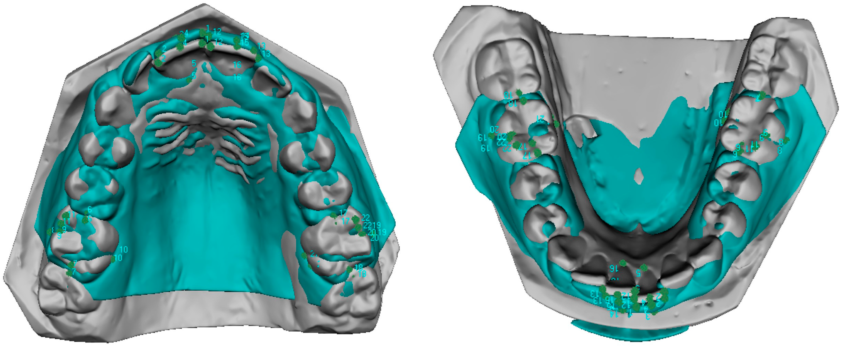
2.2. Digital 3D Analysis of Dental Casts
- (1)
- Five points were placed: the mesial and distal points of the incisal edge, two points corresponding to the gingival and incisal limits of the vestibular axis of the clinical crown (vestibular FACC), and one point corresponding to the gingival limit of the palato–lingual axis of the clinical crown (palato-lingual FACC) corresponding to the continuation of the vestibular FACC on the palatal/lingual surface [17].
- (2)
- Six points were placed: the mesial and distal points of the occlusal surface, and two points corresponding to the gingival and occlusal limits of the vestibular axis of the clinical crown (vestibular FACC). The occlusal point is positioned between the two widest cusps, and the gingival point is positioned at the gingival limit of the vestibular groove. One point corresponds to the gingival limit of the palatolingual axis of the clinical crown (vestibular FACC), the lingual axis of the clinical crown (palato-lingual FACC), and a sixth point is added at the apex of the maximum convexity of the occlusal surface of the mesio-vestibular cusp.
2.3. Statistical Analysis
3. Results
3.1. Central Incisors
3.2. First Molars
- Mandibular incisor: Height = 7.0 + 0.08 × Age
- Mandibular molar: Height = 3.9 + 0.21 × Age
- Maxillary incisor: Height = 8.1 + 0.15 × Age
- Maxillary molar: Height = 3.4 + 0.17 × Age
4. Discussion
5. Conclusions
Author Contributions
Funding
Institutional Review Board Statement
Informed Consent Statement
Data Availability Statement
Acknowledgments
Conflicts of Interest
References
- Moyers, R.E.; Linden, F.P.G.; Van der Riolo, M.L.; McNamara, J.A. Standards of Human Occlusal Development; University of Michigan: Ann Arbor, MI, USA, 1976. [Google Scholar]
- De Luca Canto, G.; Pachêco-Pereira, C.; Lagravere, M.O.; Flores-Mir, C.; Major, P.W. Intra-arch dimensional measurement validity of laser-scanned digital dental models compared with the original plaster models: A systematic review. Orthod. Craniofac. Res. 2015, 18, 65–76. [Google Scholar] [CrossRef] [PubMed]
- Rossini, G.; Parrini, S.; Castroflorio, T.; Deregibus, A.; Debernardi, C.L. Diagnostic accuracy and measurement sensitivity of digital models for orthodontic purposes: A systematic review. Am. J. Orthod. Dentofac. Orthop. 2016, 149, 161–170. [Google Scholar] [CrossRef] [PubMed]
- Levin, E.I. Dental esthetics and the golden proportion. J. Prosthet. Dent. 1978, 40, 244–252. [Google Scholar] [CrossRef]
- Burça, K.; Ruzin, U. Influence on smile attractiveness of the smile arc in conjunction with gingival display. Am. J. Orthod. Dentofac. Orthop. 2013, 144, 541–547. [Google Scholar]
- Alyami, A.H.; Sanea, J.A.; Togoo, R.A.; Ain, T.S. Aesthetic perception about gingival display on maxillary incisor inclination among Saudi dentists, orthodontist and lay persons. J. Clin. Diagn. Res. 2018, 12, 56–60. [Google Scholar] [CrossRef]
- Ioi, H.; Nakata, S.; Counts, A.L. Influence of gingival display on smile aesthetics in Japanese. Eur. J. Orthod. 2010, 32, 633–637. [Google Scholar] [CrossRef]
- Alhammadi, M.S.; Halboub, E.; Al-Dumaini, A.A.; Malhan, S.M.; Alfaife, F.; Otudi, J. Perception of dental, smile and gingival esthetic components by dental specialists, general dental practitioners, dental assistants and laypersons: A cross-sectional study. World, J. Dent. 2022, 13, 250–260. [Google Scholar]
- Sarver, D.M. Principles of cosmetic dentistry in orthodontics: Part 1. Shape and proportionality of anterior teeth. Am. J. Orthod. Dentofac. Orthop. 2004, 126, 749–753. [Google Scholar] [CrossRef]
- Atik, E.; Turkoglu, H. Does different vertical position of maxillary central incisors in women with different facial vertical height affect smile esthetics perception? Prog. Orthod. 2023, 24, 28. [Google Scholar] [CrossRef]
- Carrera, T.M.I.; Freire, A.E.N.; de Oliveira, G.J.P.L.; Dos Reis Nicolau, S.; Pichotano, E.C.; Junior, N.V.R.; Pires, L.C.; Pigossi, S.C. Digital planning and guided dual technique in esthetic crown lengthening: A randomized controlled clinical trial. Clin. Oral. Investig. 2023, 27, 1589–1603. [Google Scholar] [CrossRef]
- Sarver, D.M. Use of the 810nm Diode Laser: Soft Tissue Management and Orthodontic Applications of Innovative Technology. Pract. Proced. Aesthetic Dent. 2006, 18, 7–13. [Google Scholar]
- Garguilo, A.; Wenz, F.; Orban, B. Dimension and relation of the dento-gingival junction in humans. J. Periodontol. 1961, 32, 261–267. [Google Scholar] [CrossRef]
- Theytaz, G.A.; Kiliaridis, S. Gingival and dentofacial changes in adolescents and adults 2 to 10 years after orthodontic treatment. J. Clin. Periodontol. 2008, 35, 825–830. [Google Scholar] [CrossRef] [PubMed]
- Theytaz, G.A.; Christou, P.; Kiliaridis, S. Gingival changes and secondary tooth eruption in adolescents and adults: A longitudinal retrospective study. Am. J. Orthod. Dentofac. Orthop. 2011, 139 (Suppl. 4), 129–132. [Google Scholar] [CrossRef][Green Version]
- Andrews, L.F. Straight Wire: The Concept and Appliance; Wells, L.A., Ed.; Wells Co.: San Diego, CA, USA, 1989. [Google Scholar]
- Huanca Ghislanzoni, L.T.; Lineberger, M.; Cevidanes, L.H.S.; Mapelli, A.; Sforza, C.; McNamara, J.A. Evaluation of tip and torque on virtual study models: A validation study. Prog. Orthod. 2013, 14, 19. [Google Scholar] [CrossRef]
- Brief, J.; Behle, J.H.; Stellzig-Eisenhauer, A.; Hassfeld, S. Precision of landmark positioning on digitized models from patients with cleft lip and palate. Cleft Palate-Craniofacial J. 2006, 43, 168–173. [Google Scholar] [CrossRef]
- Natsumeda, G.; Miranda, F.; Massaro, C.; Lauris, J.R.P.; Garib, D. Aging changes in maxillary anterior teeth in untreated individuals: An observational longitudinal study. Prog. Orthod. 2023, 24, 26. [Google Scholar] [CrossRef]
- Morrow, L.A.; Robbins, J.W.; Jones, D.L.; Wilson, N.H.F. Clinical crown length changes from age 12–19 years: A longitudinal study. J. Dent. 2000, 28, 469–473. [Google Scholar] [CrossRef]
- Volchansky, A.; Cleaton-Jones, P. The position of the gingival margin as expressed by clinical crown height in children aged 6–16 years. J. Dent. 1976, 4, 116–122. [Google Scholar] [CrossRef]
- Massaro, C.; Miranda, F.; Janson, G.; Rodrigues de Almeida, R.; Pinzan, A.; Martins, D.R.; Garib, D. Maturational changes of the normal occlusion: A 40-year follow-up. Am. J. Orthod. Dentofac. Orthop. 2018, 154, 188–200. [Google Scholar] [CrossRef]
- Volchansky, A.; Cleaton-jones, P. Clinical crown height (length)—A review of published measurements. J. Clin. Periodontol. 2001, 28, 1085–1090. [Google Scholar] [PubMed]
- Woda, A.; Gourdon, A.M.; Faraj, M. Occlusal contacts and tooth wear. J. Prosthet. Dent. 1987, 57, 85–93. [Google Scholar] [CrossRef] [PubMed]
- Gillen, R.J.; Schwartz, R.S.; Hilton, T.J.; Evans, D.B. An analysis of selected normative tooth proportions. Int. J. Prosthodont. 1994, 7, 410–417. [Google Scholar] [PubMed]
- Sarver, D.M.; Yanosky, M. Principles of cosmetic dentistry in orthodontics: Part 2. Soft tissue laser technology and cosmetic gingival contouring. Am. J. Orthod. Dentofac. Orthop. 2005, 127, 85–90. [Google Scholar] [CrossRef]
- Sobouti, F.; Rakhshan, V.; Chiniforush, N.; Khatami, M. Effects of laser-assisted cosmetic smile lift gingivectomy on postoperative bleeding and pain in fixed orthodontic patients: A controlled clinical trial. Prog. Orthod. 2014, 15, 66. [Google Scholar] [CrossRef]


| Estimate | Std. Error | t. Value | p.z | |
|---|---|---|---|---|
| Intercept * | 7.01 | 0.14 | 51.94 | <0.001 |
| Age | 0.08 | 0.01 | 7.41 | <0.001 |
| Maxillaire = 1 | 1.06 | 0.11 | 9.90 | <0.001 |
| Tooth = 6 | −3.12 | 0.15 | −20.60 | <0.001 |
| Age:Maxillaire = 1 | 0.07 | 0.01 | 6.54 | <0.001 |
| Age:Tooth = 6 | 0.14 | 0.01 | 12.55 | <0.001 |
| Maxillaire = 1:Tooth = 6 | −1.51 | 0.09 | −16.96 | <0.001 |
| Age:Maxillaire = 1:Tooth = 6 | −0.11 | 0.01 | −7.67 | <0.001 |
Disclaimer/Publisher’s Note: The statements, opinions and data contained in all publications are solely those of the individual author(s) and contributor(s) and not of MDPI and/or the editor(s). MDPI and/or the editor(s) disclaim responsibility for any injury to people or property resulting from any ideas, methods, instructions or products referred to in the content. |
© 2025 by the authors. Licensee MDPI, Basel, Switzerland. This article is an open access article distributed under the terms and conditions of the Creative Commons Attribution (CC BY) license (https://creativecommons.org/licenses/by/4.0/).
Share and Cite
Huanca Ghislanzoni, L.; Boesiger, J.; Mourgues, T.; González-Olmo, M.J.; Romero, M. Analysis of Natural Clinical Crown Height Changes in Central Incisors and First Molars from Age 8 to 18: A Retrospective Digital Study. J. Clin. Med. 2025, 14, 766. https://doi.org/10.3390/jcm14030766
Huanca Ghislanzoni L, Boesiger J, Mourgues T, González-Olmo MJ, Romero M. Analysis of Natural Clinical Crown Height Changes in Central Incisors and First Molars from Age 8 to 18: A Retrospective Digital Study. Journal of Clinical Medicine. 2025; 14(3):766. https://doi.org/10.3390/jcm14030766
Chicago/Turabian StyleHuanca Ghislanzoni, Luis, Jean Boesiger, Thomas Mourgues, María José González-Olmo, and Martin Romero. 2025. "Analysis of Natural Clinical Crown Height Changes in Central Incisors and First Molars from Age 8 to 18: A Retrospective Digital Study" Journal of Clinical Medicine 14, no. 3: 766. https://doi.org/10.3390/jcm14030766
APA StyleHuanca Ghislanzoni, L., Boesiger, J., Mourgues, T., González-Olmo, M. J., & Romero, M. (2025). Analysis of Natural Clinical Crown Height Changes in Central Incisors and First Molars from Age 8 to 18: A Retrospective Digital Study. Journal of Clinical Medicine, 14(3), 766. https://doi.org/10.3390/jcm14030766


