Communication

Secure Ileal Pouch–Anal Anastomosis for Histologic Indeterminate Colitis

by
Amosy E. M’Koma
1,2,3,4,5
1
Department of Biochemistry, Cancer Biology, Neuroscience and Pharmacology, Meharry Medical College School of Medicine, Nashville, TN 37208-3500, USA
2
Department of Pathology, Anatomy and Cell Biology, Meharry Medical College School of Medicine, Nashville General Hospital, Nashville, TN 37208-3599, USA
3
Division of General Surgery, Section of Colon and Rectal Surgery, Vanderbilt University School of Medicine, Nashville, TN 37232-0260, USA
4
The American Society of Colon and Rectal Surgeons (ASCRS), 2549 Waukegan Road, #210, Bannockburn, IL 600015, USA
5
International Society of University Colon and Rectal Surgeons, 109 Partin Street, Chapel Hill, NC 27514, USA
J. Clin. Med. 2025, 14(23), 8390; https://doi.org/10.3390/jcm14238390
Submission received: 15 October 2025 / Revised: 17 November 2025 / Accepted: 20 November 2025 / Published: 26 November 2025
(This article belongs to the Special Issue Minimally Invasive Surgery for Gastrointestinal Disorder)

Abstract

Background/Objectives: Indeterminate colitis (IC) is an erroneous diagnosis for predominantly colonic inflammatory bowel disease (IBD) when there is a non-definitive foundation of the benchmark for ulcerative colitis (UC) and Crohn’s colitis (CC) after a combined state-of-the-art classification system of clinical, endoscopic, radiologic, and histologic tools are used. This confounds an effective surgical regimen; specifically pouch surgery, “the restorative proctocolectomy with ileal pouch–anal anastomosis (PRC-IPAA)”. Transforming the distinction between UC and CC in otherwise IC into authentic UC and CC requires priority attention when considering a patient’s candidacy for RPC-IPAA. RPC-IPAA is the accepted standard curative surgical procedure in the treatment for UC (and Familial Adenomatous Polyposis (FAP)). Further, inapproximate/incorrect diagnosis and treatment can sustain potential long-term morbidity from inaccurate and unnecessary surgery and cost. Methods: In trying to resolve these diagnostic ambiguities, the current study advances our understanding by showing the expression of human alpha defensin 5 (DEFA5 alias HD5) restricted in the colon crypt mucosal lining areas, and by identifying the cells of the small intestine (ileum) “colonic ileal metaplasia” in CC that may serve as a biomarker to portray/ascertain authentic CC and UC among IC cohorts, with a positive predictive value (PPV) of 96 percent. Results: Hence, the imprecise diagnosis of IC largely would be circumvented. This new diagnostic tool offers instant tangible benefits over existing diagnostic pathways. The journey toward its widespread clinical use is now subject to logistical and regulatory defiance, which all emerging molecular diagnostic technologies inevitably encounter. Conclusions: The aim of this communication is to provide a summary of the currently available diagnostic advances relating to surgical management for IC in clinical settings, and the related challenges. Further, I briefly discuss aspects of its pathophysiology, surveillance, and diagnostic assay development.

1. Introduction

Predominantly colonic inflammatory bowel disease (IBD) encompasses ulcerative colitis (UC) and Crohn’s colitis (CC), which are characterized by chronic, relapsing inflammation of the colon and rectum; involvement of the anus and perianal region is a feature specific to Crohn’s disease. Studies at Meharry Medical College, the Vanderbilt University Medical Center [1], and other institutions [2,3,4] found an estimated 15% of patients with colonic IBD to have an inaccurate diagnosis of “indeterminate colitis (IC)”, which occurs when the diagnostic classification features for UC and CC are inconclusive after a comprehensive evaluation including clinical, endoscopic, radiological, and histological examinations. The dedifferentiation of IBD often leads to unnecessary and ineffective interventional regimens, subsequent morbidity, and cost. The prevalence of IC is about 22 persons per 100,000 people, and it is commonly observed with a high incidence in female Caucasians [3]. A total proctocolectomy (TPC) or restorative proctocolectomy with ileal pouch–anal anastomosis (RPC-IPAA) is the surgical treatment option for UC patients who become refractory to pharmaceuticals [5,6,7]. Further, TPC and/or RPC-IPAA is carefully indicated for select patients with CC [8,9], and for patients with hereditary nonpolyposis colorectal cancer or synchronous colon cancers [10,11]. Furthermore, for patients with Familial Adenomatous Polyposis (FAP), a prophylactic TPC is indicated because they possess the hereditary adenomatous polyposis coli gene and inevitably develop colorectal cancer in their lifetimes [12]. Accurate diagnosis in patients presenting with colonic IBD and their mimics is consequentially essential, and it is important for a tailored surgical intervention, because each entity may require a specific approach or regimen, the delivery of which may have broad implications.
The RPC-IPAA procedure requires the replacement of the resected entire colon and rectum by a pouch formed from the terminal/distal ileum and sutured to the anal canal, above the dentine/pectinate/mucocutaneous line, a significant crucial anatomical landmark in the anal canal that separates the upper and lower parts, preserving the anal sphincters and maintaining numerous physiological and clinical features [13,14]. Briefly, the RPC-IPAA reconstruction gives the gastrointestinal tract luminal continuity, defecation, deferral, discrimination, and fertility. Early observations reported complication rates of over 50 percent and failure rates as high as 35 percent [15,16]. These observations prompted the importance of patient selection, and only patients with UC and FAP were recommended candidates for RPC-IPAA surgery [17,18]. Crohn’s colitis was contraindicated [17,18]. Over the years, however, improved diagnostic tools, technical modifications, and surgical expertise have been developed so that the procedure can now be performed safely with a low complication and failure rates of less than five percent [19,20,21]. Hence, indications for the intervention have widened, especially for older patients and those with a history of perianal disease, who can be selectively chosen as candidates or not for the RPC-IPAA procedure [22]. Intriguingly, some IBD Centers advocate for performing RPC-IPAA surgery in patients with CC [18,23]. However, though RPC-IPAA surgery is feasible with indifferent functional outcomes between CC and UC, even in highly selected patients with known preoperative CC or a proctocolectomy pathology, pouch failure rates stay higher than in patients with UC [9]. Therefore, it is not surprising that IC is not a contraindication for pouch surgery at many IBD Centers worldwide [22,23]. However, subsequent morbidity necessitating excision of the pouch is reported in 20 to 30 percent of patients [22,23]. These patients will most likely experience complications such as intra-abdominal and perineal sepsis and fistulas, and they are likely to require several operations, including one to excise their pouch, losing approximately 50 cm of the small bowel. Mortality from their complications is unlikely, and it is also unlikely that they will have subsequent short bowel syndrome (SBS) [24,25,26]. Conversely, approximately 70 percent to 80 percent of patients will have acceptably functional pouches, despite the fact that some of them may require further surgery due to adverse events [27]. The alternative to RPC-IPAA is total PTC and permanent terminal ileostomy [23,25,28].
In trying to resolve unmet diagnostic delay and ambiguity gapes in colonic IBD, the current study advances our understanding by showing the expression of human alpha defensin 5 (DEFA5 alias HD5) restricted in the colon crypt mucosal lining areas, and it identifies the cells of the small intestine (ileum) “colonic ileal metaplasia” in CC that may serve as a biomarker to characterize/delineate and ascertain authenticity for CC and UC among the IC cohorts, with a positive predictive value (PPV) of 96 percent [1]. Hence, the imprecise diagnosis of IC largely would be circumvented. This new diagnostic tool offers instant tangible benefits over existing diagnostic pathways. The journey toward its widespread clinical use is now subject to logistical and regulatory defiance, which all emerging molecular diagnostic technologies inevitably must withstand [5]. The aim of this communication is to provide a summary of the currently available diagnostic advances and surgical management for IC, and the challenges in clinical settings. Further, I briefly discuss aspects of its pathophysiology, surveillance, and diagnostic assay development advances.

2. Methods

A search of the diagnostic literature and treatment recommendation guidelines for IBD and IC was performed using predetermined protocols. The search used general engines like Google and specific databases including the Cochrane Database, MEDLINE, EMBASE, PubMed, and the Cumulative Index of Nursing and Allied Health Literature (CINAHL), covering the period between January 1980 and June 2025. The process also included a review of guidelines from IBD-associated society organizations and official agencies, i.e., the American Gastroenterological Association (AGA), the American Society for Gastrointestinal Endoscopy (ASGE), the British Society of Gastroenterology (BSG), the International Foundation for Gastrointestinal Disorders (IFGD), the American Society of Colon and Rectal Surgeons (ASCRS), the American College of Gastroenterology (ACG), the Society of American Gastrointestinal and Endoscopic Surgeons (SAGES), the International Organization for the Study of Inflammatory Bowel Disease (IOIBD), the World Health Organization (WHO), the U.S. Food and Drug Administration (USFDA), the European Medicines Agency (EMA), European Crohn’s and Colitis (ECC), American Crohn’s and Colitis (CCFA), and the Canadian Association of Gastroenterology (CAG). The quality of reporting followed guidelines for meta-analyses of observational studies (MOOSE).

Ethics

This communication was approved by the Meharry Medical College (IRB file numbers: 100916AM206 (date of 16 September 2010), 18-08-851 (date of 12 September 2018), 22-08-1235 (date of 15 August 2022), and 22-08-1244 (date of 26 August 2022) and Vanderbilt University (IRB file numbers: 080898 (date of 29 September 2008) and 100581 (date of 1 June 2010). Institutional Ethical Review Boards ensured this study was conducted in accordance with the World Medical Association (WMA) Declaration of Helsinki – Ethical Principles for Medical Research Involving Human Participants. Patient samples for the included studies were obtained from the NIH-funded Digestive Disease Research Center (PI: David A. Schwartz, MD), Vanderbilt Gastrointestinal Biospecimen Repository, and the Cooperative Human Tissue Network at Vanderbilt University Medical Center (VUMC) (PI: Mary K. Washington, MD, PhD), in collaboration with the Meharry Medical College Human Tissue Acquisition Shared Resource Core (PI: Billy R. Ballard, DDS, MD). The availability of a detailed IBD patient database registry at VUMC made a chart review and follow-up surveillance possible. Medical record data on patient demographics, preoperative variables prior to and after RPC-IPAA surgery, surveillance of endoscopic and clinical findings, and medical and surgical treatment history were retrieved retrospectively. Patient informed consent was given, and participation in this study was voluntary.

3. Indeterminate Colitis

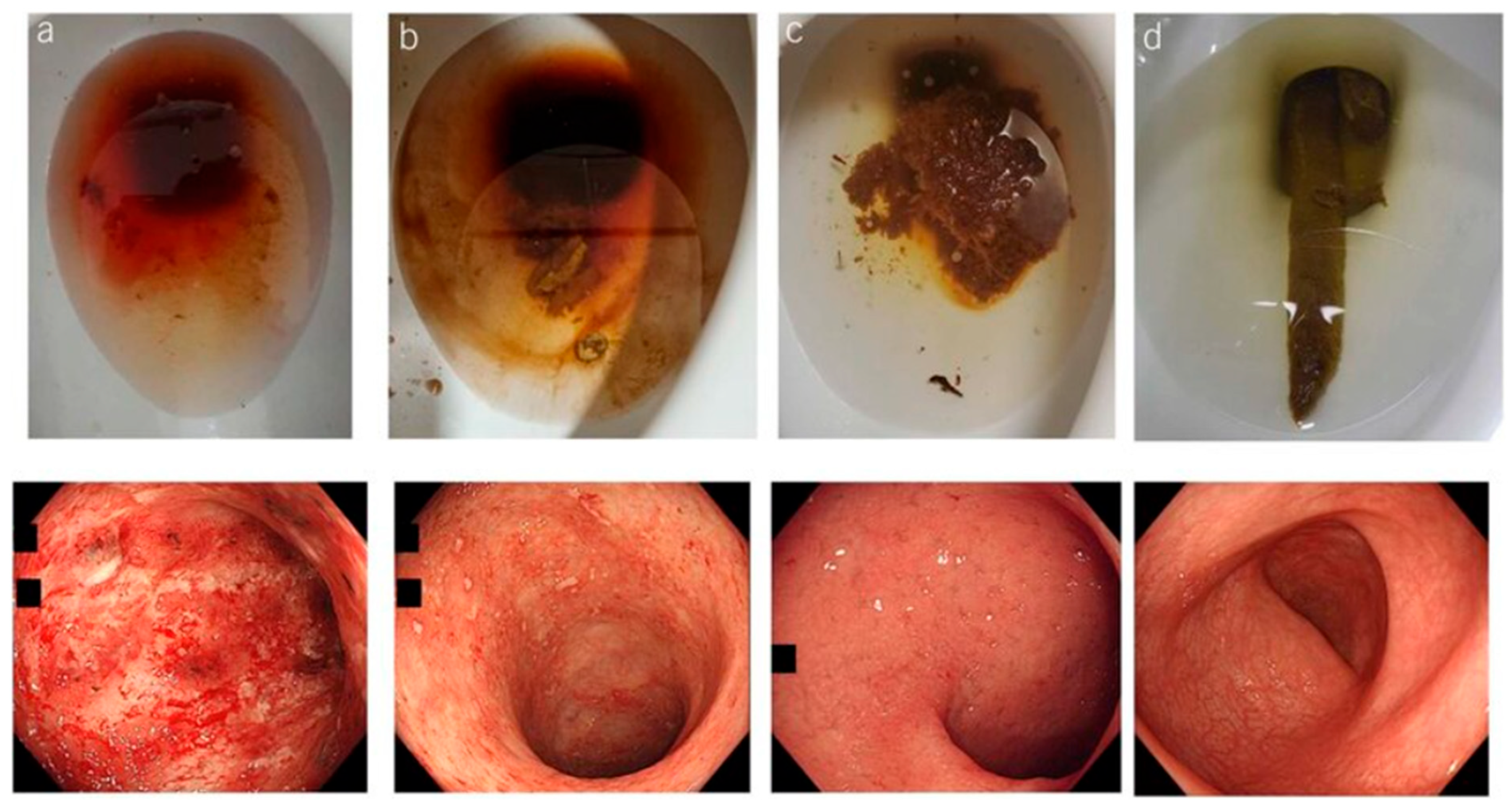
The term “indeterminate colitis (IC)” was originally introduced in 1978 to describe surgical specimens from IBD patients undergoing colectomy, when the histological features were not characteristic of either CC disease or UC [29,30,31,32,33]. Distinguishing UC from CC is often complex and challenging [34]. In particular, in the interim uncertainty prodromal-stage cases of extensive ulceration, the two diseases may visually be characteristically indistinguishable [35,36]. Inadequate differentiation of diagnostic features of UC and CC, as illustrated in Figure 1A–H may lead to an inconclusive diagnosis of IC even when a combined state-of-the-art classification systems of clinical, endoscopic, radiological, and histological tools are used [33,37,38,39]. Up to 15% of colonic IBD cases are labeled as IC when non-definitive evaluation criteria for either UC or CC have been established from colonoscopy biopsies, or at colectomy [32,33,40,41,42,43]. Most patients with IC do eventually evolve to a definite diagnosis of CC or UC after long-term follow-up surveillance, which can indeed span many years [1,25]. The approximate percentage of patients who have their diagnosis changed from presumed definitive UC to de novo Crohn’s disease (CD) after RPC-IPAA is within the range cited of 15% based on the postoperative follow-up clinical observations and histopathology changes, and development of de novo CD in the ileal pouch [17,23,25,44]. Despite the introduction of newer cutting-edge diagnostic technology modalities, these figures have not changed for over six decades [45]. One-half of these patients will require pouch excision or diversion [23]. Much of the diagnostic uncertainty arises from the overlap of clinical and histopathological features of both conditions, making CC appear like UC and vice versa, Figure 1A–H [23,34]. The characteristics of UC result in inflammation and ulcerations confined to the mucosal lining areas and scarcely extending to submucosal layer of the colon and rectum, Figure 2a–d [45,46,47]. Meanwhile, CC differs in that inflammation may result through the intestine walls (transmural) with skip lesions, Figure 3A–H [33,45,46,47,48]. Further, CC may also involve other body organs outside boundaries of the gastrointestinal tract (GIT) system through fistulation [49,50]. The current surgical treatment recommendation options for UC and CC are still debatable, and guidelines emphasize the urgency of surgical options for patients with medically refractory UC [31]. Therefore, an accurate diagnosis is of supreme importance in terms of determining the evidence-based patient candidacy for surgical intervention and personalized preoperative counseling for the possible subsequent outcome. To date, colorectal surgeons provide accounts of the challenge, where some patients with IC may appear to have UC, and others may appear to have CC. The former may be considered candidates for RPC-IPAA, whereas the latter may be advised to undergo total PTC. Another alternative approach is to perform a subtotal colectomy with terminal ileostomy initially and then, depending on the pathology results, proceed with pouch surgery if the pathological diagnosis is UC. If the pathological diagnosis is IC, one could suggest waiting to see whether small bowel or perianal manifestations develop signs, which would suggest the diagnosis of CC (severe pouchitis, proctitis, cuffitis, strictures, abscess sinus tracks, fistulae, etc.). Secondly, if one adopts a policy of performing RPC-IPAA in patients with refractory IC, it is probably reasonable to be carefully selective in the choice of candidates. It is a wise move that only patients with IC and or CC who are young, without comorbidities, and who are signed off as psychologically stable after counseling should be considered for RPC-IPAA, because morbidity rates are higher in these patients [7,8,9].
Inconsistence experiences have been reported in reviewing the cases of IC. In some reports, the disease is most likely to behave clinically like UC [51,52] and much less likely to develop into clear clinical CC [35], while others found the reverse is true [53]. Diagnostic authentication validation studies for CC versus UC among IC cohorts using the DEFA5 bioassay test with a first-clinic-visit endoscopy biopsy are underway, https://cdn.clinicaltrials.gov/large-docs/71/NCT05663671/Prot_000.pdf (accessed date 10 August 2025).

4. Criteria for Making a Diagnosis

Endoscopically, the normal colonic mucosa is lined with simple columnar epithelium, which lacks the villi and contains crypt of Lieberkühn, Figure 4. The criteria for determining a diagnosis of IBD require an experienced endoscopy gastroenterologist and gastrointestinal pathologist to review the histological pathology [35,54]. The gross and histological features quoted in the pathology literature as being useful in distinguishing the UC vs. CC are evaluated. These quotes include linear ulcers on gross examination, granulomas of the sarcoid type, terminal small intestines/ileal inflammation (including backwash ileitis), slit-shaped ulcers, patchy mucosal inflammation, skip zones, and transmural inflammation. These are considered to be present when lymphoid aggregates are noted in the subserosa and/or the deep submucosal zone [35,54,55]. Serosal inflammation that is not in the form of lymphoid follicles should be disregarded. This is present mainly in patients with fulminant colitis immediately beneath/below deep broad-based ulcers, Figure 2a [35,36].

5. Diagnostic Dilemmas for IBD

To date, there is no single diagnostic “gold standard” tool for IBD [34]. Clinicians use a comprehensive combined classification system that includes clinical, endoscopy, radiologic, and histopathology findings in order to diagnose CC and UC [45,56]. Notably, even with the combination of these diagnostic utilities, 15% to 30% of IBD patients cannot be accurately diagnosed [34,57,58]. This pitfall has a significant implication when determining a patient’s candidacy for the RPC-IPAA intervention [17,34]. Curative treatment for UC recommends RPC-IPAA [17,19,44,59,60,61,62]. The successfulness of RPC-IPAA is largely dependent on careful patient selection combined with a conscientious surgical technique and diagnosis precision [19,57,63,64]. Currently available clinical experiences suggest that it is painstaking and strenuous to identify patients with CC who are likely to have a successful outcome after RPC-IPAA surgery [17,44,57,63]. Thus, CC should remain carefully indicated for pouch surgery but is an acceptable treatment option for patients with UC and for those IC patients who are predicted to develop UC [61,65,66,67,68,69]. These findings are beyond reasonable doubt informative in notifying the role of RPC-IPAA in preoperative counseling and decision-making with candidate patients.

6. Reclassification of Indeterminate Colitis to Definitive Ulcerative Colitis or Crohn’s Colitis

Under the umbrella of predominantly colonic IBD, it is pivotal to delineate authentic CC and UC amongst IC cohorts for informed pouch surgery decisions and outcomes [1,53]. Several attempts to develop a molecular “gold-standard diagnostic tool” to identify a reliable bioassay for delineating IC into authentic CC and UC have been ongoing for decades [34,45,70,71,72,73,74,75,76]. Recently, a discriminative breakthrough was made with the discovery of aberrant expression of DEFA5 in colonic tissues linked to the distinct pathogenesis of authentic CC [1,53]. To break down patients with IC into actual UC and CC is of primary importance for RPC-IPAA decision-making [17,19,44,59,60,77,78,79]. The success of RPC-IPAA surgery depends largely on the “correct diagnosis” as UC (or FAP) [19,57,63,64]. Observational clinical experiences suggest that patients with CC have higher pouch failure outcomes subsequent to RPC-IPAA surgery compared to UC [8,9,17,44,57,61,63,65,66,67,68,69].

6.1. DEFA5 Is a Promising Diagnostic Biomarker

Among potential diagnostic biomarkers, human alpha defensin 5 (DEFA5 alias HD5) has emerged as a promising candidate due to its differential expression patterns in CC and UC, making it a valuable target for IBD-subtype diagnostic development [1,80]. In colonic mucosal crypt layers, DEFA5 is ectopically expressed in patients with CC and IC cohorts that are foreseen as CC, and it is demonstrated that this expression pattern holds high diagnostic potential in distinguishing CC versus UC pathologies amongst IC patient cohorts [1,53]. DEFA5 expression is markedly exalted 118-fold in CC when compared with UC, whereas its levels in UC are trace, distinguishing CC from UC with high specificity [1,53,80]. These differential expression patterns underscore the potential capability of DEFA5 as a biomarker for IBD-subtype classification execution.

6.2. Advances in Assay Development for IBD Diagnostics

Significant advancements are being made through the utilization of the DEFA5 biosignature, which provides precision in resolving the diagnostic ambiguity of IC into authentic CC and UC. High levels of DEFA5 are reliably linked to a diagnosis of CC among IC cohort patients to accurately classify patients who might otherwise remain without a definitive diagnosis for years [1,53,80]. These innovative inventions are protected by three granted patents, US 11427852 B2, US 12174200 B2, and US 12281351 B2, and four International patents/applications WO 2018/175913 A1 (WIPO), WO 2018/237064 A1 (WIPO), EP3602041A1/A4 and CA-3056911-A1 exclusively assigned to Meharry Medical College in the realm of IBD diagnosis biomarkers, which could change the diagnostic landscape and lend mechanistic insights into IBD pathogenesis, allowing us to move beyond traditional methods, and thus paving the way for more tailored therapies and improved patient outcomes [80].

6.3. Anti-DEFA5 Monoclonal Antibodies

Two established tested functional novel anti-DEFA5 monoclonal antibodies (mAbs), clones 1A8 and 4F5, are validated for specificity, selectivity, and cross-reactivity in recognizing the endogenous and recombinant DEFA5 protein [80]. Biotechnology development studies on mAbs, clones 1A8 and 4F5, in larger IBD cohorts are underway to establish efficacy and safety (Trial NCT05663671).

7. Transcriptome Studies

Quantitative global expression profiles of RNA levels, generated using an oligonucleotide microarray and genome-wide transcriptome analysis, were investigated to identify transcriptional signatures present in colonic biopsy tissues obtained from UC and CC mucosa and submucosal linings [71,74]. The genomic patterns noted show greater intensity in CC as compared to UC, indicative of a greater degree or different type of inflammation response in the tissues’ underlying layers [75], and these may serve as a resource for professionals involved in gene expression data mining in a variety of clinical settings, particularly for the differential diagnosis of UC and CC in IC.

8. Proteomic Profiling

Studies have developed a proteomic technology, Matrix-Assisted Laser Desorption/Ionization Mass Spectrometry (MALDI-MS) technology, for proteomic profiling of histologic mucosal and submucosal tissue layers for analyses along with bioinformatics technologies to delineating UC and CC [70,75]. These studies identified and compared protein profiles, which had the necessary (i) specificity, (ii) sensitivity, (iii) discrimination, and (iv) predictive capacity to determine the heterogeneity of IBD subtypes and the ability to delineate UC and CC molecularly [70,75]. These rare biometrics based on the lining of the colonic pathology are ectopically independent of the tissue of origin and the characteristics of the ileum, consistent with “colonic ileal metaplasia” [53], representing disease-specific inflammatory markers [70,75].

9. Blood-Based Biomarkers

In contrast to colon surgical pathology tissue resections, peripheral blood is a much more accessible source of cells that might be used to distinguish between CC and UC. Circulating peripheral blood cytokines (variables) are responsible for signs of disease surveillance. Cytokines are therefore surrogates for disease-induced gene expressions as biomarkers of the disease status [81,82]. In pursuit of this, studies on the different serum cytokine behaviors between UC and CC patients aimed to drive the development of an assay that could offer an easy, accurate, affordable, noninvasive, and fast screening test [76]. Certain cytokines were found to differ between IBD subtypes and controls [76]. A univariate analysis showed a statistically significant surge of eotaxin, GRO, and TNF-α in UC cohorts compared to controls (Ctrl); interferon γ, interleukin (IL)-6, and IL-7 in the CC group versus the Ctrl; and IL-8 in both UC and CC as compared to the Ctrl. No cytokine, chemokine, and growth factor could clearly distinguish UC from CC [76]. An analysis of the literature has shown that although several attempts have been made to define the serum cytokines, chemokine, and growth factor profiles in IBD, the results do not indicate the possibility of finding UC- versus CC-delineating biomarker(s) in the serum at this time. Despite the increased use of innovative mordent technologies, there is no single straightforward explanation for the heterogeneous results, and currently available approaches still require validation, along with confirmation on patient outcomes in large-scale clinical cohorts. Most available published reports have variably presented serum biomarkers for follow-up monitoring of the disease responses to prescribed pharmaceuticals for prognostic indicator purposes, but not for distinguishing between CC and UC or IC breakdown into authentic IBD subtypes, UC or CC [83,84,85,86,87,88,89,90,91,92,93]. Serological studies are underway, validating the development of diagnostic assays using novel anti-DEFA5 monoclonal antibodies (mAbs), clones 1A8 and 4F5 [80].

10. Discussion

The distinguishable clinical and histological features of CC and UC have previously been described thoroughly [94,95,96,97,98,99]. Additionally, in the 1980s, the Research Committee of the World Organization of Gastroenterology proposed a scoring system for the diagnosis of IBD and reported a diagnostic accuracy rate of 97 percent [96]. Despite that, to date, differentiating between CC and UC is still a challenge because of convergent pathologic characteristics [35,97,98,99,100,101]. Up to 15 percent of colectomy specimens excised for IBD fall into the zone “IC” [1,41,42,45]. If the diagnosis is IC, some institutions indicate the option of RPC-IPAA to pharmaceutical refractory patients [24,52,55,102]. In these patients, the risk of pouch dysfunction, or of having to have the pouch removed with subsequent permanent terminal ileostomy, was higher if the diagnosis was IC [17,103,104,105,106]. Therefore, performing an RPC-IPAA in a patient with IC is not nearly as comforting as performing it in patients with a diagnosis of definitive UC [25]. The need for IC classification into UC or CC is important for proper surgical management and care in patients suffering from IBD [107]. Patients with IC are mostly younger at diagnosis [108,109,110,111]; symptoms often begin at the age of 18 years or shortly after, with an identical gender disposition [97,111,112,113,114,115,116,117]. The reverse is true in UC, where there is a male dominance and a mean age at onset of 36–39 years [32,97,117,118,119]. These figures have not changed over six decades despite the introduction of newer cutting-edge diagnostic technologies [30,108,113,116,120,121]. A substantial number of patients with IC still show an unchanged diagnosis even after a follow-up of over 10 to 14 years of surveillance, with significant patient suffering in the interim [1,25,100,108,121]. The continued presence of an IC diagnosis over a long period of time partly supports the idea that IBD may represent a spectrum of diseases rather than just two entities, CC and UC [34,108]; beyond those, there may be other new unknown IBD subtypes or other mimic pathologies. Given the importance of the inexact IC diagnosis, it is critical that these patients be studied in a larger cohort to fully address whether DEFA5 may be used as a diagnostic biosignature to differentiate IC into true CC or UC or other forms of colitis with different pathological characteristics [1,53].
Patients with UC or some IC operated on with RPC-IPAA experience restored gut continuity, defecation, deferral, and discrimination [57,63,107,122,123,124,125,126]. However, CC patients are mistakenly diagnosed as UC or IC [30,35]. Thus, the differentiation between CC and UC has important therapeutic and prognostic implications. The currently available data show that 15% of IBD patients who undergo RPC-IPAA surgery for presumed definitive UC (or IC likely to develop UC) subsequently have the initial diagnosis changed to de novo CD in the ileal pouch [44,127]. The clinical experience suggests that identifying patients with CC and positive outcomes after RPC-IPAA surgery is arduous [4,44,127]. Thus, RPC-IPAA is relatively contraindicated for CC patients [4,128,129], whereas it is the standard acceptable procedure for patients with UC (and IC who are predicted likely to be UC). The reason behind this is that, inevitably, adverse pouch outcomes are significantly greater in patients with CC (+/−64%) and IC (+/−43%) compared to patients with UC (+/−22%) (p < 0.05) [23,41,42]. This diagnostic dilemma, along with the potential morbidity from a wrong diagnosis, unnecessary and/or inappropriate surgical indications, and associated costs, underscores the importance of a research strategy focused on improving the diagnostic accuracy of IBD using molecular biometrics [17,70,71,72,73,74,75,103,104,105,106,129]. The therapeutic management strategy may vary between these two pathologies, as does the prognosis. Early and accurate diagnosis and sub-classification of UC and CC is therefore the cornerstone for evidence-based, appropriate, personalized surgical interventional care [40,41,130]. This communication has discussed the long-term outcomes of RPC-IPAA surgery in patients with IC and the available strategies for the development of molecular bioassay tools that would help clinicians more accurately differentiate IC as being either authentic UC or CC, to guide appropriate treatment in clinical settings.

11. Conclusions

The DEFA5 bioassay is a reliable delineative diagnostic tool in colonic IBD, particularly for differentiating between CC and UC and in resolving ambiguous cases of IC into authentic CC and UC or a mimic colitis with different pathological characteristics. In patients with IC undergoing RPC-IPAA, those who do not develop de novo CD experience long-term complication-free outcomes identical to patients operated on for authentic UC, and they have 85% acceptable functioning pouches for life after operation. However, subsequent de novo CD of the ileal pouch following RPC-IPAA, whether it eventually develops for initially a UC or IC diagnosis, is associated with poor long-term functional outcomes. A new serological and tissue-based DEFA5 bioassay test shows the promise to change the IBD diagnostic landscape with accuracy (USPTO, US 11427852 B2, US 12174200 B2, and US 12281351 B2). Indeterminate colitis is therefore likely to be overcome.

12. Conference Presentations

This work was presented in part at the International Colorectal Research Summit (iCRS 2025) 29–31 August 2025 COEX Magok, Seoul, Korea; at the 2023/2024 Vanderbilt Digestive Diseases Research Center (VDDRC) Retreat, Vanderbilt University Medical Center, Nashville, TN, USA, 2 April 2024; at the 11th Dilemmas and Debates in Colorectal Surgery Conference (DDCRS), Bush House, King’s College London, 27 April 2024; at the 32nd Biennial Congress, the International Society of University Colon and Rectal Surgeons (ISUCRS) Seoul South Korea, 5–9 September 2024; at the Bio Century Grand Rounds, Four Season Hotel, Nashville, TN, USA, 9–11 September 2024; at the 124th American Society of Colon and Rectal Surgeons (ASCRS) Annual Scientific Congress, Seattle Convention Center, WA, USA, 3–6 June 2023; at the 10th Dilemmas & Debates in Colorectal Surgery Conference (DDCRS), Bush House, King’s College London, 12–14 January 2023; at the 122nd American Society of Colon and Rectal Surgeons (ASCRS) Annual Scientific Congress, Tampa Convention Center, FL, USA, 30 April–4 May 2022; at the 31st International Colorectal Research Summit (ICRS) and the Korean Society of Coloproctology (KSCP), 3–5 September 2021, Grand Walker Hill, Seoul, South Korea; at the European Society of Medicine’s Annual Congress (ESMED), Vienna, Austria; and at the 121st American Society of Colon and Rectal Surgeons (ASCRS) Annual Scientific Meeting, San Diego Convention Center, CA, USA, 24–28 April 2021.

Funding

This work was supported by Meharry Medical College Morphology Core NIH Grants U01NS041071, U54RR026140, U54CA091408, and S10RR0254970; Vanderbilt Institute for Clinical and Translational Research (VICTR) and Vanderbilt CTSA grant 1 UL1 RR024975-01 from NCRR/NIH; 5P30 DK58404-08 Silvio O. Conte Digestive Diseases Research Core Centers; the Cooperative Human Tissue Network (CHTN) 5U01CA094664-09; Research Foundation, American Society of Colon and Rectal Surgeons, Limited Project Grant (LPG 086); and National Institute of Health (NIH) 5R21DK095186.

Institutional Review Board Statement

This study was conducted in accordance with the second Declaration of Helsinki [46] and approved by the Ethical Committees from Meharry Medical College (MMC)’s Institutional Review Board (IRB) file numbers 100916AM206 (date of 16 September 2010), 18-08-851 (date of 12 September 2018), 22-08-1235 (date of 15 August 2022), and 22-08-1244 (date of 26 August 2022), and Vanderbilt University Medical Center (VUMC)’s IRB file number, 080898 (date of 29 September 2008)and 100581 (date of 1 June 2010).

Informed Consent Statement

Informed consent was given, and patient participation in this study was voluntary.

Data Availability Statement

The data supporting the findings of this study are included in this article. Further inquiries can be directed to the corresponding author.

Acknowledgments

The author wishes to thank the National Institute for Health Research (NIHR) for assisting in recruitment. Further, the author thanks Samuel E. Adunyah, Harold L. Moses, Billy R. Ballard, Mary K. Washington, and James R. Goldenring for their scientific guidance and administrative support. Special thanks to David L. Blum, Amos M. Sakwe, Anil Shanker and James E. K. Hildreth for DEFA5 antibody development guidance and institutional funding. Furthermore, the author thanks the Institutional Research Grant Management Officials at Meharry Medical College and Vanderbilt University Medical Center, and the Vanderbilt Institute for Clinical and Translational Research (VICTR).

Conflicts of Interest

The author declares no competing interests and that this research was conducted in the absence of any commercial or financial relationships that could be construed as a potential conflict of interest.

References

  1. Williams, A.D.; Korolkova, O.Y.; Sakwe, A.M.; Geiger, T.M.; James, S.D.; Muldoon, R.L.; Herline, A.J.; Goodwin, J.S.; Izban, M.G.; Washington, M.K.; et al. Human alpha defensin 5 is a candidate biomarker to delineate inflammatory bowel disease. PLoS ONE 2017, 12, e0179710. [Google Scholar] [CrossRef]
  2. Tontini, G.E.; Vecchi, M.; Pastorelli, L.; Neurath, M.F.; Neumann, H. Differential diagnosis in inflammatory bowel disease colitis: State of the art and future perspectives. World J. Gastroenterol. 2015, 21, 21–46. [Google Scholar] [CrossRef]
  3. Venkateswaran, N.; Weismiller, S.; Clarke, K. Indeterminate Colitis—Update on Treatment Options. J. Inflamm. Res. 2021, 14, 6383–6395. [Google Scholar] [CrossRef]
  4. Thurgate, L.E.; Lemberg, D.A.; Day, A.S.; Leach, S.T. An Overview of Inflammatory Bowel Disease Unclassified in Children. Inflamm. Intest. Dis. 2019, 4, 97–103. [Google Scholar] [CrossRef]
  5. M’koma, A.E. Inflammatory Bowel Disease: Clinical Diagnosis and Surgical Treatment-Overview. Medicina 2022, 58, 567. [Google Scholar] [CrossRef]
  6. M’Koma, A.E. Inflammatory Bowel Disease: Clinical Diagnosis and Pharmaceutical Management of Inflammatory Bowel Disease. Med. Res. Arch. 2023, 11, 18103. [Google Scholar] [CrossRef]
  7. Lightner, A.L.; Mathis, K.L.; Dozois, E.J.; Hahnsloser, D.; Loftus, E.V.; Raffals, L.E.; Pemberton, J.H. Results at Up to 30 Years After Ileal Pouch–Anal Anastomosis for Chronic Ulcerative Colitis. Inflamm. Bowel Dis. 2017, 23, 781–790. [Google Scholar] [CrossRef] [PubMed]
  8. Lightner, A.L.; Pemberton, J.H.; Loftus, E.J. Crohn’s Disease of the Ileoanal Pouch. Inflamm. Bowel Dis. 2016, 22, 1502–1508. [Google Scholar] [CrossRef] [PubMed]
  9. Lightner, A.L.; Jia, X.; Zaghiyan, K.; Fleshner, P.R. IPAA in Known Preoperative Crohn’s Disease: A Systematic Review. Dis. Colon Rectum 2020, 64, 355–364. [Google Scholar] [CrossRef]
  10. Lytvynenko, O.O.; Sorokin, B.V.; Shupyk National Healthcare University of Ukraine; Halchak, I.V.; Lishchenko, O.P.; Demianov, V.O. Synchronous Colon Cancer Associated with Idiopathic Thrombocytopenic Purpura. Clinical Case. Probl. Radiat. Med. Radiobiol. 2024, 29, 465–472. [Google Scholar] [CrossRef]
  11. Ramos, E.J.B.; Marques, H.P.; Palavecino, M.; Pawlik, T.; Adam, R.; Soubrane, O.; Herman, P.; Cotta-Pereira, R.L. Management of Synchronic Large Liver Metastasis in a Non-Occlusive Colon Tumor. Arq. Bras. Cir. Dig. 2024, 37, e1858. [Google Scholar] [CrossRef]
  12. Smith, J.C.; Schäffer, M.W.; Ballard, B.R.; Smoot, D.T.; Herline, A.J.; Adunyah, S.E.; M’kOma, A.E. Adenocarcinomas after Prophylactic Surgery for Familial Adenomatous Polyposis. J. Cancer Ther. 2013, 4, 260–270. (In English) [Google Scholar] [CrossRef] [PubMed]
  13. Parks, A.G.; Nicholls, R.J.; Belliveau, P. Proctocolectomy with ileal reservoir and anal anastomosis. Br. J. Surg. 1980, 67, 533–538. [Google Scholar] [CrossRef]
  14. Parks, A.G.; Nicholls, R.J. Proctocolectomy without ileostomy for ulcerative colitis. Br. Med. J. 1978, 2, 85–88. (In English) [Google Scholar] [CrossRef]
  15. Rothenberger, D.A.; Vermeulen, F.D.; Christenson, C.E.; Balcos, E.G.; Nemer, F.D.; Goldberg, S.M.; Belliveau, P.; Nivatvongs, S.; Schottler, J.L.; Fang, D.T.; et al. Restorative proctocolectomy with ileal reservoir and ileoanal anas-tomosis. Am. J. Surg. 1983, 145, 82–88. [Google Scholar] [CrossRef]
  16. Keighley, M.R.; Grobler, S.; Bain, I. An audit of restorative proctocolectomy. Gut 1993, 34, 680–684. [Google Scholar] [CrossRef]
  17. Keighley, M.R. The final diagnosis in pouch patients for presumed ulcerative colitis may change to Crohn’s disease: Patients should be warned of the consequences. Acta Chir. Iugosl. 2000, 47, 27–31. [Google Scholar] [PubMed]
  18. Peyrègne, V.; Francois, Y.; Gilly, F.-N.; Descos, J.-L.; Flourie, B.; Vignal, J. Outcome of ileal pouch after secondary diagnosis of Crohn’s disease. Int. J. Color. Dis. 2000, 15, 49–53. [Google Scholar] [CrossRef]
  19. M’koma, A.E.; Wise, P.E.; Muldoon, R.L.; Schwartz, D.A.; Washington, M.K.; Herline, A.J. Evolution of the restorative proctocolectomy and its effects on gastrointestinal hormones. Int. J. Color. Dis. 2007, 22, 1143–1163. [Google Scholar] [CrossRef]
  20. Cohen, Z.; McLeod, R.S.; Stephen, W.; Stern, H.S.; O’connor, B.; Reznick, R. Continuing Evolution of the Pelvic Pouch Procedure. Ann. Surg. 1992, 216, 506–512. [Google Scholar] [CrossRef] [PubMed]
  21. Ziv, Y.; Fazio, V.W.; Church, J.M.; Lavery, I.C.; King, T.M.; Ambrosetti, P. Stapled ileal pouch anal anastomoses are safer than handsewn anastomoses in patients with ulcerative colitis. Am. J. Surg. 1996, 171, 320–323. [Google Scholar] [CrossRef] [PubMed]
  22. Richard, C.S.; Cohen, Z.; Stern, H.S.; McLeod, R.S. Outcome of the pelvic pouch procedure in patients with prior perianal disease. Dis. Colon Rectum 1997, 40, 647–652. [Google Scholar] [CrossRef]
  23. Brown, C.J.; MacLean, A.R.; Cohen, Z.; MacRae, H.M.; O’Connor, B.I.; McLeod, R.S. Crohn’s Disease and Indeterminate Colitis and the Ileal Pouch-Anal Anastomosis: Outcomes and Patterns of Failure. Dis. Colon Rectum 2005, 48, 1542–1549. (In English) [Google Scholar] [CrossRef]
  24. Koltun, W.A.; Schoetz, D.J.; Roberts, P.L.; Murray, J.J.; Coller, J.A.; Veidenheimer, M.C. Indeterminate colitis predisposes to perineal complications after ileal pouch-anal anastomosis. Dis. Colon Rectum 1991, 34, 857–860. [Google Scholar] [CrossRef]
  25. Yu, C.S.; Pemberton, J.H.; Larson, D. Ileal pouch-anal anastomosis in patients with indeterminate colitis: Long-term results. Dis. Colon Rectum 2000, 43, 1487–1496. [Google Scholar] [CrossRef] [PubMed]
  26. Delaney, C.P.; Remzi, F.H.; Gramlich, T.; Dadvand, B.; Fazio, V.W. Equivalent Function, Quality of Life and Pouch Survival Rates After Ileal Pouch-Anal Anastomosis for Indeterminate and Ulcerative Colitis. Ann. Surg. 2002, 236, 43–48. [Google Scholar] [CrossRef]
  27. McLeod, R.S. Is Ileoanal the Proper Operation for Indeterminate Colitis. Inflamm. Bowel Dis. 2002, 8, 368–369. [Google Scholar] [CrossRef]
  28. Jimmo, B.; Hyman, N.H. Is Ileal pouch-anal anastomosis really the procedure of choice for patients with ulcerative colitis? Dis. Colon Rectum 1998, 41, 41–45. [Google Scholar] [CrossRef] [PubMed]
  29. Meucci, G. What is the incidence, prevalence, and natural history of indeterminate colitis? Inflamm. Bowel Dis. 2008, 14, S159–S160. [Google Scholar] [CrossRef]
  30. Burakoff, R. Indeterminate colitis: Clinical spectrum of disease. J. Clin. Gastroenterol. 2004, 38, S41–S43. (In English) [Google Scholar] [CrossRef]
  31. Guindi, M.; Riddell, R.H. Indeterminate colitis. J. Clin. Pathol. 2004, 57, 1233–1244. [Google Scholar] [CrossRef]
  32. Geboes, K.; De Hertogh, G. Indeterminate colitis. Inflamm. Bowel Dis. 2003, 9, 324–331. [Google Scholar] [CrossRef]
  33. Geboes, K.; Van Eyken, P. Inflammatory bowel disease unclassified and indeterminate colitis: The role of the pathologist. J. Clin. Pathol. 2008, 62, 201–205. [Google Scholar] [CrossRef]
  34. M’Koma, A.E. Diagnosis of inflammatory bowel disease: Potential role of molecular biometrics. World J. Gastrointest. Surg. 2014, 6, 208–219. [Google Scholar] [CrossRef] [PubMed]
  35. Price, A.B. Overlap in the spectrum of non-specific inflammatory bowel disease—‘Colitis indeterminate’. J. Clin. Pathol. 1978, 31, 567–577. [Google Scholar] [CrossRef] [PubMed]
  36. Theodossi, A.; Spiegelhalter, D.J.; Jass, J.; Firth, J.; Dixon, M.; Leader, M.; Levison, D.A.; Lindley, R.; Filipe, I.; Price, A. Observer variation and discriminatory value of biopsy features in inflammatory bowel disease. Gut 1994, 35, 961–968. [Google Scholar] [CrossRef]
  37. Kappelman, M.D.; Porter, C.Q.; Galanko, J.A.; Rifas-Shiman, S.L.; Ollendorf, D.A.; Sandler, R.S.; Finkelstein, J.A. Utilization of healthcare resources by U.S. children and adults with inflammatory bowel disease. Inflamm. Bowel Dis. 2011, 17, 62–68. [Google Scholar] [CrossRef]
  38. North American Society for Pediatric Gastroenterology, Hepatology, and Nutrition, Colitis Foundation of America; Bousvaros, A.; Antonioli, D.A.; Colletti, R.B.; Dubinsky, M.C.; Glickman, J.N.; Gold, B.D.; Griffiths, A.M.; Jevon, G.P.; Higuchi, L.M.; et al. Differentiating Ulcerative Colitis from Crohn Disease in Children and Young Adults: Report of a Working Group of the North American Society for Pediatric Gastroenterology, Hepatology, and Nutrition and the Crohn’s and Colitis Foundation of America. J. Pediatr. Gastroenterol. Nutr. 2007, 44, 653–674. [Google Scholar] [CrossRef] [PubMed]
  39. Loginov, A.S.; Parfenov, A.I.; Sivash, E.S.; Tsvetkov, V.F.; Zinov’ev, O.I. Crohn’s disease. The problem of early diagnosis. Ter. Arkh. 1992, 64, 82–85. Available online: http://www.ncbi.nlm.nih.gov/pubmed/1440317 (accessed on 20 January 2020).
  40. Tremaine, W.J. Review article: Indeterminate colitis—Definition, diagnosis and management. Aliment. Pharmacol. Ther. 2006, 25, 13–17. [Google Scholar] [CrossRef]
  41. Tremaine, W.J. Is Indeterminate Colitis Determinable? Curr. Gastroenterol. Rep. 2012, 14, 162–165. [Google Scholar] [CrossRef]
  42. Mitchell, P.J.; Rabau, M.Y.; Haboubi, N.Y. Indeterminate colitis. Tech. Coloproctology 2007, 11, 91–96. [Google Scholar] [CrossRef]
  43. Rudolph, W.G.; Uthoff, S.M.; McAuliffe, T.L.; Goode, E.T.; Petras, R.E.; Galandiuk, S. Indeterminate colitis: The real story. Dis. Colon Rectum 2002, 45, 1528–1534. [Google Scholar] [CrossRef]
  44. Wagner-Bartak, N.A.; Levine, M.S.; Rubesin, S.E.; Laufer, I.; Rombeau, J.L.; Lichtenstein, G.R. Crohn’s Disease in the Ileal Pouch After Total Colectomy for Ulcerative Colitis: Findings on Pouch Enemas in Six Patients. Am. J. Roentgenol. 2005, 184, 1843–1847. [Google Scholar] [CrossRef] [PubMed]
  45. Ballard, B.R.; M’koma, A.E. Gastrointestinal endoscopy biopsy derived proteomic patterns predict indeterminate colitis into ulcerative colitis and Crohn’s colitis. World J. Gastrointest. Endosc. 2015, 7, 670–674. [Google Scholar] [CrossRef] [PubMed]
  46. Conrad, K.; Roggenbuck, D.; Laass, M.W. Diagnosis and classification of ulcerative colitis. Autoimmun. Rev. 2014, 13, 463–466. [Google Scholar] [CrossRef]
  47. Laass, M.W.; Roggenbuck, D.; Conrad, K. Diagnosis and classification of Crohn’s disease. Autoimmun. Rev. 2014, 13, 467–471. [Google Scholar] [CrossRef] [PubMed]
  48. Jevon, G.P.; Madhur, R. Endoscopic and histologic findings in pediatric inflammatory bowel disease. Gastroenterol. Hepatol. 2010, 6, 174–180. [Google Scholar]
  49. Nosti, P.A.; Stahl, T.J.; Sokol, A.I. Surgical repair of rectovaginal fistulas in patients with Crohn’s disease. Eur. J. Obstet. Gynecol. Reprod. Biol. 2013, 171, 166–170. [Google Scholar] [CrossRef]
  50. Nielsen, O.H.; Rogler, G.; Hahnloser, D.; Thomsen, O.Ø. Diagnosis and management of fistulizing Crohn’s disease. Nat. Clin. Pr. Gastroenterol. Hepatol. 2009, 6, 92–106. [Google Scholar] [CrossRef]
  51. Nicholls, R.J.; Bartolo, D.; Mortensen, N. Restorative Proctocolectomy; Blackwell Scientific Publications: Oxford, UK, 1993; pp. 11–12. [Google Scholar]
  52. Wells, A.D.; McMillan, I.; Price, A.B.; Ritchie, J.K.; Nicholls, R.J. Natural history of indeterminate colitis. Br. J. Surg. 1991, 78, 179–181. [Google Scholar] [CrossRef] [PubMed]
  53. Breaux, W.A.; Bragg, M.A.; M’Koma, A.E. Ubiquitous Colonic Ileal Metaplasia Consistent with the Diagnosis of Crohn’s Colitis among Indeterminate Colitis Cohorts. Med. Res. Arch. 2023, 11, 4188. [Google Scholar] [CrossRef]
  54. McDowell, C.; Farooq, U.; Haseeb, M. Pathology of idiopathic inflammatory bowel disease. In Inflammatory Bowel Disease; StatPearls: Treasure Island, FL, USA, 1988; pp. 329–350. [Google Scholar]
  55. Atkinson, K.G.; Owen, D.A.; Wankling, G. Restorative proctocolectomy and indeterminate colitis. Am. J. Surg. 1994, 167, 516–518. [Google Scholar] [CrossRef]
  56. Ashton, J.J.; Coelho, T.; Ennis, S.; Vadgama, B.; Batra, A.; Afzal, N.A.; Beattie, R.M. Endoscopic Versus Histological Disease Extent at Presentation of Paediatric Inflammatory Bowel Disease. J. Pediatr. Gastroenterol. Nutr. 2016, 62, 246–251. [Google Scholar] [CrossRef]
  57. Shen, B. Crohn’s disease of the ileal pouch: Reality, diagnosis, and management. Inflamm. Bowel Dis. 2009, 15, 284–294. (In English) [Google Scholar] [CrossRef]
  58. Feakins, R.M. Ulcerative colitis or Crohn’s disease? Pitfalls and problems. Histopathology 2013, 64, 317–335. [Google Scholar] [CrossRef]
  59. Fornaro, R.; Caratto, M.; Barbruni, G.; Fornaro, F.; Salerno, A.; Giovinazzo, D.; Sticchi, C.; Caratto, E. Surgical and medical treatment in patients with acute severe ulcerative colitis. J. Dig. Dis. 2015, 16, 558–567. [Google Scholar] [CrossRef]
  60. Sugita, A.; Koganei, K.; Tatsumi, K.; Futatsuki, R.; Kuroki, H.; Yamada, K.; Arai, K.; Fukushima, T. Recent advances in medical and surgical treatment of ulcerative colitis. Nihon Geka Gakkai Zasshi 2015, 116, 99–103. Available online: http://www.ncbi.nlm.nih.gov/pubmed/26050509 (accessed on 29 March 2023).
  61. Sagar, P.M.; Dozois, R.R.; Wolff, B.G. Long-term results of ileal pouch-anal anastomosis in patients with Crohn’s disease. Dis. Colon Rectum 1996, 39, 893–898. [Google Scholar] [CrossRef] [PubMed]
  62. Zuckerman, J.; Overstraeten, A.d.B.v.; Melmed, G.Y.; Mustain, W.C.; Scow, J.S.; Otterson, M.F.; Ogilvie, J.W.; Bordeianou, L.; Brar, M.S.; Wells, K.O.; et al. Improvement in Functional Outcomes Following Ileal Pouch-Anal Anastomosis: Results from the United States Ileal Pouch-Anal Anastomosis Study. Dis. Colon Rectum 2025, 68, 1315–1326. [Google Scholar] [CrossRef] [PubMed]
  63. Shen, B.; Remzi, F.H.; Brzezinski, A.; Lopez, R.; Bennett, A.E.; Lavery, I.C.; Queener, E.; Fazio, V.W. Risk factors for pouch failure in patients with different phenotypes of Crohn’s disease of the pouch. Inflamm. Bowel Dis. 2008, 14, 942–948. [Google Scholar] [CrossRef]
  64. Shen, B.; Remzi, F.H.; Lavery, I.C.; Lashner, B.A.; Fazio, V.W. A Proposed Classification of Ileal Pouch Disorders and Associated Complications After Restorative Proctocolectomy. Clin. Gastroenterol. Hepatol. 2008, 6, 145–158. [Google Scholar] [CrossRef]
  65. Handelsman, J.C.; Gottlieb, L.M.; Hamilton, S.R. Crohn’s disease as a contraindication to Kock pouch (continent ileostomy). Dis. Colon Rectum 1993, 36, 840–843. [Google Scholar] [CrossRef]
  66. Le, Q.; Melmed, G.; Dubinsky, M.; McGovern, D.; Vasiliauskas, E.A.; Murrell, Z.; Ippoliti, A.; Shih, D.; Kaur, M.; Targan, S.; et al. Surgical Outcome of Ileal Pouch—Anal Anastomosis When Used Intentionally for Well-Defined Crohn’s Disease. Inflamm. Bowel Dis. 2013, 19, 30–36. [Google Scholar] [CrossRef]
  67. Shen, B.; Patel, S.; Lian, L. Natural history of Crohn’s disease in patients who underwent intentional restorative proctocolectomy with ileal pouch-anal anastomosis. Aliment. Pharmacol. Ther. 2010, 31, 745–753. [Google Scholar] [CrossRef] [PubMed]
  68. Martínez-Ramos, D.; Gibert-Gerez, J.; Escrig-Sos, J.; Alcalde-Sánchez, M.; Salvador-Sanchis, J.L. Ileal pouch-anal anastomosis for Crohn’s disease. Current status. Cirugia Espanola 2009, 85, 69–75. [Google Scholar] [CrossRef] [PubMed]
  69. de Oca, J.; Sánchez-Santos, R.; Ragué, J.M.; Biondo, S.; Parés, D.; Osorio, A.; del Rio, C.; Jaurrieta, E. Long-Term Results of Ileal Pouch–Anal Anastomosis in Crohn’s Disease. Inflamm. Bowel Dis. 2003, 9, 171–175. [Google Scholar] [CrossRef] [PubMed]
  70. M’Koma, A.E.; Seeley, E.H.; Washington, M.K.; Schwartz, D.A.; Muldoon, R.L.; Herline, A.J.; Wise, P.E.; Caprioli, R.M. Proteomic profiling of mucosal and submucosal colonic tissues yields protein signatures that differentiate the inflammatory colitides. Inflamm. Bowel Dis. 2011, 17, 875–883. [Google Scholar] [CrossRef]
  71. M’Koma, A.E.; Wise, P.E.; Schwartz, D.A.; Washington, M.K.; Muldoon, R.L.; El-Rifai, W.M.; Herline, A.J. Gene Expression of Colonic Submucosa Differs Between the Inflammatory Colitides. In Proceedings of the Annual Congress—The American Society of Colon and Rectal Surgeons, Minneapolis, MN, USA, 15–19 May 2010; p. 17. [Google Scholar]
  72. M’Koma, A.E.; Seeley, E.H.; Wise, P.E.; Washingtoin, M.K.; Schwartz, D.A.; Caprioli, R.M.; Muldon, R.L.; Herline, A.J. Proteomic Patterns of Colonic Submucosa Discriminates Inflammatory Colitides. In Proceedings of the Annual Congress—The American Society of Colon and Rectal Surgeons, Hollywood, FL, USA, 30 September–3 October 2009; p. 166. [Google Scholar]
  73. M’Koma, A.E.; Seeley, E.H.; Wise, P.E.; Washington, M.K.; Schwartz, D.A.; Herline, A.J.; Muldoon, R.L.; Caprioli, R.M. M1096 Proteomic Analysis of Colonic Submucosa Differentiates Crohn’s and Ulcerative Colitis. Gastroenterology 2009, 136, A-349. [Google Scholar] [CrossRef]
  74. M’Koma, A.; Wise, P.E.; Seeley, E.H.; Washington, M.K.; Schwartz, D.A.; Muldoon, R.L.; Herline, A.J.; Caprioli, R.M. Human Alpha Defensins are Differentially Expressed Between the Inflammatory Colitides. Gastroenterology 2010, 138, S-525. [Google Scholar] [CrossRef]
  75. Seeley, E.H.; Washington, M.K.; Caprioli, R.M.; M’Koma, A.E. Proteomic patterns of colonic mucosal tissues delineate Crohn’s colitis and ulcerative colitis. Proteom. Clin. Appl. 2013, 7, 541–549. [Google Scholar] [CrossRef]
  76. Korolkova, O.Y.; Myers, J.N.; Pellom, S.T.; Wang, L.; M’Koma, A.E. Characterization of Serum Cytokine Profile in Predominantly Colonic Inflammatory Bowel Disease to Delineate Ulcerative and Crohn’s Colitides. Clin. Med. Insights Gastroenterol. 2015, 8, 29–44. [Google Scholar] [CrossRef] [PubMed]
  77. Murrah, V.A.; Gilchrist, E.P.; Moyer, M.P. Attenuation of the natural course of herpes simplex virus infection in human oral epithelial cell cultures by smokeless tobacco extracts suggests the possibility of a synergistic mechanism for carcinogenesis. Oral Surg. Oral Med. Oral Pathol. Oral Radiol. Endodontol. 1996, 81, 63–69. (In English) [Google Scholar] [CrossRef]
  78. Ikeuchi, H.; Nakano, H.; Uchino, M.; Nakamura, M.; Noda, M.; Yanagi, H.; Yamamura, T. Safety of One-Stage Restorative Proctocolectomy for Ulcerative Colitis. Dis. Colon Rectum 2005, 48, 1550–1555. [Google Scholar] [CrossRef]
  79. Ikeuchi, H. Surgery for ulcerative colitis. Nihon Geka Gakkai Zasshi 2015, 116, 109–113. Available online: http://www.ncbi.nlm.nih.gov/pubmed/26050511 (accessed on 29 March 2023).
  80. Thangaiyan, R.; Sakwe, A.M.; Hawkins, A.T.; Washington, M.K.; Ballard, B.R.; Izban, M.G.; Chirwa, S.S.; Hildreth, J.E.K.; Shanker, A.; Blum, D.L.; et al. Functional characterization of novel anti-DEFA5 monoclonal antibody clones 1A8 and 4F5 in inflammatory bowel disease colitis tissues. Inflamm. Res. 2025, 74, 30. [Google Scholar] [CrossRef]
  81. Lawrance, I.C.; Fiocchi, C.; Chakravarti, S. Ulcerative colitis and Crohn’s disease: Distinctive gene expression profiles and novel susceptibility candidate genes. Hum. Mol. Genet. 2001, 10, 445–456. [Google Scholar] [CrossRef] [PubMed]
  82. Rockett, J.C.; Burczynski, M.E.; Fornace, A.J.; Herrmann, P.C.; Krawetz, S.A.; Dix, D.J. Surrogate tissue analysis: Monitoring toxicant exposure and health status of inaccessible tissues through the analysis of accessible tissues and cells. Toxicol. Appl. Pharmacol. 2004, 194, 189–199. [Google Scholar] [CrossRef] [PubMed]
  83. Langhorst, J.; Elsenbruch, S.; Koelzer, J.; Rueffer, A.; Michalsen, A.; Dobos, G.J. Noninvasive Markers in the Assessment of Intestinal Inflammation in Inflammatory Bowel Diseases: Performance of Fecal Lactoferrin, Calprotectin, and PMN-Elastase, CRP, and Clinical Indices. Am. J. Gastroenterol. 2008, 103, 162–169. (In English) [Google Scholar] [CrossRef]
  84. Shinzaki, S.; Iijima, H.; Nakagawa, T.; Egawa, S.; Nakajima, S.; Ishii, S.; Irie, T.; Kakiuchi, Y.; Nishida, T.; Yasumaru, M.; et al. IgG Oligosaccharide Alterations Are a Novel Diagnostic Marker for Disease Activity and the Clinical Course of Inflammatory Bowel Disease. Am. J. Gastroenterol. 2008, 103, 1173–1181. (In English) [Google Scholar] [CrossRef]
  85. Kader, H.A.; Tchernev, V.T.; Satyaraj, E.; Lejnine, S.; Kotler, G.; Kingsmore, S.F.; Patel, D.D. Protein Microarray Analysis of Disease Activity in Pediatric Inflammatory Bowel Disease Demonstrates Elevated Serum PLGF, IL-7, TGF-beta1, and IL-12p40 Levels in Crohn’s Disease and Ulcerative Colitis Patients in Remission versus Active Disease. Am. J. Gastroenterol. 2005, 100, 414–423. [Google Scholar] [CrossRef]
  86. Burczynski, M.E.; Peterson, R.L.; Twine, N.C.; Zuberek, K.A.; Brodeur, B.J.; Casciotti, L.; Maganti, V.; Reddy, P.S.; Strahs, A.; Immermann, F.; et al. Molecular Classification of Crohn’s Disease and Ulcerative Colitis Patients Using Transcriptional Profiles in Peripheral Blood Mononuclear Cells. J. Mol. Diagn. 2006, 8, 51–61. [Google Scholar] [CrossRef]
  87. Anand, V.; Russell, A.S.; Tsuyuki, R.; Fedorak, R. Perinuclear Antineutrophil Cytoplasmic Autoantibodies and Anti-Saccharomyces Cerevisiae Antibodies as Serological Markers Are Not Specific in the Identification of Crohn’s Disease and Ulcerative Colitis. Can. J. Gastroenterol. Hepatol. 2008, 22, 33–36. [Google Scholar] [CrossRef]
  88. Sandborn, W.J. Serologic markers in inflammatory bowel disease: State of the art. Rev. Gastroenterol. Disord. 2004, 4, 167–174. [Google Scholar] [PubMed]
  89. Fukushima, K.; Yonezawa, H.; Fiocchi, C. Inflammatory Bowel Disease-Associated Gene Expression in Intestinal Epithelial Cells by Differential cDNA Screening and mRNA Display. Inflamm. Bowel Dis. 2003, 9, 290–301. [Google Scholar] [CrossRef]
  90. Shkoda, A.; Werner, T.; Daniel, H.; Gunckel, M.; Rogler, G.; Haller, D. Differential Protein Expression Profile in the Intestinal Epithelium from Patients with Inflammatory Bowel Disease. J. Proteome Res. 2007, 6, 1114–1125. [Google Scholar] [CrossRef]
  91. Felley-Bosco, E.; André, M. Proteomics and chronic inflammatory bowel diseases. Pathol. Res. Pr. 2004, 200, 129–133. [Google Scholar] [CrossRef] [PubMed]
  92. Bossuyt, X. Serologic Markers in Inflammatory Bowel Disease. Clin. Chem. 2006, 52, 171–181. [Google Scholar] [CrossRef] [PubMed]
  93. Costa, F.; Mumolo, M.G.; Ceccarelli, L.; Bellini, M.; Romano, M.R.; Sterpi, C.; Ricchiuti, A.; Marchi, S.; Bottai, M. Calprotectin is a stronger predictive marker of relapse in ulcerative colitis than in Crohn’s disease. Gut 2005, 54, 364–368. [Google Scholar] [CrossRef]
  94. Lockhart-Mummery, H.E.; Morson, B.C. Crohn’s Disease (Regional Enteritis) of the Large Intestine and its Distinction from Ulcerative Colitis. Gut 1960, 1, 87–105. [Google Scholar] [CrossRef]
  95. Lockhart-Mummery, H.E.; Morson, B.C. Crohn’s Disease of the Large Intestine. Gut 1964, 5, 493–509. Available online: http://www.ncbi.nlm.nih.gov/pubmed/14244023 (accessed on 20 January 2020). [CrossRef]
  96. Clamp, S.E.; Myren, J.; Bouchier, I.A.; Watkinson, G.; de Dombal, F.T. Diagnosis of inflammatory bowel disease: An international multicentre scoring system. Br. Med. J. 1982, 284, 91–95. [Google Scholar] [CrossRef]
  97. Lee, K.; Medline, A.; Shockey, S. Indeterminate colitis in the spectrum of inflammatory bowel-disease. Arch. Pathol. Lab. Med. 1979, 103, 173–176. [Google Scholar] [PubMed]
  98. Lewin, K.S.J. Granulomatous colitis and atypical ulcerative colitis. Gastyroenterology 1966, 50, 211–223. [Google Scholar] [CrossRef]
  99. Berre, N.L.H.D.; Kerbaol, M.; Caulet, S.; Bretagne, J.F.; Chaperon, J.; Gosselin, M.; Ramée, M.P. Histological discrimination of idiopathic inflammatory bowel disease from other types of colitis. J. Clin. Pathol. 1995, 48, 749–753. [Google Scholar] [CrossRef]
  100. Kangas, E.; Matikainen, M.; Mattila, J. Is “indeterminate colitis” Crohn’s disease in the long-term follow-up? Int. Surg. 1994, 79, 120–123. [Google Scholar] [PubMed]
  101. Hamilton, S.R. The differential diagnosis of idiopathic inflammatory disease by colorectal biopsy. Int. J. Color. Dis. 1987, 2, 113–117. [Google Scholar] [CrossRef]
  102. Pezim, M.E.; Pemberton, J.H.; Beart, R.W., Jr.; Wolff, B.G.; Dozois, R.R.; Nivatvongs, S.; Devine, R.; Ilstrup, D.M. Outcome of “indeterminant” colitis following ileal pouch-anal anastomosis. Dis. Colon Rectum 1989, 32, 653–658. [Google Scholar] [CrossRef]
  103. Deutsch, A.A.; McLeod, R.S.; Cullen, J.; Cohen, Z. Results of the pelvic-pouch procedure in patients with Crohn’s disease. Dis. Colon Rectum 1991, 34, 475–477. [Google Scholar] [CrossRef]
  104. Grobler, S.P.; Hosie, K.B.; Affie, E.; Thompson, H.; Keighley, M.R. Outcome of restorative proctocolectomy when the diagnosis is suggestive of Crohn’s disease. Gut 1993, 34, 1384–1388. [Google Scholar] [CrossRef]
  105. Mylonakis, E.; Allan, R.N.; Keighley, M.R.B. How does pouch construction for a final diagnosis of Crohn’s disease compare with ileoproctostomy for established Crohn’s proctocolitis? Dis. Colon Rectum 2001, 44, 1137–1142. [Google Scholar] [CrossRef]
  106. Achkar, J.-P.; Shen, B. Medical management of postoperative complications of inflammatory bowel disease: Pouchitis and crohn’s disease recurrence. Curr. Gastroenterol. Rep. 2001, 3, 484–490. [Google Scholar] [CrossRef] [PubMed]
  107. Telakis, E.T. Indeterminate colitis—Definition, diagnosis, characteristics and management. Ann. Gastroenterol. 2008, 3, 173–179. [Google Scholar]
  108. Malaty, H.M.; Ferry, G.D.; Abraham, B.; Mehta, S.; Garnett, E. The natural course of inflammatory bowel disease-indeterminate from childhood to adulthood: Within a 25 year period. Clin. Exp. Gastroenterol. 2013, 6, 115–121. [Google Scholar] [CrossRef]
  109. Hildebrand, H.; Fredrikzon, B.; Holmquist, L.; Kristiansson, B.; Lindquist, B. Chronic inflammatory bowel disease in children and adolescents in Sweden. J. Pediatr. Gastroenterol. Nutr. 1991, 13, 293–297. [Google Scholar] [CrossRef]
  110. Hildebrand, H.; Brydolf, M.; Holmquist, L.; Krantz, I.; Kristiansson, B. Incidence and prevalence of inflammatory bowel disease in children in South-Western Sweden. Acta Paediatr. 2008, 83, 640–645. [Google Scholar] [CrossRef]
  111. Lindberg, E.; Lindquist, B.; Holmquist, L.; Hildebrand, H. Inflammatory bowel disease in children and adolescents in Sweden, 1984–1995. J. Pediatr. Gastroenterol. Nutr. 2000, 30, 259–264. [Google Scholar] [CrossRef]
  112. Malaty, H.M.; Fan, X.; Opekun, A.R.; Thibodeaux, C.; Ferry, G.D. Rising Incidence of Inflammatory Bowel Disease Among Children: A 12-year Study. J. Pediatr. Gastroenterol. Nutr. 2010, 50, 27–31. [Google Scholar] [CrossRef] [PubMed]
  113. Kugathasan, S.; Judd, R.H.; Hoffmann, R.G.; Heikenen, J.; Telega, G.; Khan, F.; Weisdorf-Schindele, S.; Pablo, W.S.; Perrault, J.; Park, R.; et al. Epidemiologic and clinical characteristics of children with newly diagnosed inflammatory bowel disease in wisconsin: A statewide population-based study. J. Pediatr. 2003, 143, 525–531. [Google Scholar] [CrossRef] [PubMed]
  114. Loftus, E.V., Jr. Clinical epidemiology of inflammatory bowel disease: Incidence, prevalence, and environmental influences. Gastroenterology 2004, 126, 1504–1517. (In English) [Google Scholar] [CrossRef]
  115. Turunen, P.; Kolho, K.L.; Auvinen, A.; Iltanen, S.; Huhtala, H.; Ashorn, M. Incidence of inflammatory bowel disease in Finnish chil-dren, 1987–2003. Inflamm. Bowel Dis. 2006, 12, 677–683. (In English) [Google Scholar] [CrossRef] [PubMed]
  116. Abraham, B.P.; Mehta, S.; El-Serag, H.B. Natural History of Pediatric-onset Inflammatory Bowel Disease: A systematic review. J. Clin. Gastroenterol. 2012, 46, 581–589. [Google Scholar] [CrossRef] [PubMed]
  117. Ekbom, A. Indeterminate IBD: The magnitude of the problem. Inflamm. Bowel Dis. 2000, 6, S14–S15. [Google Scholar]
  118. Wells, P.; Halliwell, M.; Skidmore, R.; Woodcock, J.; Webb, A. Tumour detection by ultrasonic Doppler blood-flow signals. Ultrasonics 1977, 15, 231–232. [Google Scholar] [CrossRef]
  119. Dayton, M.T.; Larsen, K.R.; Christiansen, D.D. Similar Functional Results and Complications After Ileal Pouch–Anal Anastomosis in Patients with Indeterminate vs Ulcerative Colitis. Arch. Surg. 2002, 137, 690–695. [Google Scholar] [CrossRef]
  120. Carvalho, R.S.; Abadom, V.; Dilworth, H.P.; Thompson, R.; Oliva-Hemker, M.; Cuffari, C. Indeterminate colitis: A significant subgroup of pediatric IBD. Inflamm. Bowel Dis. 2006, 12, 258–262. [Google Scholar] [CrossRef]
  121. Meucci, G.; Bortoli, A.; Riccioli, F.A.; Girelli, C.M.; Radaelli, F.; Rivolta, R.; Tatarella, M. Frequency and clinical evolution of indeterminate colitis: A retrospective multi-centre study in northern Italy. GSMII (Gruppo di Studio per le Malattie Infiammatorie Intestinali). Eur. J. Gastroenterol. Hepatol. 1999, 11, 909–913. [Google Scholar] [CrossRef]
  122. Tatsumi, K.; Sugita, A.; Koganei, K.; Futatsuki, R.; Kuroki, H.; Yamada, K.; Nakao, S.; Sako, M.; Kimura, H.; Arai, K.; et al. Long-term outcomes of ileal pouch-anal canal anastomosis in children with ulcerative colitis. Nihon Shokakibyo Gakkai Zasshi (Jpn. J. Gastro-Enterol.) 2013, 110, 2081–2088. [Google Scholar]
  123. Pellino, G.; Sciaudone, G.; Candilio, G.; De Fatico, G.S.; Landino, I.; Canonico, S.; Selvaggi, F. Restorative proctocolectomy with ileal pouch-anal anastomosis is safe and effective in selected very elderly patients suffering from ulcerative colitis. Int. J. Surg. 2014, 12, S56–S59. [Google Scholar] [CrossRef]
  124. Ceriati, E.; De Peppo, F.; Rivosecchi, M. Role of surgery in pediatric ulcerative colitis. Pediatr. Surg. Int. 2013, 29, 1231–1241. [Google Scholar] [CrossRef]
  125. Bikhchandani, J.; Polites, S.F.; Wagie, A.E.M.; Habermann, E.B.; Cima, R.R. National Trends of 3- Versus 2-Stage Restorative Proctocolectomy for Chronic Ulcerative Colitis. Dis. Colon Rectum 2015, 58, 199–204. [Google Scholar] [CrossRef]
  126. Pellino, G.; Sciaudone, G.; Miele, E.; Candilio, G.; De Fatico, G.S.; Riegler, G.; Staiano, A.; Canonico, S.; Selvaggi, F. Functional Outcomes and Quality of Life after Restorative Proctocolectomy in Paediatric Patients: A Case-Control Study. Gastroenterol. Res. Pr. 2014, 2014, 340341. [Google Scholar] [CrossRef] [PubMed]
  127. Gu, J.; Stocchi, L.; Kiran, R.P.; Shen, B.; Remzi, F.H. Do Clinical Characteristics of de Novo Pouch Crohn’s Disease After Restorative Proctocolectomy Affect Ileal Pouch Retention? Dis. Colon Rectum 2014, 57, 76–82. [Google Scholar] [CrossRef] [PubMed]
  128. Panis, Y. Is there a place for ileal pouch-anal anastomosis in patients with Crohn’s colitis? Neth. J. Med. 1998, 53, S47–S51. [Google Scholar] [CrossRef]
  129. Panis, Y.; Poupard, B.; Hautefeuille, P.; Valleur, P.; Nemeth, J.; Lavergne, A. Ileal pouch/anal anastomosis for Crohn’s disease. Lancet 1996, 347, 854–857. [Google Scholar] [CrossRef]
  130. Koktysz, R.; Kozłowski, W.; Trawiński, J.; Wojtuń, S.; Gil, J. Histoclinic of “indeterminate colitis”. Pol. Merkur. Lek. Organ Pol. Tow. Lek. 2007, 22, 446–448. [Google Scholar]
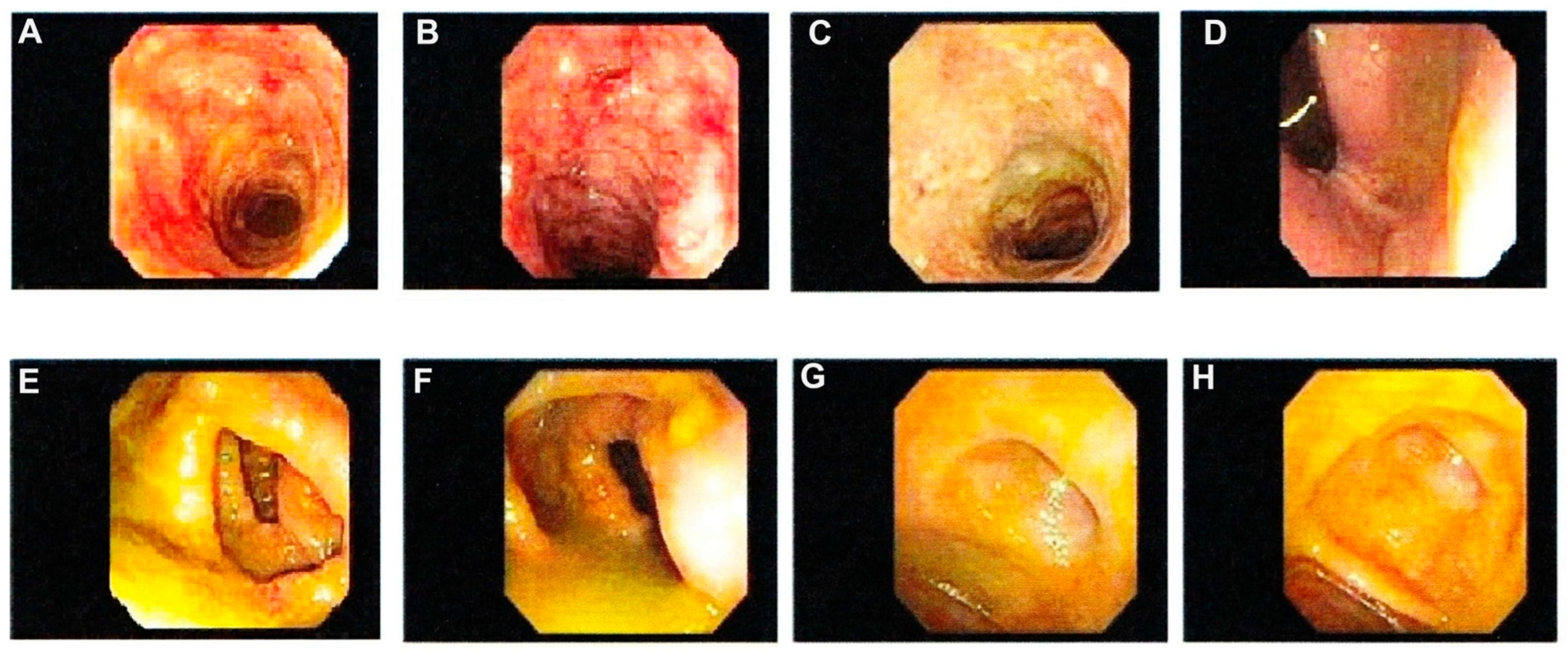
Figure 1. Depicts indeterminate colitis (IC) endoscopy images. IC is diagnosed when transmural lympoid aggregates are present. Inconsistence expiriences have been reported in reviewing the cases of IC. In some reports the disease is most likely to behave clinically like UC [51,52] and much less likely to develop into obvious clinical CC [35] while others found the reverse is true [53]. IC is provisional diagnosed for IBD when transmural lymphoid aggregates are present but it is not possible to definitively classify the disease as CC or UC. (A) fulminant IC of the transverse colon, (B) fulminant IC of the descending colon, (C) IC of the sigmoid colon, (D) IC of rectum (E) IC of the mid-ascending colon, (F) IC of the ileocecal, (G) IC of the appendiceal orifice, (H) IC of the cecum.
Jcm 14 08390 g001
Figure 2. Endoscopic photographs of patients with ulcerative colitis (UC) according to their endoscopic activity. Ulcerative colitis endoscopic index of severity (UCEIS). (a) UCEIS 7, (b) UCEIS 4, (c) UCEIS 2 and (d) UCEIS 0. Reproduced with permission from Lee JW et al., Am J Gastroenterol 120(1):213-224, 2025.
Jcm 14 08390 g002
Figure 3. Endoscopic view of colonic Crohn’s colitis (CC). (AC), view of colonic Crohn’s colitis (CC) with patchy inflammation, ulcers, and a cobblestone appearance of the mucosa; (D), CC of sigmoid colon; (E,F), CC of the Rectum; (G), Ulceration in the Rectum; (H), Rectal stricture. Reproduced with permission from GASTROLAB/SCIENCE Photo Library: Licensee—American African Health Team, LLC, https://americanafricanhealthteam.com/ (accessed on 5 August 2025).
Jcm 14 08390 g003
Figure 4. Healthy Colon. (A) Ileocaecal valve, (B) ascending colon, (C) hepatic flexure of the colon, (D) rectum. Reproduced with permission from GASTROLAB/SCIENCE Photo Library: Licensee—American African Health Team, LLC, https://americanafricanhealthteam.com/ (accessed on 5 August 2025).
Jcm 14 08390 g004
Disclaimer/Publisher’s Note: The statements, opinions and data contained in all publications are solely those of the individual author(s) and contributor(s) and not of MDPI and/or the editor(s). MDPI and/or the editor(s) disclaim responsibility for any injury to people or property resulting from any ideas, methods, instructions or products referred to in the content.