Systematic Review

Bullous Wells’ Syndrome: Case Report and Systematic Review

1
Dermatology Unit, Department of Medical of Surgical Sciences, University of Foggia, Viale L. Pinto, 1, 71122 Foggia, Italy
2
Section of Dermatology, Department of Precision and Regenerative Medicine and Jonian Area, University of Bari Aldo Moro, Piazza Giulio Cesare, 11, 70124 Bari, Italy
3
Section of Molecular Pathology, Department of Precision and Regenerative Medicine and Ionian Area (DiMePRe-J), University of Bari Aldo Moro, Piazza Giulio Cesare, 11, 70124 Bari, Italy
4
Casa di Cura Villa Montallegro, Via Monte Zovetto, 27, 16145 Genoa, Italy
*
Author to whom correspondence should be addressed.
J. Clin. Med. 2025, 14(23), 8370; https://doi.org/10.3390/jcm14238370
Submission received: 28 October 2025 / Revised: 15 November 2025 / Accepted: 22 November 2025 / Published: 25 November 2025
(This article belongs to the Section Dermatology)

Abstract

Background/Objectives: Wells’ syndrome (WS) is an uncommon cutaneous disease with unknown etiology. Itchy bullous lesions and erythematous plaques characterize the bullous WS (BWS), a rare subtype of the syndrome. We describe the case of a woman affected by chronic lymphocytic leukemia who developed BWS and responded to the classic corticosteroid treatment. We also systematically reviewed the literature, analyzing the clinical, laboratory, and histological features and treatments of this rare disease. Methods: We used the databases Ovid MEDLINE, PubMed, and EMBASE with the following search terms: ((bullous Wells’ syndrome [MeSH Terms]) OR (eosinophilic cellulitis)) OR (bullous eosinophilic dermatitis) to identify and compare case reports of BWS. Results: We analyzed 28 patients, including our case. They were primarily female adults with a median age of 44.92 years. Blood eosinophilia was common, and histologically, the tissue samples showed an eosinophilic-neutrophilic dermal infiltrate. From a clinical perspective, the bullae were typically associated with or preceded by other lesions, primarily urticarial plaques, and mainly involved the extremities. Possible triggering agents of BWS include medications, insect bites, malignancies, and autoimmune/infectious diseases. Systemic steroids constituted the first-line treatment. Recent studies described the efficacy of the anti-interleukin-5 monoclonal antibody Mepolizumab in refractory cases of WS. Conclusions: Diagnosis of BWS is often challenging due to the rarity of the disease, clinical polymorphism, and multiple differential diagnoses. Integrating clinical features with laboratory and histopathological findings is essential for achieving a definitive diagnosis. Although the causal link between WS and underlying neoplastic/autoimmune/infective conditions is not always present, this possibility should be taken into account and investigated for the best patient management.

1. Introduction

Wells syndrome (WS) is a rare inflammatory cutaneous disease first described by George Wells in 1971 as a recurrent granulomatous dermatitis with eosinophilia. The disease is also known as eosinophilic cellulitis due to its clinical resemblance to bacterial cellulitis. It affects people of all races, primarily adults, without sex predilection [1,2].
The exact etiology is unknown: among the reported cases, some appear to be idiopathic, but others suggest the presence of triggering events, as long as underlying chronic disorders. Indeed, it is still debated whether WS is a distinct entity or a reaction pattern to different stimuli. The elements known to be triggering factors include: arthropod bites (mosquitoes, fleas, ticks and spiders), cutaneous and/or systemic infections (Parvovirus B19, Herpes simplex viruses, Mumps virus, Toxocara canis and others), drugs (antibiotics, nonsteroidal anti-inflammatory drugs, vaccinations, anti-tumor necrosis factor [TNF]-α and other biologic drugs), haematological and solid organ malignancies and autoimmune diseases (Churg-Strauss syndrome and ulcerative colitis). It has been hypothesized that these conditions can lead to an increased response and migration of eosinophils in the skin [1].
Clinical manifestations are heterogeneous, and histological appearance is typical but not specific. The limbs and the extremities are the site of predilection [1,2].
Seven clinical variants of WS have been categorized based on the prevalent elementary lesions observed during physical examination: plaque-type, annular granuloma-like, urticaria-like, papulovesicular, bullous, papulo-nodular, and fixed drug eruption-like. These variants of WS may present in succession or coexist in the same patient. Plaque-type unilesional presentation mimicking erysipelas has been reported to be more frequent in children than in adults. Itching and burning sensations typically precede the sudden onset of the skin lesions. Systemic symptoms, such as arthralgia and fever, may accompany the cutaneous eruption, which typically has a chronic, relapsing course. The prognosis is good because the disease may resolve spontaneously [1,2]. In adults, erythematous annular lesions resembling annular granuloma were the most frequently documented presentation of WS. The urticaria-like variant is also common and often concurrent with the plaque-type presentation. Other manifestations of WS, like bullae, papulovesicles, and papulo-nodules, are rarer [1,2].
The histological features of WS depend on the time of biopsy and the stage of the disease.
In the very early stages, tissue eosinophilia may be associated with sub-epidermal edema (acute stage, 2–3 days from onset). Later, the eosinophil degranulation is responsible for the development of the typical ‘flame figures’ corresponding to the collagen fiber degeneration caused by the release of toxic substances from the eosinophils (sub-acute stage, week 1–3). The subsequent migration of macrophages into the inflamed dermis and the appearance of giant cells around collagen deposits, produces cutaneous granulomatous infiltrates, eventually resulting in a gradual disappearance of the eosinophilia (regressive phase, 2–8 weeks) [3,4].
The clinical and histopathological heterogeneity of the disease constitutes a diagnostic challenge for both clinicians and pathologists.
The bullous variant is a very rare presentation of WS that is clinically characterized by itchy, tense vesico-bullous lesions often associated with erythematous plaques [4,5]. The disease is complex to diagnose and to distinguish from other bullous dermatoses, especially in the absence of specific laboratory and histological analyses.
In the present work, we describe the case of a woman affected by chronic lymphocytic leukemia (CLL) who developed BWS. In addition, to draw attention to this rare form of WS, we systematically reviewed the literature, analyzing its clinical, laboratory, and histological features, as well as the proposed treatments. To date, only single case reports and a few case series about BWS have been reported and a systematic review on this topic has never been published.

2. Materials and Methods

We performed a systematic electronic literature search following the PRISMA statement (www.prisma-statment.org, accessed on 27 October 2025), Table S1 [6]. Figure 1 presents a graphically altered PRISMA flowchart that includes all relevant information of study selection in a simple visual form. Articles were screened independently by two of us (W.A.R. and G.C) with any conflicts resolved by discussion, or involvement of a third assessor (C.F.) in the case of disagreement.
The last search was conducted on 17 August 2025; results were limited to English and French. We used the databases Ovid MEDLINE, PubMed and EMBASE with the following search terms: ((bullous Wells’ syndrome [MeSH Terms]) OR (eosinophilic cellulitis)) OR (bullous eosinophilic dermatitis) to find and compare reports of cases of BWS. Patients were defined as having BWS and included in our final review if all four of the following criteria were met: (I) signs and symptoms compatible with WS; (II) clinical description of bullous lesions of the skin; (III) laboratory and/or histologic data supporting the diagnosis; (IV) data on the used treatment/treatments. The review has been submitted to the PROSPERO dataset [ID 650076].

3. Results

3.1. Case Report

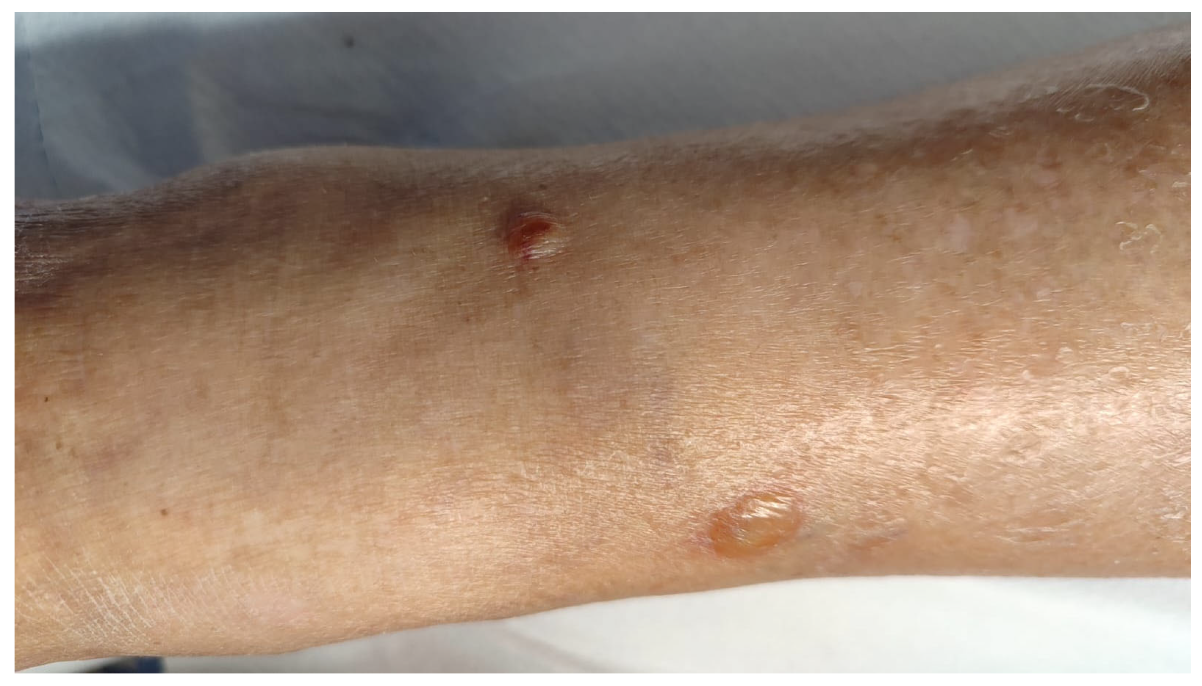
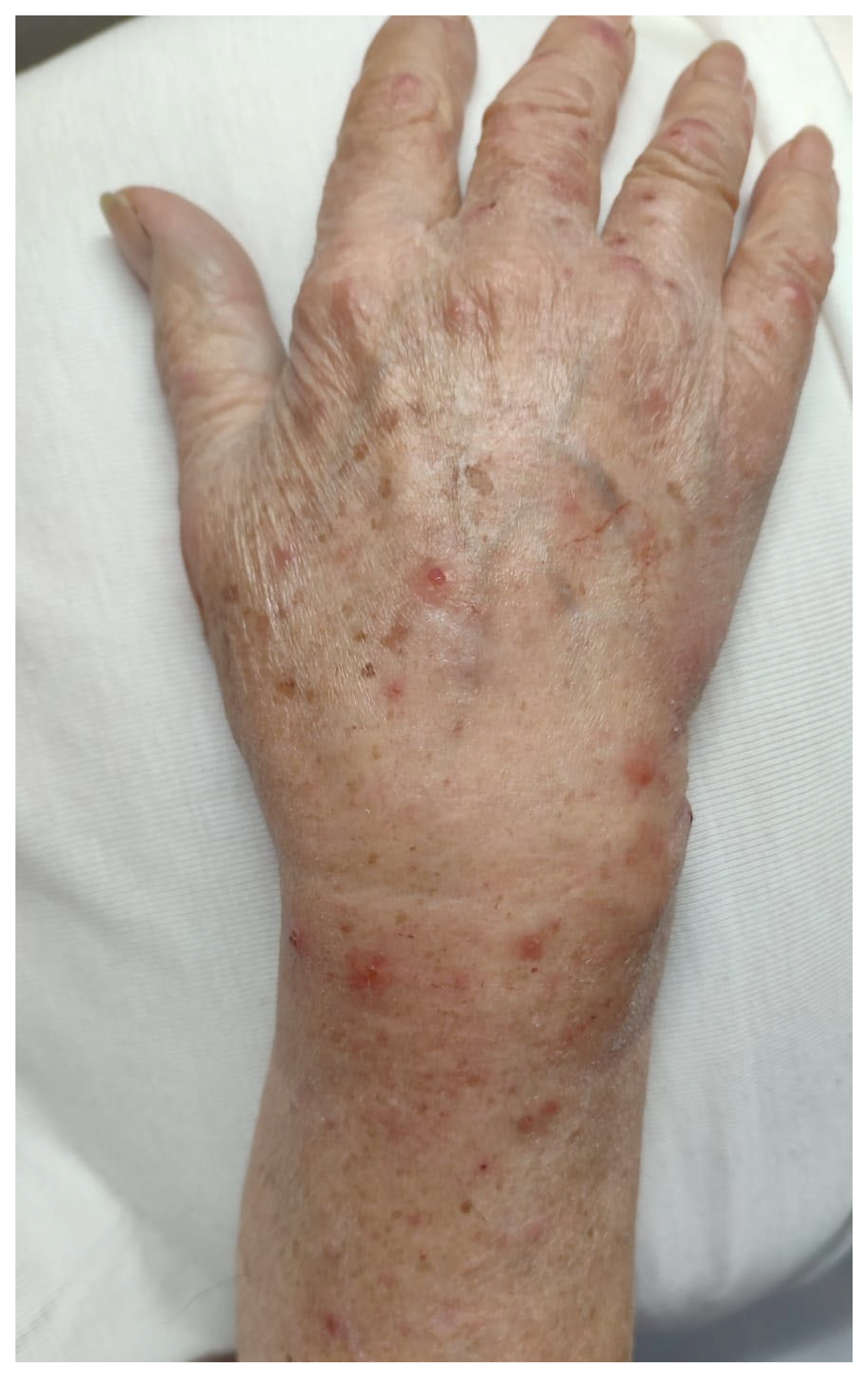
A 90-year-old female presented us with a one-year history of an itchy bullous skin eruption. The patient was affected by T cell CLL (STAGE 0) for which she was in haematological follow-up for the last 2 years. She had no personal family history of dermatological diseases and was otherwise in good health. She wasn’t undergoing any medical treatment. At the physical examination, vesicular, bullous lesions and crusts were observed on the upper and lower limbs. Isolated infiltrated erythematous plaques were also present on both hands and trunk. Laboratory investigations, including blood cell count, electrolytes, C-reactive protein, serum total IgE antibodies, antinuclear antibodies (ANA), anti-extractable nuclear antigen (ENA), anti-double-strand DNA, antibodies against bullous pemphigoid (BP)-180 antigen, and BP230 antigen, were within normal limits, as well as the anti-desmoglein-1 and 3 (Dsg1 and Dsg3) and anti-collagen VII antibodies. Two skin biopsies were performed, one from a bullous lesion of the right leg (Figure 2) and one from an eroded vesicle on the dorsum of the left hand (Figure 3).
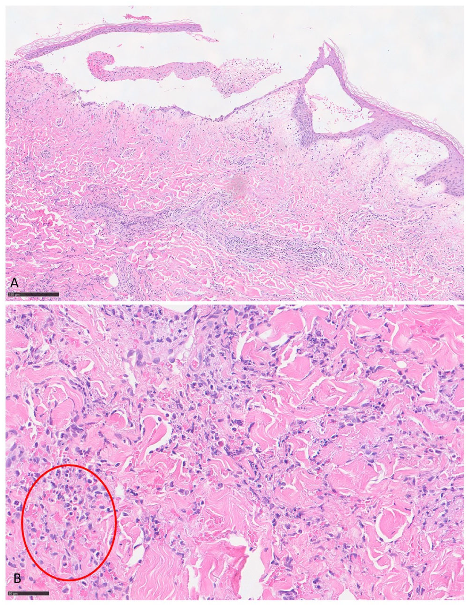
Histology of the first cutaneous sample showed keratinocyte detachment in the spinous layer of the epidermis with a fibrin-granulocytic eosinophilic blister; a marked lympho-eosinophilic inflammatory infiltration was observed in the perivascular area of the superficial and deep dermis. Histology of the second skin sample revealed epidermal vesiculation, focal erosion and marked eosinophilic granulocytic inflammatory infiltration with numerous “flame figures” in the mid-dermis. The histological picture of both samples supports the clinical diagnosis of WS” (Figure 4). Direct immunofluorescence was also performed providing negative results.
Based on the clinical features, laboratory investigations, and histological examination, BWS was diagnosed. After consultation with the hematologist, the patient received treatment with systemic corticosteroids (oral prednisone at an initial dose of 0.5 mg/kg/day), which showed benefits. At the three-month follow-up visit, the patient still presented a few eroded vesicles on the right hand; therefore, a new short cycle of systemic and topical steroids (mometasone furoate 0.1%) was prescribed. To date, the patient is still in follow-up and has not presented new lesions.
The patient gave written informed consent to publish her clinical, histological and laboratory data.

3.2. Systematic Literature Review

After reviewing the titles and abstracts of 956 articles from our initial search, we excluded 913 papers (duplicates, narrative reviews, non-human studies, basic science studies). After reviewing the full text of the remaining 43 articles, ten were excluded because they lacked detailed clinical data on bullous lesions, and six were not in English or French. We therefore analyzed 28 patients, including the case we described herein [3,5,7,8,9,10,11,12,13,14,15,16,17,18,19,20,21,22,23,24,25,26,27,28]. Clinical, laboratory, and histological features and treatments of the collected cases are summarized in Table 1.
The cases of BWS analyzed in our review have been published between 1988 and 2025, mainly by European authors. The majority of patients were female (71%) and the age of onset varied from one to ninety years, with a mean age of 44.9 years at diagnosis (range: 1–90 years). Only 5 cases (18%) referred to children under the age of twelve.
Blood eosinophil count, documented in every published case of BWS, was commonly increased; however, it was within normal ranges (0–530 cells/μL) in 36% of patients.
Conversely, the presence of the typical “flame figures” in the histological reports was almost always detected (75%).
The body areas most commonly affected by the bullous lesions were the upper and/or lower limbs: in 14 out of 28 cases (50%) only the limbs were involved, sometimes with unilateral presentation. A widespread location of the skin lesions was possible (43%), whereas the exclusive involvement of the face and/or neck was less frequent (7%).
The presence of mucosal manifestations was reported in only one of the 28 BWS cases, involving the tongue and throat.
A wide range of cutaneous lesions resulted associated with the bullae: plaques and nodules (75%), vesicles, papules, maculo-papules and purpuric lesions (Table 1). Itching was frequently reported (54%), whereas systemic signs and symptoms (fever or other flu-like symptoms) affected only one-fifth of patients (21%).
Comorbidities characterized by an infectious, inflammatory, or neoplastic etiology were documented in ten out of 28 patients (36%). In some cases of active diseases, they were considered as possible triggers for BWS (53%). More specifically, triggers were represented by infections (tinea pedis and onychomycosis, intestinal disease, upper respiratory tract infection), malignancies (CLL, colon cancer, small cell non Hodgkin’s B lymphoma, nasopharynx carcinoma), drugs (ustekinumab, lincomycin, thiopental, acetylsalicylic acid, pholcodine, tenoxicam, diclofenac sodium, amoxicillin), insect bites and autoimmune disease (Churg Strauss syndrome).
The time elapsed between the skin eruption and the potential trigger factor was highly variable, ranging from a few days for the insect bites to two years for the CLL.
The first-line treatment for BWS was represented by systemic steroids, as for the other variant of WS, in 86% of the patients, in some cases associated with the therapies administered for the underlying conditions (such as terbinafine for the tinea pedis) or with antihistamines and topical steroids in cases of diffuse skin eruption.
The disease often went into remission after treatment; however, in three patients, including ours, BWS had a relapsing course and required further treatment (Table 1 [3,5,7,8,9,10,11,12,13,14,15,16,17,18,19,20,21,22,23,24,25,26,27,28]).

4. Discussion

The diagnosis of BWS is often challenging due to the rarity of the disease and its similarity to other bullous dermatoses with hereditary, autoimmune, infectious, or drug-related etiologies. Indeed, the differential diagnoses of BWS include a wide range of cutaneous disorders such as BP, linear IgA bullous dermatitis, epidermolysis bullosa acquisita, bullous erysipelas, atypical herpes zoster, bullous scabies, bullous drug reaction with eosinophilia and systemic symptoms (DRESS), bullous Sweet syndrome, skin reactions to insect bites, porphyria, pseudoporphyria and others [2,7,29,30,31,32].
Diagnostic criteria for WS were proposed by Heelan et al. in 2013 [33]. These include major criteria (at least two out of four) and minor criteria (at least one). Major criteria are described as follows: (1) any of the reported clinical variants: plaque-type, annular granuloma-like, urticaria-like, papulovesicular, bullous, papulo-nodular, fixed-drug eruption-like; (2) relapsing, remitting course; (3) no evidence of systemic disease; (4) histology showing eosinophilic infiltrates, no vasculitis. Minor criteria include: (1) “flame figures” visible at histology; (2) granulomatous changes evident at histology; (3) peripheral eosinophilia not persistent and not greater than 1500/μL; (4) triggering factors (for example, drug intake) [33,34].
To our knowledge, the present work is the first comprehensive, systematic review of BWS. Indeed, the cases described to date were single case reports and small case series.
Notably, nine out of the 28 BWS cases reported to date were observed in Italy (32%), indicating a high level of awareness and knowledge of this rare disease in our country.
Blood eosinophilia was present to a slightly greater extent in BWS (64%) compared to all the other WS variants (50%) [2,3]. This difference may be explained with the acute nature of bullous lesions compared to other cutaneous manifestations (papules, plaques and nodules); therefore, the BWS cases might have been more frequently diagnosed during the acute or subacute phase, when the eosinophil level was usually high. However, our patient did not exhibit blood eosinophilia, likely because her bullous eruption was long-lasting and her disease in a subacute phase.
In our review, indeed, “flame figures” at histological examination occurred more frequently among BWS (75%) compared to other variants (50%), mainly indicating a subacute disease. As a matter of fact, in later stages the migration of macrophages into the dermis leads to the gradual disappearance of the tissue eosinophilia, to the absence of “flames figures” and to the formation of dermal granulomatous infiltrates [4,5,27]. Therefore, we can assume that the more frequent detection of “flame figures” in BWS compared to other WS forms may be related to the earlier execution of skin biopsy in the bullous variant of the disease.
As for clinical presentation, most BWS cases (75%) were associated with erythematous and sometimes urticarial, plaques and vesicles; skin lesions predominantly involved the lower and/or upper extremities (93%) and were often itchy (68%). The possible involvement of the oropharyngeal mucosa in patients with WS has been investigated only by a few authors, likely because few dermatologists are aware of its occurrence, as it may happen in the context of other dermatoses [30,35]. Notably, only one patient with nodular mucosal lesions of the tongue and throat, associated with diffuse skin lesions, has been reported [24].
In our review, possible triggering agents have been identified in about half of the patients, with variable timings from the trigger to BWS onset, ranging from a few days to 10 months. This aspect suggests that the association may sometimes be coincidental, although a causal link between the triggering factor and the development of BWS can be assumed when drug discontinuation led to resolution without subsequent relapses [9,22].
The association between BWS and malignancies has been documented in a consistent percentage of patients (14%). In some cases, the diagnosis of cancer and BWS were concomitant; in others, the malignancies preceded the eosinophilic disease by 1–2 years (Table 1). The pathogenic link between WS and hematologic diseases is not entirely understood. Some authors have speculated that in patients with an immunodeficiency due to hematologic disease, a trigger, such as a drug or a virus, induces cytokine production with an excess of interleukin (IL)-4 and IL-5, resulting in an altered immune response with eosinophil predominance [36,37]. One of the key events in the disease expression of WS is indeed the aberrant accumulation of eosinophils in the skin. Increased IL-5 levels observed in WS not only mobilize eosinophils from the bone marrow but also promote the homing of eosinophils by altering the expression of adhesion molecules. Tissue eosinophilia is commonly seen in cutaneous T-cell lymphoma. It has been hypothesized that the production of an eosinophil chemotactic factor from neoplastic lymphocytes may cause WS in patients with CLL [38]. Given the potential association with cancers, following a diagnosis of WS (and also BWS), we recommend obtaining an accurate medical history and conducting a detailed clinical examination to screen for underlying disorders through routine blood investigations. To date, an evidence-based malignancy screening protocol for WS patients that optimizes the risk-to-benefit ratio does not exist. Therefore, we propose that screening tests should be performed at the onset of eosinophilic disease, according to clinical findings, the patient’s medical history, age, and personal and family history of cancer [39].
Concerning the treatment, although WS may be limited to a restricted body area and may spontaneously remit, its widespread distribution and chronic-remitting course often require therapy. In all the cases of BWS that we reviewed, including our patient, systemic steroids constituted the first-line treatment, mainly administered orally but also intravenously and topically [5,7,8,10,11,12,13,14,15,16,17,18,19,20,21,22,23,24,25,26,27,28], as reported in the other variants of WS (Table 1). In patients suffering from recurrent disease, several steroid-sparing agents have been used, such as oral dapsone and cyclophosphamide.
Interestingly, only few cases of recalcitrant WS treated with Omalizumab (an anti-IgE monoclonal antibody) have been reported so far [40,41,42]. Recently, our research group documented the first patient with BWS who was successfully treated with this drug, achieving long-term remission [27]. Binding free IgE, Omalizumab downregulates the expression of the high-affinity IgE receptor (FcεRI) on mast cells and basophils. This receptor is expressed and upregulated also on skin-infiltrating eosinophils in eosinophilic conditions, such as atopic dermatitis and BP [43,44,45], which explains the successful treatment with Omalizumab in these diseases. Similarly, we can hypothesize that the overexpression of dermal eosinophils in WS upregulates FcεRI, which Omalizumab subsequently downregulates, leading to the clinical remission of the disease [41,42]. Moreover, the reduction in the level of different cytokines causing the recruitment, activation and survival of eosinophils (IL-5, IL-13, IL-4, IL-2, GM-CSF, IL-8) may also explain the anti-inflammatory efficacy of Omalizumab [44,45].
Recent studies have described the efficacy of another humanized monoclonal antibody in refractory cases of WS, specifically the anti-IL-5 Mepolizumab [46,47]. Indeed, this drug modulates IL-5, one of the central cytokines involved in eosinophil chemotaxis, maturation, and survival. Mepolizumab, therefore, acts to reduce eosinophil levels in the blood and lesional skin, thereby mitigating the cutaneous signs and symptoms of WS [46,47]. No cases of the bullous subtype of WS treated with Mepolizumab have been described to date; however, this treatment, together with Omalizumab, may represent a new therapeutic strategy in cases of relapsing disease, especially when it is necessary to limit the use of corticosteroids.
Mild and transient side effects have been occasionally reported following Omalizumab (slight fatigue and loose bowel movements a few days after injection [42]) and Mepolizumab treatment (headache, back pain, and reactions at the site of injection [47]). However, these adverse effects did not cause drug discontinuation. We therefore suggest that these monoclonal antibodies should be considered as second-line treatments in cases of WS that are refractory to first-line corticosteroid therapy or in cases where the side effects of corticosteroids outweigh their benefits.

5. Conclusions

Our article adds a comprehensive review of the clinical, laboratory, histologic, and therapeutic features of BWS to the current literature. Indeed, beyond single case reports and small case series, no systematic review has been published to date specifically addressing this topic.
In the presence of cutaneous bullous lesions, primarily when associated with other manifestations like erythematous papules, plaques and vesicles, after excluding common cutaneous bullous diseases (bullous erysipelas and BP), the diagnosis of BWS should not be neglected. Our review found that blood eosinophilia and the presence of “flame figures” in skin samples are more common in BWS compared to the other WS variants. Therefore, measuring total IgE in the patient’s serum and searching for the dermal eosinophilic infiltrate and “flame figures” in the skin tissue may help clinicians and pathologists achieve a definite diagnosis, which always derives from integrating the clinical features with laboratory and histopathological findings.
Although the causal link between WS and underlying neoplastic/autoimmune/infective conditions is not always evident, this possibility should be taken into account and investigated for the best patient management.
Our work may support the efficacy of systemic steroids as first-line treatment for BWS, but it also highlights that monoclonal antibodies targeting IgE and IL-5, key molecules involved in the pathogenesis of WS, are emerging as effective therapies for relapsing cases. Based on the results presented in our review, especially those regarding the most recently published case reports [33,46,47], we propose that Omalizumab and Mepolizumab may be promising treatment options for refractory WS, including its bullous subtype. Further studies on treating this rare disease with such drugs are warranted.
The limitations of case report-based systematic reviews include the fact that milder cases are less likely to be reported in the literature. Therefore, the sample size, especially in rare diseases like BWS, may be small, which limits the possibility of generalizing the study’s validity.

Supplementary Materials

The following supporting information can be downloaded at: https://www.mdpi.com/article/10.3390/jcm14238370/s1, Table S1: “PRISMA checklist”.

Author Contributions

Conceptualization, G.C. (Giulia Ciccarese), W.A.R., F.D. and C.F.; methodology, G.C. (Giulia Ciccarese), L.L., W.A.R. and C.F.; formal analysis, G.C. (Gerardo Cazzato), A.R.M., G.S., G.L. and A.D.M.; investigation, G. L., G.C. (Gerardo Cazzato) and A.D.M.; resources, W.A.R., A.R.M. and D.B.; data curation, G.S., L.L., F.D. and R.F.; writing—original draft preparation: G.C. (Giulia Ciccarese), L.L., A.D.M. and R.F.; writing—review and editing, G.C. (Giulia Ciccarese), F.D., G.S. and D.B.; visualization, G.L., L.L., A.R.M., G.L. and D.B.; supervision, C.F., L.L., G.C. (Giulia Ciccarese), G.C. (Gerardo Cazzato) and R.F. All authors have read and agreed to the published version of the manuscript.

Funding

This research received no external funding.

Institutional Review Board Statement

The study was conducted in accordance with the Declaration of Helsinki. Ethical review and approval were waived for this study due to the fact that is a systematic review of the already available literature.

Informed Consent Statement

Even if the described patient cannot be identified, written informed consent has been obtained from the patient to publish this paper.

Data Availability Statement

Data available on reasonable request.

Conflicts of Interest

The authors declare no conflicts of interest.

Abbreviations

The following abbreviations are used in this manuscript:
WSWells’ syndrome
BWSBullous Wells’ syndrome
ILInterleukin
DRESSDrug reaction with eosinophilia and systemic symptoms
BPBullous pemphigoid

References

  1. Long, H.; Zhang, G.; Wang, L.; Lu, Q. Eosinophilic Skin Diseases: A Comprehensive Review. Clin. Rev. Allergy Immunol. 2016, 50, 189–213. [Google Scholar] [CrossRef]
  2. Marzano, A.V.; Genovese, G. Eosinophilic Dermatoses: Recognition and Management. Am. J. Clin. Dermatol. 2020, 21, 525–539. [Google Scholar] [CrossRef]
  3. Caputo, R.; Marzano, A.V.; Vezzoli, P.; Lunardon, L. Wells syndrome in adults and children: A report of 19 cases. Arch. Dermatol. 2006, 142, 1157–1161. [Google Scholar] [CrossRef]
  4. Weins, A.B.; Biedermann, T.; Weiss, T.; Weiss, J.M. Wells syndrome. J. Dtsch. Dermatol. Ges. 2016, 14, 989–993. [Google Scholar] [CrossRef]
  5. Spinelli, M.; Frigerio, E.; Cozzi, A.; Garutti, C.; Garavaglia, M.; Altomare, G. Bullous Wells’ syndrome associated with non-Hodgkin’s lymphocytic lymphoma. Acta Derm. Venereol. 2008, 88, 530–531. [Google Scholar] [CrossRef] [PubMed]
  6. Page, M.J.; McKenzie, J.E.; Bossuyt, P.M.; Boutron, I.; Hoffmann, T.C.; Mulrow, C.D.; Shamseer, L.; Tetzlaff, J.M.; Akl, E.A.; Brennan, S.E.; et al. The PRISMA 2020 statement: An updated guideline for reporting systematic reviews. BMJ 2021, 372, 71. [Google Scholar] [CrossRef] [PubMed]
  7. Feliciani, C.; Motta, A.; Tortorella, R.; De Benedetto, A.; Amerio, P.; Tulli, A. Bullous Wells syndrome. J. Eur. Acad. Dermatol. Venereol. 2006, 20, 1021–1022. [Google Scholar] [CrossRef] [PubMed]
  8. Peckruhn, M.; Elsner, P.; Tittelbach, J. Bullous Wells Syndrome. Dtsch. Arztebl. Int. 2019, 116, 470. [Google Scholar] [CrossRef]
  9. Kim, Y.; Bae, K.N.; Son, J.H.; Shin, K.; Kim, H.; Ko, H.-C.; Kim, B.; Kim, M.-B. Bullous Wells Syndrome Induced by Ustekinumab. Ann. Dermatol. 2023, 35, S180–S181. [Google Scholar] [CrossRef]
  10. Guglielmo, A.; Filippi, F.; Pileri, A.; Misciali, C.; Bardazzi, F. Bullous Wells Syndrome: A needle in the haystack. Int. J. Dermatol. 2020, 60, e150–e153. [Google Scholar] [CrossRef]
  11. Lieberman, J.A. Wells Syndrome with Bullous Lesions. J. Allergy Clin. Immunol. Pract. 2017, 5, 1410. [Google Scholar] [CrossRef] [PubMed]
  12. Lim, C.E.; Ng, S.K.; Thng, S.T. Bullous Presentation of Idiopathic Wells Syndrome (Eosinophilic Cellulitis). Ann. Acad. Med. Singap. 2017, 46, 324–326. [Google Scholar] [CrossRef] [PubMed]
  13. Katoulis, A.C.; Bozi, E.; Samara, M.; Kalogeromitros, D.; Panayiotides, I.; Stavrianeas, N.G. Idiopathic bullous eosinophilic cellulitis (Wells’ syndrome). Clin. Exp. Dermatol. 2009, 34, e375–e376. [Google Scholar] [CrossRef] [PubMed]
  14. Shams, M.; Hudgens, J.; Lesher, J.L., Jr.; Florentino, F. Wells’ syndrome presenting as a noninfectious bullous cellulitis in a child. Pediatr. Dermatol. 2012, 29, 224–226. [Google Scholar] [CrossRef]
  15. Verma, P.; Singal, A.; Sharma, S. Idiopathic bullous eosinophilic cellulitis (Wells syndrome) responsive to topical tacrolimus and antihistamine combination. Indian J. Dermatol. Venereol. Leprol. 2012, 78, 378–380. [Google Scholar] [CrossRef]
  16. Kamiyama, T.; Watanabe, H.; Sueki, H. Bullous eosinophilic cellulitis with subcorneal pustules. Indian J. Dermatol. Venereol. Leprol. 2015, 81, 301–303. [Google Scholar] [CrossRef]
  17. Soua, Y.; Akkari, H.; Sriha, B.; Belhadjali, H.; Zili, J. Lésions bulleuses des poignets [Bullous lesions on the wrists]. Ann. Dermatol. Venereol. 2014, 141, 717–719. [Google Scholar] [CrossRef]
  18. Schuttelaar, M.L.; Jonkman, M.F. Bullous eosinophilic cellulitis (Wells’ syndrome) associated with Churg-Strauss syndrome. J. Eur. Acad. Dermatol. Venereol. 2003, 17, 91–93. [Google Scholar] [CrossRef]
  19. Gilliam, A.E.; Bruckner, A.L.; Howard, R.M.; Lee, B.P.; Wu, S.; Frieden, I.J. Bullous “cellulitis” with eosinophilia: Case report and review of Wells’ syndrome in childhood. Pediatrics 2005, 116, e149–e155. [Google Scholar] [CrossRef]
  20. Moon, S.H.; Shin, M.K. Bullous Eosinophilic Cellulitis in a Child Treated with Dapsone. Pediatr. Dermatol. 2013, 30, e46–e47. [Google Scholar] [CrossRef]
  21. Ferrier, M.C.; Janin-Mercier, A.; Souteyrand, P.; Bourges, M.; Hermier, C. Eosinophilic cellulitis (Wells’ syndrome): Ultrastructural study of a case with circulating immune complexes. Dermatologica 1988, 176, 299–304. [Google Scholar] [CrossRef] [PubMed]
  22. Seçkin, D.; Demirhan, B. Drugs and Wells’ syndrome: A possible causal relationship? Int. J. Dermatol. 2001, 40, 138–140. [Google Scholar] [CrossRef] [PubMed]
  23. Arca, E.; Köse, O.; Karslioğlu, Y.; Taştan, H.B.; Demïrïz, M. Bullous eosinophilic cellulitis succession with eosinophilic pustular folliculitis without eosinophilia. J. Dermatol. 2007, 34, 80–85. [Google Scholar] [CrossRef] [PubMed]
  24. Ling, T.C.; Antony, F.; Holden, C.A.; Al-Dawoud, A.; Coulson, I. Two cases of bullous eosinophilic cellulitis. Br. J. Dermatol. 2002, 146, 160–161. [Google Scholar] [CrossRef]
  25. Li, M.; Liu, Q.; Chen, X. Bullous Wells syndrome in a patient with occult nasopharyngeal carcinoma. Lancet Oncol. 2025, 26, e122. [Google Scholar] [CrossRef]
  26. Bao, W.W.; Chow, E.Y. Breaking the Blister: A Case Report of Bullous Wells’ Syndrome Resolved with Oral Terbinafine. Cureus 2025, 17, e80060. [Google Scholar] [CrossRef]
  27. Ciccarese, G.; Rosato, W.A.; Drago, F.; Meduri, A.R.; Ambrogio, F.; De Marco, A.; Cazzato, G.; Foti, C. Bullous Wells’ Syndrome Successfully Treated with Omalizumab. JDDG J. Dtsch. Dermatol. Ges. 2025, in press. [Google Scholar]
  28. Papaetis, G.S.; Politou, V.N.; Panagiotou, S.M.; Georghiou, A.A.; Antonakas, P.D. Recurrent Cellulitis-Like Episodes of the Lower Limbs and Acute Diarrhea in a 30-Year-Old Woman: A Case Report. Am. J. Case Rep. 2021, 22, e932732. [Google Scholar] [CrossRef]
  29. Drago, F.; Cogorno, L.; Agnoletti, A.F.; Ciccarese, G.; Parodi, A. A retrospective study of cutaneous drug reactions in an outpatient population. Int. J. Clin. Pharm. 2015, 37, 739–743. [Google Scholar] [CrossRef]
  30. Drago, F.; Ciccarese, G.; Merlo, G.; Trave, I.; Javor, S.; Rebora, A.; Parodi, A. Oral and cutaneous manifestations of viral and bacterial infections: Not only COVID-19 disease. Clin. Dermatol. 2021, 39, 384–404. [Google Scholar] [CrossRef]
  31. Drago, F.; Ciccarese, G.; Agnoletti, A.F.; Sarocchi, F.; Parodi, A. Neuro sweet syndrome: A systematic review. A rare complication of Sweet syndrome. Acta Neurol. Belg. 2017, 117, 33–42. [Google Scholar] [CrossRef] [PubMed]
  32. Drago, F.; Herzum, A.; Ciccarese, G.; Broccolo, F.; Rebora, A.; Parodi, A. Acute pain and postherpetic neuralgia related to Varicella zoster virus reactivation: Comparison between typical herpes zoster and zoster sine herpete. J. Med. Virol. 2019, 91, 287–295. [Google Scholar] [CrossRef] [PubMed]
  33. Heelan, K.; Ryan, J.F.; Shear, N.H.; Egan, C.A. Wells syndrome (eosinophilic cellulitis): Proposed diagnostic criteria and a literature review of the drug-induced variant. J. Dermatol. Case Rep. 2013, 7, 113–120. [Google Scholar] [CrossRef] [PubMed]
  34. Heinig, B.; Vojvocic, A.; Lotti, T.; Tirant, M.; Wollina, U. Wells Syndrome—An Odyssey. Open Access Maced. J. Med. Sci. 2019, 7, 3002–3005. [Google Scholar] [CrossRef]
  35. Ciccarese, G.; Broccolo, F.; Rebora, A.; Parodi, A.; Drago, F. Oropharyngeal lesions in pityriasis rosea. J. Am. Acad. Dermatol. 2017, 77, 833–837.e4. [Google Scholar] [CrossRef]
  36. Plötz, S.G.; Abeck, D.; Behrendt, H.; Simon, H.U.; Ring, J. Eosinophilic cellulitis (Wells syndrome). Hautarzt 2000, 51, 182–186. [Google Scholar]
  37. Shin, D.; Kim, D.Y. Chronic relapsing eosinophilic cellulitis associated, although independent in severity, with chronic lymphocytic leukemia. J. Eur. Acad. Dermatol. Venereol. 2016, 30, 159–161. [Google Scholar] [CrossRef]
  38. Gambichler, T.; Othlinghaus, N.; Rotterdam, S.; Altmeyer, P.; Stücker, M. Impetiginized Wells’ syndrome in a patient with chronic lymphocytic leukaemia. Clin. Exp. Dermatol. 2009, 34, e274–e275. [Google Scholar] [CrossRef]
  39. Santos, R.P.; Carvalho, S.D.; Ferreira, O.; Brito, C. Wells syndrome associated with lung cancer. BMJ Case Rep. 2017, 2017. [Google Scholar] [CrossRef]
  40. Coattrenec, Y.; Ibrahim Yasmine, L.; Harr, T.; Spoerl, D.; Jandus, P. Long-term Remission of Wells Syndrome With Omalizumab. J. Investig. Allergol. Clin. Immunol. 2020, 30, 58–59. [Google Scholar] [CrossRef]
  41. Egeland, Ø.; Balieva, F.; Undersrud, E. Wells syndrome: A case of successful treatment with omalizumab. Int. J. Dermatol. 2018, 57, 994–995. [Google Scholar] [CrossRef]
  42. Kaplan, A.P.; Giménez-Arnau, A.M.; Saini, S.S. Mechanisms of action that contribute to efficacy of omalizumab in chronic spontaneous urticaria. Allergy 2017, 72, 519–533. [Google Scholar] [CrossRef]
  43. Tanaka, Y.; Takenaka, M.; Matsunaga, Y.; Okada, S.; Anan, S.; Yoshida, H.; Ra, C. High affinity IgE receptor (Fc epsilon RI) expression on eosinophils infiltrating the lesions and mite patch tested sites in atopic dermatitis. Arch. Dermatol. Res. 1995, 287, 712–717. [Google Scholar] [CrossRef] [PubMed]
  44. Messingham, K.N.; Holahan, H.M.; Frydman, A.S.; Fullenkamp, C.; Srikantha, R.; A Fairley, J. Human eosinophils express the high affinity IgE receptor, FcεRI, in bullous pemphigoid. PLoS ONE 2014, 9, e107725. [Google Scholar] [CrossRef] [PubMed]
  45. Perotin, J.M.; Barnig, C. Mécanismes d’action de l’omalizumab: Au-delà de l’action anti-IgE [Omalizumab: Beyond anti-IgE properties]. Rev. Mal. Respir. 2017, 34, 121–133. [Google Scholar] [CrossRef] [PubMed]
  46. Terhorst-Molawi, D.; Altrichter, S.; Röwert, J.; Magerl, M.; Zuberbier, T.; Maurer, M.; Bergmann, K.C.; Metz, M. Effective treatment with mepolizumab in a patient with refractory Wells syndrome. J. Dtsch. Dermatol. Ges. 2020, 18, 737–739. [Google Scholar] [CrossRef]
  47. Herout, S.; Bauer, W.M.; Schuster, C.; Stingl, G. Eosinophilic cellulitis (Wells syndrome) successfully treated with mepolizumab. JAAD Case Rep. 2018, 4, 548–550. [Google Scholar] [CrossRef]
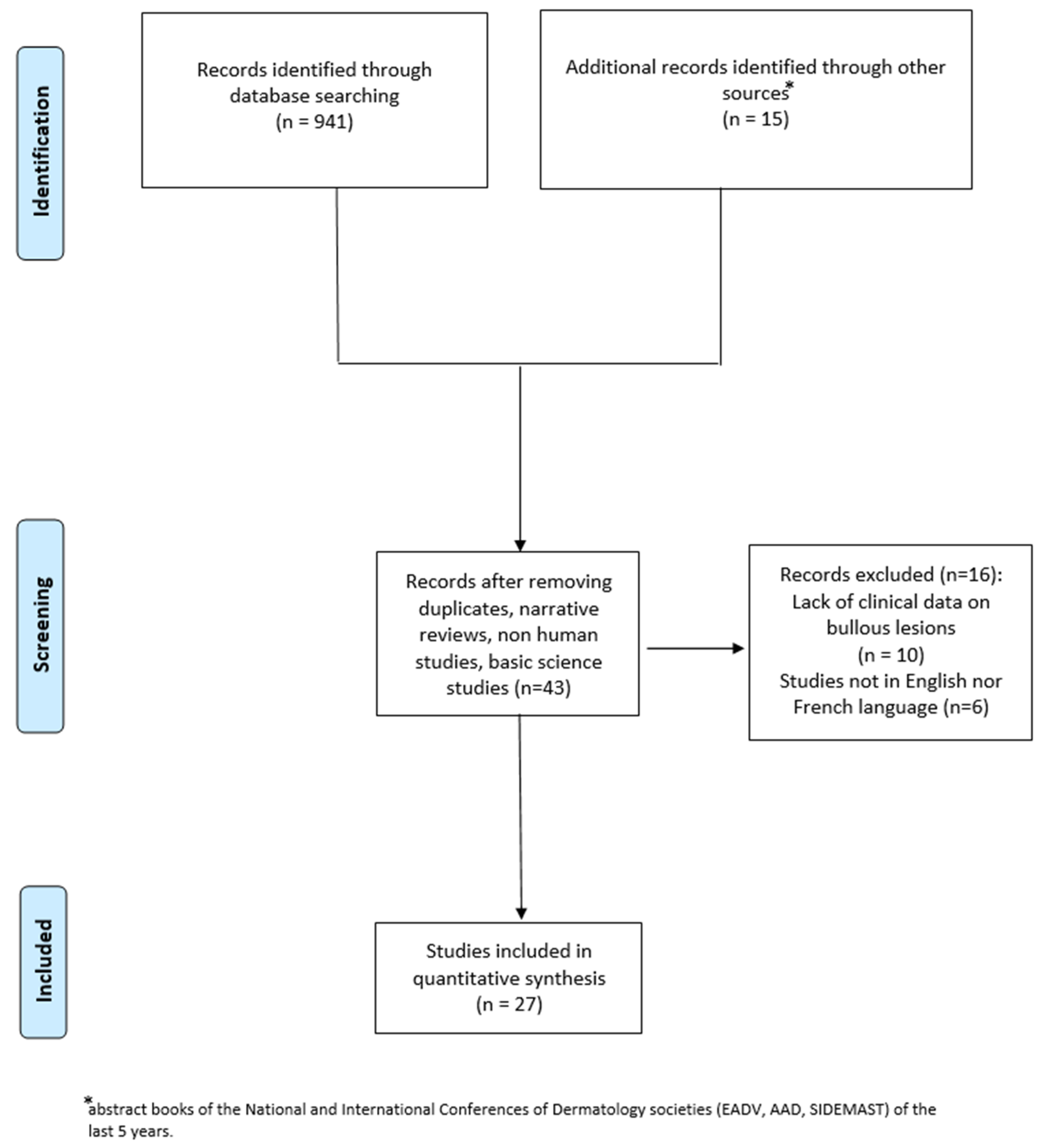
Figure 1. Selection of studies.
Jcm 14 08370 g001
Figure 2. Bullous lesions of the right leg.
Jcm 14 08370 g002
Figure 3. Erythematous papules and eroded vesicles on the dorsum of the left hand.
Jcm 14 08370 g003
Figure 4. (A) Histology showed keratinocyte detachment in the spinous layer of the epidermis with formation of a fibrin-granulocytic eosinophilic blister (Hematoxylin-Eosin, 10× magnification); (B) a marked chronic lympho-eosinophilic inflammatory infiltration was observed in the perivascular area of the superficial and deep dermis (Hematoxylin-Eosin, 40× magnification); “flame figures” correspond to an amorphous eosinophilic structure composed of proteinaceous material and cellular debris, surrounded by degenerated eosinophils and histiocytes (red circle).
Jcm 14 08370 g004
Table 1. Clinical, laboratory, and histological features and treatments of the cases of BWS. WNR: within normal ranges; INR: information not reported; normal range for eosinophilic blood count: 0–530 cells/µL; 0–4.4%.
ReferencesCountryAgeSexBlood Eosinophils (Cells/µL) (%)Other Cutaneous SignsInvolved SitesSkin
Symptoms
Mucosal InvolvementSystemic InvolvementComorbiditiesHistological Flames FiguresPossible TriggerTime from Trigger to EruptionTreatmentOutcomeFollow up (Months)
Our patientItaly90FWNRvesiclesLimbsitchingnonochronic lymphocytic leukemiayeschronic lymphocytic leukemia2 yearsoral steroid (prednisolone 25 mg/day), mometasone furoate cream (twice daily)relapsing disease then remission6
Bao et al. (2025) [26]Canada52F1100macules, papulesfeetitchingINRINRepilepsy and osteoarthrosisyestinea pedis, onychomycosis-oral terbinafine (250 mg/day) for 3 months; ciclopirox cream, bilastine (40 mg twice day), prednisone(30 mg/day) for five daysremission18
Ciccarese et al. (2025) [27]Italy36F1770 (18%)urticarial plaques,
vesicles
trunk, lower limbsitchingnononoyes--Oral steroid (prednisone 0.5 mg/kg/day), Omalizumab 300 mg/4 weeksremission36
Peckruhn et al. (2019) [8]Germany5F8850plaques, nodulesFeetPainINRINRINRyesInsect bite14–21 daysOral steroid (prednisolone 100 mg/day), dapsone (50 mg/day), dimetindene twice dailyremission<1
Kim et al. (2023) [9]Sud Korea64FWNRpatchesLeft limbsItching/burning sensationINRINRPsoriasisyesUstekinumab10 daysDrug discontinuationremissionINR
Papaetis et al. (2021) [28]Cyprus30FWNRpapules, nodules, plaques,
urticaria, vesicles
left lower limbitchingINRfeverC. difficile intestinal
infection
yesintestinal infection-mometasone furoate cream (twice day), levocetirizine (10 mg day) for 1 monthremission24
Guglielmo et al. (2020) [10]Italy82MWNRpatches, plaques,
pustules
LimbsINRINRINRnoyes--Oral methylprednisolone
(0.5 mg/kg/day)
remissionINR
Guglielmo et al. (2020) [10]Italy70FWNRplaquesLimbsPainINRINRDiabetesyesInsect bite7 daysOral methylprednisolone
(0.5 mg/kg/day)
remissionINR
Lieberman JA (2017) [11]Tennessee11M730patches, plaquesLower limbINRINRINRINRINRInsect bite2 daysOral steroids, topical mupirocinremissionINR
Feliciani et al. (2006) [7]Italy88FWNRplaques, vesiclestrunk, folds, limbsItching/burning
sensation
INRINRColon carcinomaINRcolon carcinoma10 monthsOral methylprednisolone (40 mg/day)remission36
Lim et al. (2017) [12]Singapore44F810papules, plaques, vesiclesface, trunk,
upper limbs
ItchingINRnonoINR--Oral prednisolone (20 mg/day)remission6
Katoulis et al. (2009) [13]Greece64F700plaques, vesiclesneckItchingINRINRUterine
fibromyomas, osteoporosis
no--Topical steroidremission12
Shams et al. (2012) [14]Louisiana11F3129 (21%)plaques, erosions, crustsface, neck,
upper limbs
ItchingINRFevernoyesInsect bite1 monthIntravenous steroidINRINR
Verma et al. (2012) [15]India40MWNRplaques, vesiclestrunk, upper
limbs
Itchingnononoyes--Oral antihistamine, topical
tacrolimus
remission6
Kamiyama et al. (2015) [16]Japan39F1260 (15%)plaques, vesicles, pustulestrunk, upper and lower
limbs
Itching, tendernessINRnonoyes--Oral prednisolone (0.5 mg/kg/day), fexofenadine 60 mg twice daily, topical steroidremission6
Soua et al. (2014) [17]Tunisia61FWNRvesiclesupper limbsItchingnononoyes--Oral prednisolone (40
mg/day), antihistamine 10 mg/day
remissionINR
Spinelli et al. (2008) [5]Italy73F1740 (14.3%)purpuralower limbsItchingINRFever, lymph node
swelling
sigma adenocarcinoma 2 years earlieryesSmall-cell non-Hodgkin’s B lymphomaconcomitant diagnosisOral methylprednisolone (20 mg/day), antihistamine, topical fusidic acid creamremissionINR
Schuttelaar et al. (2003) [18]Netherlands55M5780 (34%)plaques, vesicles, purpuralimbsItching, painINRMalaise, fever, arthralgiaNasal polyposis, asthma, peripheral eosinophiliayesChurg-Strauss syndrome-Intravenous dexamethasone (200 mg for 3 days), Doxycycline (100 mg twice daily), cyclophosphamide (150 mg/day)remissionINR
Gilliam et al. (2005) [19]USA1F14,400 (48%)plaqueslower limbsItchingINRnonoyes--Oral steroid (2 mg/kg), topical
steroid
remission12
Moon et al. (2013) [20]Sud Korea9M4770 (24.5%)vesiclestrunk, limbsItchingINRnonoyesUpper respiratory
infection
21 daysOral prednisolone (40 mg/day), cetirizine, dapsone, topical steroidremissionINR
Caputo et al. (2006) [3]Italy64F(32%)notrunk, lower extremitiesINRINRINRnono--Oral betamethasone sodium phosphate (4 mg/day),
oral amoxicillin
remission120
Caputo et al. (2006) [3]Italy34F(19%)plaquesfaceINRINRINRnono--Oral betamethasone
sodium phosphate (4 mg/day), ceftriaxone sodium (2 g/day)
relapsing disease60
Ferrier et al. (1988) [21]France42F1460 (8%)plaquesface, trunk, upper limbsItchingINRnoINRyeslincomycin, thiopental, acetylsalicylic c acid,
pholcodine
-Oral betamethasone (8 mg/day), dapsonerelapsing disease then remission48
Seçkin et al. (2001) [22]Turkey28F990 (16.5%)papules, vesicles, crustsLimbsItchingINRINRINRyestenoxicam and diclofenac sodium and/or
amoxicillin
-Oral prednisolone(40 mg/day)remissionINR
Arca et al. (2007) [23]Turkey20MWNRvesicles, pustules, plaqueslimbsItchingINRnonoyes--Oral prednisone (60 mg/kg), tetracycline 500 mg twice dailyremission12
Ling et al. (2002) [24]UK45F6180 (46.5%)plaquesface, trunk, upper limbsINRINRnonono--Oral prednisolone (30 mg/day), cetirizine (10 mg
twice day)
remission12
Ling et al. (2002) [24]UK42MWNRplaquesface, limbsPaintongue, throatInfluenza-like illnessINRyes--Oral prednisolone(40 mg/day)lost to follow-uplost to follow up
Li M et al. (2025) [25]China58M750papules, plaques, vesiclesneck, trunk,
upper limbs
INRINRfever, lymph
node swelling
nasopharynx
carcinoma
yesnasopharynx
carcinoma
concomitant
diagnosis
Oral prednisone (15 mg/day)remissionINR
Disclaimer/Publisher’s Note: The statements, opinions and data contained in all publications are solely those of the individual author(s) and contributor(s) and not of MDPI and/or the editor(s). MDPI and/or the editor(s) disclaim responsibility for any injury to people or property resulting from any ideas, methods, instructions or products referred to in the content.