Review

Pulmonary Sequestration in Adults: Endovascular and Hybrid Treatment Strategies—A Systematic Review

Department of Interventional Radiology, Heart and Vascular Center, Semmelweis University, 1122 Budapest, Hungary
*
Author to whom correspondence should be addressed.
J. Clin. Med. 2025, 14(21), 7493; https://doi.org/10.3390/jcm14217493
Submission received: 28 July 2025 / Revised: 30 September 2025 / Accepted: 21 October 2025 / Published: 23 October 2025
(This article belongs to the Section Vascular Medicine)

Abstract

Background and Objectives: Pulmonary sequestration (PS) is a rare congenital lung malformation. In adults, intralobar disease with recurrent infection or hemoptysis predominates. Cross-sectional imaging (CTA/MRA) is central to mapping the aberrant systemic supply; catheter angiography is used when noninvasive imaging is inconclusive or when an endovascular procedure is planned. We aimed to synthesize adult PS cases treated with endovascular or hybrid approaches and to summarize case selection, techniques, and outcomes. Methods: We conducted a PRISMA-2020-informed systematic review. We searched PubMed and Scopus from 1 January 2000 to 31 May 2025. Two reviewers extracted data independently; due to heterogeneity, we performed a narrative synthesis and a JBI-adapted qualitative risk-of-bias appraisal. Eligible studies enrolled adults (≥18 years) with imaging-confirmed PS treated with embolization, stent-graft exclusion, or hybrid therapy; prespecified outcomes included technical and clinical success, complications, recurrence, and re-intervention. The review was not registered. Results: Of 93 records screened, 41 publications reporting 48 adults were included. Twenty-five patients were managed endovascularly and 23 with hybrid therapy. Intralobar sequestration predominated (36/48); feeding arteries most often arose from the descending thoracic aorta (28/48). Complications were reported in 10 cases, mostly minor; three embolization cases required re-intervention. Conclusions: Endovascular therapy is useful for selected anatomies and urgent bleeding control, while hybrid strategies may benefit large, complex, or aneurysmal feeding arteries. The evidence base is limited to small case reports/series with heterogeneous outcome definitions and follow-up, precluding quantitative synthesis. Standardized outcome definitions, structured follow-up, and prospective registries are needed.

1. Introduction

Pulmonary sequestration (PS) is a congenital anomaly of the lower respiratory tract characterized by dysplastic, nonfunctioning lung tissue that lacks communication with the tracheobronchial tree and is supplied by aberrant systemic arteries [1,2]. Although PS is the second most common congenital lung anomaly, it remains rare overall, representing roughly 0.15–6.4% of congenital pulmonary malformations at birth, with population-level prevalence estimates of approximately 0.15–1.8% and probable under-recognition in mild disease [3,4,5,6]. In adults, intralobar sequestration (ILS) predominates, while extralobar sequestration (ELS) is less common [2,5,7]. Key clinicopathologic distinctions between ILS and ELS that influence diagnosis and management are summarized in Table 1.
Accurate delineation of arterial supply and venous drainage is central to diagnosis and treatment planning. Computed tomography angiography (CTA) is the principal modality for anatomic mapping; magnetic resonance angiography is used in selected contexts, and digital subtraction angiography (DSA) is reserved when noninvasive imaging is inconclusive or when an endovascular procedure is planned [1,5,8,9]. Resection remains definitive therapy in many adults and is frequently achieved via minimally invasive approaches when anatomy permits [10,11,12,13,14,15]. Endovascular strategies expand options for bleeding control, downstaging before surgery, or, in selected small, simple anatomies and in high-risk surgical candidates, definitive treatment [14,16,17,18,19,20,21,22,23,24]. To provide a coherent synthesis for adult care, this systematic review collates 93 screened records and 41 included publications reporting 48 adult cases, integrating patient and lesion features with procedural details and outcomes.
Table 1. Principal distinctions between intralobar and extralobar sequestration relevant to adult management [1,2,3,4,5,6,8,25].
CharacteristicIntralobar SequestrationExtralobar Sequestration
Pleural investmentShares the visceral pleura with adjacent lung (no separate pleural covering)Has its own, separate pleural covering
Venous drainageUsually to pulmonary veinsOften to systemic veins (azygos, hemiazygos, inferior vena cava)
Typical locationLower lobes; posterobasal segments; left predominanceLower lobes; variable extrapulmonary attachments
Systemic arterial supplyDescending thoracic or abdominal aorta and branchesSimilar origins; may involve diaphragmatic or abdominal branches
Associated congenital anomaliesLess frequent (~10–15%)More frequent (~50–65%)
Clinical presentationRecurrent infections, cough, chest/back pain, hemoptysis; may be incidentalOften neonatal/infantile symptoms; adult presentation less common
Age at diagnosisAdolescence/adulthood commonNeonatal/infancy predominant; occasional later diagnosis
Side predominanceLeft more commonLeft somewhat more common
Risk of infectionHigher due to shared pleura and retained secretionsLower; pleural separation limits contamination

Research Question (PICOS)

We sought to synthesize adult PS cases treated with endovascular or hybrid approaches and to summarize selection criteria, techniques, and outcomes. PICOS framework. Population: adults (≥18 years) with imaging-confirmed PS. Interventions: endovascular embolization (coils, vascular plugs, particles, or liquid agents), stent-graft exclusion, or hybrid strategies (endovascular therapy plus surgical resection). Comparator: not required (rare condition and predominance of single-arm designs). Outcomes: technical success (immediate angiographic or CTA confirmation of complete exclusion of aberrant systemic inflow), clinical success (durable resolution or improvement of index symptoms without unplanned re-intervention over follow-up), complications (minor/major), recurrence, re-intervention, imaging-based lesion evolution, and follow-up duration. Study design: case reports/series/cohorts with an endovascular or hybrid arm.

2. Materials and Methods

2.1. Protocol and Registration

The review followed PRISMA-2020 reporting principles. The study was not prospectively registered because the evidence base originated from a precompiled corpus assembled during preliminary scoping. For transparency, prespecified protocol elements and any amendments were recorded in an internal protocol; the full document is on file with the authors and available upon reasonable request. To mitigate selection bias, we executed full, reproducible searches in PubMed and Scopus (1 January 2000–31 May 2025), deduplicated records, applied a priori eligibility criteria with dual independent screening and data extraction, and maintained an audit trail of inclusion/exclusion decisions. Full database search strategies are provided in Supplementary Methods S1, and reasons for title/abstract screening exclusions are summarized in Supplementary Table S1; there were no full-text exclusions.

2.2. Eligibility Criteria and Outcomes

We included English-language human reports describing adults aged ≥18 years with intralobar or extralobar PS confirmed by imaging and, where available, intraoperative findings. Eligible interventions comprised endovascular embolization using coils, vascular plugs, particles, or liquid agents; aortic stent-graft exclusion of aberrant arterial origins; and hybrid strategies combining endovascular therapy with surgical resection. Comparators were not required owing to the rarity of adult PS and the predominance of single-arm designs. Prespecified outcomes included technical success (immediate angiographic or CTA confirmation of complete exclusion of aberrant systemic inflow), clinical success (durable resolution or improvement of index symptoms without unplanned re-intervention over follow-up), complications (minor/major), recurrence (imaging or clinical relapse), re-intervention (any unplanned endovascular or surgical procedure after the index treatment), and follow-up duration, including imaging-based lesion evolution (Table 2).

2.3. Information Sources and Search Strategy

We searched PubMed and Scopus for articles published from 1 January 2000, through 31 May 2025, using MeSH terms (PubMed) and free-text keywords related to PS, adults, endovascular treatment, embolization, endografts, stent-grafts, and hybrid procedures. We identified 93 records and applied systematic screening and data extraction (Figure 1). A completed PRISMA-2020 checklist (Checklist S1) and the PRISMA for Abstracts checklist (Checklist S2) are available in the Supplementary Materials; reasons for title/abstract screening exclusions are summarized in Supplementary Table S1. Full database search strategies (PubMed and Scopus) are provided in Supplementary Methods S1.

2.4. Study Selection and Data Collection

Two reviewers independently screened titles, abstracts, and full texts, resolving disagreements by consensus. Reasons for exclusion were recorded qualitatively and most commonly reflected pediatric-only cohorts, surgery-only management, non-English publications, and insufficient procedural or outcome detail. A piloted extraction form captured demographics, PS type and location, arterial supply, imaging modalities, endovascular and surgical techniques, complications, recurrence, re-intervention, follow-up duration, and imaging-based lesion evolution. Missing information was documented as not reported without imputation. No automation tools were used for study selection or data extraction; study authors were not contacted.

2.5. Risk-of-Bias Assessment

Given the predominance of case reports and small series, we conducted a qualitative risk-of-bias appraisal adapted from the JBI Critical Appraisal tools for case reports/series, using five domains: (i) case definition (adequacy and consistency with accepted criteria); (ii) diagnostic ascertainment (imaging and, where applicable, intraoperative confirmation); (iii) intervention description (sufficient procedural detail to permit replication); (iv) outcome ascertainment (clarity and consistency of technical/clinical success and complications); and (v) follow-up adequacy (duration and imaging/clinical surveillance). Two reviewers judged each domain as Yes (adequate/low concern), No (inadequate/high concern), or NR (not reported/unclear), resolving disagreements by consensus. We present a per-study domain matrix in Supplementary Table S2 and an aggregate domain-level summary in Table 3 (PRISMA-2020 Item 18). We avoided study-level summary scores and did not pool comparative effect measures, in keeping with the heterogeneous, single-arm evidence base.

2.6. Synthesis Methods

We performed a narrative synthesis structured by intervention type (endovascular vs. hybrid) and by anatomic features (e.g., feeding artery origin and diameter, PS subtype, presence of aneurysmal feeders, urgency/bleeding). For each synthesis, studies were eligible if they reported the relevant outcome with a clear denominator for descriptive aggregation. No meta-analysis was attempted due to clinical/methodological heterogeneity, small sample sizes, and the absence of comparable effect measures across single-arm designs. No data transformations or imputations were undertaken; missing items were coded as not reported. Continuous variables were summarized as reported (means ± SD); categorical results were presented as descriptive numerators and denominators. Results were tabulated and summarized descriptively; no synthesis software was used. No sensitivity analyses were planned or feasible given the evidence base.

2.7. Reporting Bias Assessment

Risk-of-bias due to missing results (reporting bias) was assessed qualitatively. Because all included evidence comprised single-arm case reports or small series with heterogeneous outcomes and follow-up, statistical assessments of small-study effects (e.g., funnel plots, Egger’s test, selection models) were not feasible. Instead, we recorded qualitative signals of potential reporting bias (e.g., non-consecutive inclusion or unclear case ascertainment, selective outcome reporting, incomplete/short follow-up, and non-reporting of technical failures or adverse events) and discussed them narratively in the Discussion. The presence and magnitude of any reporting bias could not be quantified in this evidence base.

3. Results

3.1. Risk-of-Bias Summary

Reporting was heterogeneous across case-based reports, particularly for explicit outcome definitions and standardized follow-up horizons. Overall, most reports provided clear case definitions and diagnostic imaging, whereas outcome ascertainment and follow-up adequacy were variably documented. The distribution of domain-level judgments is summarized in Table 3, and per-study assessments are provided in Supplementary Table S2. These considerations informed our choice of a narrative synthesis with descriptive numerators and denominators rather than inferential statistics.

3.2. Study Selection

From 93 records screened, 41 publications met inclusion and collectively described 48 adult cases treated with endovascular or hybrid strategies [20,21,22,23,24,26,27,28,29,30,31,32,33,34,35,36,37,38,39,40,41,42,43,44,45,46,47,48,49,50,51,52,53,54,55,56,57,58,59,60,61] (Figure 1). Exclusions mainly reflected pediatric-only cohorts, surgical-only reports lacking an endovascular or hybrid component, non-English publications, and studies with inadequate procedural or outcome detail.

3.3. Patient Characteristics and Clinical Presentation

Of the 48 cases, sex was male in 29, female in 18, and not reported in one; the mean (±SD) age was 44.9 ± 14.9 years (range, 18–76 years). Most patients were symptomatic (44/48), with hemoptysis the most frequent symptom (22/48, 45.8%), followed by chest, back, or abdominal pain (18/48, 37.5%) and recurrent infections (15/48, 31.3%). Two cases were asymptomatic [42,43] and two lacked symptom details [40,52]. CTA was used in 47/48 cases, with adjunctive DSA in 27/48 [20,21,22,23,26,28,29,30,33,34,35,36,37,38,39,41,42,43,45,49,51,52,54,57] (Table 4).

3.4. Anatomy, Lesion Characteristics, and Arterial Supply

Intralobar sequestration was the predominant type (36/48), with ELS confirmed in two cases [35,41]; the subtype was unspecified in the remainder (10/48). The left lower lobe was most commonly involved (24/48), followed by the right lower lobe (19/48). The aberrant systemic artery most frequently originated from the descending thoracic aorta (28/48), with additional origins from the celiac trunk (8/48) and abdominal aorta (7/48). Other arterial sources included the renal and inferior phrenic arteries [26,31,55]; aneurysmal feeding arteries were described in 12 cases [21,34,40,42,44,52,53,54,55,56,57,61] (Table 5).

3.5. Interventions and Outcomes

Twenty-five patients underwent endovascular treatment [20,21,22,23,24,26,28,29,30,33,34,35,36,37,38,39,40,41,42,43,44,45] and 23 received hybrid therapy [26,27,31,32,46,47,48,49,50,51,52,53,54,55,56,57,58,59,60,61]. Endovascular approaches included embolization alone (n = 21), stent-graft implantation alone (n = 2), and a combined approach (n = 2). Hybrid care most commonly comprised embolization followed by surgery; video-assisted thoracoscopic surgery (VATS) was favored when feasible [27,46,47,48,50,53,54,58,60,61], and lobectomy was the predominant resection type [27,46,49,51,52,54,55,56,57,58,59,60,61] (Table 6).
Across 39 embolization events (endovascular and hybrid settings combined), coils and microcoils were most frequently used [20,21,22,24,26,27,28,29,30,31,32,34,35,36,39,40,42,43,44,47,48,49,50,51,53,54], followed by vascular plugs [24,29,34,37,41,45,46,47,52] and polyvinyl alcohol (PVA) particles [20,21,26,30,33,38,39,43]. Liquid agents such as N-butyl cyanoacrylate were reported in selected cases, and gelatin sponge was also employed [20,40,45,50]. Stent-graft exclusion was typically chosen for large or aneurysmal feeding arteries or when the ostium was amenable to aortic coverage [23,42,55,56,57,61].
Technical success (immediate exclusion of aberrant systemic inflow) was not consistently reported; clinical recovery was common, although formal definitions and follow-up horizons varied. Complications occurred in 10 cases, predominantly minor. Three embolization cases required re-intervention [29,39,44] (Table 6).

4. Discussion

4.1. Summary of Evidence

This review consolidates adult experiences with endovascular and hybrid management of PS and places them in the context of the largely surgical literature. Three patterns emerged. First, ILS predominates in adults and most often involves the left lower lobe; aberrant systemic inflow typically arises from the descending thoracic aorta, consistent with historic compilations and contemporary imaging overviews [1,5,8,25]. Second, symptomatic presentation is common (hemoptysis, recurrent infections, and chest or back pain predominate), yet clinical severity, anatomic complexity, and comorbidities vary widely [10,11,15,17]. Third, while resection remains definitive treatment in many centers (often via minimally invasive approaches), endovascular and hybrid strategies have assumed key roles for anatomies in which embolization or stent-graft exclusion can provide hemostasis, reduce arterial inflow (downstage vascularity), or, in selected small/simple lesions, serve as definitive therapy [10,11,12,13,14,17,20,21,22,23,24,46].

4.2. Clinical Implications

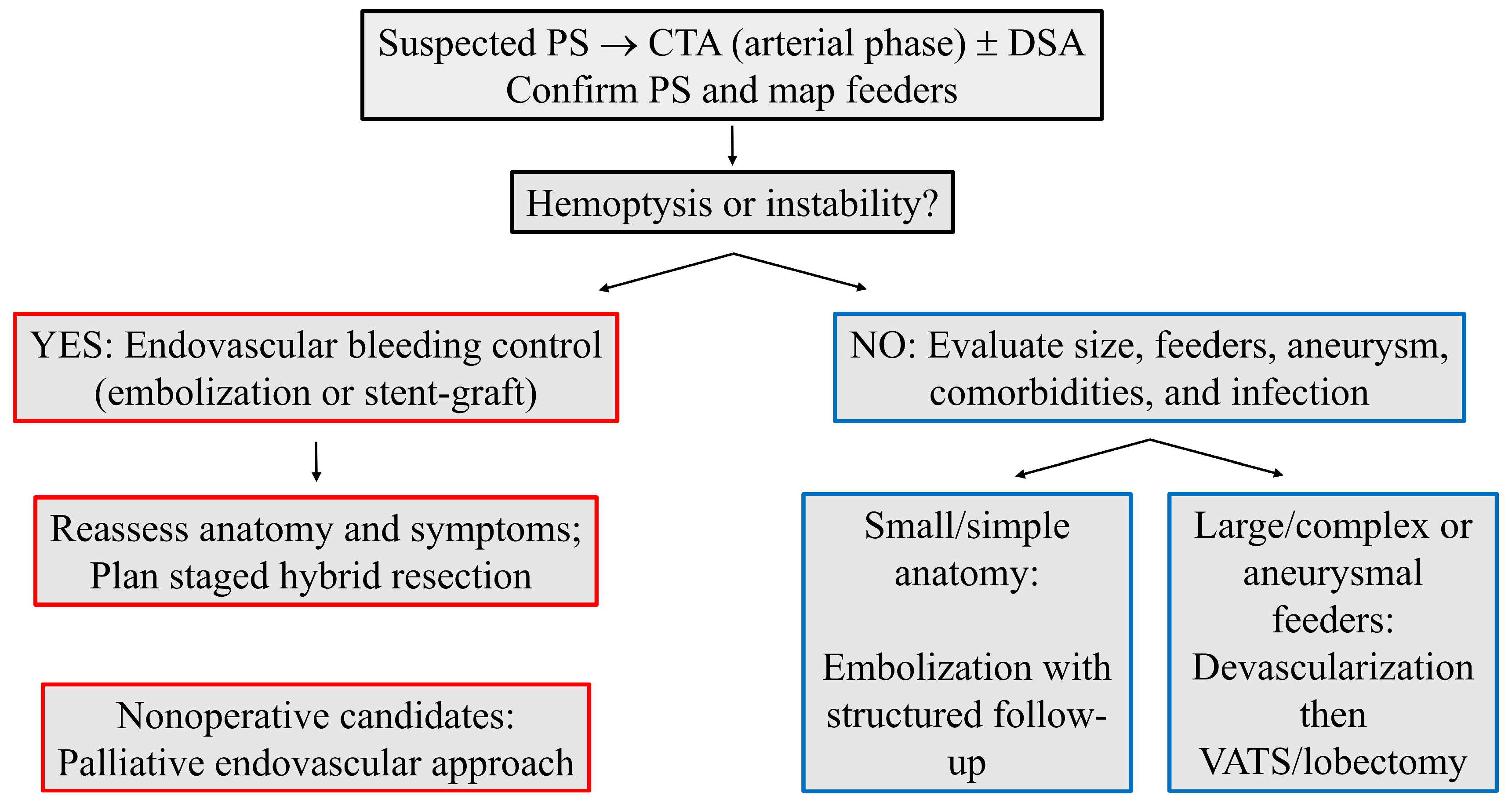
Clinical decision-making is therefore anatomy- and symptom-driven. Smaller lesions supplied by a single, nonaneurysmal systemic artery with a favorable take-off and course are plausible candidates for definitive embolization, provided that postprocedural imaging confirms complete exclusion and serial follow-up documents progressive involution [14,17,28,29,30,31,32,33,35,36]. In contrast, larger or complex lesions; multiple feeding arteries; short ostial necks at the aorta (limited proximal landing zones); or aneurysmal change favor a planned hybrid pathway (devascularization followed by resection) both to reduce intraoperative bleeding and to improve operative visualization [31,32,33,46,47,48,49,50,51,52,53,54,55,56,57,58,59,60,61]. Nonoperative candidates with persistent symptoms can benefit from palliative endovascular control with symptom-guided surveillance.

4.3. Technique Selection

Technique selection is dictated by vascular geometry and flow dynamics. Coils and microcoils are versatile in small, tortuous, or branching feeding arteries and allow a distal-to-proximal strategy; vascular plugs provide rapid, secure occlusion in larger or straighter channels and can buttress coil constructs; and particles (e.g., PVA) achieve more distal penetration but require caution in high-flow beds [21,29,34,35,36,41,47,48,49,50,51,52,59]. Aneurysmal dilation of aberrant systemic arteries appears relatively frequent in adult cohorts undergoing endovascular or hybrid therapy, likely reflecting chronic exposure of pulmonary-type vessels to systemic pressures, adverse take-off angles with turbulent flow, and a publication bias toward more complex cases [42,62]. In such settings, and for feeding arteries with aortic ostia, stent-graft exclusion of the ostium (often combined with distal embolization to prevent retrograde perfusion) may offer durable vascular control, mitigate rupture risk, and serve as a bridge or adjunct to surgery while facilitating subsequent resection if needed [23,24,55,56,57].

4.4. Timing Within Hybrid Care

Timing within hybrid care is individualized. Same-session or short-interval surgery (hours to a few days) may minimize the risk of recanalization, limit infectious complications in chronically inflamed parenchyma, and capitalize on diminished arterial inflow to reduce blood loss [31,32,57]. Longer intervals of weeks can be reasonable for clinical stabilization, optimization, or complex planning, although adhesions or collateralization may accrue and complicate resection [51,55]. These trade-offs emphasize the value of multidisciplinary planning.

4.5. Complications and Re-Intervention

Across adult reports, complications following endovascular therapy are mostly minor and self-limited (postembolization pain, fever, and pleuritic symptoms) and typically resolve within about two weeks with supportive care [2,5,17,20,21,22,23,38,39]. Re-intervention, when required, usually reflects residual branches, collateral recruitment, device migration, or recanalization [29,39,44]. Several lesion and vessel features appear to coincide with higher procedural complexity and postprocedural events: (i) large sequestration size and chronic parenchymal inflammation; (ii) multiple or short-neck systemic feeding arteries; (iii) aneurysmal dilation and high-flow dynamics; and (iv) active infection at presentation or recurrent hemoptysis requiring urgent control. These observations, while hypothesis-generating and limited by case report designs, support an anatomy-based selection framework favoring hybrid devascularization plus resection for large, complex, or aneurysmal feeding arteries and reserving definitive embolization for small, single-feeder, nonaneurysmal anatomies with reliable follow-up imaging (Figure 2).

4.6. Practice Implications

Given the heterogeneity and reporting gaps across source studies, the following practice implications should be interpreted as descriptive signals rather than comparative effect estimates.
We recommend explicit definitions for technical success (angiographic or CTA exclusion of all systemic inflows), clinical success (durable resolution or improvement of index symptoms without unplanned re-intervention), and recurrence (imaging or clinical relapse requiring further treatment), accompanied by a structured imaging schedule at 1, 3, 6, and 12 months for nonresected lesions [14,17]. Patient selection can be operationalized by anatomy and physiology: definitive embolization for small, single-feeder, nonaneurysmal lesions in suitable candidates; hybrid devascularization plus VATS or limited thoracotomy for large, complex, or aneurysmal feeding arteries; and palliative endovascular control for nonoperative patients, with attention to infection control and symptom-guided follow-up [10,12,13,14,23,24,30,31,32,45,55,56,57].

4.7. Limitations

The evidence base is constrained by heterogeneity in design and reporting. Most source studies are single cases or small series with nonstandardized definitions of technical and clinical success, inconsistent imaging protocols, incomplete outcome reporting, and widely variable follow-up horizons, increasing susceptibility to selection and detection biases. In addition, because the dataset originated from a precompiled corpus, residual selection and reporting biases may persist despite deduplication, dual independent screening/extraction, and publication of full search strings. We therefore refrained from quantitative synthesis and instead presented explicit numerators and denominators, treating percentages as descriptive signals rather than estimates of comparative effect sizes.

4.8. Future Work

Future work should prioritize multicenter registries and prospective observational cohorts with harmonized outcome definitions, minimum imaging datasets, and lesion-level descriptors (type, size, feeder diameter and tortuosity, aneurysmal change, and venous drainage). Carefully adjusted comparative designs are needed to refine selection thresholds among endovascular, hybrid, and surgical pathways and to examine optimal timing within hybrid care. Agreement on a core outcome set (including standardized definitions for technical and clinical success, recurrence, and imaging-based involution) would substantially improve interpretability across studies.

5. Conclusions

Endovascular therapy is an important component of adult PS care (critical for urgent hemostasis and, in anatomically simple lesions, a potential definitive strategy), while hybrid approaches appear advantageous for large, complex, or aneurysmal feeding arteries by improving operative safety. Standardized outcome definitions and structured imaging follow-up, along with prospective registries, are essential to refine selection, timing, and durability.

Supplementary Materials

The following supporting information can be downloaded at: https://www.mdpi.com/article/10.3390/jcm14217493/s1, Table S1: Studies excluded at full-text review with reasons (none); title/abstract screening exclusions by reason (summary); Table S2: Risk-of-bias appraisal (JBI-adapted); Methods S1: Full database search strategies; Checklist S1: PRISMA-2020 checklist; Checklist S2: PRISMA-2020 for abstracts–checklist (narrative systematic review).

Author Contributions

Conceptualization, F.É.S. and E.D.; methodology, F.É.S.; validation, Á.B., B.B.N., Á.S. and E.D.; formal analysis, M.P.; investigation, F.É.S. and Á.B.; writing—original draft preparation, F.É.S. and Á.S.; writing—review and editing, F.É.S., Á.S. and E.D.; visualization, F.É.S. and Á.B.; supervision, E.D. All authors have read and agreed to the published version of the manuscript.

Funding

This study was supported by the National Research, Development, and Innovation Fund of Hungary (grant NKFI-K-146929 was awarded to E.D. and grant EKÖP-2024-220 was awarded to Á.S.).

Institutional Review Board Statement

Not applicable. This systematic review used publicly available published data and did not involve human participants or animals.

Informed Consent Statement

Not applicable.

Data Availability Statement

Full database search strategies, a summary of title/abstract screening exclusions by reason, and the per-study risk-of-bias table are provided as Supplementary Files. The full protocol can be obtained from the corresponding author upon reasonable request. No analysis code was generated.

Conflicts of Interest

The authors declare no conflicts of interest.

References

  1. Durhan, G.; Ardali Duzgun, S.; Akpınar, M.G.; Demirkazık, F.; Arıyürek, O.M. Imaging of congenital lung diseases presenting in the adulthood: A pictorial review. Insights Imaging 2021, 12, 153. [Google Scholar] [CrossRef]
  2. Chakraborty, R.K.; Modi, P.; Sharma, S. Pulmonary Sequestration; StatPearls Publishing: Treasure Island, FL, USA, 2023. [Google Scholar]
  3. Abbey, P.; Das, C.J.; Pangtey, G.S.; Seith, A.; Dutta, R.; Kumar, A. Imaging in bronchopulmonary sequestration. J. Med. Imaging Radiat. Oncol. 2009, 53, 22–31. [Google Scholar] [CrossRef]
  4. Corbett, H.J.; Humphrey, G.M. Pulmonary sequestration. Paediatr. Respir. Rev. 2004, 5, 59–68. [Google Scholar] [CrossRef]
  5. Gabelloni, M.; Faggioni, L.; Accogli, S.; Aringhieri, G.; Neri, E. Pulmonary sequestration: What the radiologist should know. Clin. Imaging 2021, 73, 61–72. [Google Scholar] [CrossRef]
  6. Walker, C.M.; Wu, C.C.; Gilman, M.D.; Godwin, J.D., 2nd; Shepard, J.A.; Abbott, G.F. The imaging spectrum of bronchopulmonary sequestration. Curr. Probl. Diagn. Radiol. 2014, 43, 100–114. [Google Scholar] [CrossRef] [PubMed]
  7. Pham, L.H.; Hamdaoui, Y.; Zeron, G.; El-Bershawi, A.; Alazzeh, A. Separating Out Pulmonary Sequestration. Cureus 2024, 16, e53190. [Google Scholar] [CrossRef]
  8. Wei, Y.; Li, F. Pulmonary sequestration: A retrospective analysis of 2625 cases in China. Eur. J. Cardiothorac. Surg. 2011, 40, e39–e42. [Google Scholar] [CrossRef] [PubMed]
  9. Sotto Mayor, J.; Rocha, D.; Esperança, S.; Oliveira e Silva, A. Intralobar pulmonary sequestration: Diagnostic expertise. BMJ Case Rep. 2015, 2015, bcr2015212384. [Google Scholar] [CrossRef] [PubMed]
  10. Alsumrain, M.; Ryu, J.H. Pulmonary sequestration in adults: A retrospective review of resected and unresected cases. BMC Pulm. Med. 2018, 18, 97. [Google Scholar] [CrossRef]
  11. Ren, S.; Yang, L.; Xiao, Y.; Tong, Z.; Wang, L.; Hu, Y. Pulmonary sequestration in adult patients: A single-center retrospective study. Respir. Res. 2023, 24, 13. [Google Scholar] [CrossRef]
  12. Liu, C.; Pu, Q.; Ma, L.; Mei, J.; Xiao, Z.; Liao, H.; Liu, L. Video-assisted thoracic surgery for pulmonary sequestration compared with posterolateral thoracotomy. J. Thorac. Cardiovasc. Surg. 2013, 146, 557–561. [Google Scholar] [CrossRef]
  13. Wang, L.M.; Cao, J.L.; Hu, J. Video-assisted thoracic surgery for pulmonary sequestration: A safe alternative procedure. J. Thorac. Dis. 2016, 8, 31–36. [Google Scholar]
  14. Zhang, Y.; Qiu, Y.; Li, Y. Debating the embolization of a large aberrant systemic artery for pulmonary sequestration using an Amplatzer duct occluder: A case report and literature review. Cardiol. Young 2022, 32, 331–336. [Google Scholar] [CrossRef]
  15. Berna, P.; Cazes, A.; Bagan, P.; Riquet, M. Intralobar sequestration in adult patients. Interact. Cardiovasc. Thorac. Surg. 2011, 12, 970–972. [Google Scholar] [CrossRef]
  16. Park, S.T.; Yoon, C.H.; Sung, K.B.; Yoon, H.K.; Goo, D.E.; Kim, K.S.; Pi, S.Y.; Auh, Y.H. Pulmonary sequestration in a newborn infant: Treatment with arterial embolization. J. Vasc. Interv. Radiol. 1998, 9, 648–650. [Google Scholar] [CrossRef] [PubMed]
  17. Zhang, S.X.; Wang, H.D.; Yang, K.; Cheng, W.; Wu, W. Retrospective review of the diagnosis and treatment of pulmonary sequestration in 28 patients: Surgery or endovascular techniques? J. Thorac. Dis. 2017, 9, 5153–5160. [Google Scholar] [CrossRef] [PubMed]
  18. Lee, B.S.; Kim, J.T.; Kim, E.A.; Kim, K.S.; Pi, S.Y.; Sung, K.B.; Yoon, C.H.; Goo, H.W. Neonatal pulmonary sequestration: Clinical experience with transumbilical arterial embolization. Pediatr. Pulmonol. 2008, 43, 404–413. [Google Scholar] [CrossRef] [PubMed]
  19. Lee, K.H.; Sung, K.B.; Yoon, H.K.; Ko, G.Y.; Yoon, C.H.; Goo, H.W.; Kim, E.A.; Kim, K.S.; Pi, S.Y. Transcatheter arterial embolization of pulmonary sequestration in neonates: Long-term follow-up results. J. Vasc. Interv. Radiol. 2003, 14, 363–367. [Google Scholar] [CrossRef]
  20. Zener, R.; Bottoni, D.; Zaleski, A.; Fortin, D.; Malthaner, R.A.; Inculet, R.I.; Mujoomdar, A. Transarterial embolization of intralobar pulmonary sequestration in a young adult with hemoptysis. J. Thorac. Dis. 2017, 9, E188–E193. [Google Scholar] [CrossRef] [PubMed]
  21. Healy, J.; Healey, A.; Kitley, C. Embolization of symptomatic intralobar pulmonary sequestration—A minimally invasive treatment option. Radiol. Case Rep. 2019, 14, 759–762. [Google Scholar] [CrossRef]
  22. Borzelli, J.; Brahmbhatt, S.; Desmond, D.; Ching, B.; Hostler, J. Coil embolization of intralobar pulmonary sequestration—An alternative to surgery: A case report. J. Med. Case Rep. 2018, 12, 375. [Google Scholar]
  23. Chen, Y.; Liu, B.; Shao, J.; Liu, D.; Zheng, Y. Endovascular treatment of pulmonary sequestration with thoracic endograft: Two case reports. Medicine 2019, 98, e16666, Erratum in Medicine 2019, 98, e17603. [Google Scholar] [CrossRef]
  24. Wilder, F.G.; Minasyan, S.Z. Thoracic Stent Graft Accompanied by Coil Embolization for Pulmonary Sequestration. Innovations 2019, 14, 168–173. [Google Scholar] [CrossRef]
  25. Savic, B.; Birtel, F.J.; Tholen, W.; Funke, H.D.; Knoche, R. Lung sequestration: Report of seven cases and review of 540 published cases. Thorax 1979, 34, 96–101. [Google Scholar] [CrossRef]
  26. He, B.; Sun, M.S.; Niu, Y.; Zhang, J.B.; Nie, Q.Q.; Zheng, X.; Fan, X.Q.; Liu, P. Hybrid and Endovascular Treatment of Pulmonary Sequestration: Two Case Reports and Literature Review. Ann. Vasc. Surg. 2020, 69, 447.e1–447.e8. [Google Scholar] [CrossRef]
  27. Zhou, H.; Tang, S.; Fu, Q.; Yu, L.; Liu, L. Hybrid surgery in treatment of pulmonary sequestration with abdominal aorta feeding vessel: A case report. J. Cardiothorac. Surg. 2018, 13, 44. [Google Scholar] [CrossRef]
  28. Mohapatra, M.; Mishra, S.; Jena, P. Massive hemoptysis in a case of intralobar pulmonary sequestration associated with pulmonary hypoplasia and meandering right pulmonary vein: Diagnosis and management. Case Rep. Pulmonol. 2012, 2012, 960948. [Google Scholar] [CrossRef] [PubMed]
  29. Marine, L.M.; Valdes, F.E.; Mertens, R.M.; Bergoeing, M.R.; Kramer, A. Endovascular treatment of symptomatic pulmonary sequestration. Ann. Vasc. Surg. 2011, 25, 696.e11–696.e15. [Google Scholar] [CrossRef] [PubMed]
  30. Madhusudhan, K.S.; Das, C.J.; Dutta, R.; Kumar, A.; Bhalla, A.S. Endovascular embolization of pulmonary sequestration in an adult. J. Vasc. Interv. Radiol. 2009, 20, 1640–1642. [Google Scholar] [CrossRef]
  31. Goto, T.; Toya, K.; Wakaki, M.; Kato, R. Resection of intralobar pulmonary sequestration after coil embolization of aberrant arteries: Report of a case. Surg. Today 2013, 43, 923–925. [Google Scholar] [CrossRef] [PubMed]
  32. Wang, L.; Balasubramanian, S.; Jackson, R.; Agrawal, D. Combined endovascular and surgical approaches to treat intralobar pulmonary sequestration: A case report. Ann. R. Coll. Surg. Engl. 2021, 103, e35–e37. [Google Scholar] [CrossRef] [PubMed]
  33. Chataut, D.; Katwal, S.; Suwal, S.; Thapa, A.; Bhusal, A. Endovascular embolization for massive hemoptysis in intralobar pulmonary sequestration with celiac artery supply: A comprehensive case report. Radiol. Case Rep. 2024, 19, 2239–2244. [Google Scholar] [CrossRef]
  34. Marine, L.; Torrealba, J.I.; Valdes, F.; Mertens, R.; Vargas, F.; Bergoeing, M.; Vallejos, D. Endovascular treatment of a right pulmonary sequestration supplied by an aneurysmal aberrant artery originating from the abdominal aorta. J. Vasc. Bras. 2022, 21, e20190160. [Google Scholar] [CrossRef] [PubMed]
  35. Deng, Y.; Fang, X.; Wu, B. Coil embolization to treat pulmonary sequestration in the right upper lobe. Interact. Cardiovasc. Thorac. Surg. 2022, 35, ivac178. [Google Scholar] [CrossRef]
  36. Borzelli, A.; Paladini, A.; Giurazza, F.; Tecame, S.; Giordano, F.; Cavaglià, E.; Amodio, F.; Corvino, F.; Beomonte Zobel, D.; Frauenfelder, G.; et al. Successful endovascular embolization of an intralobar pulmonary sequestration. Radiol. Case Rep. 2017, 13, 125–129. [Google Scholar] [CrossRef] [PubMed]
  37. Tartaglia, D.; Cascione, F.; Modesti, M.; Gianardi, D.; Caputo, R.; Galatioto, C.; Chiarugi, M. Hemoptysis caused by pulmonary sequestration in perforated appendicitis: A rare case report. Ulus. Travma Acil Cerrahi Derg. 2016, 22, 569–571. [Google Scholar]
  38. Ojha, V.; Samui, P.P.; Dakshit, D. Role of endovascular embolization in improving the quality of life in a patient suffering from complicated intralobar pulmonary sequestration—A case report. Respir. Med. Case Rep. 2015, 16, 24–28. [Google Scholar] [CrossRef]
  39. Kim, T.E.; Kwon, J.H.; Kim, J.S. Transcatheter embolization for massive hemoptysis from an intralobar pulmonary sequestration: A case report. Clin. Imaging 2014, 38, 326–329. [Google Scholar] [CrossRef]
  40. Greben, C.R.; Goldstein, G.E.; Gandras, E.J.; Setton, A. Pulmonary sequestration aneurysm embolization. J. Vasc. Interv. Radiol. 2012, 23, 477. [Google Scholar] [CrossRef]
  41. Leoncini, G.; Rossi, U.G.; Ferro, C.; Chessa, L. Endovascular treatment of pulmonary sequestration in adults using Amplatzer® vascular plugs. Interact. Cardiovasc. Thorac. Surg. 2011, 12, 98–100. [Google Scholar] [CrossRef]
  42. Nemoto, M.; Koyama, K.; Tadokoro, Y.; Watanabe, T.; Suzuki, H.; Kiyoshima, M.; Yoshimi, F. Treatment of an Aberrant Arterial Aneurysm with Intralobar Pulmonary Sequestration: A Case Report. Ann. Vasc. Surg. 2020, 69, 453.e11–453.e14. [Google Scholar] [CrossRef]
  43. Türk, İ.; Özdemir, M.; Çetin, M.; Bıçakçıoğlu, P. Coil embolization for pulmonary sequestration may serve as an alternative to surgical treatment: Under which circumstances? Indian J. Thorac. Cardiovasc. Surg. 2025, 41, 1464–1468. [Google Scholar] [CrossRef]
  44. Hordijk, M.; Buimer, M.G. Rupture of an Aneurysmal Pulmonary Sequestration Artery. Vasc. Endovasc. Surg. 2025, 59, 338–341. [Google Scholar] [CrossRef] [PubMed]
  45. Kim, Y.J.; Cho, Y.K.; Lee, J.H.; Woo, J.J.; Choi, Y.S. Endovascular Treatment of Intralobar Pulmonary Sequestration with Vascular Plug: A Case Report. J. Korean Soc. Radiol. 2012, 67, 105–108. [Google Scholar] [CrossRef]
  46. Grossi, W.; Londero, F.; Vit, A.; De Franceschi, E.; Masullo, G.; Sponza, M.; Morelli, A. Hybrid minimally invasive treatment of intralobar pulmonary sequestration: A single-centre experience. Interact. Cardiovasc. Thorac. Surg. 2022, 34, 255–257. [Google Scholar] [CrossRef]
  47. Hakiri, S.; Fukui, T.; Chen-Yoshikawa, T.F. Combined surgical therapy for pulmonary sequestration and aberrant artery from the abdominal aorta. Gen. Thorac. Cardiovasc. Surg. 2021, 69, 1031–1034. [Google Scholar] [CrossRef] [PubMed]
  48. Sakai, M.; Ozawa, Y.; Konishi, T.; Watanabe, A.; Shiigai, M. Endostapling the aberrant artery filled with embolized coils for intralobar pulmonary sequestration: A report of two cases. J. Thorac. Dis. 2018, 10, E304–E308. [Google Scholar] [CrossRef]
  49. Petty, L.; Joseph, A.; Sanchez, J. Case report: Pulmonary sequestration in an adult. Radiol. Case Rep. 2017, 13, 21–23. [Google Scholar] [CrossRef]
  50. Fabbri, N.; Tamburini, N.; Galeotti, R.; Quarantotto, F.; Maniscalco, P.; Rinaldi, R.; Salviato, E.; Cavallesco, G. A rare case of intralobar pulmonary sequestration: Combined endovascular and video-assisted thoracoscopic approach. AME Case Rep. 2018, 2, 19. [Google Scholar] [CrossRef]
  51. Ferland, N.; Couture, C.; Provencher, S. Near-fatal haemoptysis as presentation of a giant intralobar pulmonary sequestration. Eur. Respir. Rev. 2015, 24, 155–156. [Google Scholar] [CrossRef]
  52. Ragusa, M.; Vannucci, J.; Lenti, M.; Cieri, E.; Cao, P.; Puma, F. Pulmonary sequestration supplied by giant aneurysmal aortic branch. Ann. Thorac. Surg. 2010, 89, e7–e8. [Google Scholar] [CrossRef]
  53. Nahal, C.C.; Lackland, T.; Lowe, H.; Platz, J. Presentation and Management of Pulmonary Sequestration with an Aneurysmal Aberrant Pulmonary Artery. Cureus 2024, 16, e60225. [Google Scholar] [CrossRef]
  54. Monfregola, A.; De Angelis, L.; Comune, R.; Arienzo, F.; Barbato, G.; Di Stasio, M.; Pourmolkara, D.; Rosano, N.; Picchi, S.G.; Galluzzo, M.; et al. Interlobar pulmonary sequestration with celiac aberrant artery in an elderly patient treated with combined endovascular and video-assisted thoracoscopic approach. Radiol. Case Rep. 2024, 19, 3418–3424. [Google Scholar] [CrossRef] [PubMed]
  55. Yamamoto, M.; Okada, H.; Nakashima, J.; Anayama, T. Thoracic endovascular aortic repair of an aberrant arterial aneurysm with pulmonary sequestration. Interact. Cardiovasc. Thorac. Surg. 2020, 30, 156–158. [Google Scholar] [CrossRef] [PubMed]
  56. Sridhar, P.; Hardouin, S.; Cheng, T.; Farber, A.; Suzuki, K.; Jones, D.W. Endovascular exclusion and open resection of aberrant pulmonary artery aneurysm associated with intralobar pulmonary sequestration. J. Vasc. Surg. 2019, 70, 1328–1329. [Google Scholar] [CrossRef]
  57. Hewett, L.; Kwon, J.; Adams, J.D.; Denlinger, C.E.; Klapper, J.A. Intralobar Pulmonary Sequestration With Aneurysmal Feeding Vessel: Use of Hybrid Surgical Management. Ann. Thorac. Surg. 2016, 102, e533–e535. [Google Scholar] [CrossRef]
  58. Yamasaki, M.; Suzuki, M.; Misumi, H.; Abe, K.; Ito, J.; Kawazoe, K. Hybrid surgery for intralobar pulmonary sequestration with aortic aneurysm. Ann. Thorac. Surg. 2014, 98, e11–e13. [Google Scholar] [CrossRef]
  59. Nakagiri, T.; Sawabata, N.; Kuratani, T.; Okumura, M. Endovascular stent-graft implantation for a cecum of an aberrant artery from a systemic arterial supply to the basal segment of the left pulmonary lobe. Gen. Thorac. Cardiovasc. Surg. 2010, 58, 640–643. [Google Scholar] [CrossRef]
  60. Porez, F.; Singhal, S.; Mercier, O.; Fabre, D. One-stage hybrid minimally invasive treatment by thoracic endovascular repair and video-assisted thoracoscopic surgery for symptomatic pulmonary sequestration. JTCVS Tech. 2025, 30, 8–10. [Google Scholar] [CrossRef] [PubMed]
  61. Fukuyama, K.; Funakoshi, Y. Successful Concomitant Thoracic Endovascular Aortic Repair and Lobectomy for Pulmonary Sequestration. Ann. Thorac. Surg. Short Rep. 2023, 1, 483–485. [Google Scholar] [CrossRef]
  62. Tatli, S.; Yucel, E.K.; Couper, G.S.; Henderson, J.M.; Colson, Y.L. Aneurysm of an aberrant systemic artery to the lung. AJR Am. J. Roentgenol. 2005, 184, 1241–1244. [Google Scholar] [CrossRef] [PubMed]
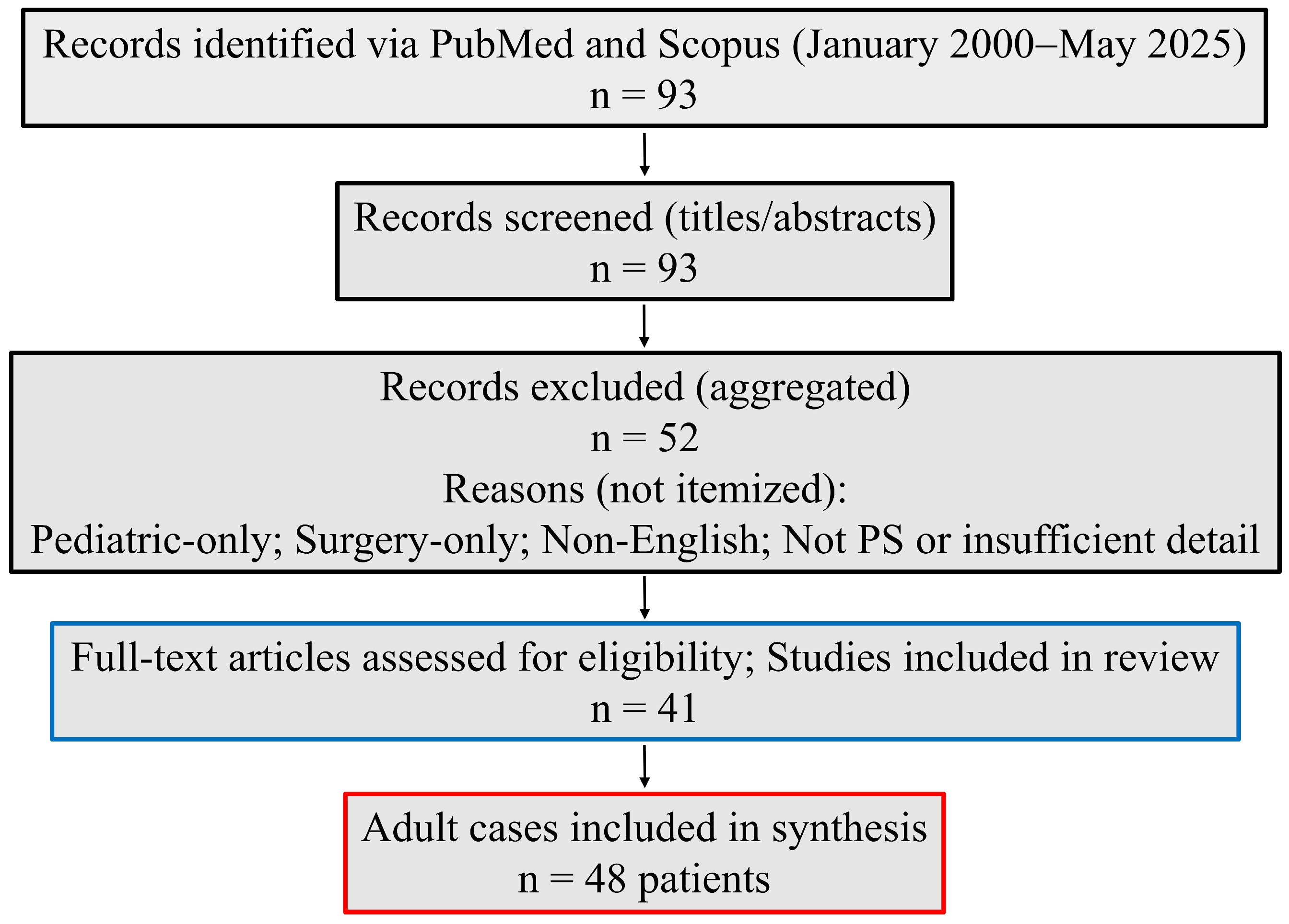
Figure 1. PRISMA-style flow diagram. Records identified via PubMed and Scopus (January 2000–May 2025) (n = 93); records excluded at title/abstract screening (n = 52; pediatric-only; surgery-only; non-English; not PS or insufficient detail); full-text articles included (n = 41 publications). Adult cases included in synthesis: n = 48 patients. No full-text exclusions. PS, Pulmonary sequestration.
Jcm 14 07493 g001
Figure 2. Clinical decision pathway for adult PS. After CTA (arterial phase) ± DSA to confirm PS and map systemic feeding arteries, urgent hemoptysis or instability prompts endovascular bleeding control (embolization or aortic stent-graft), followed by reassessment and planned staged hybrid resection; nonoperative candidates may receive palliative endovascular therapy. In stable patients, small/simple anatomy favors embolization with structured imaging follow-up, whereas large/complex or aneurysmal feeding arteries favor hybrid devascularization followed by VATS/lobectomy. CTA, Computed tomography angiography; DSA, digital subtraction angiography; PS, pulmonary sequestration; VATS, video-assisted thoracoscopic surgery.
Jcm 14 07493 g002
Table 2. Eligibility criteria, outcomes, and exclusions used in this review.
DomainDefinition
PopulationAdults (≥18 years) with intralobar or extralobar PS confirmed by imaging and, where available, intraoperative findings.
InterventionsEndovascular embolization (coils, vascular plugs, particles, or liquid agents); aortic stent-graft exclusion of aberrant arterial origins; or hybrid strategies combining endovascular therapy with surgical resection.
ComparatorsNot required; single-arm reports included due to rarity and design constraints.
OutcomesTechnical success, clinical success, complications, recurrence, re-intervention, imaging-based lesion evolution, and follow-up duration.
Study designsEnglish-language human case reports, case series, and cohorts including an endovascular or hybrid arm.
ExclusionsPediatric-only reports; surgery-only reports without an endovascular or hybrid component; non-PS lesions; non-English publications; insufficient detail to classify intervention or outcomes; duplicate reports merged where identified.
PS, Pulmonary sequestration.
Table 3. Risk-of-bias summary by domain (JBI-adapted; percentages calculated as n/41 studies).
DomainYes, n (%)No, n (%)NR, n (%)
Case definition38 (92.7)3 (7.3)0 (0)
Diagnostic ascertainment40 (97.6)1 (2.4)0 (0)
Intervention description39 (95.1)2 (4.9)0 (0)
Outcome ascertainment36 (87.8)5 (12.2)0 (0)
Follow-up adequacy23 (56.1)7 (17.1)11 (26.8)
Codes: Yes = clearly described and appropriate (lower concern); No = inadequately described or inappropriate (higher concern); NR = not reported/unclear. Per-study domain matrix: see Supplementary Table S2.
Table 4. Patient characteristics and clinical presentation across included cases and treatment subgroups.
CharacteristicAll
(n = 48)
Endovascular Treatment
(n = 25)
Hybrid Treatment
(n = 23)
Embolization
(n = 21)
Stent-Graft
(n = 2)
Embolization + Stent-Graft
(n = 2)
Surgery + Embolization
(n = 16)
Surgery + Stent-Graft
(n = 7)
Sex (male/female/not reported)29/18/111/9/12/0/02/0/07/9/07/0/0
Age (years), mean ± SD44.9 ± 14.940 ± 14.636.5 ± 0.739 ± 5.747.3 ± 14.858.1 ± 11
Symptomatic, n (%)44 (91.7)19 (90.5)2 (100)1 (50)15 (93.8)7 (100)
Hemoptysis, n (%)22 (45.8)15 (71.4)0 (0)1 (50)5 (31.3)1 (14.3)
Chest, back, or abdominal pain, n (%)18 (37.5)7 (33.3)1 (50)1 (50)6 (37.5)3 (42.9)
Recurrent infections, n (%)15 (31.3)7 (33.3)1 (50)0 (0)3 (18.8)4 (57.1)
Cough, expectoration, n (%)9 (18.8)4 (19)0 (0)0 (0)5 (31.3)0 (0)
Fever, n (%)7 (14.6)2 (9.5)0 (0)0 (0)5 (31.3)0 (0)
Percentages are calculated within each column (denominators as shown in the headers). SD, Standard deviation.
Table 5. Lesion characteristics and arterial supply by treatment subgroup.
CharacteristicAll
(n = 48)
Endovascular Treatment
(n = 25)
Hybrid Treatment
(n = 23)
Embolization
(n = 21)
Stent-Graft
(n = 2)
Embolization + Stent-Graft
(n = 2)
Surgery + Embolization
(n = 16)
Surgery + Stent-Graft
(n = 7)
Type
ILS, n (%)36 (75)13 (61.9)0 (0)2 (100)15 (93.8)6 (85.7)
ELS, n (%)2 (4.2)2 (9.5)0 (0)0 (0)0 (0)0 (0)
NR, n (%)10 (20.8)6 (28.6)2 (100)0 (0)1 (6.3)1 (14.3)
Location
Left lower lobe, n (%)24 (50)9 (42.9)2 (100)2 (100)7 (43.8)4 (57.1)
Right lower lobe, n (%)19 (39.6)9 (42.9)0 (0)0 (0)8 (50)2 (28.6)
Other, n (%)5 (10.4)3 (14.3)0 (0)0 (0)1 (6.3)1 (14.3)
PS maximum diameter (mm), mean ± SD67.1 ± 31.5
(n = 12)
51.5 ± 36.1
(n = 2)
NRNR74.5 ± 33
(n = 8)
53 ± 25.5
(n = 2)
Feeding artery origin
Thoracic aorta, n (%)28 (58.3)14 (66.7)2 (100)2 (100)4 (25)6 (85.7)
Celiac trunk, n (%)8 (16.7)2 (9.5)0 (0)0 (0)6 (37.5)0 (0)
Abdominal aorta, n (%)7 (14.6)3 (14.3)0 (0)0 (0)4 (25)0 (0)
Other or no data, n (%)5 (10.4)2 (9.5)0 (0)0 (0)2 (12.5)1 (14.3)
Aneurysmal feeding artery, n (%)12 (25)4 (19)0 (0)1 (50)3 (18.8)4 (57.1)
Feeding artery maximum diameter (mm), mean ± SD30.6 ± 38.5
(n = 21)
26.2 ± 41.8
(n = 9)
NR27
(n = 1)
37 ± 52.2
(n = 5)
32.3 ± 29.5
(n = 6)
Percentages are calculated within each column (denominators as shown in the headers). For PS maximum diameter and feeding artery maximum diameter, n reflects the number of reports with available data in that column. ELS, Extralobar sequestration; ILS, intralobar sequestration; NR, not reported; PS, pulmonary sequestration; SD, standard deviation.
Table 6. Procedural and post-procedural parameters of endovascular and hybrid treatments.
ParameterAll
(n = 48)
Endovascular Treatment
(n = 25)
Hybrid Treatment
(n = 23)
Embolization
(n = 21)
Stent-Graft
(n = 2)
Embolization + Stent-Graft
(n = 2)
Surgery + Embolization
(n = 16)
Surgery + Stent-Graft
(n = 7)
Embolic agents used
Coils, n (%)29 (60.4)15 (71.4)-2 (100)12 (75)-
Vascular plug, n (%)12 (25)6 (28.6)-1 (50)5 (31.3)-
PVA, n (%)8 (16.7)8 (38.1)-0 (0)0 (0)-
Other agents, n (%)4 (8.3)3 (14.3)-0 (0)1 (6.3)-
Hospitalization (days), mean ± SD4.1 ± 2.7
(n = 36)
2.9 ± 3
(n = 15)
3
(n = 1)
4 ± 4.2
(n = 2)
5.4 ± 2.1
(n = 13)
4.8 ± 1.9
(n = 5)
Complications, n (%)10 (20.8)8 (38.1)0 (0)1 (50)1 (6.3)0 (0)
Recurrence, n (%)2 (4.2)2 (9.5)0 (0)0 (0)0 (0)0 (0)
Re-intervention, n (%)3 (6.3)3 (14.3)0 (0)0 (0)0 (0)0 (0)
Follow-up (months), mean ± SD15.3 ± 15.4
(n = 32)
20.8 ± 17.5
(n = 18)
7.5 ± 6.4
(n = 2)
0.5 ± 0.7
(n = 2)
8 ± 5.3
(n = 7)
13.7 ± 15
(n = 3)
Imaging-based size reduction, involution, n9 (of 48)7 (of 21)1 (of 2)1 (of 2)--
Percentages are calculated within each column (denominators as shown in the headers). For length-of-stay and follow-up, n reflects the number of reports with available data in that column. PVA, Polyvinyl alcohol; SD, standard deviation.
Disclaimer/Publisher’s Note: The statements, opinions and data contained in all publications are solely those of the individual author(s) and contributor(s) and not of MDPI and/or the editor(s). MDPI and/or the editor(s) disclaim responsibility for any injury to people or property resulting from any ideas, methods, instructions or products referred to in the content.