Article

Predictive Factors for 24-h Survival After Perioperative Cardiopulmonary Resuscitation: Single-Center Retrospective Cohort Study

by
Soontarin Chungsaengsatitayaporn
1,
Tanyong Pipanmekaporn
1,2,*,
Jiraporn Khorana
2,3,4,
Prangmalee Leurcharusmee
1,
Settapong Boonsri
1 and
Visith Siriphuwanun
1
1
Department of Anesthesiology, Faculty of Medicine, Chiang Mai University, Intavarorote Rd., Muang Chiang Mai District, Chiang Mai 50200, Thailand
2
Department of Biomedical Informatics and Clinical Epidemiology, Faculty of Medicine, Chiang Mai University, Chiang Mai 50200, Thailand
3
Division of Pediatric Surgery, Department of Surgery, Faculty of Medicine, Chiang Mai University, Chiang Mai 50200, Thailand
4
Clinical Surgical Research Center, Department of Surgery, Faculty of Medicine, Chiang Mai University, Chiang Mai 50200, Thailand
*
Author to whom correspondence should be addressed.
J. Clin. Med. 2025, 14(2), 599; https://doi.org/10.3390/jcm14020599
Submission received: 13 December 2024 / Revised: 12 January 2025 / Accepted: 14 January 2025 / Published: 17 January 2025
(This article belongs to the Section Clinical Research Methods)

Abstract

:
Background: Perioperative cardiac arrest (POCA) remains a major challenge in surgical settings, with low survival after cardiopulmonary resuscitation (CPR). This study aims to identify predictive factors for 24 h survival after CPR and cause of POCA. Method: A retrospective, single-center study was conducted on patients aged ≥18 years who experienced POCA and received CPR in the operating room or within 2 h postoperatively at Chiang Mai University Hospital from 2010 to 2019. The multivariable analysis of independent survival predictors was performed using risk regression models. Results: There were 288 cases of cardiopulmonary arrest requiring CPR, with 61 patients surviving. Significant predictors of survival after perioperative CPR included the American Society of Anesthesiologists physical status classification (ASA) 1–2 (RR 2.53; 95%CI 1.69–3.77; p < 0.001), preoperative hemoglobin ≥ 8 g/dL (RR 2.27; 95%CI 1.05–4.89; p = 0.036), preoperative oxygen saturation ≥ 90% (spontaneous breathing in room air) (RR 3.19; 95%CI 1.21–8.41; p = 0.019), initial end-tidal carbon dioxide between 35 and 45 mmHg (RR 1.55; 95%CI 0.98–2.44; p = 0.059), and duration of CPR ≤ 30 min (RR 3.68; 95%CI 1.51–8.98; p = 0.004). The major cause of POCA was hypovolemia (74.30%). Conclusions: This study identifies several critical predictors for 24 h survival following POCA, which can inform pre-operative optimization and perioperative management. Timely interventions, such as blood transfusions and volume resuscitation, are crucial in improving survival outcomes, particularly in trauma and high-risk patients. Further multi-center studies are needed to validate these findings and explore long-term outcomes to refine perioperative cardiac arrest management.

1. Introduction

Perioperative cardiopulmonary arrest (POCA) is a life-threatening event that occurs in the operating room or shortly after surgery [1,2] with outcomes that often include survival with or without complication or death [3,4,5,6,7,8]. POCA is distinct from other cardiac arrests in the hospital setting arrests due to its close association with anesthesia and surgical interventions, which are challenges for timely diagnosis, intervention, and management. Cardiac arrest refers to the absence of circulation, requiring cardiopulmonary resuscitation (CPR) whether by chest compression or, in rare cases, open cardiac massage [6,8,9,10,11]. Although POCA is relatively rare compared to other adverse events, its incidence and outcomes vary globally, often reflecting the complexity of healthcare environments [12]. Developed countries report better post-CPR outcomes due to advanced healthcare systems, resources, and comprehensive research support [13], while the incidence of POCA in these settings is reported to be between 2.63 and 6.8 per 10,000 cases [9,14,15,16,17]. In Thailand, the Perioperative Anesthetic Adverse Events in Thailand (PAAd Thai) study reported a higher incidence of POCA at 15.5 per 10,000 cases, signaling a need to understand the risk factors and outcomes more deeply [3].
The causes of POCA are often sudden and directly linked to intraoperative events, including hypovolemia from surgical blood loss, airway obstruction, hypoxia, and anesthetic-related cardiovascular suppression. These intraoperative events demand rapid, specialized interventions to prevent irreversible damage or death, setting POCA apart from general in-hospital cardiac arrests, which often result from chronic conditions and present in less-controlled environments like general wards. Continuous monitoring such as continuous end-tidal CO2 (ETCO2), pulse oximetry, and electrocardiography (ECG), and the readiness and awareness of anesthesiologists, surgeons, and trained nursing staff in resuscitation enables immediate resuscitation efforts [18,19,20] which lead to a quicker response, potentially improving the chances of successful intervention by utilizing the readily available medical resources in operating rooms [3,12,21,22,23]. Although the controlled environment and rapid response capabilities provide some advantage for POCA cases, survival rates are far from consistent, with outcomes influenced by a complex interplay of patient-specific factors, such as pre-existing comorbidities and the urgency of the surgery [8,14,18,19,20]. Despite the available resources, research on POCA survival predictors remains inconclusive, with findings on factors like The American Society of Anesthesiologists (ASA) physical status, surgical urgency, and initial arrest rhythms often varying widely due to the differences in the study methodologies and patient characteristics [11,22,24,25,26,27,28]. Furthermore, while short-term survival (within 24 h post-CPR) is an essential indicator of immediate intervention success, few studies have focused on this critical timeframe [11,29,30]. For perioperative clinicians, understanding specific predictors within the first 24 h is crucial for preoperative optimization, real-time intraoperative monitoring, and tailoring resuscitation efforts to patient needs, thus highlighting an important gap in current knowledge.
This study addresses these gaps by analyzing the key clinical predictors associated with 24 h survival following POCA. By examining detailed variables such as ASA classification, preoperative oxygen saturation, initial ETCO2 levels, and CPR duration, this study aims to provide actionable insights that can refine perioperative protocols and resuscitation practices. Ultimately, identifying these predictors will support better risk stratification, improve clinical decision making, and contribute to a safer perioperative environment, enhancing patient outcomes after cardiac arrest.

2. Material and Methods

2.1. Study Design

This retrospective cohort study was conducted according to the STROBE guidelines, ensuring the transparent reporting of observational studies. The Ethics Committee of the Faculty of Medicine at Chiang Mai University approved this study (Research ID: 8823, Study Code: ANE:2565-08823), and a waiver of informed consent was granted due to the retrospective design.

2.2. Setting

Data were collected from Chiang Mai University Hospital, Thailand, covering POCA cases between January 2010 and December 2019. A summary of the study protocol and collected data is available in Supplementary File S1.

2.3. Participants

This study included adult patients aged 18 years and older who experienced POCA during non-cardiac surgeries under general anesthesia, regional anesthesia, or monitored anesthesia care (MAC). Exclusion criteria encompassed patients undergoing organ transplantation, pregnant patients, and cases with incomplete POCA documentation.

2.4. Variables

Data extraction and analysis were carried out by the research team, consisting of two research assistants and anesthesiologists, who reviewed each case to ensure consistency and accuracy. Baseline characteristics were carefully extracted from anesthesia records, including demographic data (age, gender), The American Society of Anesthesiologists physical status, and documented preoperative comorbidities. Comorbidities noted included respiratory diseases, diabetes, hypertension, cardiovascular disease, and renal dysfunction. Additionally, airway management strategies, recorded prior to entering the operating room, were documented as part of the baseline data.
Surgical data encompassed the nature of the surgery (emergency vs. elective), specific surgical sites (e.g., intrathoracic, intra-abdominal, major vascular, intracranial, and major orthopedic procedures), trauma status, specifying whether cases involved multiple injuries, whether perioperative data were collected on blood loss (measured in milliliters), the need for blood transfusion, and patient positioning during surgery. Preoperative laboratory values, obtained from patient records, included blood glucose, hemoglobin, platelet count, the neutrophil–to-lymphocyte ratio (NLR), serum albumin, creatinine, sodium, potassium, and 12-lead electrocardiogram (ECG) readings to provide a comprehensive baseline profile.
Intraoperative parameters and vital signs: Upon arrival in the operating room (OR), initial vital signs were recorded to establish baseline conditions before any cardiac events. These parameters included heart rate (HR), systolic blood pressure (SBP), diastolic blood pressure (DBP), mean arterial pressure (MAP), and oxygen saturation (SpO2) with levels over 90% considered stable, for patients breathing spontaneously in a room air environment. ETCO2 was measured immediately following successful intubation. This set of baseline vital signs was crucial in assessing the patient condition upon entry into the OR.
Details of POCA events were characterized by detailed timing, diagnosis, and physiological status at the moment of arrest. The occurrence of POCA was noted with respect to working hours (08:00 a.m.–04:00 p.m.) or non-working hours (04:00 p.m.–08:00 a.m.) to account for potential variations in resource availability. Diagnosis included initial cardiac rhythms, categorized as shockable (ventricular fibrillation, pulseless ventricular tachycardia), or non-shockable (asystole, pulseless electrical activity). The duration of CPR was measured from the onset of absent cardiac rhythm until the return of spontaneous circulation (ROSC), providing insight into the effectiveness of resuscitative efforts.
Causes and contributing factors of POCA: The authors used the mnemonic “5Hs and 5Ts”, which serves to identify reversible factors in cardiac arrest as shown in Figure 1 [31].
Other notable causes outside the “5Hs and 5Ts” framework were also recorded, such as local anesthetic systemic toxicity and malignant hyperthermia, a rare but critical anesthetic-related syndrome that is a significant cause of anesthetic-related morbidity and mortality in an otherwise healthy patient [24,32,33].

2.5. Data Sources/Measurement

Data were extracted from electronic and paper medical records, including anesthesia records, CPR records, and laboratory reports, with measurements standardized according to previous studies (Supplementary Table S1) [34,35,36,37,38,39,40,41]. Initial vital signs and ETCO2 were recorded upon arrival in the operating room, ensuring that baseline values reflected pre-arrest conditions as closely as possible.

2.6. Bias

Selection bias was minimized by defining clear inclusion and exclusion criteria and utilizing standardized data collection protocols. The research assistants reviewed and validated data entries to maintain consistency and reduce potential errors in recorded measurements.

2.7. Study Size

The sample size was estimated using STATA version 16.0 (Stata Corp LP, College Station, TX, USA). A two-proportion test with an alpha level of 0.05 and a power of 0.80 indicated a requirement of 77 participants per group, resulting in a minimum total of 154 patients. This study included 288 eligible cases, ensuring robust statistical power to detect differences across predictor variables.

2.8. Statistical Methods

Data analysis was conducted using STATA version 16.0 (Stata Corp LP, College Station, TX, USA). Categorical data are presented as frequencies and percentages, while continuous data are reported as mean with standard deviation or median with interquartile range, depending on the distribution. The Shapiro–Wilk test was applied to assess the normality of continuous data. Categorical variables were compared using Fisher’s exact test, and continuous variables were analyzed using the Student’s t-test or the Mann–Whitney U test, based on data distribution.
Variables with p-values less than 0.1 in univariable analysis were selected for further examination using stepwise multivariable risk regression to determine risk ratios (RRs), with additional variables identified from relevant literature. Statistical significance was defined as a p-value of less than 0.05.
Subgroup analysis was performed to compare the survival rates among patients aged 65 years or older versus those younger than 65, as age was associated with higher survival rates. Specifically, for patients undergoing trauma surgery, detailed age comparisons are presented in Supplementary Table S2, subgroup analysis between age and type of surgery is presented in Supplementary Table S3, and subgroup analysis between age and the type of operations in the trauma group (149 patients) is presented in Supplementary Table S4.

3. Results

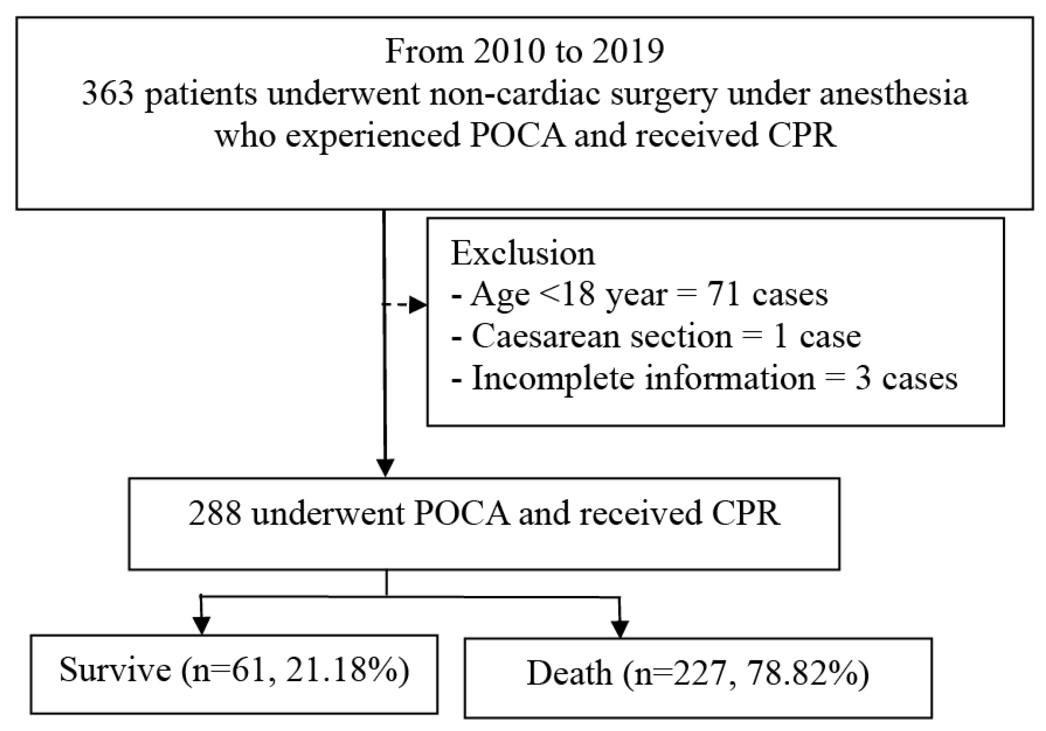
In this 10-year retrospective study, a total of 181,905 patients underwent surgery with anesthesia, including general anesthesia, regional anesthesia, and MAC. The incidence of POCA was found to be 19.95 per 10,000 cases (363 of 181,905 patients). After applying the inclusion criteria for non-cardiac surgeries with POCA, 363 cases were identified, among which 75 cases met the exclusion criteria, leaving 288 patients included in the final analysis. Among these, 61 patients (21.18%) survived within 24 h following POCA (Figure 2).
All patients who experienced POCA had received general anesthesia, with only one initially receiving an infraclavicular brachial plexus block and two others undergoing MAC. Patients aged 65 years and older demonstrated a higher survival rate compared to those younger than 65 years (RR 1.67; 95%CI 1.07–2.59; p = 0.023). Patients with ASA physical status classification 1–2 had a higher likelihood of survival compared to those with ASA 3–5 (RR 4.92; 95%CI 3.46–6.99; p < 0.001). Patients undergoing non-trauma surgery demonstrated a higher survival rate compared to surgery for trauma with multiple injuries (RR 2.04; 95%CI 1.26–3.28; p = 0.003). Patients with no preoperative signs of shock before experiencing POCA had better survival rates than those who were in shock upon arrival in the operating room (RR 2.41; 95% CI 1.34–4.33; p = 0.003) (Table 1 and Table 2).
Regarding the preoperative laboratory investigation, hemoglobin levels of ≥8 g/dL (RR 2.96; 95%CI 1.40–6.23; p = 0.04) was the most significant predictor linked to survival (Table 3).
The vital signs upon arrival to the operating room are crucial predictors of CPR outcomes, with preoperative SPO2 being particularly important. Patients with SPO2 ≥ 90% had a significantly higher survival rate compared to those with preoperative SPO2 measured by pulse oximetry while breathing room air less than 90% (RR 4.82; 95% CI 1.99–11.61; p < 0.001). Patients with initial ETCO2 levels between 35 and 45 mmHg had a better chance of survival compared to those with levels outside this range (RR 2.41; 95% CI 1.53–3.80; p < 0.001) (Table 4).
Patients with an initial shockable rhythm, such as pulseless ventricular tachycardia (pVT) or ventricular fibrillation (VF), had better survival outcomes compared to those with an unshockable rhythm (RR 1.65; 95% CI 0.99–2.73; p = 0.050). Additionally, a CPR duration of less than 30 min was significantly associated with improved survival compared to CPR lasting more than 30 min (RR 4.66; 95% CI 1.75–12.42; p = 0.002) (Table 5).
The leading cause of POCA was hypovolemic shock, as presented in Table 6.
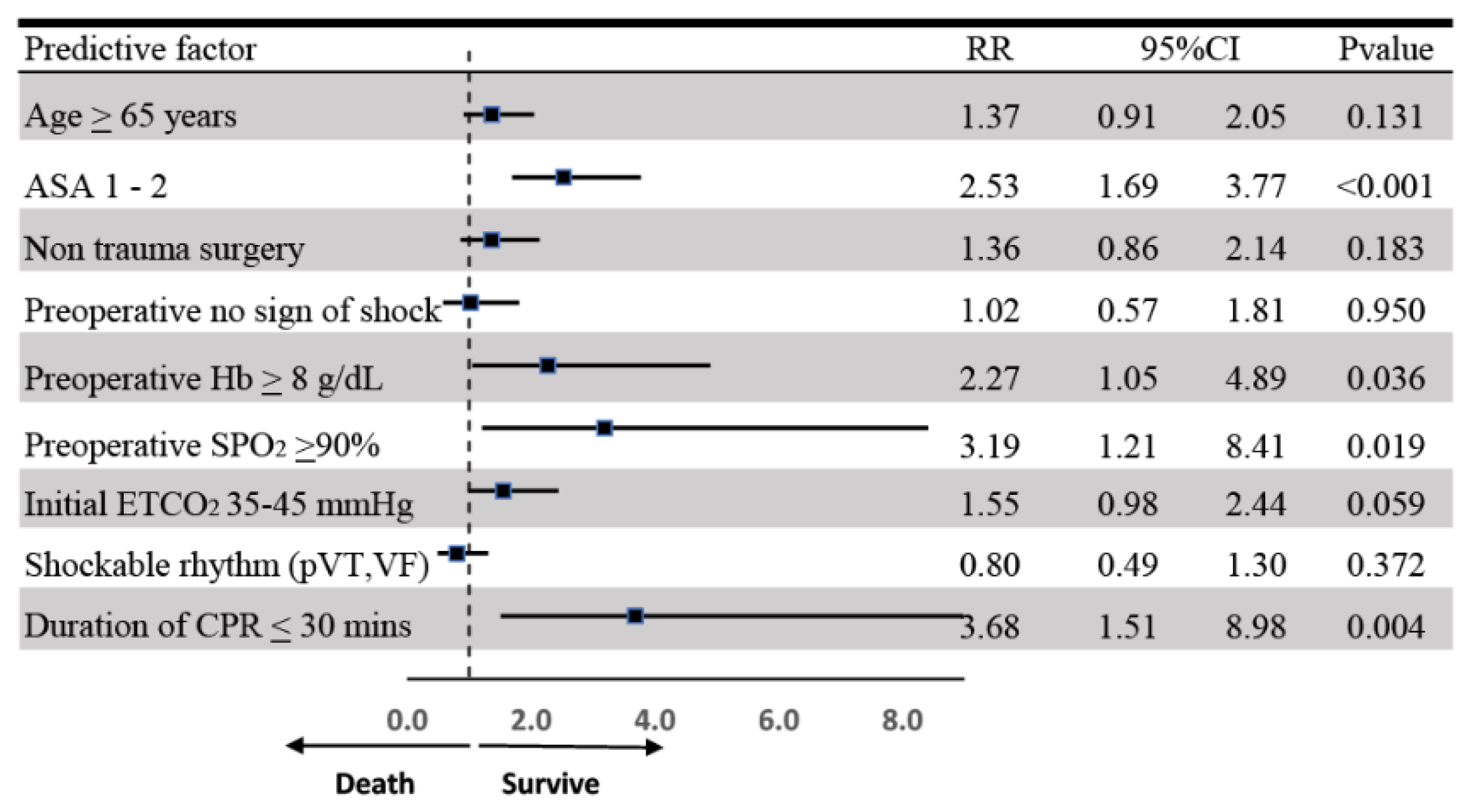
The multivariable risk regression analysis identified strong predictors of survival within 24 h after receiving CPR (Figure 3). These predictors include age greater than 65 years (RR 1.73; 95%CI 1.11–2.09; p = 0.014), ASA 1–2 (RR 2.71; 95%CI 1.80–4.08; p < 0.001), preoperative SPO2 measured by pulse oximetry while breathing room air greater than 90% (RR 3.26; 95%CI 1.23–8.64; p = 0.018), initial ETCO2 levels within the normal range (RR 1.63; 95%CI 1.03–2.57; p = 0.035), and a CPR duration equal to or less than 30 min (RR 3.8; 95%CI 1.59–9.11; p = 0.003).

4. Discussion

This study found a 24 h survival rate of 21% following perioperative CPR (Figure 2), emphasizing the critical nature of the condition. This survival rate is significantly lower than previously reported rates, which range from 32% to 51.4% in the OR settings within 24 h after perioperative CPR [6,10,42,43]. Previous studies indicate that survival rates for cardiac arrest are significantly higher in the OR compared to general wards or ICUs. This is primarily due to the swift recognition of patient condition changes through continuous monitoring and the prompt initiation of resuscitation measures [12,44,45]. The prior research typically examines short- or long-term survival outcomes, but the identification of predictive factors for survival rarely remains [15,46,47]. The causes of POCA in the OR are distinct from those in other settings, frequently arising from anesthetic effects, patient comorbidities, and the complexity of surgical procedures [6,44]. Nonetheless, inadequate preoperative assessments remain a critical challenge, as they can contribute to suboptimal decisions and poor intraoperative outcomes, even in this resource-intensive environment [12,48]. Our findings aim to improve risk stratification and guide evidence-based decision-making during resuscitation efforts by examining factors such as physiological parameters and preoperative conditions. These insights not only provide a foundation for enhancing perioperative protocols but also underscore the importance of targeted interventions to optimize patient outcomes following cardiac arrest. Ultimately, these findings contribute to a more comprehensive understanding of survival determinants in POCA and highlight opportunities to improve perioperative care practices.
Our study demonstrates that patients aged 65 and older, particularly those undergoing elective or non-emergent surgeries, have significantly higher survival rates. This contrasts with much of the existing literature, where advanced age generally correlates with poorer survival outcomes following cardiac arrest [16,22,23,26,28]. In our cohort, older patients were frequently admitted for planned, elective procedures, allowing for better preoperative optimization and lower ASA scores. Elderly patients in this study demonstrated greater stability and higher survival rates compared to younger trauma patients, who faced a markedly high 24 h mortality rate (89.84%) due to severe, multi-system injuries requiring urgent intervention (see Supplementary Table S4).
The ASA physical status score is a preoperative rapid risk assessment tool routinely used for all anesthetized patients to predict perioperative complications and mortality [47]. The ASA score of less than 3 was associated with a 14% increase in survival probability [2], highlighting the role of lower ASA scores in improving outcomes. Conversely, higher ASA scores indicate compromised physiological reserves, often reflecting the severity of acute and high-risk conditions [10,18,22,23,26,30,45,49,50,51].
Non-trauma surgery has low risks associated with trauma surgeries, where controlled surgical environments and thorough preoperative planning contribute to significantly higher survival rates. Because of trauma surgeries often conducted under emergency conditions, limit the opportunity for stabilization, amplifying perioperative risks [16]. Consistent with prior studies [12,22,25,26], which showed trauma as the primary cause of POCA and the third leading contributor to mortality among younger patients, and that injuries frequently involved the brain, chest, abdomen, or major bones, often resulting in hypovolemic shock, cardiac tamponade, or respiratory failure [52,53].
No preoperative sign of shock upon arrival to the operating room emerged as a crucial predictor of survival following POCA. Patients presenting without clinical signs of shock demonstrated a significant survival advantage, aligning with studies emphasizing the importance of stable hemodynamics for effective responses to surgical stress and resuscitation efforts [19,23,54]. In the perioperative context, it is important to recognize that shock is characterized by severe hypotension, impaired perfusion, and organ dysfunction, reflecting that hemodynamic instability and borderline low BP without meeting the criteria for shock may still indicate underlying instability. Such cases, particularly in trauma patients, may represent a compensatory phase where perfusion is maintained through increased vascular resistance, masking the early stages of hemodynamic compromise [19,44].
Preoperative hemoglobin levels are critical for assessing patient stability before surgery. In this study, hemoglobin levels above 8 g/dL were strongly associated with improved 24 h survival rates because of adequate oxygen-carrying capacity in maintaining organ perfusion and supporting effective resuscitation during CPR [55,56,57]. However, it is important to note that preoperative hemoglobin levels may not fully reflect the patient’s status during POCA, particularly in trauma cases where massive blood loss can lead to a rapid decline in hemoglobin levels. This relationship between hemoglobin levels and survival highlights the necessity of preoperative optimization, especially for high-risk patients undergoing trauma surgeries where significant bleeding is anticipated. Maintaining sufficient preoperative hemoglobin reserves can improve physiological resilience to perioperative stress and enhance survival outcomes in crises [58].
Preoperative SPO2 levels above 90% with spontaneous breathing on room air upon arrival to the OR was identified as a significant predictor of 24 h survival following POCA. Higher SPO2 levels were associated with better hemodynamic stability under perioperative stress, reflecting adequate baseline oxygen reserves [59]. Conversely, low preoperative SPO2 (<90%), especially in trauma cases, was linked to compromised oxygen delivery to vital organs. This condition, compounded by intraoperative blood loss, exacerbates hypoxia, decreases perfusion, and heightens the risk of hypovolemia and myocardial ischemia due to reduced venous return, stroke volume, and cardiac output [60]. Preoperative SPO2 ≥ 90% serves as an actionable and easy-to-use parameter for identifying high-risk patients and optimizing perioperative care [25,26].
Initial ETCO2 values ranging from 35 to 45 mmHg, which were measured immediately following successful intubation, were strongly associated with survival, underscoring their role as a real-time marker of effective ventilation and perfusion. This parameter provides important feedback on the adequacy of CPR efforts, allowing clinicians to adjust resuscitative interventions to optimize outcomes and achieve ROSC [12,61]. The importance of ETCO2 monitoring in POCA situations lies in its dual function as a diagnostic and predictive tool, enabling the early recognition of ineffective resuscitation and guiding timely adjustments to improve outcomes. These findings are consistent with the broader utility of ETCO2 in emergency and perioperative settings, emphasizing its value in supporting high-quality resuscitation efforts. Combined with other predictors such as high SPO2 and stable hemoglobin levels, ETCO₂ forms part of an integrated set of perioperative indicators that can be continuously monitored and optimized [12]. Together, these parameters provide clinicians with actionable insights into the patient’s physiological status, enhancing their ability to deliver precise, effective care, and ultimately improving survival rates in POCA scenarios.
The presence of an initial shockable rhythm, such as pVT or VF, was significantly associated with improved survival outcomes, likely due to their distinct pathophysiology. Shockable rhythms reflect a state of electrical disorganization in the myocardium, which, if promptly treated with defibrillation [13,45,62,63], can be rapidly converted into an organized and perfusing rhythm. The rapid recognition of shockable rhythms enables targeted interventions, such as defibrillation, to provide circulation support and good outcomes [25,44,50].
The duration of CPR ≤ 30 min was associated with improved outcomes. This finding is consistent with prior studies demonstrating that shorter resuscitation times contribute to better neurological and physiological recovery, likely due to the earlier restoration of ROSC and reduced exposure to hypoxic injury [29,63,64]. The OR environment is particularly suited for continuous physiological monitoring, including blood pressure, EKG, SpO2, ETCO2, and in cases of regional anesthesia or MAC, even the patient’s level of consciousness. These real-time assessments enable the rapid detection of hemodynamic instability and the immediate identification of cardiac arrest, facilitating the early initiation of CPR and the optimization of resuscitation efforts [19]. The presence of skilled personnel and access to advanced equipment, such as defibrillators and emergency medications, further ensures the delivery of high-quality, timely interventions [18,19,20]. By integrating advanced monitoring with rapid access to resources, the OR environment minimizes delays and enhances the precision and effectiveness of resuscitative efforts, contributing to reduced CPR duration and improved survival outcomes [44,45].
Hypovolemia due to massive blood loss was identified as the leading cause of POCA in our study, accounting for 74.30% of cases, consistent with prior research [43] that highlights its critical role in surgical emergencies. The high prevalence of hemorrhagic shock emphasizes the necessity of continuous hemodynamic and respiratory monitoring for the early detection of instability. The real-time tracking of parameters such as blood pressure, oxygen saturation, ETCO2, and HR facilitates the identification of reversible conditions, such as hypovolemia and hypoxia, allowing for timely interventions, including fluid resuscitation, blood transfusion, and ventilatory adjustments, to prevent progression to cardiac arrest [12,43]. The OR offers a uniquely equipped environment to manage such crises, with advanced monitoring systems, healthcare teams trained in advanced cardiovascular life support (ACLS), and basic life support (BLS), and immediate access to defibrillators, blood products, and medications. These resources, combined with vigilant surveillance, enable clinicians to rapidly address the root causes of hemodynamic instability, particularly in trauma and high-risk surgeries, where significant blood loss is anticipated. The integration of early detection, continuous monitoring, and targeted interventions underscores the importance of a proactive approach to perioperative management, ultimately enhancing survival outcomes in high-risk scenarios [3,12,13,18,19,20,22,23].
This study has several limitations. First, as a retrospective, single-center analysis, the findings may not be generalizable to institutions with varying resources, protocols, or patient populations. Second, the absence of an electronic medical record (EMR) system during the study period posed challenges in ensuring consistent and precise data collection, particularly for continuous monitoring parameters during POCA events. This reliance on manually recorded data may have introduced variability, especially in documenting vital signs and other critical parameters in real time. Additionally, predictors such as preoperative SPO2 and ETCO2 were measured upon arrival to the OR and may not accurately represent the patient’s physiological state during the POCA event itself, as these parameters can fluctuate due to intraoperative management. Another limitation is that this study focuses solely on 24 h survival outcomes, leaving questions about long-term survival, the recurrence of POCA, and post-resuscitation complications unanswered. Future research should prioritize multi-center studies.

5. Conclusions

Despite the low 24 h survival rate following POCA, this study identifies key predictors that can inform clinical decision making and improve outcomes. These predictors include ASA 1–2, preoperative hemoglobin ≥ 8 g/dL, SpO2 ≥ 90%, initial ETCO2 levels between 35 and 45 mmHg, CPR duration ≤ 30 min, no preoperative sign of shock, and non-trauma surgeries. Hypovolemia due to hemorrhagic shock was the leading cause of mortality, emphasizing the need for early detection and appropriate fluid and blood replacement. The findings from this study should be further developed into predictive models and validated in multi-center, prospective studies to ensure their generalizability across diverse clinical settings. Such efforts are crucial for optimizing perioperative cardiac arrest management and improving both survival rates and long-term patient outcomes.

Supplementary Materials

The following supporting information can be downloaded at https://www.mdpi.com/article/10.3390/jcm14020599/s1, Supplementary File S1. Details of the collected data including information about the hospital staff and their competencies. Supplementary Table S1. Data measurement and quantitative variables. Supplementary Table S2. Subgroup analysis between age and trauma surgery. Supplementary Table S3. Subgroup analysis between age and type of surgery. Supplementary Table S4. Subgroup analysis between age and type of operations in trauma group.

Author Contributions

S.C.: Study conception and design; Data analysis and interpretation; Writing and finalizing of the manuscript. T.P., J.K., P.L., S.B. and V.S.: Study conception and design; Data analysis and interpretation; Review, editing and finalizing of the manuscript. All authors have read and agreed to the published version of the manuscript.

Funding

This research was supported by Faculty of Medicine Research Fund, grant no. mc012-2565.

Institutional Review Board Statement

This research was conducted in accordance with the Ethics Committee of the Faculty of Medicine at Chiang Mai University approved this study (research ID: 8823, Study Code: ANE:2565-08823.

Informed Consent Statement

A waiver of informed consent was granted due to the retrospective design.

Data Availability Statement

Datasets generated or analyzed in this study are accessible upon reasonable request from the corresponding author.

Conflicts of Interest

The authors declare no conflicts of interest.

Abbreviation

STROBEStrengthening the Reporting of Observational studies in Epidemiology

References

  1. Allen, M.B.; Orkaby, A.R.; Justice, S.; Hall, D.E.; Hu, F.Y.; Cooper, Z.; Bernacki, R.E.; Bader, A.M. Frailty and Outcomes Following Cardiopulmonary Resuscitation for Perioperative Cardiac Arrest. JAMA Netw. Open 2023, 6, e2321465. [Google Scholar] [CrossRef] [PubMed]
  2. Shang, H.; Chu, Q.; Ji, M.; Guo, J.; Ye, H.; Zheng, S.; Yang, J. A retrospective study of mortality for perioperative cardiac arrests toward a personalized treatment. Sci. Rep. 2022, 12, 13709. [Google Scholar] [CrossRef] [PubMed]
  3. Charuluxananan, S.; Sriraj, W.; Punjasawadwong, Y.; Pitimana-aree, S.; Lekprasert, V.; Werawatganon, T.; Wasinwong, W.; Ratanachai, P.; Sriramatr, D.; Atichat, S.; et al. Perioperative and Anesthetic Adverse events in Thailand (PAAd Thai) incident reporting study: Anesthetic profiles and outcomes. Asian Biomed. 2017, 11, 21–32. [Google Scholar]
  4. Nair, A.; Naik, V.; Rayani, B. Letters to the Editor. Indian J. Crit. Care Med. 2017, 21, 713. [Google Scholar] [CrossRef] [PubMed]
  5. Sprung, J.; Flick, R.; Gleich, S.; Weingarten, T. Perioperative Cardiac Arrests. Signa Vitae 2008, 3, 8–12. [Google Scholar]
  6. Constant, A.L.; Montlahuc, C.; Grimaldi, D.; Pichon, N.; Mongardon, N.; Bordenave, L.; Soummer, A.; Sauneuf, B.; Ricome, S.; Misset, B.; et al. Predictors of functional outcome after intraoperative cardiac arrest. Anesthesiology 2014, 121, 482–491. [Google Scholar] [CrossRef]
  7. Vane, M.F.; Carmona, M.J.C.; Pereira, S.M.; Kern, K.B.; Timerman, S.; Perez, G.; Vane, L.A.; Otsuki, D.A.; Auler, J.O.C., Jr. Predictors and their prognostic value for no ROSC and mortality after a non-cardiac surgery intraoperative cardiac arrest: A retrospective cohort study. Sci. Rep. 2019, 9, 14975. [Google Scholar] [CrossRef]
  8. Goswami, S.; Brady, J.E.; Jordan, D.A.; Li, G. Intraoperative Cardiac Arrests in Adults Undergoing Noncardiac Surgery: Incidence, Risk Factors, and Survival Outcome. Anesthesiology 2012, 117, 1018–1026. [Google Scholar] [CrossRef]
  9. Gong, C.L.; Hu, J.P.; Qiu, Z.L.; Zhu, Q.Q.; Hei, Z.Q.; Zhou, S.L.; Li, X. A study of anaesthesia-related cardiac arrest from a Chinese tertiary hospital. BMC Anesthesiol. 2018, 18, 127. [Google Scholar] [CrossRef]
  10. Nunes, J.C.; Braz, J.R.C.; Oliveira, T.S.; de Carvalho, L.R.; Castiglia, Y.M.M.; Braz, L.G. Intraoperative and Anesthesia-Related Cardiac Arrest and Its Mortality in Older Patients: A 15-Year Survey in a Tertiary Teaching Hospital. PLoS ONE 2014, 9, e104041. [Google Scholar] [CrossRef]
  11. Carlucci, M.T.O.; Braz, J.R.C.; do Nascimento, P., Jr.; de Carvalho, L.R.; Castiglia, Y.M.M.; Braz, L.G. Intraoperative Cardiac Arrest and Mortality in Trauma Patients. A 14-Yr Survey from a Brazilian Tertiary Teaching Hospital. PLoS ONE 2014, 9, e90125. [Google Scholar] [CrossRef] [PubMed]
  12. Hinkelbein, J.; Andres, J.; Böttiger, B.W.; Brazzi, L.; De Robertis, E.; Einav, S.; Gwinnutt, C.; Kuvaki, B.; Krawczyk, P.; McEvoy, M.D.; et al. Cardiac arrest in the perioperative period: A consensus guideline for identification, treatment, and prevention from the European Society of Anaesthesiology and Intensive Care and the European Society for Trauma and Emergency Surgery. Eur. J. Trauma Emerg. Surg. Off. Publ. Eur. Trauma Soc. 2023, 49, 2031–2046. [Google Scholar] [CrossRef] [PubMed]
  13. Alao, D.O.; Mohammed, N.A.; Hukan, Y.O.; Al Neyadi, M.; Jummani, Z.; Dababneh, E.H.; Cevik, A.A. The epidemiology and outcomes of adult in-hospital cardiac arrest in a high-income developing country. Resusc. Plus 2022, 10, 100220. [Google Scholar] [CrossRef] [PubMed]
  14. Hohn, A.; Machatschek, J.N.; Franklin, J.; Padosch, S.A. Incidence and risk factors of anaesthesia-related perioperative cardiac arrest: A 6-year observational study from a tertiary care university hospital. Eur. J. Anaesthesiol. 2018, 35, 266–272. [Google Scholar] [CrossRef] [PubMed]
  15. Kaiser, H.A.; Saied, N.N.; Kokoefer, A.S.; Saffour, L.; Zoller, J.K.; Helwani, M.A. Incidence and prediction of intraoperative and postoperative cardiac arrest requiring cardiopulmonary resuscitation and 30-day mortality in non-cardiac surgical patients. PLoS ONE 2020, 15, e0225939. [Google Scholar] [CrossRef]
  16. Kan, H.; Ding, Y.; Wu, S.; Zhang, Z. Retrospective study of perioperative cardiac arrest from a Chinese tertiary hospital. Medicine 2021, 100, e26890. [Google Scholar] [CrossRef]
  17. Morimatsu, H. Incidence of accidental events during anesthesia from 2012 to 2016: Survey on anesthesia-related events by the Japanese Society of Anesthesiologists. J. Anesth. 2021, 35, 206–212. [Google Scholar] [CrossRef]
  18. Braz, L.G.; Braz, J.R.C.; Modolo, M.P.; Corrente, J.E.; Sanchez, R.; Pacchioni, M.; Cury, J.B.; Soares, I.B.; Braz, M.G. Perioperative and anesthesia-related cardiac arrest and mortality rates in Brazil: A systematic review and proportion meta-analysis. PLoS ONE 2020, 15, e0241751. [Google Scholar] [CrossRef]
  19. Siriphuwanun, V.; Punjasawadwong, Y.; Lapisatepun, W.; Charuluxananan, S.; Uerpairojkit, K.; Patumanond, J. The initial success rate of cardiopulmonary resuscitation and its associated factors in patients with cardiac arrest within 24 hours after anesthesia for an emergency surgery. Risk Manag. Healthc. Policy 2014, 7, 65–76. [Google Scholar]
  20. Khan, N.U.; Razzak, J.A.; Ahmed, H.; Furqan, M.; Saleem, A.F.; Alam, H.; ul Huda, A.; Khan, U.R.; Rehmani, R. Cardiopulmonary resuscitation: Outcome and its predictors among hospitalized adult patients in Pakistan. Int. J. Emerg. Med. 2008, 1, 27–34. [Google Scholar] [CrossRef]
  21. Berry, W.R. Cardiac resuscitation in the operating room: Reflections on how we can do better. Can. J. Anesth. 2012, 59, 522–526. [Google Scholar] [CrossRef] [PubMed]
  22. Aloweidi, A.; Alghanem, S.; Bsisu, I.; Ababneh, O.; Alrabayah, M.; Al-Zaben, K.; Qudaisat, I. Perioperative Cardiac Arrest: A 3-Year Prospective Study from a Tertiary Care University Hospital. Drug Healthc. Patient Saf. 2022, 14, 1–8. [Google Scholar] [CrossRef] [PubMed]
  23. Braz, L.; Morais, A.; Sanchez, R.; Porto, D.; Pacchioni, M.; Serafim, W.; Módolo, N.; Nascimento, P.D., Jr.; Braz, M.; Braz, J. Epidemiology of perioperative cardiac arrest and mortality in Brazil: Systematic review. Braz. J. Anesthesiol. (Engl. Ed.) 2020, 70, 82–89. [Google Scholar] [CrossRef]
  24. Murray, C.; Sasaki, S.S.; Berg, D. Local Anesthesia and Malignant Hyperthermia: Review of the Literature and Recommendations for the Dermatologic Surgeon. Dermatol. Surg. 1999, 25, 626–630. [Google Scholar] [CrossRef]
  25. Siriphuwanun, V.; Punjasawadwong, Y.; Lapisatepun, W.; Charuluxananan, S.; Uerpairojkit, K. Prognostic factors for death and survival with or without complications in cardiac arrest patients receiving CPR within 24 hours of anesthesia for emergency surgery. Risk Manag. Healthc. Policy 2014, 7, 199–210. [Google Scholar]
  26. Braz, L.G.; Módolo, N.S.P.; do Nascimento, P., Jr.; Bruschi, B.A.M.; Castiglia, Y.M.M.; Ganem, E.M.; de Carvalho, L.R.; Braz, J.R.C. Perioperative cardiac arrest: A study of 53 718 anaesthetics over 9 yr from a Brazilian teaching hospital. BJA Br. J. Anaesth. 2006, 96, 569–575. [Google Scholar] [CrossRef]
  27. Choi, Y.J.; Han, S.U.; Woo, S.; Ro, Y.J.; Yang, H.S. Perioperative cardiac arrest in 457,529 anesthetized patients at a single teaching hospital in Korea: A retrospective study. Anesth. Pain Med. 2014, 9, 144–151. [Google Scholar]
  28. Sobreira-Fernandes, D.; Teixeira, L.; Lemos, T.S.; Costa, L.; Pereira, M.; Costa, A.C.; Couto, P.S. Perioperative cardiac arrests—A subanalysis of the anesthesia-related cardiac arrests and associated mortality. J. Clin. Anesth. 2018, 50, 78–90. [Google Scholar] [CrossRef]
  29. Fernando, S.M.; Tran, A.; Cheng, W.; Rochwerg, B.; Taljaard, M.; Vaillancourt, C.; Rowan, K.M.; Harrison, D.A.; Nolan, J.P.; Kyeremanteng, K.; et al. Pre-arrest and intra-arrest prognostic factors associated with survival after in-hospital cardiac arrest: Systematic review and meta-analysis. BMJ 2019, 367, l6373. [Google Scholar] [CrossRef]
  30. Pignaton, W.; Braz, J.R.C.; Kusano, P.S.; Módolo, M.P.; de Carvalho, L.R.; Braz, M.G.; Braz, L.G. Perioperative and Anesthesia-Related Mortality: An 8-Year Observational Survey From a Tertiary Teaching Hospital. Medicine 2016, 95, e2208. [Google Scholar] [CrossRef]
  31. Kuan, K.K.; Rahalkar, K. Beyond 5Hs and 5Ts: A rare cause of cardiac arrest. Singap. Med. J. 2023, 64, 146–148. [Google Scholar] [CrossRef]
  32. Kaur, H.; Katyal, N.; Yelam, A.; Kumar, K.; Srivastava, H.; Govindarajan, R. Malignant Hyperthermia. Mo. Med. 2019, 116, 154–159. [Google Scholar] [PubMed]
  33. Gupta, P.K.; Bilmen, J.G.; Hopkins, P.M. Anaesthetic management of a known or suspected malignant hyperthermia susceptible patient. BJA Educ. 2021, 21, 218–224. [Google Scholar] [CrossRef] [PubMed]
  34. Newland, M.C.; Ellis, S.J.; Lydiatt, C.A.; Peters, K.R.; Tinker, J.H.; Romberger, D.J.; Ullrich, F.A.; Anderson, J.R. Anesthetic-related cardiac arrest and its mortality: A report covering 72,959 anesthetics over 10 years from a US teaching hospital. Anesthesiology 2002, 97, 108–115. [Google Scholar] [CrossRef] [PubMed]
  35. Wang, C.-H.; Huang, C.-H.; Chang, W.-T.; Tsai, M.-S.; Yu, P.-H.; Wang, A.-Y.; Chen, N.-C.; Chen, W.-J. Association between hemoglobin levels and clinical outcomes in adult patients after in-hospital cardiac arrest: A retrospective cohort study. Intern. Emerg. Med. 2016, 11, 727–736. [Google Scholar] [CrossRef]
  36. Brown, L.M.; Call, M.S.; Margaret Knudson, M.; Cohen, M.J.; Holcomb, J.B.; Wade, C.E.; Brasel, K.J.; Vercruysse, G.; MacLeod, J.; Dutton, R.P.; et al. A normal platelet count may not be enough: The impact of admission platelet count on mortality and transfusion in severely injured trauma patients. J. Trauma 2011, 71 (Suppl. S3), S337–S342. [Google Scholar] [CrossRef]
  37. Chae, Y.J.; Lee, J.; Park, J.H.; Han, D.G.; Ha, E.; Yi, I.K. Late Mortality Prediction of Neutrophil-to-Lymphocyte and Platelet Ratio in Patients with Trauma Who Underwent Emergency Surgery: A Retrospective Study. J. Surg. Res. 2021, 267, 755–761. [Google Scholar] [CrossRef]
  38. Hong, S.I.; Kim, Y.J.; Cho, Y.J.; Huh, J.W.; Hong, S.B.; Kim, W.Y. Predictive value of pre-arrest albumin level with GO-FAR score in patients with in-hospital cardiac arrest. Sci. Rep. 2021, 11, 10631. [Google Scholar] [CrossRef]
  39. Barnett, S.; Moonesinghe, S.R. Clinical risk scores to guide perioperative management. Postgrad. Med. J. 2011, 87, 535. [Google Scholar] [CrossRef]
  40. Han, F.; Wang, Y.; Wang, Y.; Dong, J.; Nie, C.; Chen, M.; Hou, L. Intraoperative cardiac arrest: A 10-year study of patients undergoing tumorous surgery in a tertiary referral cancer center in China. Medicine 2017, 96, e6794. [Google Scholar] [CrossRef]
  41. Moitra, V.; Einav, S.; Thies, K.-C.; Nunnally, M.; Gabrielli, A.; Maccioli, G.; Weinberg, G.; Banerjee, A.; Ruetzler, K.; Dobson, G.; et al. Cardiac Arrest in the Operating Room: Resuscitation and Management for the Anesthesiologist Part 1. Anesth. Analg. 2018, 127, e49–e50. [Google Scholar] [CrossRef] [PubMed]
  42. Ramachandran, S.K.; Mhyre, J.; Kheterpal, S.; Christensen, R.E.; Tallman, K.; Morris, M.; Chan, P.S. Predictors of survival from perioperative cardiopulmonary arrests: A retrospective analysis of 2,524 events from the Get With The Guidelines-Resuscitation registry. Anesthesiology 2013, 119, 1322–1339. [Google Scholar] [CrossRef] [PubMed]
  43. Sprung, J.; Warner, M.E.; Contreras, M.G.; Schroeder, D.R.; Beighley, C.M.; Wilson, G.A.; Warner, D.O. Predictors of survival following cardiac arrest in patients undergoing noncardiac surgery: A study of 518,294 patients at a tertiary referral center. Anesthesiology 2003, 99, 259–269. [Google Scholar] [CrossRef] [PubMed]
  44. Kalkman, S.; Hooft, L.; Meijerman, J.M.; Knape, J.T.A.; van Delden, J.J.M. Survival after Perioperative Cardiopulmonary Resuscitation: Providing an Evidence Base for Ethical Management of Do-not-resuscitate Orders. Anesthesiology 2016, 124, 723–729. [Google Scholar] [CrossRef]
  45. Rukewe, A.; Fatiregun, A.; Osunlaja, T.O. Cardiac arrest during anesthesia at a university Hospital in Nigeria. Niger. J. Clin. Pract. 2014, 17, 28–31. [Google Scholar] [CrossRef]
  46. Krittayaphong, R.; Saengsung, P.; Chawaruechai, T.; Yindeengam, A.; Udompunturak, S. Factors predicting outcome of cardiopulmonary resuscitation in a developing country: The Siriraj cardiopulmonary resuscitation registry. J. Med. Assoc. Thail. = Chotmaihet Thangphaet 2009, 92, 618–623. [Google Scholar]
  47. Markovic, D.; Jevtovic-Stoimenov, T.; Stojanovic, M.; Vukovic, A.; Dinic, V.; Markovic-Zivkovic, B.; Jankovic, R.J. Addition of clinical risk scores improves prediction performance of American Society of Anesthesiologists (ASA) physical status classification for postoperative mortality in older patients: A pilot study. Eur. Geriatr. Med. 2018, 9, 51–59. [Google Scholar] [CrossRef]
  48. Salottolo, K.M.; Mains, C.W.; Offner, P.J.; Bourg, P.W.; Bar-Or, D. A retrospective analysis of geriatric trauma patients: Venous lactate is a better predictor of mortality than traditional vital signs. Scand. J. Trauma Resusc. Emerg. Med. 2013, 21, 7. [Google Scholar] [CrossRef]
  49. An, J.-X.; Zhang, L.-M.; Sullivan, E.A.; Guo, Q.-L.; Williams, J.P. Intraoperative cardiac arrest during anesthesia: A retrospective study of 218 274 anesthetics undergoing non-cardiac surgery in a US teaching hospital. Chin. Med. J. 2011, 124, 227–232. [Google Scholar]
  50. Hanif, A.A.; Rachman, I.A.; Yuwono, H.S. Factors Influencing the Success Rate of Cardiopulmonary Resuscitation. Althea Med. J. 2015, 2, 615–619. [Google Scholar] [CrossRef]
  51. Ahmed, A.; Ali, M.; Khan, F.A.; Khan, M.U. An Audit of Perioperative Cardiac Arrests in a Southeast Asian University Teaching Hospital Over 15 Years. Anaesth. Intensive Care 2008, 36, 710–716. [Google Scholar] [CrossRef] [PubMed]
  52. Leidel, B.A.; Kanz, K.G.; Kirchhoff, C.; Bürklein, D.; Wismüller, A.; Mutschler, W. Cardiac arrest following blunt chest injury. Emergency thoracotomy without ifs or buts? Der Unfallchirurg 2007, 110, 884–890. [Google Scholar] [CrossRef]
  53. Fitzgerald, M.; Spencer, J.; Johnson, F.; Marasco, S.; Atkin, C.; Kossmann, T. Definitive management of acute cardiac tamponade secondary to blunt trauma. Emerg. Med. Australas. EMA 2005, 17, 494–499. [Google Scholar] [CrossRef] [PubMed]
  54. Yongyukantorn, K.; Oofuvong, M. Risk factors of intraoperative and 24-hour postoperative cardiac arrest in geriatric patients in non-cardiac surgery. J. Gerontol. Geriatr. 2020, 68, 159–167. [Google Scholar] [CrossRef]
  55. Kim, I.J.; Yang, P.S.; Kim, T.H.; Uhm, J.S.; Pak, H.N.; Lee, M.H.; Sung, J.H.; Joung, B. Relationship Between Anemia and the Risk of Sudden Cardiac Arrest—A Nationwide Cohort Study in South Korea. Circ. J. Off. J. Jpn. Circ. Soc. 2018, 82, 2962–2969. [Google Scholar] [CrossRef]
  56. Shor, L.; Helviz, Y.; Einav, S. Anemia before in-hospital cardiac arrest and survival from cardio-pulmonary resuscitation—A retrospective cohort study. J. Anesth. Analg. Crit. Care 2022, 2, 51. [Google Scholar] [CrossRef]
  57. Eagle, K.A.; Berger, P.B.; Calkins, H.; Chaitman, B.R.; Ewy, G.A.; Fleischmann, K.E.; Fleisher, L.A.; Froehlich, J.B.; Gusberg, R.J.; Leppo, J.A.; et al. ACC/AHA Guideline Update for Perioperative Cardiovascular Evaluation for Noncardiac Surgery—Executive Summary A Report of the American College of Cardiology/American Heart Association Task Force on Practice Guidelines (Committee to Update the 1996 Guidelines on Perioperative Cardiovascular Evaluation for Noncardiac Surgery). J. Am. Coll. Cardiol. 2002, 105, 1257–1267. [Google Scholar]
  58. Weil, I.A.; Kumar, P.; Seicean, S.; Neuhauser, D.; Seicean, A. Platelet count abnormalities and peri-operative outcomes in adults undergoing elective, non-cardiac surgery. PLoS ONE 2019, 14, e0212191. [Google Scholar] [CrossRef]
  59. Wheeler, I.; Price, C.; Sitch, A.; Banda, P.; Kellett, J.; Nyirenda, M.; Rylance, J. Early warning scores generated in developed healthcare settings are not sufficient at predicting early mortality in Blantyre, Malawi: A prospective cohort study. PLoS ONE 2013, 8, e59830. [Google Scholar] [CrossRef]
  60. Blankush, J.M.; Freeman, R.; McIlvaine, J.; Tran, T.; Nassani, S.; Leitman, I.M. Implementation of a novel postoperative monitoring system using automated Modified Early Warning Scores (MEWS) incorporating end-tidal capnography. J. Clin. Monit. Comput. 2017, 31, 1081–1092. [Google Scholar] [CrossRef]
  61. Paiva, E.F.; Paxton, J.H.; O’Neil, B.J. The use of end-tidal carbon dioxide (ETCO(2)) measurement to guide management of cardiac arrest: A systematic review. Resuscitation 2018, 123, 1–7. [Google Scholar] [CrossRef] [PubMed]
  62. Chomchoey, C.; Thawitsri, T. The incidence of postoperative cardiac arrest and pre-resuscitation factors associated with post- cardiopulmonary resuscitation mortality: A single-center study in Thailand. Clin. Crit. Care 2021, 29, e0007. [Google Scholar] [CrossRef]
  63. Okubo, M.; Komukai, S.; Andersen, L.W.; Berg, R.A.; Kurz, M.C.; Morrison, L.J.; Callaway, C.W. Duration of cardiopulmonary resuscitation and outcomes for adults with in-hospital cardiac arrest: Retrospective cohort study. BMJ 2024, 384, e076019. [Google Scholar] [CrossRef] [PubMed]
  64. Maeda, M.; Hirata, N.; Chaki, T.; Yamakage, M. Risk factors of cardiac arrest and failure to achieve return of spontaneous circulation during anesthesia: A 20-year retrospective observational study from a tertiary care university hospital. J. Anesth. 2022, 36, 221–229. [Google Scholar] [CrossRef]
Figure 1. Cause of cardiac arrest.
Jcm 14 00599 g001
Figure 2. Study flowchart. Abbreviations: POCA (perioperative cardiac arrest); CPR (cardiopulmonary resuscitation).
Jcm 14 00599 g002
Figure 3. Multivariable risk regression analysis pertinent to survival within 24 h after perioperative cardiopulmonary resuscitation. Abbreviations: ASA: American Society of Anesthesiologists, Hb: hemoglobin, SPO2: oxygen saturation, ETCO2: the end-tidal carbon dioxide that was measured immediately following successful intubation, pVT: pulseless ventricular tachycardia, VF: ventricular fibrillation, CPR: cardiopulmonary resuscitation.
Jcm 14 00599 g003
Table 1. Baseline characteristics of the study population and univariable analysis of prognostic factors for 24 h survival after perioperative CPR (n = 288).
Variables.Survival
(n = 61)
Death
(n = 227)
RR95%CIp-Value
Age (years)
      ≥65, n (%)28 (45.90)69 (30.40)1.671.07–2.590.024 *
      <65, n (%)33 (54.10)158 (69.60)Ref.
Mean ± SD59.16 ± 19.7753.09 ± 20.03
Gender
      Female, n (%)27 (44.26)67 (29.52)1.631.05–2.550.028 *
      Male, n (%)34 (55.74)160 (70.48)Ref.
ASA physical status, n (%)
      1–220 (32.79)6 (2.64)4.923.46–6.99<0.001 *
      3–541 (67.21)221 (97.36)Ref.
Non-smoker, n (%)
      Yes52 (85.25)189 (83.26)1.130.59–2.130.713
      No9 (14.75)38 (16.74)Ref.
Preoperative comorbidities, n (%)
  Diabetes mellitus9 (14.75)28 (12.33)1.170.63–2.180.611
  Hypertension29 (47.54)57 (25.11)2.131.38–3.29<0.001 *
  Congestive heart failure3 (4.92)14 (6.17)0.820.28–2.360.720
  Ischemic heart disease6 (9.84)15 (6.61)1.390.67–2.840.371
  Respiratory disease9 (14.75)28 (12.33)1.170.63–2.180.611
  Renal disease15 (24.59)29 (12.78)1.811.11–2.940.017 *
  TIA/stroke5 (8.20)13 (5.73)1.340.61–2.930.464
  Sepsis4 (6.56)13 (5.73)1.120.46–2.720.805
  Solid cancer13 (21.31)14 (6.17)2.621.64–4.18<0.001 *
Current medication, n (%)
  Anticoagulant15 (24.59)28 (12.33)1.861.14–3.010.012 *
  Antihypertensive drug20 (32.79)55 (24.23)1.380.87–2.210.170
  Steroid7 (11.48)9 (3.96)2.201.20–4.040.011 *
  Insulin2 (3.28)3 (1.32)1.920.64–5.760.245
Preoperative sign of shock, n (%)
  No49 (80.33)132(58.15)2.411.34–4.330.003 *
  Yes12 (19.67)95 (41.85)Ref.
Preoperative mechanical ventilation, n (%)
  No22 (36.07)48 (21.15)1.761.12–2.750.014 *
  Yes39 (63.93)179 (78.85)Ref.
Values are presented as median (Q1, Q3), number (%), or mean ± SD. Abbreviations: ASA: American Society of Anesthesiologists. *: p-value ≤ 0.05 shows statistical significance.
Table 2. Details of surgical procedures and univariable analysis of prognostic factors for 24 h survival following perioperative CPR (n = 288).
VariablesSurvival
(n = 61)
Death
(n = 227)
RR95%CIp-Value
Elective surgery, n (%)
      Yes16 (26.23)14 (6.17)3.061.99–4.69<0.001 *
      No45 (73.77)213 (93.83)Ref.
Site of operation, n (%)
      Upper intra-abdominal16 (26.23)83 (36.56)0.670.40–1.140.132
      Major vascular 5 (8.20)42 (18.50)0.450.19–1.080.053
      Intracranial9 (14.75)37 (16.30)0.910.48–1.720.769
      Intrathoracic11 (18.03)38 (16.74)1.070.60–1.910.811
      Orthopedic7 (11.48)9 (3.96)2.201.20–4.030.023 *
      Others13 (21.31)20 (8.81)2.091.27–3.43 0.006 *
Non-trauma surgery, n (%)
      Yes40 (63.57)99 (43.61)2.041.26–3.280.003 *
      No21 (34.43)128 (56.39)Ref.
Supine position, n (%)
      Yes 49 (83.05)212 (93.39)2.131.24–3.670.006 *
      No10 (16.95)15 (6.61)Ref.
Intraoperative blood loss, n (%)
      ≤3000 mL50 (81.97)166 (73.13)1.520.84–2.750.172
      >3000 mL11 (18.03)61 (26.87)Ref.
Median [Q1, Q3]500 [200, 2000]1500 [200, 3900]
Values are presented as median (Q1, Q3), number (%), or mean ± SD, RR: risk ratio, CI: confidence interval, SD: standard deviation. *: p-value ≤ 0.05 shows statistical significance.
Table 3. Preoperative laboratory investigation and univariable analysis of prognostic factors for 24 h survival after perioperative CPR (n = 288).
VariablesSurvival
(n = 61)
Death
(n = 227)
RR95%CIp-ValueMissing
Data, n (%)
Blood glucose level mg/dL, n (%) 35 (12.51)
      ≤701 (2.08)14 (6.90)1.970.19- 20.260.570
      71–23945 (93.75)132 (65.02)7.501.88–29.970.004 *
      ≥2402 (4.17)57 (28.08)Ref.
Mean ± SD147.23 ± 66.59192.41 ± 98.19
Hemoglobin ≥ 8 g/dL, n (%) 3 (1.04)
      Yes53 (86.89)130 (58.04)3.691.83–7.47<0.001 *
      No8 (13.11)94 (41.96)Ref.
Mean + SD10.59 ± 2.598.83 ± 3.58
Platelet count ≥ 100 × 103 cells/µL, n (%) 3 (1.04)
      Yes 54 (88.52) 151 (67.41)3.011.43–6.340.004 *
      No7 (11.48)73 (32.59)Ref.
Median [Q1, Q3]201 [153, 291]137 [86.5, 217]
Neutrophil–to-lymphocyte ratio (NLR) ≤ 9, n (%) 3 (1.04)
      Yes 42 (70)166 (74.11)0.850.52–1.390.520
      No18 (30)58 (25.89)Ref.
Median [Q1, Q3]5.63 [2.62, 12.38]4.71 [2.37, 10.13]
Serum albumin > 3.5 g/dL, n (%) 22 (7.66)
      Yes21 (37.50)49 (23.33)1.681.05–2.680.030 *
      No35 (62.50)161 (76.67)Ref.
Mean ± SD3.06 ± 0.922.46 ± 1.13
Serum creatinine < 2 mg/dL, n (%) 2 (0.69)
      Yes47 (77.05)173 (76.89)1.00 0.52–1.980.979
      No14 (22.95)52 (23.11)Ref.
Median [Q1, Q3] 1.2 [0.8, 1.9]1.3 [0.9, 1.9]
Sodium (mmol/L), n (%) 2 (0.69)
      <1358 (13.11)26 (11.56)3.591.21–10.420.026 *
      135–14549 (80.33)142 (63.11)3.911.16–11.070.006 *
      >1454 (6.56)57 (25.33)Ref.
Mean ± SD139.11 ± 5.38141.84 ± 7.50
Potassium (mmol/L), n (%) 2 (0.69)
      ≤39 (14.75)41 (18.22)2.070.68–6.280.199
      3.1–4.948 (78.69)142 (63.11)2.911.10–7.660.031 *
      ≥54 (6.56)42 (18.67)Ref.
Mean ± SD3.82 ± 0 744.09 ± 1.19
Values are presented as median (Q1, Q3), number (%), or mean ± SD, cells/µL: cells per microliter, g/dL: grams per deciliter, mg/dL: milligrams per deciliter. *: p-value ≤ 0.05 shows statistical significance.
Table 4. Initial vital signs upon arrival to the operating room and univariable analysis of prognostic factors for 24 h survival after perioperative CPR (n = 288).
VariablesSurvival
(n = 61)
Death
(n = 227)
RR95%CIp-ValueMissing Data, n (%)
Heart rate (bpm), n (%) 8 (2.78)
      >10019 (31.15)115 (52.51)1.708.40–3.43<0.001 *
      51–10042 (68.85)92 (42.01)2.211.36–3.590.001 *
      ≤500 (0)12 (5.48)Ref.
Mean ± SD96.62 ± 21.16101.82 ± 28.58
Systolic blood pressure (mmHg) 12 (4.16)
      >14013 (21.31)35 (16.28)3.721.47–7.470.004 *
      81–14040 (65.57)90 (41.86)3.771.85–7.69<0.001 *
      ≤808 (13.11)90 (41.86)Ref.
Mean ± SD114.98 ± 26.6998.65 ± 38.17
Diastolic blood pressure (mmHg) 12 (4.16)
      >808 (13.11)97 (45.12)3.941.74–8.930.001 *
      51–8041 (67.21)90 (411.86)4.112.01–8.30<0.001 *
      <5012 (19.67)28 (13.02)Ref.
Mean ± SD 69.18 ± 16.7457.81 ± 25.15
Mean arterial pressure (mmHg) 12 (4.16)
      >9514 (22.95)41 (19.07)2.881.33–6.240.007 *
      65–9538 (62.30)81 (37.67)3.621.84–7.13<0.001 *
      <659 (14.75)93 (43.26)Ref.
Mean ± SD84.44 ± 19.0971.14 ± 28.45
Preoperative oxygen saturation 12 (4.16)
      ≥90%56 (91.80)137(63.72)4.821.99–11.61<0.001 *
      <90%5 (8.20)78 (36.28)Ref.
Median [Q1, Q3] 99 [96, 100]97 [86, 100]
The initial end-tidal carbon-dioxide (mmHg) 37 (12.89)
      35–4516 (29.09)18 (10.06)2.411.53–3.80<0.001 *
      <3539 (70.91)161 (89.94)Ref
Mean ± SD29.34 ± 7.9723.91 ± 8.91
Values are presented as median (Q1, Q3), number (%), or mean ± SD, mmHg: millimeters of mercury, bpm: beats per minute. *: p-value ≤ 0.05 shows statistical significance.
Table 5. Details of POCA events and perioperative CPR, and univariable analysis of prognostic factors for 24 h survival following perioperative CPR (n = 288).
VariablesSurvival
(n = 61)
Death
(n = 227)
RR95%CIp-Value
Time of cardiac arrest
      Working hours33 (54.10)87 (38.33)1.651.05–2.580.028 *
      Non-working hours28 (45.90)140 (61.67)Ref.
Initial rhythm at time of CPR
      Shockable rhythm (pVT/VF)14 (22.95)30 (13.22)1.650.99–2.730.050 *
      Non-shockable rhythm
        (PEA/asystole)47 (77.05)197 (86.78)Ref.
Place where the cardiac arrest occurred
      In the operating room58 (95.08)194 (85.46)2.760.91–8.370.072
      ICU/PACU3 (4.92)33 (14.54)Ref.
Duration of CPR
      ≤30 min57 (93.44)160 (70.48)4.661.75–12.420.002 *
      >30 min4 (6.56)67 (29.52)Ref.
Median [Q1, Q3]15 [5, 20]20 [10, 35]
Values are presented as median (Q1, Q3), number (%), or mean ± SD, RR: risk ratio, CI: confidence interval, SD: standard deviation, pVT: pulseless ventricular tachycardia, VF: ventricular fibrillation, ICU: intensive care unit, PACU: postanesthetic care unit. *: p-value ≤ 0.05 shows statistical significance.
Table 6. Cause of POCA (n = 288).
Cause of POCATotal
(n = 288)
Survive
(n = 61)
Death
(n = 227)
Hypovolemia214 (74.30%)37 (60.66)177 (77.97)
Hypoxia28 (9.72%)9 (14.75)19 (8.37)
Hydrogen ions (acidosis)18 (6.25%)5 (8.20)13 (5.73)
Hypothermia3 (1.04%)0 (0)3 (1.32)
Cardiac tamponade1 (0.35%)1 (1.64)0 (0)
Tension pneumothorax4 (1.39%)1 (1.64)3 (1.32)
Pulmonary thrombosis1 (0.35%)0 (0)1 (0.44)
Coronary thrombosis16 (5.56%)5 (8.20)11 (4.85)
Toxins/anaphylactic3 (1.04%)3 (4.92)0 (0)
Disclaimer/Publisher’s Note: The statements, opinions and data contained in all publications are solely those of the individual author(s) and contributor(s) and not of MDPI and/or the editor(s). MDPI and/or the editor(s) disclaim responsibility for any injury to people or property resulting from any ideas, methods, instructions or products referred to in the content.