Review

Arthroscopic Management of Patellar Instability in Skeletally Immature Patients: Current Concepts and Future Directions

by
Alexandria Mallinos
1,2,* and
Kerwyn Jones
2
1
Rebecca D. Considine Research Institute, Akron Children’s Hospital, Akron, OH 44302, USA
2
Department of Orthopedic Surgery, Akron Children’s Hospital, Akron, OH 44302, USA
*
Author to whom correspondence should be addressed.
J. Clin. Med. 2025, 14(19), 7085; https://doi.org/10.3390/jcm14197085
Submission received: 22 July 2025 / Revised: 2 October 2025 / Accepted: 3 October 2025 / Published: 7 October 2025
(This article belongs to the Special Issue Clinical Application of Knee Arthroscopy)

Abstract

Background/Objectives: Patellar instability is a common orthopedic condition affecting pediatric and adolescent populations, particularly during periods of rapid growth and increased sports participation. Recurrent patellar dislocation in skeletally immature patients is frequently associated with underlying anatomical risk factors such as patella alta, trochlear dysplasia, or increased tibial tubercle–trochlear groove distance. Methods: This narrative review summarizes the current evidence on the epidemiology, diagnostic approach, and arthroscopic management of patellar instability in skeletally immature patients. Results: Arthroscopy has become an essential tool in both the diagnosis and treatment of patellar instability, allowing for minimally invasive assessment of patellofemoral alignment, chondral pathology, and ligament integrity. It also enables precise surgical interventions such as physeal-sparing medial patellofemoral ligament reconstruction, which remains the preferred stabilization technique for patients with open physes due to its safety and efficacy. Emerging innovations, including robotic-assisted tunnel placement, bioengineered scaffolds for cartilage repair, and three-dimensional modeling for surgical planning, have the potential to improve outcomes and arthroscopic surgical precision in this population. Despite these advances, major challenges such as a lack of pediatric-specific outcome measures, variability in surgical indications and rehabilitation protocols, and limited long-term follow-up data remain. Conclusions: Optimizing outcomes in pediatric and adolescent patients with patellar instability requires individualized growth-aware strategies and multidisciplinary collaborations. By integrating technological innovation with patient-centered care, clinicians can continue to refine the arthroscopic management of patellofemoral instability in young patients.

1. Introduction

Patellar instability is a common cause of knee dysfunction in pediatric and adolescent populations. In the United States, the incidence of patellar dislocation is 31 per 100,000 individuals between the ages of 10 and 19 years [1]. This condition has a substantial impact on the patient’s function, activity level, and quality of life. Patellar instability has been associated with high levels of recurrence rates, especially among skeletally immature individuals where underlying anatomical risk factors, such as patella alta, troclear dysplasia, generalized ligament laxity, and increased tibial tubercle to trochlear groove (TT-TG) distance, have been observed [2,3,4,5,6,7].
Although non-operative management remains the standard of care after a first-time dislocation, especially when there are no high-risk anatomical factors or osteochondral injuries, many patients will continue to experience recurrent instability that requires surgical intervention [8].
Arthroscopy has advanced the diagnosis and treatment of common intra-articular pathologies that frequently accompany patellar dislocation such as loose bodies and chondral lesions [9]. Arthroscopic techniques have enabled minimally invasive approaches for cartilage preservation, the evaluation of patellar tracking, and medial patellofemoral ligament (MPFL) reconstruction [10,11].
In skeletally immature patients, careful surgical planning is required to avoid physeal damage during surgery. Techniques such as growth-aware graft fixation and physeal-sparing MPFL reconstruction have been increasingly utilized in order to protect the open growth plate [12,13,14]. However, despite these efforts, variations in surgical techniques and outcome reporting remain a challenge due to a lack of high-quality, long-term, pediatric-specific data. In addition, rehabilitation protocols customized to patients with open physes are vital to restore function while protecting surgical outcomes in this population.
Recent innovations, such as patient-specific instrumentation, robotic-assisted arthroscopy, and three-dimensional (3D) preoperative planning, have the potential to improve surgical accuracy and outcomes [14,15,16,17,18]. Additionally, biological augmentation using tissue scaffolds or orthobiologics may offer alternative strategies to enhance healing in instances where cartilage damage or recurrent instability are present [19]. When integrated with established arthroscopic techniques, these technologies have the potential to further improve care and surgical outcomes for patients with patellar instability.
The purposes of this narrative review were to summarize current clinical evidence, surgical advancements, and pediatric-specific considerations for arthroscopic management of patellar instability and to inform best practices, support individualized care strategies, and guide future research to improve outcomes in skeletally immature patients with patellar instability.

2. Methods

This narrative review was conducted using a structured search and appraisal approach to ensure comprehensive coverage of the literature. PubMed and Google Scholar databases were queried for studies published between January 2000 and June 2025. English language search terms consisted of: “patellar instability”, “adolescent”, “skeletally immature”, “pediatric”, “arthroscopy”, “arthroscopic management”, “medial patellofemoral ligament reconstruction”, and “growth-sparing techniques”.
Eligible studies included patients younger than 18 years of age with patellar instability who underwent arthroscopic or arthroscopy-assisted procedures. Inclusion was limited to articles reporting clinical outcomes, technical innovations, and diagnostic approaches. Studies restricted to skeletally mature populations, editorials, single case studies, and expert opinion without clinical data were excluded.
Evidence quality was appraised according to the Oxford Centre of Evidence-Based Medicine (OCEBM) criteria, with most key pediatric patellar instability studies representing Level III (retrospective cohort) or Level IV (case series) evidence. Fewer prospective studies (Level II) were obtained and no randomized controlled trials specific to arthroscopic physeal-sparing techniques were utilized. Thus, this relative strength of evidence was considered when summarizing treatment recommendations and knowledge gaps.

3. Epidemiology and Etiology of Patellar Instability in Skeletally Immature Patients

Patellar instability is most common in individuals between 10 and 17 years of age, with the highest incidence reported in early adolescence [20]. Females have a higher incidence of patellar instability than males [21]. The age of first-time patellar dislocation coincides with periods of increased participation in sports and rapid skeletal growth [2,20]. Nearly 70% of initial patellar dislocations for this patient demographic occur due to external trauma caused by a sporting activity [22]. Recurrence rates after nonoperative management can range from 30 to 70%, with a higher likelihood of occurring if underlying anatomical abnormalities are present [23].
There are several biomechanical and anatomical factors that contribute to the development of patellar instability in pediatric and adolescent patients. Some of these risk factors include patella alta, trochlear dysplasia, increased TT-TG and tibial tubercle-posterior cruciate ligament (TT-PCL) distance, ligament laxity, and femoral anteversion [2,3,4,5,6,7]. Trochlear dysplasia, in particular, has been found in up to 85% of patients with recurrent instability [24]. In skeletally immature populations, a larger TT-TG distance is associated with lateralization of the extensor mechanism, which further contributes to patellar instability [25]. Analogous to TT-TG, elevated TT-PCL values suggest significant lateral and rotational malignment [26,27,28,29]. In instances where patella alta is present, the risk of lateral subluxation is increased as the bony constraint offered by the trochlear groove during knee flexion is reduced [14].
These risks factors are further complicated by the presence of open growth plates in skeletally immature patients, thus influencing surgical decision-making and constraining certain interventions. Additionally, the presence of neuromuscular conditions, such as cerebral palsy or ligamentous laxity syndromes, may increase the likelihood of recurrent dislocation, further complicating standard treatment approaches [7,30]. Classification and recognition of these anatomical and biomechanical risk factors are essential to determine the appropriate course of care and treatment, ultimately resulting in tailored, patient-specific interventions to the pediatric population. Emerging imaging techniques, such as weight-bearing magnetic resonance imaging (MRI) [31] and 3D computational modeling [32] may enhance the precision of preoperative planning and provide additional insight into complex anatomic contributors to instability.

4. Diagnostic Approach to Patellar Instability

4.1. Clinical Evaluation

Accurate diagnosis of patellar instability in pediatric and adolescent populations requires a combination of thorough clinical evaluation and advanced imaging techniques. Clinical history should document the mechanism of injury, the number of prior subluxation events, and functional limitations during movement. Physical examination often reveals tenderness along the MPFL, a positive apprehension test, lateral patellar hypermobility, and an increased passive lateral translation of the patella during early flexion angles [33,34].

4.2. Radiographic Evaluation

Imaging plays a critical role in confirming the diagnosis and identifying any anatomical risk factors that may contribute to patellar instability (Table 1). Standard radiographs should include anteroposterior (AP), lateral, axial (sunrise or Merchant) views, as well as views that evaluate trochlear morphology, patellar height, and determine if there is evidence of osteochondral injury [35]. Patella alta is frequently quantified using the Caton–Deschamps index (or the Insall–Salvati ratio in skeletally mature populations) with values greater than 1.2 suggestive of abnormal patellar positioning [36,37] (Figure 1).

4.3. Updated Dejour Classification and the Menu à la Carte

The Dejour classification system has historically been used to categorize trochlear dysplasia into types A through D based on lateral radiographic findings such as the presence of double contours, supratrochlear spurs, and a crossing sign [41,42]. However, recently Dejour and colleagues introduced an updated classification that redefines trochlear dysplasia by integrating radiographic and MRI features with functional assessment of patellofemoral tracking [40]. This revised framework emphasizes dynamic evaluation and incorporates new MRI-based parameters that enhance diagnostic precision and risk stratification (Table 2).
This revised framework links imaging findings with the updated menu à la carte that associates specific dysplasia patterns with tailored treatment options offering a more precise basis for risk stratification and surgical decision-making, particularly regarding the indications for tracheoplasty.
The concept of the menu à la carte has been central to guiding surgical decision-making from patellofemoral instability since it was first introduced by Dejour and colleagues in the 1990s, when the original classification of trochlear dysplasia was paired with corresponding surgical procedures to address distinct anatomic abnormalities [41]. During the 2000s and 2010s, the framework evolved to incorporate emerging procedures, most notably MPFL reconstruction and TTO, reflecting increasing recognition of the role of the soft tissue stabilization and extensor mechanism realignment in restoring patellar stability [41,43,44]. In 2021, Dejour and colleagues published an updated version of the menu à la carte, expanding the algorithm to integrate multimodal imaging and clarifying the indications for trochleoplasty relative to soft tissue procedures [45].
The most recent update in 2025 [40] represents a major advance, as it introduced both a redefined Dejour classification of trochlear dysplasia and a revised menu à la carte that explicitly links diagnostic parameters with individualized treatment pathways. Unlike earlier iterations, which relied heavily on static radiographic morphology, the updated classification incorporates MRI-based assessment, functional evaluation of patellofemoral tracking, and dynamic risk stratification.
The decision logic now emphasizes combining or sequencing procedures such as MPFL reconstruction, soft tissue balancing, realignment osteotomies, and tracheoplasty based on a comprehensive appraisal of dysplasia severity, patient-specific factors, and alignment abnormalities. This historical progression underscores the transformation of the menu à la carte from a descriptive classification tool into a dynamic, clinically integrated framework that continues to shape the management of patellofemoral instability worldwide.

4.4. MRI-Based Exploration

MRI provides a detailed evaluation of the MPFL, articular cartilage, and bone bruising patterns consistent with recent subluxation events [5,46,47,48]. In addition, MRI can be used to measure TT-TG and TT-PCL distance. Measurements greater than 20 mm are considered pathologic in most patients [25,29]. However, in skeletally immature populations, values between 15 and 20 mm may fall within a borderline range [39] and should be interpreted within the context of skeletal maturity, trochlear morphology, patellar height, and the presence of clinical instability. Because anatomical variation is more pronounced in growing individuals, the TT-TG distance should be considered alongside other risk factors rather than as an isolated indicator for surgical intervention. MRI is also useful to identify osteochondral fractures or loose bodies that may necessitate arthroscopic intervention [9]. MRI should be obtained when a loose body of any size is seen on plain radiographs as even a small loose body may involve a rather large articular component that is more accurately evaluated on MRI.
MRI critically allows visualization of the distal femoral and proximal tibial physes, informing growth-sparing surgical strategies. Assessment of the growth plates is essential when evaluating and managing patellar instability in skeletally immature patients. Surgical planning must account for the location and status of the physes to avoid iatrogenic growth disturbances which can lead to angular deformities or limb-length discrepancies [49]. MRI is the preferred imaging modality for physeal evaluation because it provides high-resolution visualization of cartilaginous structures and ossification centers without radiation exposure. In the context of MPFL reconstruction, identification of the femoral physis is critical in preventing tunnel placement that compromises the growth plate. Techniques that place the femoral graft fixation distal to the physis or utilize epiphyseal or physeal-sparing constructions are recommended on skeletally immature patients [50]. Accurate physeal assessment also helps stratify patients into growth-based treatment classifications and allows for the determination of timing surgical intervention. Ultimately, incorporating growth plate evaluation into the diagnostic process supports safe, individualized care that balances the need for stabilization with the preservation of future growth potential.

4.5. Advanced and Emerging Imaging Modalities

Emerging technologies have enhanced diagnostic precision. Weight-bearing and dynamic MRI techniques offer improved evaluation of patellofemoral tacking and joint congruity under physiologic conditions, although they are currently not widely used in pediatric settings [31,51,52]. Similarly, 3D reconstruction from MR and CT imaging can provide accurate assessments of trochlear morphology in patients with patellar instability [53,54]. Functional video-based assessments and gait analysis may complement structural imaging in complex or recurrent cases by evaluating dynamic malalignment [55,56].

4.6. Diagnostic Arthroscopy

While non-invasive imaging remains the standard for initial assessment, diagnostic arthroscopy serves as a valuable adjunct in select cases, particularly when symptoms or imaging findings are inconclusive. Arthroscopy enables direct visualization of the articular cartilage, MPFL attachment sites, and the patellofemoral articulation under dynamic conditions, allowing for real-time assessment of patellar tracking and engagement within the trochlear groove [57]. This is especially advantageous in individuals with mechanical symptoms, such as catching, persistent pain, locking, and when chondral injuries may be suspected but inconclusive on MRI. Arthroscopy can also aid in confirming intra-articular lesions, characterizing cartilage damage severity [58,59], thus guiding treatment decisions, such as loose body removal, cartilage debridement, or early surgical stabilization [59,60]. In the context of recurrent instability, arthroscopy can also be combined with definitive procedures such as MPFL reconstruction or lateral release, providing both diagnostic and therapeutic value [59]. Although not routinely used as a first-line tool, arthroscopy remains an important diagnostic option, particularly in complex, symptomatic, or high-demand pediatric and adolescent patients.

4.7. Indications for Surgical Intervention and Decision-Making

Indications of surgical intervention typically include first-time dislocations accompanied by substantial loose bodies and recurrent dislocations that markedly impact functionality and quality of life [9,61]. A first-time patellar dislocation should be treated conservatively unless it is accompanied by substantial loose bodies, which may necessitate immediate surgical intervention to prevent further cartilage damage [62]. Recurrent dislocations significantly impair knee function and quality of life, often requiring surgical stabilization, particularly when conservative management has failed to prevent recurrent instability [63].
An algorithmic approach for surgical decision-making involves evaluating several key factors. Patient history is crucial. Differentiating traumatic versus atraumatic dislocations guides urgency and the appropriate surgical technique. Traumatic dislocations typically result from direct injuries or major external forces, often necessitating immediate intervention to address acute damage, such as osteochondral fractures or large loose bodies. Conversely, atraumatic dislocations are usually associated with underlying anatomical or ligamentous deficiencies and recurrent instability, prompting a thorough assessment of alignment and ligamentous integrity to guide elective surgical stabilization.
Alignment assessment across multiple planes is necessary. Coronal plane alignment identifies conditions such as genu valgum or varum. Axial plane analysis involves TT-TG and TT-PCL distances, highlighting lateralization and rotational abnormalities. The sagittal plane assessment evaluates patellar height and patellar tilt. Additionally measuring generalized ligamentous laxity informs whether soft tissue procedures or additional stabilization might be required [64].

5. Surgical Techniques

Arthroscopy plays a vital role in the surgical management of recurrent patellar instability. Minimally invasive, arthroscopic surgical interventions provide visualization, access, and treatment for patellofemoral pathology. In pediatric populations, arthroscopic techniques must be adapted to accommodate open physes and variable skeletal maturity. While physeal-sparing MPFL reconstruction remains the gold-standard surgical intervention in pediatric populations, additional procedures such as tibial tubercle osteotomy (TTO) or trochleoplasty may be contraindicated for skeletally immature patients with chronic instability or severe dysplasia.
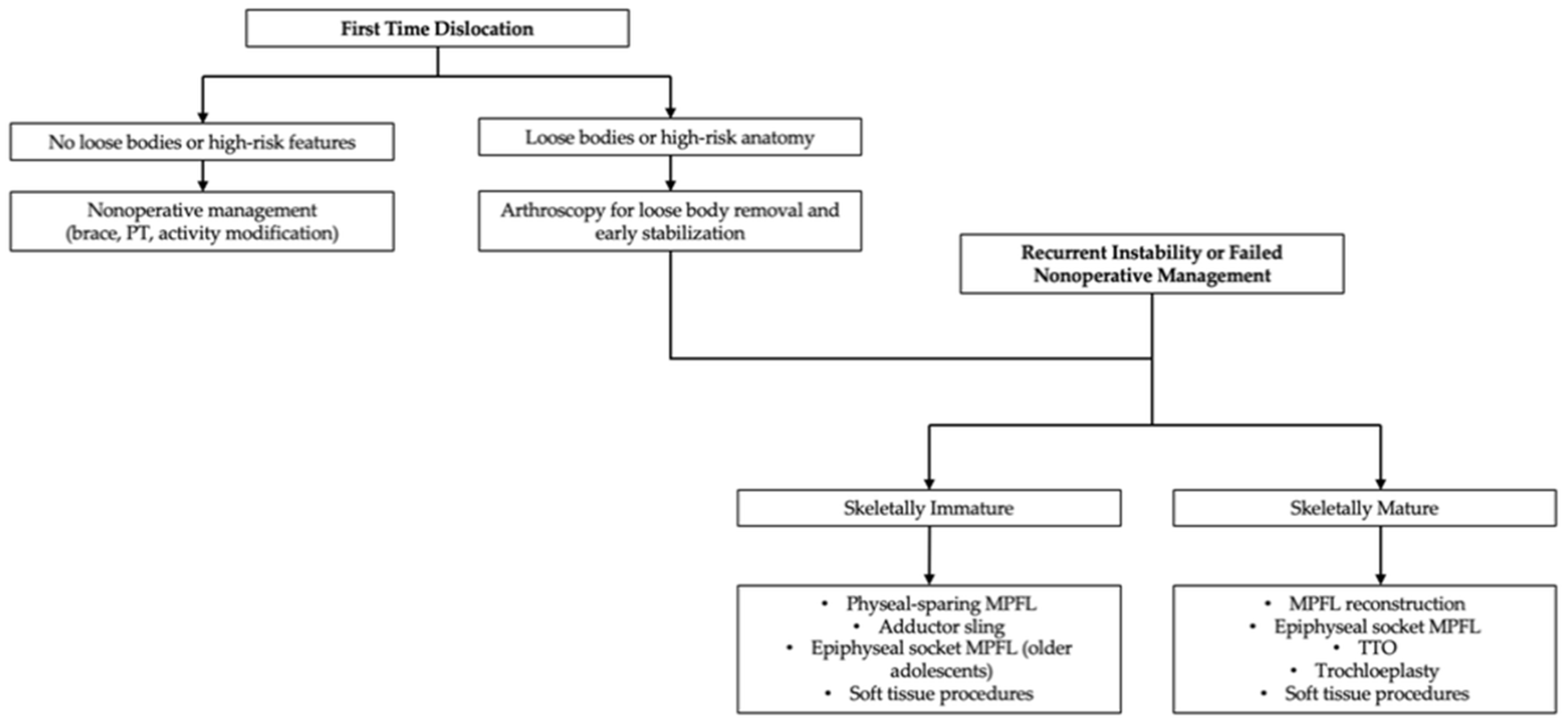
Soft tissue arthroscopic procedures, including lateral release or imbrication, may be performed arthroscopically to address abnormal patellar tracking and reduce lateral patellar tilt. These techniques adjust the tension in surrounding soft tissues to enhance stability. Conversely, bony procedures like TTO involve repositioning of the tibial tubercle to correct biomechanical alignment and reduce lateralization of the extensor mechanism. However, due to the proximity of the growth plates, such bony interventions carry risks of physeal injury and resultant growth disturbances and are typically reserved for patients nearing skeletal maturity. Similarly, trochleoplasty, which reshapes the trochlear groove, is contraindicated in younger patients due to the risk of damage to developing cartilage and growth plates. Thus, this section outlines key arthroscopic techniques and their pediatric-specific considerations, emphasizing the delicate balance required to effectively manage patellar instability while preserving growth potential. Building upon these diagnostic considerations, the following treatment pathway (Figure 2) outlines evidence-based management options tailored to skeletal maturity.

5.1. Medial Patellafeoral Ligament Reconstruction

MPFL reconstruction is the most commonly performed surgical intervention for recurrent lateral patellar instability. The MPFL serves as the primary constraint to lateral translation of the patella between 0° and 30° of knee flexion, with its insufficiency nearly universal after a first-time subluxation [65]. Reconstruction is indicated in patients with recurrent instability or a first-time dislocation accompanied by the presence of anatomical risk factors, loose bodies, or chondral damage [43].
Graft options for MPFL reconstruction include autografts such as the gracilis or semitendinosus tendon, allograft tissue, or synthetic materials. Most pediatric studies favor allografts due to improved graft survivorship, low cost and revision rate, and clinical outcome scores, and reduced donor-site morbidity [66]. Graft fixation techniques vary and may include interference screws, suture anchors, or cortical buttons [67,68]. Autografts remain acceptable alternatives when tendon quality is sufficient. Pediatric reconstructions generally use a 5–6 mm diameter double graft (18–22 cm length), balancing adequate strength with minimized patellar fracture risk [66].
The femoral tunnel position is critical for restoring isometry and avoiding graft over-constraint as incorrect placement may result in altered patellar tracking or increased contact pressures [67,68]. In skeletally immature patients, physeal-sparing MPFL reconstruction is essential to avoid iatrogenic damage to the distal femoral physis [13]. Given the proximity of the MPFLs anatomical femoral insertion (Schöttle point) to the physis, conventional tunnel-based fixation techniques used in adults pose a significant risk if adapted directly in pediatric patients. To mitigate this risk, a number of surgical strategies have been developed that respect the growth plate while restore patellofemoral stability. Techniques include using an epiphyseal femoral socket, soft tissue fixation (adductor sling), and partial tunnel constructs that avoid transphyseal drilling [13,69,70]. These approaches have shown favorable short-term outcomes with low reinjury rates and minimal complications [13,69,70]. Adductor sling techniques utilize the adductor magnus tendon as a point of graft fixation by looping the graft around it, avoiding the need for bony tunnels altogether. This method has been shown to preserve femoral physis integrity and is particularly suitable for younger children with substantial growth remaining [69,70,71,72]. This technique is relatively straightforward, reproducible, and eliminates the risk of physeal injury. Outcomes have demonstrated re-dislocation rates below 10–15% and favorable return-to-sport rates in short- to mid-term series [69,70,71,72]. However, limitations include the potential for graft lengthening or creep over time due to soft-tissue fixation, which may increase the risk of recurrent laxity [71].
Another approach involves creating a small epiphyseal socket, allowing for anatomical femoral fixation within the epiphysis without violating the physis. This technique is generally suited for older children and adolescents nearing skeletal maturity, when the distal femoral epiphysis provides adequate bone stock for a short tunnel. The socket is typically 4–6 mm in diameter and 15–20 mm in depth [73], drilled entirely under fluoroscopic or navigational guidance to ensure accurate placement and to avoid physeal violation [70]. Reported outcomes are favorable, with re-dislocation rates in the 5–15% range and good functional recovery [70]. Complications, though uncommon, include tunnel malposition with epiphyseal breach, hardware irritation, and graft over-constraint if the femoral socket is placed too proximally or posteriorly.
Some surgeons have also used soft tissue-only constructs which involve anchoring the graft to the medial epicondyle periosteum or employing suture anchors placed in the distal femoral epiphysis [74,75,76]. These methods are particularly useful in very young patients with limited epiphyseal bone stock, where even a short socket may be unsafe. Early outcomes demonstrate re-dislocation rates typically under 20%, with satisfactory scores and pain reduction [77]. However, because fixation depends on soft tissue purchase, complications include anchor pull-out, gradual graft laxity, and higher reoperation rates compared to bony fixation [77]. Despite these limitations, soft-tissue fixation remains a valuable physeal-sparing option in the youngest or most skeletally immature patients.
When it comes to patellar fixation, several options exist; however, strategies that minimize fracture risk are recommended in pediatrics. Two low-profile medial anchors or small unicortical sockets are often favored, as they limit bone removal and allow independent limb tensioning [78,79]. Trans-osseous sutures through short, protected tunnels are another alternative, though they carry greater risk of fracture if oversized or malpositioned [80]. Large full-thickness transverse tunnels, while biomechanically strong, are generally avoided in skeletally immature patients due to high fracture risk [80].
Graft fixation is typically performed at 30–45° of knee flexion, with the patella centered in the trochlear groove to preserve near-isometric behavior [81]. Cadaveric biomechanical data confirms that fixation at full extension (0°) raises medial patellofemoral contact pressures, whereas fixation at 30° or 60° restores both pressure profiles and patellar tracking to near-normal levels [82]. To avoid over-constraint, intraoperative tension is set to allow about one quadrant of lateral translation at 30°, with smooth tracking confirmed at 0°, 30°, and 60° [83]. This technique reduces the risk of over-constraint and excessive contact pressures, which can occur if the graft is fixed in extension or over-tensioned.
Following graft fixation, intraoperative checks should confirm isometry through the arc of motion, with minimal length change from 0 to 60°. Patellar tracking should be smooth without excessive medial tightness or an exaggerated J-sign. Fluoroscopic documentation of femoral tunnel positioning and photographic evidence of patellar tracking is recommended for the operative record. Importantly, routine lateral release is discouraged, as it risks overcorrection and has been associated with high rates of postoperative medial instability [84,85,86,87]. In cases where significant lateral retinacular tethering persists despite appropriate graft reconstruction, a graded lateral lengthening may be selectively considered as a safer adjunct [88]. Technical pitfalls that can compromise outcomes include femoral tunnel malposition (too proximal or transphyseal) [89], graft over-tensioning [90], fixation at inappropriate flexion angles [91], patellar fracture from oversized tunnels [89], and neglecting to verify isometry intraoperatively.
When it comes to skeletally immature patients, MRI-based physeal mapping is recommended preoperatively to assess the location and extent of open growth plates and to tailor the surgical approach accordingly. This imaging also aids in identifying other concurrent anatomical risk factors which may influence graft tensioning and positioning. While long-term outcome data remain limited, the literature has reported re-dislocation rates below 25%, with higher rates of return-to-sports and minimal complication profiles in patients undergoing physeal-sparing reconstruction [69,74,92,93].
As surgical instrumentation and imaging techniques continue to evolve, newer methods such as 3D personalized navigation templates [94] and all-epiphyseal tunnel drilling with navigation [13] may help further improve surgical precisions, reproducibility, and patient outcomes with this demographic. Ultimately, MPFL reconstruction in skeletally immature patients demands a careful balance between growth plate preservation and anatomic restoration. Table 3 summarizes fixation strategies for physeal-sparing MPFL reconstruction in skeletally immature patients, highlighting their indications, advantages, limitations, and level of evidence.

5.2. Tibial Tubercle Osteotomy: Caution in Skeletally Immature Patients

TTO is an arthroscopic procedure that is typically reserved for skeletally mature patients with persistent instability and lateralization of the extensor mechanism, in combination with anatomical risk factors such as TT-TG greater than 20 mm and significant patella alta [44]. The goal of TTO is to distalize, medialize, or anteriorize the tibial tubercle to normalize patellar tracking and reduce lateral patellofemoral joint stress [44].
In skeletally immature patients, TTO is contraindicated due to the proximity of the osteotomy site to the tibial tubercle apophysis and the proximal tibial physis [100]. Thus, disruption to the open physes increases the risk of angular deformities, growth disturbances, and physeal arrest [49]. Even with patients in late adolescence, careful assessment of skeletal maturity is essential prior to considering surgical interventions such as TTO to avoid iatrogenic damage to the growth plates. When indicated in skeletally mature adolescents, arthroscopic-assisted planning with radiographic measurements (Caton–Deschamps index, TT-TG, etc.) should help guide the extent and direction of the correction.
One safe method to correct coronal plane malalignment as documented by increased TT-TG or TT-PCL distance is the Roux Goldthwait procedure in which the patellar tendon is split longitudinally in the central portion of the tendon, the lateral half is detached distally and then passed under the medial portion to resecure it with suture stabilization to the periosteum of the proximal tibia just medial to the native medial portion. This can be used in combination with the MPFL reconstruction or in isolation [101].

5.3. Trochleoplasty

In recent years, arthroscopic trochleoplasty has emerged as a minimally invasive alternative to traditional open techniques for the correction of high-grade trochlear dysplasia [102]. This technique is indicated for symptomatic patients with Dejour 2025 high-grade dysplasia [40] (types requiring surgical containment procedures) in whom nonoperative care of isolated soft-tissue procedures are insufficient and should only be considered in adolescents or young adults nearing skeletal maturity. It remains contraindicated in skeletally immature patients with open physes, given the risk of physeal injury and long-term growth disturbances.
Among the most widely described methods is the thin-flap arthroscopic trochleoplasty popularized by Capella and colleagues [102]. This technique involves a stepwise approach: (1) diagnostic arthroscopy to evaluate patellofemoral tracking and associated chondral pathology; (2) use of a C-arm (fluoroscopic) system to localize the limits of the dysplastic trochlea; (3) creation of a thin osteochondral flap over the supratrochlear spur using an arthroscopic burr; (4) recession of the subchondral bone to deepen and reshape the trochlear groove; and (5) fixation of the thin flap back into position, ensuring congruent contour and stability.
Clinical outcomes of arthroscopic trochleoplasty have been increasingly reported. Blønd and Barfod [103] demonstrated that arthroscopic recession trochleoplasty, when performed in conjunction with MPFL reconstruction, significantly improved trochlear morphology on post-operative MRI and led to improve patient-reported outcome scores. These findings suggest that arthroscopic approaches can achieve comparable short- to mid-term outcomes relative to open techniques, with potential benefits in terms of cosmesis and recovery.
Compared with open trochleoplasty, the arthroscopic approach is less invasive, preserves the surrounding soft tissues, and allows for concurrent assessment and treatment of intra-articular pathology. Fluoroscopic assistance provides accurate visualization of the bony contour, reducing the risk of over- or under-correction. Patients may benefit from decreased postoperative pain, faster rehabilitation, and fewer wound-related complications.
However, some challenges should be addressed. Arthroscopic trochleoplasty remains technically demanding, with a steep learning curve. Risks include the creation of an unstable flap if subchondral support is insufficient, or conversely inadequate deepening that leaves residual dysplasia. Over correction may lead to excessive trochlear constraint, while under-correction fails to restore stability. Post-operatively, protocols emphasize protected weight-bearing and progressive return-to-sport after 4–6 months, balancing flap healing with functional recovery [104]. Long-term outcome data are limited with most published studies reporting short- to mid-term follow-up [105,106,107]. Additionally, the safety and durability of the thin-flap technique in younger patients with higher activity levels remain to be validated.
Overall, arthroscopic trochleoplasty represents a promising, less invasive alternative to open trochleoplasty for high-grade dysplasia in skeletally mature adolescents and young adults. Future multicenter studies and long-term follow-up are required to determine its role relative to established open procedures.

5.4. Soft Tissue Procedures: Lateral Release and Imbrication

Soft tissue procedures, such as lateral release and imbrication, can be performed arthroscopically to address patellar maltracking and instability, particularly when excessive lateral retinacular tightness or patellar tilt is identified. Although typically supplementary rather than primary treatments, these methods can be beneficial in patients with less severe instability or minimal anatomical abnormalities [14,61].
Lateral release involves arthroscopically transecting the lateral retinaculum to reduce lateral tethering forces acting on the patella. This can help correct lateral patellar tilt and mildly improve medial patellar tracking. However, isolated lateral release has been increasingly scrutinized due to its inconsistent outcomes, high recurrence rates, and risk of iatrogenic medial instability if overly aggressive or improperly indicated [72,75]. Despite these limitations, lateral release remains useful when appropriately selected for patients with significant lateral retinacular tightness and minimal underlying anatomical deformities, especially when used in conjunction with MPFL reconstruction [61,75].
Medial imbrication involves tightening or reinforcing the medial retinacular and capsular structures, typically performed in combination with MPFL reconstruction or other medial stabilization techniques. This method aims to enhance medial patellar support by restoring proper medial soft tissue tension. Medial imbrication is advantageous for skeletally immature patients where growth plate preservation restricts bony corrective interventions. When effectively executed, medial imbrication can improve patellar tracking and reduce instability without substantial risk to the physes [14,74].
Careful patient selection, based on detailed anatomical assessments and growth considerations, is crucial to optimize the outcomes of these procedures. Anatomical assessments should include thorough evaluation of patellar height, trochlear groove morphology, alignment of the lower extremity, and rotational abnormalities. In skeletally immature patients, careful consideration of growth plate status and potential implications for future growth and development is paramount. Overly aggressive soft tissue correction can lead to postoperative complications like medial over-constraint, which may result in stiffness or limited patellar mobility, requiring further interventions, or reduced patellar tracking that negatively impacts functional outcomes. Therefore, individualized treatment planning that integrates both preoperative imaging findings and clinical examinations, along with precise arthroscopic evaluation, remain essential for successful implementation of these adjunctive soft tissue techniques [14,61,74]. Collaborative approaches involving pediatric orthopedic specialists, rehabilitation therapists, and imaging specialists can further enhance patient outcomes and minimize the risks associated with these procedures.
A summary of patellar instability procedures can be found in Table 4.

6. Innovations and Future Directions

Standard surgical techniques for patellar instability in skeletally immature patients, while effective in many cases, face important limitations that create gaps in clinical application. Conventional MPFL reconstruction, though widely performed, carries risks of physeal injury given the proximity of the femoral insertion into the distal femoral physis. Outcomes may also be compromised by tunnel malposition or graft over-constraint, leading to recurrent instability or altered patellar tracking. Similarly, TTO and trochleoplasty, though effective in skeletally mature patients, are generally contraindicated in younger populations due to the risk of growth disturbances and long-term joint sequelae. Even with physeal-sparing adaptations, soft tissue-only fixation methods may result in graft creep or recurrence, while reliance on two-dimensional imaging can limit the ability to account for complex three-dimensional risk factors such as rotational malalignment. Collectively, these challenges underscore the need for strategies that improve surgical precision, preserve growth potential, and address associated chondral damage.
Advancements in surgical techniques, imaging modalities, and biologic therapies continue to expand treatment options for patellar instability, particularly in skeletally immature patients. Recent innovations, such as robotic-assisted navigation, bioengineered scaffolds, and 3D patient-specific surgical planning, offer promising improvements to enhance tunnel placement accuracy, address the unmet need of concomitant chondral damage, and provide detailed visualization enabling individualized alignment corrections.
Together, these novel approaches (Table 5) hold promise to refine current practices by improving safety in skeletally immature patients, enhancing surgical reproducibility, and offering biologically restorative solutions where standard reconstruction falls short.

6.1. Robot-Assisted Navigation

Robot-assisted navigation is an emerging and increasingly utilized orthopedic procedure used to enhance surgical precision. In the context of MPFL reconstruction, robotic systems facilitate accurate tunnel placement, minimizing the risk of iatrogenic physeal damage in skeletally immature patients. By precisely navigating anatomical landmarks and growth plate boundaries, robotic assistance helps avoid inadvertent damage while optimizing graft positioning and tensioning. A study in 2025 confirmed the effectiveness and safety of robot-assisted individualized MPFL reconstruction, demonstrating accurate femoral tunnel placement within the distal epiphysis, reduced operative variability, and favorable early clinical outcomes, underscoring its potential as a standard adjunct in pediatric patellar instability management [18]. Although early outcomes are encouraging, current data remains limited to small single-center retrospective series (e.g., n = 20–40 patients, follow-up less than 2 year) [18]. These studies demonstrate proof of concept and improved tunnel accuracy but lack long-term multicenter validation.

6.2. Bioengineered Scaffolds for Cartilage Restoration

Bioengineered scaffolds are at the forefront of cartilage repair strategies to restore articular cartilage integrity in patients with recurrent patellar instability. Designed to mimic the extracellular matrix, these scaffolds support chondrocyte proliferation and matrix deposition. Recent advancements include the development of zonal osteochondral scaffolds with bioactive cartilage zones, ultimately resulting in the promotion of endogenous cell recruitment and cartilage regeneration [108]. Such scaffolds can be integrated into arthroscopic procedures offering a minimally invasive solution for cartilage restoration in skeletally immature patients. This technology is particularly advantageous in pediatric orthopedic patients, where preserving joint health and function during crucial development periods is essential for optimal long-term outcomes. However, most published work [108] remains confined to preclinical animal models and early feasibility trials, with very limited pediatric clinical data. Sample sizes are small (less than n = 30), and long-term graft integration and durability remain unproven.

6.3. 3D Modeling and Patient-Specific Surgical Planning

3D modeling and patient-specific surgical planning have revolutionized the way complex orthopedic conditions are approached, particularly in pediatric patients with patellar instability. Through advanced imaging modalities, 3D reconstructions of the patellofemoral joint can be generated to visualize the bony and cartilaginous anatomy in detail [53,109]. These models allow surgeons to evaluate critical anatomical risk factors more accurately than with traditional two-dimensional (2D) imaging alone.
For arthroscopic procedures, 3D modeling improves preoperative planning and intraoperative navigation, especially when treating high-grade trochlear dysplasia or complex instability patterns. In skeletally immature patients, where preserving open growth plates is essential, 3D models help identify safe zones for tunnel placement during MPFL reconstruction, reducing the risk of iatrogenic physeal damage. By simulating different surgical scenarios such as femoral tunnel trajectories or graft lengths, surgeons can individualize procedures based on the patient’s anatomy resulting in safer and more effective surgical outcomes [110,111,112].
Beyond surgical execution, 3D modeling facilitates better communication with patients and families by offering a tangible visualization of the pathology and planned intervention. This is especially valuable in pediatric populations, where patient and parent understanding and engagement are crucial components of informed consent and postoperative compliance.
As the integration of artificial intelligence and automated segmentation tools advances, the efficiency and accessibility of 3D modeling will continue to improve. The incorporation of these technologies into pediatric knee arthroscopy not only enhances surgical safety and precision, but also represents a step towards personalized, data-driven orthopedic care.
Unfortunately, modeling approaches largely derive from case series and pilot studies (sample sizes typically less than 50), that are retrospective in nature. While 3D modeling improves anatomical understanding and operative planning, robust prospective studies comparing outcomes to standard care are lacking [53,109,110,111,112]. Cost and limited access to advanced imaging and software may also restrict widespread adoption.

7. Knowledge Gaps and Research Needs

Despite the growing adoption of arthroscopic techniques for managing patellar instability, important knowledge gaps remain, particularly in skeletally immature populations. Many current surgical workflows and rehabilitation protocols are derived from adult studies, with limited pediatric-specific data available to guide individualized care. Addressing these deficiencies is essential for optimizing outcomes and ensuring safe, evidence-based management of patellar instability in skeletally immature patients.

7.1. Lack of Pediatric-Specific Outcome Measures

Most studies evaluating outcomes after MPFL reconstruction or trochleoplasty utilize adult-derived patient-reported outcome measures (PROMs), such as the Kujala or Lysholm scores. While these tools assess symptoms and functional limitations, they may not fully capture the unique activity demands, growth-related concerns, or psychosocial factors relevant to children and adolescents. There is a need for validated pediatric-specific PROMs that can assess return-to-play, developmental milestones, and long-term joint health following orthopedic interventions, such as patellofemoral surgery [113,114,115]. Thus, incorporating such tools would improve postoperative tracking and allow for more meaningful comparisons across age groups and arthroscopic treatment modalities.

7.2. Need for Long-Term Studies on Skeletally Immature Patients

The long-term impact of MPFL reconstruction, particularly when performed using arthroscopic physeal-sparing techniques, remains relatively understudied. While short-term outcomes have shown low re-dislocation rates and improved function [69,74,92,93], there is limited data on physeal growth patterns, durability of graft integrity, and the development of patellofemoral osteoarthritis over time. Most pediatric studies involve fewer than 5 years of follow-up and focus on recurrence rates, leaving questions about cartilage preservation, growth disturbance, and patient-reported function unanswered. Longitudinal, prospective cohort studies are necessary to establish the safety and efficacy of these procedures, particularly those using arthroscopic techniques designed to minimize physeal injury.

7.3. Standardization of Indications and Rehabilitation Protocols

Variation in clinical practice is common, both in decision to operate and in postoperative management. In the literature, inconsistencies in the threshold for surgical intervention, especially following first-time dislocations in skeletally immature patients have been documented [116]. Anatomical risk factors such as trochlear dysplasia, patella alta, and TT-TG distance are considered but no universally accepted scoring system or decision workflow exists to guide when to proceed with surgery in children. Arthroscopy has become increasingly utilized not only as a therapeutic modality but also as a diagnostic adjunct in equivocal or recurrent cases, enabling real-time assessment of patellofemoral tracking, MPFL integrity under physiological loading conditions, and chondral damage. This capability may support earlier identification of intraarticular pathology not captured on static MRI, thereby influencing operation timing and surgical technique selection.
However, arthroscopic indications themselves remain non-standardized. In some clinics, diagnostic arthroscopy is routinely performed to evaluate for loose bodies and cartilage lesions even after first-time subluxation, while in others, it is reserved for patients with chronic instability and mechanical symptoms or may not used at all. This lack of consensus impacts clinical comparability and research reproducibility.
Likewise, rehabilitation protocols following arthroscopic MPFL reconstruction, lateral release, or trochleoplasty vary widely across institutions. Differences exist in initial weight-bearing status, the use of bracing, timing of range-of-motion exercises, and return-to-sports timelines. Pediatric patients, who differ from adults in healing capacity, compliance, and skeletal maturity, may benefit from arthroscopy-specific, age-adjusted rehabilitation protocols. This highlights the need for consensus-driven, pediatric-specific guidelines to optimize functional recovery and reduce the risk of recurrence or over-restriction during key growth periods [117]. Standardizing arthroscopic indications, procedural approaches, and rehabilitation pathways will be essential to improve outcomes and ensure consistency in the care of skeletally immature patients with patellar instability.

8. Clinical Considerations and Challenges

Managing patellar instability in skeletally immature patients presents several unique clinical challenges that require careful consideration to determine the best procedure for each individual patient. There are three large categories contributing to instability: traumatic injury, mechanical malalignment, and hypermobility syndromes. It is critical to understand the source of instability for each patient. In general, patients who sustain a traumatic injury will report a history of a sports related contact injury or higher energy injury such as a fall from a bicycle. Patients with hyperligamentous laxity syndromes can be detected by a history of other joint instabilities as well as on physical exams, including an assessment of Beighton’s Criteria for ligamentous laxity. It is also important to recognize that any given patient with patellar instability can have sources of instability relevant to two or three of these general classifications. As an example, a 14-year-old girl with hyperligamentous laxity may also have pathology related to mechanical malalignment. Furthermore, it is important to recognize that some patients with patellar instability may have malalignment in more than one plane. It is not uncommon for adolescents with a condition known as “Miserable Malalignment” to have femoral anteversion (malalignment in the axial plane), valgus knee alignment (malalignment in the coronal plane), and pronated feet (this contributes to malalignment in both the coronal and axial planes). Any physical therapy program or surgical plan for treatment must consider all of these factors in order to maximize the potential for success.

9. Rehabilitation

Post-operative rehabilitation following patellar instability reconstruction in skeletally immature patients should follow a structured, phase-based progression with criteria-driven milestones rather than arbitrary time points. Guidelines from leading pediatric institutions such as Massachusetts General Brigham outline a five-phase protocol [118,119] beginning with phase I (0–2 weeks) focusing on surgical protection, pain and swelling control, restoration of full active knee extension and gradual flexion to 60°, with partial weight-bearing and brace locked in extension during ambulation. Phase II (2–6 weeks) progresses to weight-bearing as tolerated, unlocking the brace as quadriceps control permits, achieving knee flexion towards 110–120°, and introducing close-chain strengthening and proprioceptive exercises [118].
Beyond six weeks, criteria-based progression continues, emphasizing limb symmetry in quadriceps strength (≥80% via isokinetic testing) and neuromuscular control assessed via single-leg squat mechanics, before advancing to running and agility drills [104,118,119,120]. Return-to-sports is typically targeted at around 10 months post-op [121], contingent on passing objective functional tests including hop testing (leg symmetry index ≥ 90%), strength symmetry, and absence of pain or effusion.
However, a recent work [121] highlights a critical limitation of hop-based testing. The leg symmetry index may be inappropriate for patients with patellar instability because bilateral deficits in strength and function can normalize the index and mask residual impairments [121]. This suggests that while hop testing remains useful, clinicians should supplement limb symmetry index with absolute performance measures, side-by-side comparisons of mechanics, and validated pediatric-specific PROMs. Notably, Saper et al. [122] reported that only 32% of adolescents achieve satisfactory hop test results at eight months, further underscoring the heterogeneity of recovery and the importance of individualized, multimodal clearance criteria. Together, these findings support a cautious, criteria-driven approach to return to sport that integrates objective strength and functional testing, highlighting the need for PROMs tailored to pediatric populations.

10. Strengths and Limitations

This review provides a comprehensive and clinically relevant synthesis of contemporary approaches to patellar instability in skeletally immature patients. By incorporating innovative techniques such as robotic-assisted navigation, 3D modeling, and bioengineered scaffolds, this review highlights cutting-edge strategies that may soon influence practice. The focus on pediatric-specific considerations, including physeal-sparing MPFL reconstruction, distinguishes this work from prior general reviews. Furthermore, the detailed historical framing of the Dejour classification and menu à la carte provides valuable context for understanding the evolution of surgical decision-making.
However, limitations of this paper should be acknowledged. First, the review is narrative in design, without quantitative meta-analysis, which may introduce selection bias. Second, the available literature is heterogeneous, spanning retrospective series, case reports, and small single-center studies, limiting the generalizability of conclusions. Third, there is a paucity of long-term follow-up, with most studies reporting less than 5 years of outcomes, leaving growth effects and osteoarthritis risk incompletely understood. Fourth, rehabilitation protocols remain variable across institutions, with limited evidence to guide standardized pediatric-specific pathways. Finally, there is a lack of validated pediatric outcome measures, which hampers cross-study comparisons and high-quality evidence synthesis.

11. Conclusions

Arthroscopy has become an essential tool in the diagnosis and management of patellar instability in pediatric and adolescent patients. It enables minimally invasive evaluation of patellofemoral anatomy, identification of chondral lesions, and treatment of intraarticular pathology, such as loose bodies. Arthroscopic techniques also facilitate reconstruction procedures, such as MPFL reconstruction, with improved visualization and precision. When appropriately applied, these procedures can significantly improve knee stability, restore function, and reduce recurrence rates in skeletally immature patients.
Management strategies for patellar instability must be tailored to the unique anatomical and developmental characteristics of each patient. Individualized, growth-aware approaches, such as physeal-sparing techniques, growth modulation procedures, and patient-specific surgical planning, are critical to preserving joint integrity and preventing long-term complications. Preoperative planning using 3D modeling, and advances in biologics and cartilage restoration, further support safe and effective care.
Despite these advancements, important challenges remain. The field would benefit from the development of pediatric-specific outcome measures, standardization of surgical indications, and long-term studies to assess durability and safety. Collaborative, interdisciplinary research teams are essential to address these knowledge gaps. Innovation in imaging, surgical instrumentation, and biologic augmentation should be integrated with clinical expertise to advance care for young patients with patellar instability.
Future studies should prioritize the following: (1) validation of pediatric-specific PROMs capable of capturing activity demands, growth concerns, and psychological impact; (2) multicenter prospective trials evaluating the safety and efficacy of physeal-sparing MPFL techniques; (3) development of international registries to pool large datasets and enable cross-population analyses; (4) cost-effectiveness studies comparing emerging technologies such as robotics and 3D planning to standard approaches; and (5) long-term follow-up (≥10 years) assessing growth plate effects, graft durability, and the risk of secondary patellofemoral osteoarthritis. Together these efforts will create the evidence base necessary to translate current innovation into sustainable, evidence-driven pediatric orthopedic care.
Arthroscopy offers a powerful platform for both current treatment and future innovation. With continued refinement to surgical techniques and research protocols, the field is well positioned to improve the quality, safety, and personalization of care for pediatric and adolescent patients with patellofemoral instability.

Author Contributions

Conceptualization, K.J.; investigation, A.M.; writing—original draft preparation, A.M. and K.J.; writing—review and editing, A.M. and K.J. All authors have read and agreed to the published version of the manuscript.

Funding

This research received no external funding.

Institutional Review Board Statement

Not applicable.

Informed Consent Statement

Not applicable.

Data Availability Statement

Not applicable.

Conflicts of Interest

The authors declare no conflicts of interest.

Abbreviations

The following abbreviations are used in this manuscript:
TT-TGTibial tuberosity to trochlear groove
TT-PCLTibial tuberosity to posterior cruciate ligament
MPFLMedial patellofemoral ligament
3DThree-dimensional
MRIMagnetic resonance imaging
CTComputed tomography
TTOTibial tubercle osteotomy
2DTwo-dimensional
PROMsPatient-reported outcome measures
APAnteroposterior

References

  1. Waterman, B.R.; Belmont, P.J.; Owens, B.D. Patellar Dislocation in the United States: Role of Sex, Age, Race, and Athletic Participation. J. Knee Surg. 2012, 25, 51–58. [Google Scholar] [CrossRef]
  2. Stefancin, J.J.; Parker, R.D. First-Time Traumatic Patellar Dislocation: A Systematic Review. Clin. Orthop. Relat. Res. 2007, 455, 93–101. [Google Scholar] [CrossRef] [PubMed]
  3. Lewallen, L.W.; McIntosh, A.L.; Dahm, D.L. Predictors of Recurrent Instability after Acute Patellofemoral Dislocation in Pediatric and Adolescent Patients. Am. J. Sports Med. 2013, 41, 575–581. [Google Scholar] [CrossRef] [PubMed]
  4. Höhne, S.; Gerlach, K.; Irlenbusch, L.; Schulz, M.; Kunze, C.; Finke, R. Patella Dislocation in Children and Adolescents. Z. Orthop. Unf. 2017, 155, 169–176. [Google Scholar] [CrossRef] [PubMed]
  5. Seeley, M.; Bowman, K.F.; Walsh, C.; Sabb, B.J.; Vanderhave, K.L. Magnetic Resonance Imaging of Acute Patellar Dislocation in Children: Patterns of Injury and Risk Factors for Recurrence. J. Pediatr. Orthop. 2012, 32, 145–155. [Google Scholar] [CrossRef]
  6. Barzan, M.; Maine, S.; Modenese, L.; Lloyd, D.G.; Carty, C.P. Patellofemoral Joint Alignment Is a Major Risk Factor for Recurrent Patellar Dislocation in Children and Adolescents: A Systematic Review. J. ISAKOS 2018, 3, 287–297. [Google Scholar] [CrossRef]
  7. Hasler, C.C.; Studer, D. Patella Instability in Children and Adolescents. EFORT Open Rev. 2016, 1, 160–166. [Google Scholar] [CrossRef]
  8. Jaquith, B.P.; Parikh, S.N. Predictors of Recurrent Patellar Instability in Children and Adolescents after First-Time Dislocation. J. Pediatr. Orthop. 2017, 37, 484–490. [Google Scholar] [CrossRef]
  9. Migliorini, F.; Marsilio, E.; Cuozzo, F.; Oliva, F.; Eschweiler, J.; Hildebrand, F.; Maffulli, N. Chondral and Soft Tissue Injuries Associated to Acute Patellar Dislocation: A Systematic Review. Life 2021, 11, 1360. [Google Scholar] [CrossRef]
  10. Grantham, W.J.; Aman, Z.S.; Brady, A.W.; Rosenberg, S.I.; Turnbull, T.L.; Storaci, H.W.; Dornan, G.J.; LaPrade, R.F. Medial Patellotibial Ligament Reconstruction Improves Patella Tracking When Combined with Medial Patellofemoral Reconstruction: An in Vitro Kinematic Study. Arthrosc. J. Arthrosc. Relat. Surg. 2020, 36, 2501–2509. [Google Scholar] [CrossRef]
  11. Madry, H.; Grün, U.W.; Knutsen, G. Cartilage Repair and Joint Preservation: Medical and Surgical Treatment Options. Dtsch. Arztebl. Int. 2011, 108, 669. [Google Scholar] [PubMed]
  12. Sahin, E.; Tandogan, R.; Liebensteiner, M.; Demey, G.; Kayaalp, A. Management of Patellar Instability in Skeletally Immature Patients. EFORT Open Rev. 2024, 9, 60–68. [Google Scholar] [CrossRef] [PubMed]
  13. Stavinoha, T.J.; Shea, K.G. Physeal Sparing Approaches for MPFL Reconstruction. Curr. Rev. Musculoskelet. Med. 2023, 16, 598–606. [Google Scholar] [CrossRef] [PubMed]
  14. Popkin, C.A.; Bayomy, A.F.; Trupia, E.P.; Chan, C.M.; Redler, L.H. Patellar Instability in the Skeletally Immature. Curr. Rev. Musculoskelet. Med. 2018, 11, 172–181. [Google Scholar] [CrossRef]
  15. Sheng, X.; Guo, L.; Jiang, J.; Liu, Z.; Chen, Y.; Wang, X.; Zhang, X.; Zhao, L.; Wu, M.; Geng, B.; et al. Robot-Assisted Medial Patellofemoral Ligament Reconstruction in the Treatment of Recurrent Patellar Dislocation Can Improve Tunnel Accuracy but Yields Similar Outcome Compared with Traditional Technique. Arthrosc. J. Arthrosc. Relat. Surg. 2025, 41, 2999–3009. [Google Scholar] [CrossRef]
  16. Fritz, B.; Fucentese, S.F.; Zimmermann, S.M.; Tscholl, P.M.; Sutter, R.; Pfirrmann, C.W.A. 3D-Printed Anatomic Models of the Knee for Evaluation of Patellofemoral Dysplasia in Comparison to Standard Radiographs and Computed Tomography. Eur. J. Radiol. 2020, 127, 109011. [Google Scholar] [CrossRef]
  17. Kristin, E.Y.; Beitler, B.; Cooperman, D.R.; Frumberg, D.; Schneble, C.; McLaughlin, W.; Fulkerson, J.P. Three-Dimensional Reproductions for Surgical Decision-Making in the Treatment of Recurrent Patella Dislocation. Arthrosc. Tech. 2023, 12, e807–e811. [Google Scholar]
  18. Liang, Q.; Liao, C.; Zhao, Z.; Li, J.; Zhan, H.; Liu, P.; Kang, X.; Ren, B.; Tian, B.; Zhang, L.; et al. Robot-Assisted Individualized Medial Patellofemoral Ligament Reconstruction in Skeletally Immature Patients with Recurrent Patellar Dislocation: A Single-Center Retrospective Study. Orthop. J. Sports Med. 2025, 13, 23259671251339496. [Google Scholar] [CrossRef]
  19. Mavrogenis, A.F.; Karampikas, V.; Zikopoulos, A.; Sioutis, S.; Mastrokalos, D.; Koulalis, D.; Scarlat, M.M.; Hernigou, P. Orthobiologics: A Review. Int. Orthop. 2023, 47, 1645–1662. [Google Scholar] [CrossRef]
  20. McFarlane, K.H.; Coene, R.P.; Feldman, L.; Miller, P.E.; Heyworth, B.E.; Kramer, D.E.; Kocher, M.S.; Yen, Y.-M.; Milewski, M.D. Increased Incidence of Acute Patellar Dislocations and Patellar Instability Surgical Procedures across the United States in Paediatric and Adolescent Patients. J. Child. Orthop. 2021, 15, 149–156. [Google Scholar] [CrossRef]
  21. Poorman, M.; Talwar, D.; SanJuan, J.; Baldwin, K.; Sutliff, N.; Franklin, C.C. Increasing Hospital Admissions for Patellar Instability: A National Database Study from 2004–2017. Orthop. J. Sports Med. 2019, 7, 2325967119S00145. [Google Scholar] [CrossRef]
  22. VandenBerg, C.D.; Sarkisova, N.; Pace, J.L.; Rhodes, J.; Perea, S.H.; Green, D.W. Current Practice Trends in the Surgical Management of Patellofemoral Instability: A Survey of the Paediatric Research in Sports Medicine (PRiSM) Society. J. Child. Orthop. 2021, 15, 571–576. [Google Scholar] [CrossRef]
  23. Lewallen, L.; McIntosh, A.; Dahm, D. First-Time Patellofemoral Dislocation: Risk Factors for Recurrent Instability. J. Knee Surg. 2015, 28, 303–310. [Google Scholar] [CrossRef] [PubMed]
  24. DeVries, C.A.; Bomar, J.D.; Pennock, A.T. Prevalence of Trochlear Dysplasia and Associations with Patellofemoral Pain and Instability in a Skeletally Mature Population. J. Bone Jt. Surg. 2021, 103, 2126–2132. [Google Scholar] [CrossRef] [PubMed]
  25. Park, S.J.; Won, S.H.; Park, M.S.; Sung, K.H. Normative Values of Tibial Tubercle–Trochlear Groove Distance and Tibial Tubercle–Posterior Cruciate Ligament Distance in Children. Am. J. Sports Med. 2023, 51, 1785–1791. [Google Scholar] [CrossRef] [PubMed]
  26. Daynes, J.; Hinckel, B.B.; Farr, J. Tibial Tuberosity—Posterior Cruciate Ligament Distance. J. Knee Surg. 2016, 29, 471–477. [Google Scholar] [CrossRef]
  27. Su, P.; Jian, N.; Mao, B.; Zhang, Z.; Li, J.; Fu, W. Defining the Role of TT-TG and TT-PCL in the Diagnosis of Lateralization of the Tibial Tubercle in Recurrent Patellar Dislocation. BMC Musculoskelet. Disord. 2021, 22, 52. [Google Scholar] [CrossRef]
  28. Brady, J.M.; Rosencrans, A.S.; Shubin Stein, B.E. Use of TT-PCL versus TT-TG. Curr. Rev. Musculoskelet. Med. 2018, 11, 261–265. [Google Scholar] [CrossRef]
  29. Seitlinger, G.; Scheurecker, G.; Högler, R.; Labey, L.; Innocenti, B.; Hofmann, S. Tibial Tubercle–Posterior Cruciate Ligament Distance: A New Measurement to Define the Position of the Tibial Tubercle in Patients with Patellar Dislocation. Am. J. Sports Med. 2012, 40, 1119–1125. [Google Scholar] [CrossRef]
  30. Rethlefsen, S.A.; Nguyen, D.T.; Wren, T.A.L.; Milewski, M.D.; Kay, R.M. Knee Pain and Patellofemoral Symptoms in Patients with Cerebral Palsy. J. Pediatr. Orthop. 2015, 35, 519–522. [Google Scholar] [CrossRef]
  31. Becher, C.; Fleischer, B.; Rase, M.; Schumacher, T.; Ettinger, M.; Ostermeier, S.; Smith, T. Effects of Upright Weight Bearing and the Knee Flexion Angle on Patellofemoral Indices Using Magnetic Resonance Imaging in Patients with Patellofemoral Instability. Knee Surg. Sports Traumatol. Arthrosc. 2017, 25, 2405–2413. [Google Scholar] [CrossRef] [PubMed]
  32. Alvarez, O.; Steensen, R.N.; Rullkoetter, P.J.; Fitzpatrick, C.K. Computational Approach to Correcting Joint Instability in Patients with Recurrent Patellar Dislocation. J. Orthop. Res. 2020, 38, 768–776. [Google Scholar] [CrossRef] [PubMed]
  33. Ahmad, C.S.; Stein, B.E.S.; Matuz, D.; Henry, J.H. Immediate Surgical Repair of the Medial Patellar Stabilizers for Acute Patellar Dislocation: A Review of Eight Cases. Am. J. Sports Med. 2000, 28, 804–810. [Google Scholar] [CrossRef] [PubMed]
  34. Hawkins, R.J.; Bell, R.H.; Anisette, G. Acute Patellar Dislocations. Am. J. Sports Med. 1986, 14, 117–120. [Google Scholar] [CrossRef] [PubMed]
  35. Palmu, S.; Kallio, P.E.; Donell, S.T.; Helenius, I.; Nietosvaara, Y. Acute Patellar Dislocation in Children and Adolescents: A Randomized Clinical Trial. J. Bone Jt. Surg. 2008, 90, 463–470. [Google Scholar] [CrossRef]
  36. Patel, N.B.; Oak, S.R.; Rogers, K.; Crawford, E.A. The Onset and Development of Patella Alta in Children with Patellar Instability. J. Pediatr. Orthop. 2023, 43, 447–452. [Google Scholar] [CrossRef]
  37. Insall, J.; Salvati, E. Patella Position in the Normal Knee Joint. Radiology 1971, 101, 101–104. [Google Scholar] [CrossRef]
  38. Kurowecki, D.; Shergill, R.; Cunningham, K.M.; Peterson, D.C.; Takrouri, H.S.R.; Habib, N.O.; Ainsworth, K.E. A Comparison of Sagittal MRI and Lateral Radiography in Determining the Insall–Salvati Ratio and Diagnosing Patella Alta in the Pediatric Knee. Pediatr. Radiol. 2022, 52, 527–532. [Google Scholar] [CrossRef]
  39. Suomalainen, J.-S.; Regalado, G.; Joukainen, A.; Kääriäinen, T.; Könönen, M.; Manninen, H.; Sipola, P.; Kokki, H. Effects of Knee Flexion and Extension on the Tibial Tuberosity–Trochlear Groove (TT–TG) Distance in Adolescents. J. Exp. Orthop. 2018, 5, 31. [Google Scholar] [CrossRef]
  40. Dejour, D.H.; Mazy, D.; Pineda, T.; Cance, N.; Dan, M.J.; de Sanctis, E.G. Patellar Instability: Current Approach. EFORT Open Rev. 2025, 10, 378–387. [Google Scholar] [CrossRef]
  41. LaPrade, R.F.; Cram, T.R.; James, E.W.; Rasmussen, M.T. Trochlear Dysplasia and the Role of Trochleoplasty. Clin. Sports Med. 2014, 33, 531–545. [Google Scholar] [CrossRef]
  42. Kazley, J.M.; Banerjee, S. Classifications in Brief: The Dejour Classification of Trochlear Dysplasia. Clin. Orthop. Relat. Res. 2019, 477, 2380–2386. [Google Scholar] [CrossRef]
  43. Huntington, L.S.; Webster, K.E.; Devitt, B.M.; Feller, J.A. Risk Assessment and Management of Primary Patellar Dislocation Is Complex and Multifactorial: A Survey of Australian Knee Surgeons. J. ISAKOS 2021, 6, 333–338. [Google Scholar] [CrossRef] [PubMed]
  44. Stokes, D.J.; Elrick, B.P.; Carpenter, M.L.; Raji, Y.; McQuivey, K.S.; Sherman, S.L.; Frank, R.M. Tibial Tubercle Osteotomy: Indications, Outcomes, and Complications. Curr. Rev. Musculoskelet. Med. 2024, 17, 484–495. [Google Scholar] [CrossRef] [PubMed]
  45. Dejour, D.H.; Mesnard, G.; de Sanctis, E. Updated Treatment Guidelines for Patellar Instability: “Un Menu à La Carte”. J. Exp. Orthop. 2021, 8, 109. [Google Scholar] [CrossRef] [PubMed]
  46. Zaidi, A.; Babyn, P.; Astori, I.; White, L.; Doria, A.; Cole, W. MRI of Traumatic Patellar Dislocation in Children. Pediatr. Radiol. 2006, 36, 1163–1170. [Google Scholar] [CrossRef]
  47. Askenberger, M.; Arendt, E.A.; Ekström, W.; Voss, U.; Finnbogason, T.; Janarv, P.-M. Medial Patellofemoral Ligament Injuries in Children with First-Time Lateral Patellar Dislocations: A Magnetic Resonance Imaging and Arthroscopic Study. Am. J. Sports Med. 2016, 44, 152–158. [Google Scholar] [CrossRef]
  48. Wilson, A.; Afarin, A.; Shaw, C.; Shirley, E.; Pierce, J.; Slakey, J.B. Magnetic Resonance Imaging Findings After Acute Patellar Dislocation in Children. Orthop. J. Sports Med. 2013, 1, 2325967113512460. [Google Scholar] [CrossRef]
  49. Mallinos, A.; Jones, K. The Double-Edged Sword: Anterior Cruciate Ligament Reconstructions on Adolescent Patients—Growth Plate Surgical Challenges and Future Considerations. J. Clin. Med. 2024, 13, 7522. [Google Scholar] [CrossRef]
  50. Shamrock, A.G.; Day, M.A.; Duchman, K.R.; Glass, N.; Westermann, R.W. Medial Patellofemoral Ligament Reconstruction in Skeletally Immature Patients: A Systematic Review and Meta-Analysis. Orthop. J. Sports Med. 2019, 7, 2325967119855023. [Google Scholar] [CrossRef]
  51. Mariani, S.; La Marra, A.; Arrigoni, F.; Necozione, S.; Splendiani, A.; Di Cesare, E.; Barile, A.; Masciocchi, C. Dynamic Measurement of Patello-Femoral Joint Alignment Using Weight-Bearing Magnetic Resonance Imaging (WB-MRI). Eur. J. Radiol. 2015, 84, 2571–2578. [Google Scholar] [CrossRef]
  52. Hansen, P.; Harving, M.; Øhlenschlæger, T.; Brinch, S.; Lavard, P.; Krogsgaard, M.; Boesen, M. Comparison between Conventional MRI and Weight-Bearing Positional MRI Reveals Important Differences in Radiological Measurements of the Patellofemoral Joint. Skelet. Radiol. 2023, 52, 1525–1534. [Google Scholar] [CrossRef]
  53. Schneble, C.A.; Yu, K.; Venkadesan, M.; Cooperman, D.; Beitler, B.; Sieberer, J.; Fulkerson, J. Three-Dimensional Imaging of the Patellofemoral Joint Improves Understanding of Trochlear Anatomy and Pathology and Planning of Realignment. Arthrosc. J. Arthrosc. Relat. Surg. 2025, 41, 130–140. [Google Scholar] [CrossRef]
  54. Pennock, A.T.; Chang, A.; Doan, J.; Bomar, J.D.; Edmonds, E.W. 3D Knee Trochlear Morphology Assessment by Magnetic Resonance Imaging in Patients with Normal and Dysplastic Trochleae. J. Pediatr. Orthop. 2020, 40, 114–119. [Google Scholar] [CrossRef] [PubMed]
  55. Tanaka, M.J.; Elias, J.J.; Williams, A.A.; Demehri, S.; Cosgarea, A.J. Characterization of Patellar Maltracking Using Dynamic Kinematic CT Imaging in Patients with Patellar Instability. Knee Surg. Sports Traumatol. Arthrosc. 2016, 24, 3634–3641. [Google Scholar] [CrossRef] [PubMed]
  56. McNally, E.G.; Ostlere, S.J.; Pal, C.; Phillips, A.; Reid, H.; Dodd, C. Assessment of Patellar Maltracking Using Combined Static and Dynamic MRI. Eur. Radiol. 2000, 10, 1051–1055. [Google Scholar] [CrossRef] [PubMed]
  57. Hermanowicz, K.; Mrozek, T.; Jancewicz, P.; Sar, M.; Hermanowicz, J.; Szajwa, L.; Góralczyk, A. All-Arthroscopic Management of Lateral Patellar Instability. Arthrosc. Tech. 2024, 13, 103055. [Google Scholar] [CrossRef]
  58. Vollnberg, B.; Koehlitz, T.; Jung, T.; Scheffler, S.; Hoburg, A.; Khandker, D.; Hamm, B.; Wiener, E.; Diederichs, G. Prevalence of Cartilage Lesions and Early Osteoarthritis in Patients with Patellar Dislocation. Eur. Radiol. 2012, 22, 2347–2356. [Google Scholar] [CrossRef]
  59. Tanaka, M.J.; Mirochnik, K.; Esfahani, S.A.; Lubberts, B.; Waryasz, G.; Bhimani, R. Arthroscopic Patellofemoral Measurements Can Reliably Assess Patellar Instability. Arthrosc. J. Arthrosc. Relat. Surg. 2022, 38, 902–910. [Google Scholar] [CrossRef]
  60. Mazzolaa, C.; Mantovani, D. Patellofemoral Malalignment and Chondral Damage: Current Concepts. Joints 2013, 1, 27. [Google Scholar]
  61. Hinckel, B.; Smith, J.; Tanaka, M.J.; Matsushita, T.; Martinez-Cano, J.P. Patellofemoral Instability Part 1 (When to Operate and Soft Tissue Procedures): State of the Art. J. ISAKOS 2025, 10, 100278. [Google Scholar] [CrossRef]
  62. Jain, N.P.; Khan, N.; Fithian, D.C. A Treatment Algorithm for Primary Patellar Dislocations. Sports Health 2011, 3, 170–174. [Google Scholar] [CrossRef]
  63. Straume-Næsheim, T.M.; Randsborg, P.-H.; Mikaelsen, J.R.; Sivertsen, E.A.; Devitt, B.; Granan, L.-P.; Årøen, A. Recurrent Lateral Patella Dislocation Affects Knee Function as Much as ACL Deficiency–However Patients Wait Five Times Longer for Treatment. BMC Musculoskelet. Disord. 2019, 20, 318. [Google Scholar] [CrossRef]
  64. Kutschke, M.J.; Albright, J.A.; Winschel, J.M.; He, E.W.; Cruz, A.I.; Daniels, A.H.; Owens, B.D. Increased Risk of Patellofemoral Instability Events and Surgical Management in Patients with Joint Hypermobility Syndromes: A Matched Cohort Analysis. Arthrosc. Sports Med. Rehabil. 2024, 6, 100995. [Google Scholar] [CrossRef] [PubMed]
  65. Nomura, E.; Inoue, M.; Osada, N. Anatomical Analysis of the Medial Patellofemoral Ligament of the Knee, Especially the Femoral Attachment. Knee Surg. Sports Traumatol. Arthrosc. 2005, 13, 510–515. [Google Scholar] [CrossRef] [PubMed]
  66. Hendawi, T.; Godshaw, B.; Flowers, C.; Stephens, I.; Haber, L.; Waldron, S. Autograft vs Allograft Comparison in Pediatric Medial Patellofemoral Ligament Reconstruction. Ochsner J. 2019, 19, 96–101. [Google Scholar] [CrossRef] [PubMed]
  67. Tscholl, P.M.; Ernstbrunner, L.; Pedrazzoli, L.; Fucentese, S.F. The Relationship of Femoral Tunnel Positioning in Medial Patellofemoral Ligament Reconstruction on Clinical Outcome and Postoperative Complications. Arthrosc. J. Arthrosc. Relat. Surg. 2018, 34, 2410–2416. [Google Scholar] [CrossRef]
  68. Stephen, J.M.; Kaider, D.; Lumpaopong, P.; Deehan, D.J.; Amis, A.A. The Effect of Femoral Tunnel Position and Graft Tension on Patellar Contact Mechanics and Kinematics after Medial Patellofemoral Ligament Reconstruction. Am. J. Sports Med. 2014, 42, 364–372. [Google Scholar] [CrossRef]
  69. Alm, L.; Krause, M.; Mull, C.; Frosch, K.-H.; Akoto, R. Modified Adductor Sling Technique: A Surgical Therapy for Patellar Instability in Skeletally Immature Patients. Knee 2017, 24, 1282–1288. [Google Scholar] [CrossRef]
  70. Uppstrom, T.J.; Price, M.; Black, S.; Gausden, E.; Haskel, J.; Green, D.W. Medial Patellofemoral Ligament (MPFL) Reconstruction Technique Using an Epiphyseal Femoral Socket with Fluoroscopic Guidance Helps Avoid Physeal Injury in Skeletally Immature Patients. Knee Surg. Sports Traumatol. Arthrosc. 2019, 27, 3536–3542. [Google Scholar] [CrossRef]
  71. Zhang, Q.; Ying, L.; Han, D.; Ye, L.; Tung, T.-H.; Liang, J.; Liu, P.; Zhou, X. Arthroscopic Reconstruction of the Medial Patellofemoral Ligament in Skeletally Immature Patients Using the Modified Sling Procedure: A Novel Technique for MPFL Reconstruction. J. Orthop. Surg. Res. 2023, 18, 334. [Google Scholar] [CrossRef]
  72. Redler, L.H.; Wright, M.L. Surgical Management of Patellofemoral Instability in the Skeletally Immature Patient. J. Am. Acad. Orthop. Surg. 2018, 26, e405–e415. [Google Scholar] [CrossRef]
  73. Ladenhauf, H.N.; Berkes, M.B.; Green, D.W. Medial Patellofemoral Ligament Reconstruction Using Hamstring Autograft in Children and Adolescents. Arthrosc. Tech. 2013, 2, e151–e154. [Google Scholar] [CrossRef]
  74. Bremond, N.; Prima, R.; Rabattu, P.-Y.; Accadbled, F.; Chotel, F.; Konkel, M.; Eid, A.; Philippe, C.; Godinho, A.; Turati, M.; et al. Isolated MPFL Reconstruction with Soft Tissue Femoral Fixation Technique in 54 Skeletally Immature Patients: Clinical Outcomes at 2 Years Follow-up. A French Multicenter Retrospective Study. Orthop. Traumatol. Surg. Res. 2023, 109, 103530. [Google Scholar] [CrossRef]
  75. Song, J.-G.; Kang, S.-B.; Oh, S.-H.; Han, J.-H.; Shah, D.; Park, H.-J.; Kholmurodov, U.T.; Nha, K.-W. Medial Soft-Tissue Realignment versus Medial Patellofemoral Ligament Reconstruction for Recurrent Patellar Dislocation: Systematic Review. Arthrosc. J. Arthrosc. Relat. Surg. 2016, 32, 507–516. [Google Scholar] [CrossRef] [PubMed]
  76. Arendt, E.A. MPFL Reconstruction for PF Instability. The Soft (Tissue) Approach. Rev. Chir. Orthopédique Traumatol. 2009, 95, 371–374. [Google Scholar] [CrossRef]
  77. Morgan, V.K.; Warrier, A.A.; Credille, K.; Wang, Z.; Elias, T.; Haneberg, E.; Hevesi, M.; Yanke, A.B. Medial Patellofemoral Ligament Reconstruction in Skeletally Immature Patients: A Systematic Review of Outcomes by Fixation Technique. Orthop. J. Sports Med. 2025, 13, 23259671251322724. [Google Scholar] [CrossRef] [PubMed]
  78. Monaco, E.; Criseo, N.; Annibaldi, A.; Carrozzo, A.; Pagnotta, S.M.; Cantagalli, M.R.; Orlandi, P.; Daggett, M. Medial Patellofemoral Ligament Reconstruction Using Gracilis Tendon Graft and “All Suture” Knotless Anchors for Patellar Fixation. Arthrosc. Tech. 2023, 12, e2329–e2334. [Google Scholar] [CrossRef]
  79. Migliorini, F.; Driessen, A.; Quack, V.; Schenker, H.; Tingart, M.; Eschweiler, J. Patellar Fixation Graft via Suture Anchors versus Tunnel Techniques during Isolated MPFL Reconstruction for Recurrent Patellofemoral Instability: A Systematic Review of the Literature. Arch. Orthop. Trauma. Surg. 2020, 140, 1201–1210. [Google Scholar] [CrossRef]
  80. Deasey, M.J.; Moran, T.E.; Lesevic, M.; Burnett, Z.R.; Diduch, D.R. Small, Short, Oblique Patellar Tunnels for Patellar Fixation Do Not Increase Fracture Risk or Complications in MPFL Reconstruction: A Retrospective Cohort Study. Orthop. J. Sports Med. 2020, 8, 2325967120954430. [Google Scholar] [CrossRef]
  81. Burrus, M.T.; Werner, B.C.; Conte, E.J.; Diduch, D.R. Troubleshooting the Femoral Attachment during Medial Patellofemoral Ligament Reconstruction: Location, Location, Location. Orthop. J. Sports Med. 2015, 3, 2325967115569198. [Google Scholar] [CrossRef]
  82. Stephen, J.M.; Kittl, C.; Williams, A.; Zaffagnini, S.; Marcheggiani Muccioli, G.M.; Fink, C.; Amis, A.A. Effect of Medial Patellofemoral Ligament Reconstruction Method on Patellofemoral Contact Pressures and Kinematics. Am. J. Sports Med. 2016, 44, 1186–1194. [Google Scholar] [CrossRef] [PubMed]
  83. Elias, J.J.; Jones, K.C.; Lalonde, M.K.; Gabra, J.N.; Rezvanifar, S.C.; Cosgarea, A.J. Allowing One Quadrant of Patellar Lateral Translation during Medial Patellofemoral Ligament Reconstruction Successfully Limits Maltracking without Overconstraining the Patella. Knee Surg. Sports Traumatol. Arthrosc. 2018, 26, 2883–2890. [Google Scholar] [CrossRef] [PubMed]
  84. Schneider, D.K.; Grawe, B.; Magnussen, R.A.; Ceasar, A.; Parikh, S.N.; Wall, E.J.; Colosimo, A.J.; Kaeding, C.C.; Myer, G.D. Outcomes after Isolated Medial Patellofemoral Ligament Reconstruction for the Treatment of Recurrent Lateral Patellar Dislocations: A Systematic Review and Meta-Analysis. Am. J. Sports Med. 2016, 44, 2993–3005. [Google Scholar] [CrossRef] [PubMed]
  85. Desio, S.M.; Burks, R.T.; Bachus, K.N. Soft Tissue Restraints to Lateral Patellar Translation in the Human Knee. Am. J. Sports Med. 1998, 26, 59–65. [Google Scholar] [CrossRef]
  86. McCarthy, M.A.; Bollier, M.J. Medial Patella Subluxation: Diagnosis and Treatment. Iowa Orthop. J. 2015, 35, 26. [Google Scholar]
  87. Christoforakis, J.; Bull, A.M.J.; Strachan, R.K.; Shymkiw, R.; Senavongse, W.; Amis, A.A. Effects of Lateral Retinacular Release on the Lateral Stability of the Patella. Knee Surg. Sports Traumatol. Arthrosc. 2006, 14, 273–277. [Google Scholar] [CrossRef]
  88. Levy, B.J.; Jimenez, A.E.; Fitzsimmons, K.P.; Pace, J.L. Medial Patellofemoral Ligament Reconstruction and Lateral Retinacular Lengthening in the Skeletally Immature Patient. Arthrosc. Tech. 2020, 9, e737–e745. [Google Scholar] [CrossRef]
  89. Sanguanjit, P.; Rujiraphum, P.; Apivatgaroon, A.; Chernchujit, B. Medium to Long-Term Outcomes of Medial Patellofemoral Ligament Reconstruction Using the Superficial Quadriceps versus a Hamstring Autograft in Patellar Instability Patients. Sci. Rep. 2023, 13, 13353. [Google Scholar] [CrossRef]
  90. Duke, A.J.; Dai, A.; Botros, D.; Leatherwood, W.; Montemurro, N.J.; Richardson, M.; Grossman, M. A Patella-Sided Tensioning Technique for Medial Patellofemoral Ligament Reconstruction. Arthrosc. Tech. 2023, 12, e483–e489. [Google Scholar] [CrossRef]
  91. Lorbach, O.; Zumbansen, N.; Kieb, M.; Efe, T.; Pizanis, A.; Kohn, D.; Haupert, A. Medial Patellofemoral Ligament Reconstruction: Impact of Knee Flexion Angle during Graft Fixation on Dynamic Patellofemoral Contact Pressure—A Biomechanical Study. Arthrosc. J. Arthrosc. Relat. Surg. 2018, 34, 1072–1082. [Google Scholar] [CrossRef]
  92. Zirbes, C.F.; Henriquez, A.; Amanah, A.; Therien, A.D.; Perez-Espina, S.; Dorrestein, E.; Zheng, D.; Lilly, J.; Luo, E.J.; Fox, M.A.; et al. Physeal-Sparing Soft Tissue Realignment in Pediatric Patellofemoral Instability Patients: A Review of Treatment Options and Outcomes. J. Clin. Med. 2025, 14, 1116. [Google Scholar] [CrossRef]
  93. Husen, M.; Milbrandt, T.A.; Shah, V.; Krych, A.J.; Stuart, M.J.; Saris, D.B.F. Medial Patellofemoral Ligament Reconstruction Using Allografts in Skeletally Immature Patients. Am. J. Sports Med. 2023, 51, 1513–1524. [Google Scholar] [CrossRef] [PubMed]
  94. Zhang, W.; Mou, L.; Zhang, S.; Liu, W.; Remila, A.; Han, M.; Xiang, W.; Fang, R. 3D-Printed Individualized Navigation Template versus the Fluoroscopic Guide to Defining the Femoral Tunnel for Medial Patellofemoral Ligament Reconstruction: A Retrospective Study. Medicine 2023, 102, e32729. [Google Scholar] [CrossRef] [PubMed]
  95. Liles, J.; Johnston, T.; Hu, J.; Riboh, J.C. Physeal-Sparing Medial Patellofemoral Ligament Reconstruction with Suture Anchor for Femoral Graft Fixation. Arthrosc. Tech. 2020, 9, e889–e895. [Google Scholar] [CrossRef] [PubMed]
  96. Makovicka, J.L.; Hartigan, D.E.; Patel, K.A.; Tummala, S.V.; Chhabra, A. Medial Patellofemoral Ligament Reconstruction Using All-Soft Suture Anchors for Patellar Fixation. Arthrosc. Tech. 2018, 7, e231–e237. [Google Scholar] [CrossRef]
  97. Uboldi, F.; Gaetano, A.C.S.O.T.; Pini-CTO, M.; Pieroni, A.; Puglia, F.; Ferrua, P.; Priano, D.; Memeo, A. Medial Patellofemoral Ligament Surgery in the Pediatric Population: A Literature Review on the Current State of the Art. Joints 2024, 2, e869. [Google Scholar]
  98. Alshomrani, Y. A Landscape of Recent Literature on the Predictors of Success and Failure in Medial Patellofemoral Ligament Reconstruction. Orthop. Rev. 2025, 17, 138208. [Google Scholar] [CrossRef]
  99. Ye, M.; Zhang, H.; Liang, Q. Clinical Outcomes after Medial Patellofemoral Ligament Reconstruction Using Transosseous Sutures versus Suture Anchors: A Prospective Nonrandomized Controlled Trial. Orthop. J. Sports Med. 2020, 8, 2325967120917112. [Google Scholar] [CrossRef]
  100. Vellios, E.E.; Trivellas, M.; Arshi, A.; Beck, J.J. Recurrent Patellofemoral Instability in the Pediatric Patient: Management and Pitfalls. Curr. Rev. Musculoskelet. Med. 2020, 13, 58–68. [Google Scholar] [CrossRef]
  101. Marsh, J.S.; Daigneault, J.P.; Sethi, P.; Polzhofer, G.K. Treatment of Recurrent Patellar Instability with a Modification of the Roux-Goldthwait Technique. J. Pediatr. Orthop. 2006, 26, 461–465. [Google Scholar] [CrossRef]
  102. Capella, M.; Sabatini, L.; Camazzola, D.; Risitano, S.; D’Antonio, D.; Rea, A.; Massè, A. C-Arm Guidance During Thin Flap Arthroscopic Trochleoplasty for Chronic Patellar Instability. Arthrosc. Tech. 2023, 12, e1579–e1588. [Google Scholar] [CrossRef] [PubMed]
  103. Blønd, L.; Barfod, K.W. Trochlear Shape and Patient-Reported Outcomes after Arthroscopic Deepening Trochleoplasty and Medial Patellofemoral Ligament Reconstruction: A Retrospective Cohort Study Including MRI Assessments of the Trochlear Groove. Orthop. J. Sports Med. 2023, 11, 23259671231171376. [Google Scholar] [CrossRef] [PubMed]
  104. Hysing-Dahl, T.; Inderhaug, E. Rehabilitation after Surgery for Patellar Instability. J. Exp. Orthop. 2024, 11, e12062. [Google Scholar] [CrossRef] [PubMed]
  105. Riedl, G.; Holzer, L.A.; Smekal, V. Comparable Postoperative Outcomes in Patients Treated with Either Open or Arthroscopic Trochleoplasty for Patella Dysplasia. Knee Surg. Relat. Res. 2024, 36, 41. [Google Scholar] [CrossRef]
  106. Ferrua, P.; Compagnoni, R.; Calanna, F.; Randelli, P.S.; Dejour, D. Good Patient Satisfaction with Low Complications Rate after Trochleoplasty in Patellofemoral Instability. Knee Surg. Sports Traumatol. Arthrosc. 2022, 30, 3444–3450. [Google Scholar] [CrossRef]
  107. Høj, S.; Lundegaard, J.K.; Blønd, L.; Lavard, P.; Rechter, A.S.; Dippmann, C.; Barfod, K.W. Open and Arthroscopic Deepening Trochleoplasty Improves Post-Operative Outcomes: A Systematic Review of the Literature Reveals Lack of Comparability between Techniques. Knee Surg. Sports Traumatol. Arthrosc. 2025. online ahead of print. [Google Scholar] [CrossRef]
  108. Golebiowska, A.A.; Intravaia, J.T.; Sathe, V.; Kumbar, S.G.; Nukavarapu, S.P. Engineered Osteochondral Scaffolds with Bioactive Cartilage Zone for Enhanced Articular Cartilage Regeneration. Ann. Biomed. Eng. 2025, 53, 597–611. [Google Scholar] [CrossRef]
  109. Beitler, B.G.; Kunsel, K.; Kristin, E.Y.; Wang, A.; Tommasini, S.M.; Wiznia, D.H.; Fulkerson, J.P. Three-Dimensional Printing of Models of Patellofemoral Joint Articular Cartilage in Patients with Patella Instability for Observing Joint Congruity. Arthrosc. Tech. 2023, 12, e1853–e1858. [Google Scholar] [CrossRef]
  110. Shea, C.; Rustad, A.; Anchustegui, N.G.; Troyer, S.; Dingel, A.; Shea, K.; Ganley, T.J.; Milbrandt, T.A. MPFL Surgical Simulation with Femoral Drill Hole Allowing for Adjustable Graft Tension. Orthop. J. Sports Med. 2019, 7, 2325967119S00044. [Google Scholar] [CrossRef]
  111. Watson, N.A.D.; Duchman, K.R.; Bollier, M.J.; Grosland, N.M. A Finite Element Analysis of Medial Patellofemoral Ligament Reconstruction. Iowa Orthop. J. 2015, 35, 13. [Google Scholar]
  112. Elias, J.J.; Kelly, M.J.; Smith, K.E.; Gall, K.A.; Farr, J. Dynamic Simulation of the Effects of Graft Fixation Errors during Medial Patellofemoral Ligament Reconstruction. Orthop. J. Sports Med. 2016, 4, 2325967116665080. [Google Scholar] [CrossRef]
  113. Morris, E.J.; Gray, K.; Gibbons, P.J.; Grayson, J.; Sullivan, J.; Amorim, A.B.; Burns, J.; McKay, M.J. Evaluating the Use of PROMs in Paediatric Orthopaedic Registries. Children 2023, 10, 1552. [Google Scholar] [CrossRef]
  114. Kunze, K.N.; Madjarova, S.; Jayakumar, P.; Nwachukwu, B.U. Challenges and Opportunities for the Use of Patient-Reported Outcome Measures in Orthopaedic Pediatric and Sports Medicine Surgery. J. Am. Acad. Orthop. Surg. 2023, 31, e898–e905. [Google Scholar] [CrossRef] [PubMed]
  115. Phillips, L.; Carsen, S.; Vasireddi, A.; Mulpuri, K. Use of Patient-Reported Outcome Measures in Pediatric Orthopaedic Literature. J. Pediatr. Orthop. 2018, 38, 393–397. [Google Scholar] [CrossRef] [PubMed]
  116. Khormaee, S.; Kramer, D.E.; Yen, Y.-M.; Heyworth, B.E. Evaluation and Management of Patellar Instability in Pediatric and Adolescent Athletes. Sports Health 2015, 7, 115–123. [Google Scholar] [CrossRef] [PubMed]
  117. Machado, S.A.F.; Pinto, R.A.P.; Antunes, A.J.A.M.; de Oliveira, P.A.R. Patellofemoral Instability in Skeletally Immature Patients. Porto Biomed. J. 2017, 2, 120–123. [Google Scholar] [CrossRef]
  118. Massachusetts General Brigham Sports Medicine Pediatric Rehabilitation Protocol for Medial Patellofemoral Ligament (MPFL) Reconstruction 2024. Available online: https://www.massgeneral.org/assets/mgh/pdf/orthopaedics/sports-medicine/physical-therapy/pediatric-mpfl-protocol.pdf (accessed on 2 October 2025).
  119. Lampros, R.E.; Wiater, A.L.; Tanaka, M.J. Rehabilitation and Return to Sport after Medial Patellofemoral Complex Reconstruction. Arthrosc. Sports Med. Rehabil. 2022, 4, e133–e140. [Google Scholar] [CrossRef]
  120. Meynard, P.; Malatray, M.; Sappey-Marinier, E.; Magnussen, R.A.; Bodiou, V.; Lustig, S.; Servien, E. Medial Patellofemoral Ligament Reconstruction for Recurrent Patellar Dislocation Allows a Good Rate to Return to Sport. Knee Surg. Sports Traumatol. Arthrosc. 2022, 30, 1865–1870. [Google Scholar] [CrossRef]
  121. Hysing-Dahl, T.; Magnussen, L.H.; Faleide, A.G.H.; Inderhaug, E. Feasibility of Return to Sports Assessment 6 Months after Patellar Instability Surgery. BMC Musculoskelet. Disord. 2023, 24, 662. [Google Scholar] [CrossRef]
  122. Saper, M.G.; Fantozzi, P.; Bompadre, V.; Racicot, M.; Schmale, G.A. Return-to-Sport Testing after Medial Patellofemoral Ligament Reconstruction in Adolescent Athletes. Orthop. J. Sports Med. 2019, 7, 2325967119828953. [Google Scholar] [CrossRef]
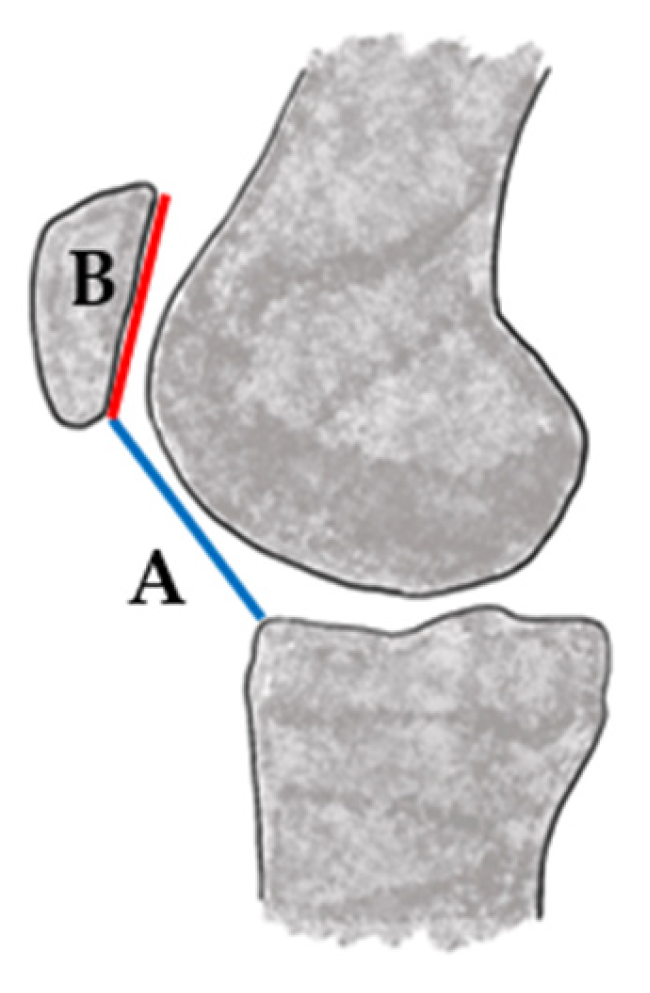
Figure 1. Caton–Deschamps index (A/B): obtained by dividing the distance from the inferior pole of the patella to the tibial plateau (A—blue) by the length of the patellar articular surface (B—red).
Jcm 14 07085 g001
Figure 2. Treatment pathway for patellar instability management.
Jcm 14 07085 g002
Table 1. Diagnostic Classification Systems and Thresholds for Patellar Instability. Italicized term is a French phrase that has been incorporated into English usage but is still treated as a loan expression.
Classification/
Measure
Risk FactorMethodThreshold for
Abnormality
NotesLimitations
Caton–Deschamps Index [36]Patella altaRatio of distance from inferior patellar pole to tibial plateau divided by the length of the patellar articular surface on lateral radiographGreater than 1.2 indicates patella altaPreferred in pediatric populations due to growth plate visibility
Sensitive to knee flexion angle at the time of imaging; does not account for trochlear morphology or dynamic engagement
Insall–Salvati Ratio [38]Patella altaRatio of patellar tendon length to patellar lengthGreater than 1.2 indicates patella altaCommon in adults, but less reliable in immature knees. Some success in skeletally immature populations [38]
Inconsistent reproducibility in pediatric knees
TT-TG Distance [25]Lateralization to tibial tuberacleMeasured on axial MRI or computer tomography (CT) as horizontal distance between tibial tubercle and trochlear grooveGreater than 20 mm considered abnormalGreater than 20 mm associated with instability; 15–20 mm may be borderline and context-dependent in pediatric cases [39]
Values vary with imaging modality (MRI vs. CT) and knee flexion angle
Dejour Classification (2025) [40]Trochlear dysplasiaIntegrated assessment using lateral radiographs and MRI; emphasizes functional evaluation of patellofemoral trackingRedefined dysplasia categories with emphasis on dynamic morphology; linked directly to updated menu à la carte treatment framework. Supersedes earlier A-D system; provides improved risk stratification and clearer surgical indications (trochleoplasty reserves for select high-grade dysplasia, not skeletally immature patients) [40]Requires advanced imaging; some parameters prone to inter-observer variability
TT-PCL [26,27,28,29]Rotational alignment, lateralization of the tibial tubercleMeasured on axial MRI or CT as the distance from the tibial tubercle to the posterior cruciate ligament insertionGreater than 20 mm is typically considered abnormal [29]Used alongside TT-TG distance to evaluate rotational malalignment and refine surgical planning, especially important in skeletally immature patientsNormative pediatric values not well established
Italicized term (menu à la carte) is a French phrase that has been incorporated into English usage but is still treated as a loan expression.
Table 2. Updated Dejour [40] MRI Parameters for Patellofemoral Instability.
ParameterDefinitionImaging PlaneThresholdClinical Significance
Spur heightBony supratrochlear spur heightSagittal>5 mmIndicates high-grade trochlear dysplasia
Cranial trochlear orientationOrientation of proximal trochlea vs. posterior condylesAxialPositive valueReflects cranial malorientation; predicts patellar maltracking
Trochlear groove heightAngle between trochlear groove and posterior condylar axisCoronal>11°Excessive obliquity indicates severe dysplasia
Patellar height indexRatio of patellar length to articular contact lengthSagittal>1.16Identifies patella alta, risk of delayed trochlear engagement
Sagittal patellofemoral engagementRatio of engaged patella length to total patellar lengthSagittal<0.38Low value = poor engagement, instability risk
TT-TG distanceHorizontal distance tibial tubercle to trochlear grooveAxial≥14 mmPathologic lateralization, informs consideration for TTO
Table 3. Comparative Fixation Strategies for Physeal-Sparing MPFL Reconstruction in Skeletally Immature Patients.
SiteFixationIndicationsAdvantagesLimitations/RisksEase of UseEvidence
FemurShort epiphyseal socket + interface screwsEpiphyseal anatomy permits short tunnel distal to physisAnatomic fixation; firm stabilityRisk of physeal breach; hardware in small epiphysis
Moderate
Level IV
FemurShort socket + cortical button/adjustable loopNeed for fine tension control; physeal respectMicro-adjustable tension; preserves epiphysisRequires cortical bridge; hardware prominence
Moderate
Level IV [13,95]
FemurAdductor sling (soft tissue loop)Younger patients; desire to avoid drillingNo bone tunnels; fully physeal sparingCreep/lengthening; soft tissue isometry dependent
Moderate
Level IV [69,70,71,72]
Femur Epiphyseal suture anchorsTunnel-free physeal sparingMinimal bone removalPull-out risk; limited long-tern outcomesEasy-ModerateLevel III–IV [95,96]
PatellaMedial anchors (2 low profile) Most pediatric casesMinimizes fracture risk; small socketsAnchor pull-out; costEasy-ModerateLevel III–IV [78,79,97,98]
PatellaProtected unicortical tunnelsWhen anchors unavailableStrong fixation; inexpensiveRisk of fracture if bicortical or oversizedModerateLevel IV [98,99]
PatellaFull-thickness transverse tunnelsHistorically usedHigh stiffnessHigh fracture risk in pediatricsModerateConsensus to avoid [80]
Table 4. Comparison of Patellar Instability Procedures by Skeletal Maturity.
TechniqueIndicationSkeletally ImmatureAdvantagesLimitationsLevel of Evidence
Physeal-sparing MPFLRecurrent instability, open physesPreferredPreserves growth plates; good outcomesTechnical complexity; limited long-term dataLevel III–IV [43,65,66,67,68]
Adductor slingYounger children, avoid tunnelsYesNo bone drilling; physeal safeGraft creep riskLevel IV [69,70,71,72]
Epiphyseal socket MPFLOlder adolescents; nearing maturitySelectiveAnatomic fixation
Risk of epiphyseal breach
Level IV [13,69,70]
TTOTT-TG > 20 mm, patella altaContraindicatedCorrects alignmentGrowth disturbance riskLevel III [44,49,100,101]
TrochleoplastySevere trochlear dysplasiaContraindicatedCorrects bony dysplasiaContraindicated in open physesLevel III [102,103,104,105,106,107]
Soft tissue procedures Adjunctive instabilityYesMinimally invasiveVariable outcomesLevel IV [14,61,72,74,75]
Table 5. Technology Readiness and Evidence Quality for Innovations in Pediatric Patellar Instability.
InnovationEvidence StrengthClinical AvailabilityCostLearning Curve
Robot-assisted navigation [18]Small single-center retrospective studies (n = 20–40; <2 years follow up)Available in tertiary centers onlyHigh capital/equipment costRequires specialized training
Bioengineers Scaffolds [108]Mostly preclinical and animal modelsExperimental, not widely availableVariable, currently highStraightforward arthroscopic integration if validated
3D Modeling and Patient-Specific Planning [53,109,110,111,112]Small retrospective series (n < 50); promising feasibilityGrowing availability at academic centersModerate (software)Moderate learning curve, aids planning
Disclaimer/Publisher’s Note: The statements, opinions and data contained in all publications are solely those of the individual author(s) and contributor(s) and not of MDPI and/or the editor(s). MDPI and/or the editor(s) disclaim responsibility for any injury to people or property resulting from any ideas, methods, instructions or products referred to in the content.