Abstract
Objectives: Among the spectrum of spinal injuries, Spinal Cord Injury Without Radiographic Abnormality (SCIWORA) occupies a unique and challenging position. SCIWORA presents diagnostic and therapeutic challenges due to its broad clinical and radiological heterogeneity. While most children recover favorably with conservative treatment, a subset may require surgery based on imaging findings. The findings underscore the need for standardized diagnostic criteria, MRI-based classification systems, and evidence-based treatment algorithms to improve consistency in care and long-term neurological outcomes. Methods: A systematic search of PubMed, Cochrane, Scopus, and Embase databases was performed through June 2025 following PRISMA guidelines. Inclusion criteria encompassed studies of pediatric SCIWORA (age < 18 years) reporting demographics, clinical and radiological features, management, and outcomes. Results: Sixty studies encompassing a total of 848 pediatric patients were included. The mean patient age was 9.33 years (±2.52), with a slight male predominance. The most common trauma mechanisms were road traffic accidents (40.3%), sports injuries (22%), and falls (18.8%). MRI findings were available in 399 cases: 46% had intraneural lesions (Type IIb), 39% showed no abnormality on MRI (Type I, or “real SCIWORA”), 9% had combined lesions (Type IIc), and 6% had extraneural abnormalities (Type IIa). Neurological severity at presentation was primarily ASIA Grade A (46.25%), but follow-up data showed substantial improvement, with ASIA E (normal function) increasing to 49.78%. Overall, 66.2% of patients experienced neurological improvement, while 33.8% remained stable. Conservative treatment was employed in 95.41% of cases. Only 4.59% underwent surgery, which was typically reserved for MRI-positive lesions demonstrating spinal instability or compression. Conclusions: Pediatric SCIWORA remains an uncommon but potentially devastating injury, with an outcome highly dependent on MRI findings and initial neurological status. This systematic review aims to clarify the contemporary understanding of pediatric SCIWORA, delineating “real” SCIWORA from other SCIWORA-like entities, and synthesizing the latest evidence regarding epidemiology, mechanisms, clinical presentation, MRI findings, and management in children.
1. Introduction
Traumatic spinal cord injury in children is a rare but potentially devastating event, often resulting in significant lifelong sequelae with profound social and psychological impact. Among the spectrum of spinal injuries, Spinal Cord Injury Without Radiographic Abnormality (SCIWORA) occupies a unique and challenging position. Initially defined in 1982 by Pang and Wilberger, SCIWORA described cases of traumatic myelopathy in children who presented with objective neurological deficits after trauma, but with no evidence of vertebral fracture or dislocation on X-rays or computed tomography (CT) [1]. With the subsequent advent and widespread use of magnetic resonance imaging (MRI), the concept and boundaries of SCIWORA have evolved—now encompassing a heterogeneous group of injuries and sparking debate over its precise definition [2,3,4]. The reported incidence of SCIWORA among pediatric spinal cord injuries varies widely, ranging from 13% to 42%, due in part to differences in imaging protocols and diagnostic criteria. Young children are especially vulnerable, attributable to unique anatomical and biomechanical features of the immature spine: greater ligamentous laxity, shallow and horizontally oriented facet joints, incomplete ossification, and a relatively large head-to-body ratio. These features allow for a high degree of vertebral column motion and “stretchability,” potentially surpassing the physiological limits of the spinal cord itself, thus predisposing to spinal cord damage in the absence of radiographically detectable skeletal injury [5,6,7]. Despite advances in imaging, uncertainty persists regarding the nosology of SCIWORA. While MRI is now considered mandatory for all suspected cases, studies reveal that a considerable proportion (“real” SCIWORA) show no abnormality even on high-resolution MRI, raising questions about the underlying pathophysiology and whether other conditions might be misclassified under this label, such as spinal cord concussion or transient neuropraxia [8,9,10,11]. Conversely, the identification of subtle intramedullary or extraneural lesions on MRI has led some authors to advocate for alternative nomenclature and more granular classification schemes [12,13,14]. Clinically, pediatric SCIWORA displays a broad spectrum: from transient and rapidly resolving neurological deficits to severe, permanent paralysis. The timing of symptom onset is variable, with some patients exhibiting delayed deficits, further complicating diagnosis [15,16,17,18,19]. MRI findings, when present, are highly heterogeneous and include cord edema, hemorrhage, contusion, or soft tissue injury patterns; however, the prognostic implications of these imaging features are incompletely understood. Several studies and consensus statements, including those by international neurosurgical societies, have attempted to refine diagnostic criteria and propose severity classifications, yet a universally accepted definition remains elusive [20,21]. Given the ongoing ambiguities in definition, diagnosis, and optimal management, the primary aim of this systematic review is to clarify the contemporary understanding of pediatric SCIWORA. Specifically, this review aims to establish what truly constitutes SCIWORA in the era of advanced imaging, delineating “real” SCIWORA from other SCIWORA-like entities, and synthesizing the latest evidence regarding epidemiology, mechanisms, clinical presentation and course, and MRI findings in children [22,23,24].
2. Materials and Methods
2.1. Search Strategy and Eligibility Criteria
A systematic literature search was performed following PRISMA guidelines. Electronic databases (PubMed, Cochrane, Scopus, Embase) were searched up to June 2025, using terms including “SCIWORA,” “spinal cord injury without radiographic abnormality,” “spinal cord injury without radiological abnormality,” “spinal cord injury with normal radiographs,” “MRI-negative spinal cord injury,” “pediatric,” “children,” and “child,”. Studies were included if they: (1) enrolled patients aged < 18 years, (2) described cases of acute spinal cord injury following trauma, (3) had no radiographically visible fractures or dislocations on plain films/CT, and (4) provided data on demographics, mechanism, imaging, clinical presentation, management, or outcome. Reviews, animal studies, and case series/individual cases lacking sufficient detail were excluded.
2.2. Data Collection Process
After the removal of duplicates, an initial screening of titles and abstracts was performed independently by two authors (D.P. and M.G.). Articles without an available abstract or lacking the relevant data were excluded. A full-text review of the remaining studies was then conducted to determine eligibility. In cases of disagreement between the reviewers, a senior author (L.O.) was consulted to reach a final decision.
2.3. Risk of Bias Assessment
The risk of bias of the included studies was systematically assessed using the Joanna Briggs Institute (JBI) critical appraisal tools [25]. Specifically, the JBI checklist for case reports and case series was applied, according to the study design. Two reviewers (D.P and M.G) independently evaluated each study, and disagreements were resolved through discussion or consultation with a senior author (L.O).
2.4. Data Extraction and Synthesis
Data extracted included: study design; sample size; age and sex; injury mechanism; spinal level(s) involved; American Spinal Injury Association Impairment Scale (ASIA) at admission and discharge; management approach; and outcomes (neurological improvement, complications, mortality).
MRI findings were classified according to the system proposed by Boese and colleagues [14]. Four imaging patterns were distinguished: Type I, with no detectable abnormalities; Type IIa, showing extraneural abnormalities (e.g., ligamentous or disc lesions); Type IIb, characterized by intraneural abnormalities such as cord edema, hemorrhage, or contusion; and Type IIc, with combined intra- and extraneural abnormalities. This classification was applied to improve comparability across studies and to explore potential correlations between imaging patterns and clinical outcomes.
2.5. Data Analysis
Data were summarized descriptively. For categorical variables (e.g., mechanism, level, outcome), frequencies and percentages were calculated. For continuous variables (e.g., age), means and standard deviations were recorded. Tables were prepared to present patient demographics, injury characteristics, MRI findings, management strategies, and outcomes.
3. Results
3.1. Study Selection
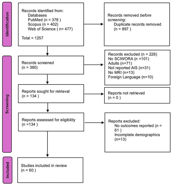
After removal of duplicates, 1257 articles were identified for title/abstract screening. 360 reports underwent full-text review, of which 60 met all criteria and were included (Figure 1 PRISMA flowchart).

Figure 1.
PRISMA flowchart diagram.
3.2. Risk of Bias Assessment Results
Most of the included studies fulfilled the main JBI criteria, with clear objectives, adequate description of populations, and consistent outcome reporting. Only a minority lacked detailed follow-up information or explicit inclusion criteria. Overall, the methodological quality was acceptable and allowed a reliable descriptive synthesis of the available evidence. Results are summarized in Supplementary Tables S1 and S2.
3.3. Study Characteristics and Population Overview
A total of 848 pediatric patients diagnosed with SCIWORA were analyzed from 60 included studies. As shown in Table 1, the mean age was 9.33 years (±2.52), with a slight male predominance (54% male, 46% female).

Table 1.
Patients’ demographics, clinical and radiological data. Trauma mechanism: sport/fall/RTA (road traffic accident); MRI types: I, no abnormalities; IIa, extraneural abnormalities; IIb, intraneural abnormalities; IIc, intraneural and extraneural abnormalities; Onset: negative sign (−): immediate onset; positive sign (+), delayed onset; Therapy: C, conservative; S, surgical; (S). NA, data not available.
3.4. Trauma Mechanisms and MRI Findings
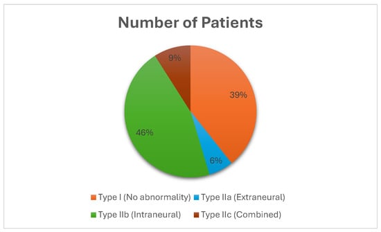
Road traffic accidents (RTAs) were the predominant trauma mechanism, accounting for 40.3% of cases, followed by sports-related injuries (22%), falls (18.8%), and other causes (13.8%). MRI findings were reported for 399 patients. Intraneural abnormalities (Type IIb) represented the most common MRI lesion (46%), followed by “real SCIWORA” cases with no abnormalities (Type I, 39%). Combined lesions (Type IIc, 9%) and extraneural abnormalities (Type IIa, 6%) were less frequent, as shown in Figure 2.

Figure 2.
This pie chart illustrates the distribution of MRI findings in 848 pediatric patients diagnosed with SCIWORA. Only 399 MRI results were reported. Intraneural abnormalities (Type IIb) were the most frequent finding (46%), followed by cases with no radiological abnormalities (Type I, 39%), referred to as “real SCIWORA.” Combined lesions (Type IIc) accounted for 9%, and extraneural abnormalities (Type IIa) were the least frequent (6%). These results underscore the heterogeneity of radiological patterns and the diagnostic value of MRI in guiding prognosis and management.
3.5. Neurological Outcomes
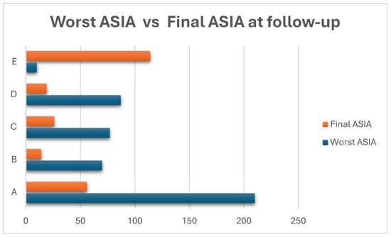
Table 2 and Figure 3 show the neurological initial and final status of the reported cases. Neurological outcomes were assessed in 454 patients using the ASIA impairment scale. At presentation, ASIA Grade A was the most frequent classification (46.25%), indicating complete spinal cord injury. However, substantial neurological improvement was observed at follow-up, with ASIA Grade A decreasing to 24.45% and ASIA Grade E (normal neurological function) increasing significantly from 4.54% to 49.78%. Overall, 66.2% of patients showed neurological improvement, while 33.8% remained stable.

Table 2.
ASIA grading at admission and discharge.

Figure 3.
This horizontal bar chart illustrates the distribution of ASIA grades at the time of worst neurological impairment during the hospitalization (blue bars) and at final follow-up (red bars) in 454 pediatric SCIWORA patients. At presentation, nearly half of the patients (46.25%) were classified as ASIA A, indicating complete spinal cord injury. However, at follow-up, the number of patients remaining in ASIA A decreased substantially to 24.45%, suggesting partial or complete neurological recovery in a significant portion. Conversely, the number of patients with normal neurological function (ASIA E) increased from only 4.54% at baseline to nearly 50% at follow-up. The remaining ASIA grades (B, C, and D) followed a similar trend, with decreases in severe grades and increases in less severe or normal findings.
3.6. Conservative Management and Surgical Treatment
Out of 848 patients, 95.41% were treated conservatively with medical therapy, while 39 (4.59%) underwent surgical intervention primarily for cervical lesions (Table 3). Types of surgical procedures included halo-gravity traction, decompressive laminectomy, fusion, and stabilization. Controversially, three patients underwent lysis of the filum terminale for associated tight filum, highlighting ongoing ambiguity in surgical indications for SCIWORA cases [63].

Table 3.
Operative cases and surgery type.
4. Discussion
The original term, as coined by Pang and Wilberger, refers to traumatic myelopathy with normal plain radiographs and CT scans. With MRI now routine, the landscape is more complex: of 848 pediatric cases aggregated in our review, 39% had “real SCIWORA” (normal MRI, Type I), while the majority disclosed some form of MRI-detectable abnormality (intraneural 46%, extraneural 6%, combined 9%). Such variability reflects the ongoing dispute in the literature, where SCIWORA retains its significance and complexity, particularly in pediatrics.
Some authors now reserve “real SCIWORA” solely for cases entirely negative on X-ray, CT, and MRI, while “SCIWORA-like” encompasses patients with neural or extraneural MRI changes absent on earlier imaging. As multiple consensus statements and recent meta-analyses highlight, the lack of a uniform definition creates inconsistent reporting, complicates inter-study comparisons, and directly impacts management strategies and prognostication. This systematic review underscores this, with a substantive minority (39% overall) qualifying as “real SCIWORA,” nearly reflecting the 43% of patients reported in previous reviews [14].
4.1. Mechanisms of Trauma, Clinical Presentation, and MRI
Out of 848 patients, trauma mechanism analysis (Table 1; Figure 2) reported sports injuries (41%), motor vehicle accidents (RTA; 22%), and falls from height (19%) as the prevalent causes, closely aligning with robust epidemiological studies [6,10,48].
Age also plays a central role: the mean age in the cohort is 9.33 ± 2.52 years, but mechanisms and injury levels differ with age. Young children (especially those < 8 years) have a higher proportion of high cervical and thoracic injuries and are more likely to suffer from motor vehicle accidents and falls. In contrast, older children and adolescents are predominantly affected by sports trauma. Consistent with the literature, delayed onset of neurological symptoms after injury (defined as >6 h post-trauma) was noted in 18% of the reported cases.
Crucially, MRI has shifted the diagnostic paradigm: only 39% of reported SCIWORA cases now have normal scans, with the remainder showing varying patterns of cord edema, hemorrhage, or extraneural injury. 60.6% of the patients reported had an MRI positivity, based on the MRI classification proposed by Boese et al., which robustly stratifies risk [14].
Clinical correlation is strong: Type I cases in this review (including the 32-patient series by Freigang et al.) mostly had full neurologic recovery, affirming Boese’s and Carroll’s findings that normal MRI predicts excellent prognosis in pediatric SCIWORA. Conversely, Type IIb/IIc patients, particularly those with intramedullary hemorrhage, had persistently worse outcomes [14,45,46,47,48,51,56].
Beyond these prognostic categories, several radiological studies have emphasized the importance of more detailed MRI assessment in pediatric SCIWORA [61,64]. Advanced analyses have shown that not only the presence, but also the extent of cord involvement, including lesion length and maximum cross-sectional area, strongly correlates with neurological recovery [61]. Farrell et al. further underlined that the choice of MRI sequences and timing of acquisition may critically influence the detection of subtle abnormalities, while at the same time, issues of sedation in young children remain a practical challenge for protocol optimization [64].
4.2. ASIA Outcomes, Neurological Improvement Rates, and Comparative Analysis
Neurological severity as measured by the American Spinal Injury Association Impairment Scale at admission strongly influences both short- and long-term outcomes. Across both our data and the literature, the spectrum at presentation ranges from complete injury (ASIA A) through incomplete (ASIA B, C, D), with a higher proportion of complete lesions in younger children [34,61]. Notably, in this series, two-thirds of patients achieved at least one-grade improvement on the ASIA at latest follow-up, a finding that closely mirrors the 39–67% improvement rates cited in large series and meta-analyses [31,54,62].
The literature and results reported here agree: the best prognoses are observed in children with normal MRIs or isolated cord edema, especially those with incomplete initial deficits [5,29,62,65]. In contrast, complete injuries (ASIA A) and those with cord hemorrhage rarely show substantial improvement, underscoring the value of early and precise prognostication. However, long-term sequelae, including neurogenic bladder and progressive spinal deformities, are common, particularly in those with poor initial grade [14,22].
4.3. Lack of Consensus and General Management Approaches
Management of pediatric SCIWORA remains equally controversial and poorly standardized. While most patients are managed conservatively with immobilization and activity restriction, the optimal duration and modality of immobilization, the role of surgery in specific subtypes, and even the potential use (or harm) of high-dose corticosteroids are all debated. Early literature advocated for prolonged rigid immobilization to prevent the recurrence of injury, yet more recent data suggest that individualized therapy informed by MRI may be more appropriate [20,44].
Conservative management, characterized by immobilization and rehabilitation, was predominant in this study (95.41%), while in only 4.59% of cases was surgical intervention required. This aligns with most major series affirming strict immobilization and “watchful waiting” in the absence of MRI evidence for instability or extraneural compression [14,62].
Steroid administration was given in 34% of cases, primarily in those with incomplete injuries and acute onset, reflecting ongoing variability and lack of clear consensus [38]. No statistically significant correlation was reported in the literature between steroid use and neurological recovery, as shown in recent systematic recommendation [14,63], which suggest steroids cannot be considered standard care in pediatric SCI.
Recent reviews and clinical practice guidelines further highlight the lack of strong evidence supporting corticosteroid use in acute pediatric SCI [14,63,65,66]. Dudney and Sherburn emphasized that most available data derive from case reports and series, with inconsistent reporting and methodological limitations, precluding definitive conclusions on efficacy [10]. Similarly, the most recent AO Spine guidelines prioritize hemodynamic optimization as the only non-surgical intervention with potential benefit, while not recommending corticosteroids as part of standard management [25,66] Other contemporary guidelines also focus on early surgical decompression and intensive care management, without endorsing routine steroid therapy [67]. Taken together, these sources confirm that corticosteroid administration cannot be considered a general recommendation, as its efficacy remains uncertain and its use varies across clinical practice.
Surgical intervention was reserved for cases with clear cord compression by extraneural lesions (disc herniation or ligamentous injury seen on MRI), again reflecting the low intervention rates reported in the literature [54,62,63]. However, as highlighted in this review and notably in the case series of Liang et al. [63], surgical indications can be controversial: three cases described there involved sectioning the filum terminale after detecting a tight filum in children without pre-existing tethered cord syndrome manifestations, challenging the validity of filum surgery in such scenarios and reflecting a broader lack of consensus.
Essentially, even in observable extraneural MRI changes, the natural history can be variable; this review shows that many patients improve without surgical intervention. This highlights the call for more cautious, individualized surgical decision-making [25,37,53,63,65,67,68,69].
On the other hand, current clinical practice guidelines for acute SCI, including the most recent AO Spine recommendations, recommend early decompression and strict hemodynamic optimization as strategies to improve outcomes in adults [25,65,66]. Nevertheless, these guidelines are not specific to pediatric cases and are extrapolated from adult data, leaving uncertainty as to whether the timing of surgery and hemodynamic targets should be applied uniformly in children [66,67]. This lack of pediatric-focused recommendations underscores the need for further research and consensus to tailor management strategies for this distinct patient population.
4.4. Study Limitations and Future Directions
This systematic review offers a current overview of pediatric SCIWORA, compiling data from 848 patients across 60 studies. Major strengths include its rigorous PRISMA-based methodology, comprehensive aggregation of data, and detailed analysis of trauma mechanisms, MRI findings, and neurological outcomes. The review’s large sample size and stratified approach help identify both areas of consensus and ongoing clinical controversies, providing a practical reference for diagnosis and management.
However, several limitations should be acknowledged. There is significant heterogeneity among included studies regarding diagnostic criteria, MRI protocols, and follow-up durations. Most studies are retrospective, potentially introducing selection and reporting biases. Furthermore, the marked heterogeneity of the included studies, many of which were single case reports or small series, precluded the possibility of performing a formal meta-analysis. The wide variability in study design, sample size, and outcome reporting, and the absence of controlled or comparative studies on therapeutic strategies, therefore, limited quantitative synthesis, and our results are presented descriptively rather than statistically pooled. Additionally, evolving imaging technologies and a lack of standardized definitions contribute to inconsistencies across the published literature.
In summary, while this review offers robust and relevant insights, these inherent limitations and the need for further prospective, standardized research must be considered.
5. Conclusions
Pediatric SCIWORA remains a complex and diagnostically challenging entity characterized by diverse clinical presentations, varied trauma mechanisms, and heterogeneous MRI findings. MRI plays a crucial role in diagnosis and prognosis, distinguishing “real SCIWORA” (MRI-negative cases) from those with detectable abnormalities. While conservative management predominates and shows favorable outcomes in most patients, surgical interventions remain controversial and lack clear guidelines. This systematic review highlights the need for standardized diagnostic criteria, MRI classification schemes, and management protocols to optimize clinical outcomes in pediatric SCIWORA.
Supplementary Materials
The following supporting information can be downloaded at https://www.mdpi.com/article/10.3390/jcm14176338/s1, Table S1: JBI critical appraisal tool for case series’ risk of bias assessment; Table S2: JBI critical appraisal tool for case reports’ risk of bias assessment.
Author Contributions
Conceptualization, D.P.; methodology, M.G., P.B., S.D.S. and L.O.; validation, L.O. and T.D.S.D.; formal analysis, P.B., S.D.S. and M.G.; investigation, M.G., D.P. and M.G.; data curation, D.P., P.B. and M.G.; writing—original draft preparation, P.B. and D.P.; writing—review and editing, L.O., T.D.S.D. and M.G.; visualization, L.O., L.M., G.T. and D.P.; supervision, L.O., L.M., G.T. and T.D.S.D.; project administration, D.P. All authors have read and agreed to the published version of the manuscript.
Funding
This research received no external funding.
Institutional Review Board Statement
Ethical approval was not required for this study.
Data Availability Statement
All data supporting the findings of this study are available within the paper.
Conflicts of Interest
The authors declare no conflicts of interest.
Abbreviations
The following abbreviations are used in this manuscript:
| SCIWORA | Spinal Cord Injury Without Radiographic Abnormality |
| MRI | Magnetic Resonance Imaging |
| CT | Computed Tomography |
| ASIA | American Spinal Injury association Impairment Scale |
| RTA | Road Traffic Accident |
| CNS | Central Nervous System |
References
- Pang, D.; Wilberger, J.E. Spinal Cord Injury Without Radiographic Abnormalities in Children. J. Neurosurg. 1982, 57, 114–129. [Google Scholar] [CrossRef]
- Matsumura, A.; Meguro, K.; Tsurushima, H.; Kikuchi, Y.; Wada, M.; Nakata, Y. Magnetic Resonance Imaging of Spinal Cord Injury Without Radiologic Abnormality. Surg. Neurol. 1990, 33, 281–283. [Google Scholar] [CrossRef] [PubMed]
- Beck, A.; Gebhard, F.; Kinzl, L.; Rüter, A.; Hartwig, E. Spinal Cord Injury Without Radiographic Abnormalities in Children and Adolescents: Case Report of a Severe Cervical Spine Lesion and Review of Literature. Knee Surg. Sports Traumatol. Arthrosc. 2000, 8, 186–189. [Google Scholar] [CrossRef]
- Bosch, P.P.; Vogt, M.T.; Ward, W.T. Pediatric Spinal Cord Injury Without Radiographic Abnormality (SCIWORA): The Absence of Occult Instability and Lack of Indication for Bracing. Spine 2002, 27, 2788–2800. [Google Scholar] [CrossRef]
- Pollack, I.F.; Pang, D.; Sclabassi, R. Recurrent Spinal Cord Injury Without Radiographic Abnormalities in Children. J. Neurosurg. 1988, 69, 177–182. [Google Scholar] [CrossRef]
- Riviello, J.J.; Marks, H.G.; Faerber, E.N.; Steg, N.L. Delayed Cervical Central Cord Syndrome after Trivial Trauma. Pediatr. Emerg. Care 1990, 6, 113–117. [Google Scholar] [CrossRef]
- Trumble, J.; Myslinski, J. Lower Thoracic SCIWORA in a 3-Year-Old Child: Case Report. Pediatr. Emerg. Care 2000, 16, 91–93. [Google Scholar] [CrossRef]
- Bondurant, C.P.; Oró, J.J. Spinal Cord Injury Without Radiographic Abnormality and Chiari Malformation. J. Neurosurg. 1993, 79, 833–838. [Google Scholar] [CrossRef] [PubMed]
- Grabb, P.A.; Pang, D. Magnetic resonance imaging in the evaluation of spinal cord injury without radiographic abnormality in children. Neurosurgery 1994, 35, 406–414. [Google Scholar] [CrossRef]
- Duprez, T.; De Merlier, Y.; Clapuyt, P.; De Cléty, S.C.; Cosnard, G.; Gadisseux, J.-F. Early Cord Degeneration in Bifocal SCIWORA: A Case Report. Pediatr. Radiol. 1998, 28, 186–188. [Google Scholar] [CrossRef]
- Boockvar, J.A.; Durham, S.R.; Sun, P.P. Cervical Spinal Stenosis and Sports-Related Cervical Cord Neurapraxia in Children. Spine 2001, 26, 2709–2713. [Google Scholar] [CrossRef]
- Yamaguchi, S.; Hida, K.; Akino, M.; Yano, S.; Saito, H.; Iwasaki, Y. A Case of Pediatric Thoracic SCIWORA Following Minor Trauma. Child’s Nerv. Syst. 2002, 18, 241–243. [Google Scholar] [CrossRef]
- Fregeville, A.; Dumas De La Roque, A.; De Laveaucoupet, J.; Mordefroid, M.; Gajdos, V.; Musset, D. Traumatisme médullaire sans anomalie radiologique visible (SCIWORA): À propos d’un cas et revue de la littérature. J. Radiol. 2007, 88, 904–906. [Google Scholar] [CrossRef]
- Boese, C.K.; Lechler, P. Spinal cord injury without radiologic abnormalities in adults: A systematic review. J. Trauma Acute Care Surg. 2013, 75, 320–330. [Google Scholar] [CrossRef] [PubMed]
- Dickman, C.A.; Zabramski, J.M.; Hadley, M.N.; Rekate, H.L.; Sonntag, V.K. Pediatric spinal cord injury without radiographic abnormalities: Report of 26 cases and review of the literature. J. Spinal Disord. 1991, 4, 296–305. [Google Scholar] [CrossRef] [PubMed]
- Meuli, M.; Sacher, P.; Lasser, U.; Boltshauser, E. Traumatic spinal cord injury: Unusual recovery in 3 children. Eur. J. Pediatr. Surg. 1991, 1, 240–241. [Google Scholar] [CrossRef] [PubMed]
- Pollina, J.; Li, V. Tandem Spinal Cord Injuries Without Radiographic Abnormalities in a Young Child. Pediatr. Neurosurg. 1999, 30, 263–266. [Google Scholar] [CrossRef]
- Buldini, B.; Amigoni, A.; Faggin, R.; Laverda, A.M. Spinal Cord Injury Without Radiographic Abnormalities. Eur. J. Pediatr. 2006, 165, 108–111. [Google Scholar] [CrossRef]
- Kalra, V.; Gulati, S.; Kamate, M.; Garg, A. SCIWORA-Spinal Cord Injury Without Radiological Abnormality. Indian J. Pediatr. 2006, 73, 829–831. [Google Scholar] [CrossRef]
- Grubenhoff, J.A.; Brent, A. Case Report: Brown-Séquard Syndrome Resulting from a Ski Injury in a 7-Year-Old Male. Curr. Opin. Pediatr. 2008, 20, 341–344. [Google Scholar] [CrossRef]
- Zou, Z.; Kang, S.; Hou, Y.; Chen, K. Pediatric Spinal Cord Injury with Radiographic Abnormality: The Beijing Experience. Spine J. 2023, 23, 403–411. [Google Scholar] [CrossRef] [PubMed]
- Yalcin, N.; Dede, O.; Alanay, A.; Yazici, M. Surgical Management of Post-SCIWORA Spinal Deformities in Children. J. Child. Orthop. 2011, 5, 27–33. [Google Scholar] [CrossRef] [PubMed]
- Kim, S.H.; Yoon, S.H.; Cho, K.H.; Kim, S.H. Spinal Cord Injury Without Radiological Abnormality in an Infant with Delayed Presentation of Symptoms After a Minor Injury. Spine 2008, 33, E792–E794. [Google Scholar] [CrossRef] [PubMed]
- Bansal, K.R.; Chandanwale, A.S. Spinal Cord Injury without Radiological Abnormality in an 8 Months Old Female Child: A Case Report. J. Orthop. Case Rep. 2016, 6, 8. [Google Scholar]
- Kwon, B.K.; Tetreault, L.A.; Martin, A.R.; Arnold, P.M.; Marco, R.A.W.; Newcombe, V.F.J.; Zipser, C.M.; McKenna, S.L.; Korupolu, R.; Neal, C.J.; et al. A Clinical Practice Guideline for the Management of Patients with Acute Spinal Cord Injury: Recommendations on Hemodynamic Management. Glob. Spine J. 2024, 14 (Suppl. 3), 187S–211S. [Google Scholar] [CrossRef] [PubMed] [PubMed Central]
- Ferguson, J.; Beattie, T.F. Occult Spinal Cord Injury in Traumatized Children. Injury 1993, 24, 83–84. [Google Scholar] [CrossRef]
- Felsberg, G.J.; Tien, R.D.; Osumi, A.K.; Cardenas, C.A. Utility of MR Imaging in Pediatric Spinal Cord Injury. Pediatr. Radiol. 1995, 25, 131–135. [Google Scholar] [CrossRef]
- Koestner, A.J.; Hoak, S.J. Spinal cord injury without radiographic abnormality (SCIWORA) in children. J. Trauma Nurs. 2001, 8, 101–108. [Google Scholar] [CrossRef]
- Mortazavi, M.M.; Mariwalla, N.R.; Horn, E.M.; Tubbs, R.S.; Theodore, N. Absence of MRI Soft Tissue Abnormalities in Severe Spinal Cord Injury in Children: Case-Based Update. Child’s Nerv. Syst. 2011, 27, 1369–1373. [Google Scholar] [CrossRef]
- Dare, A.O.; Dias, M.S.; Li, V. Magnetic Resonance Imaging Correlation in Pediatric Spinal Cord Injury Without Radiographic Abnormality. J. Neurosurg. Spine 2002, 97, 33–39. [Google Scholar] [CrossRef]
- Ergun, A.; Oder, W. Pediatric Care Report of Spinal Cord Injury Without Radiographic Abnormality (SCIWORA): Case Report and Literature Review. Spinal Cord 2003, 41, 249–253. [Google Scholar] [CrossRef][Green Version]
- Liao, C.-C.; Lui, T.-N.; Chen, L.-R.; Chuang, C.-C.; Huang, Y.-C. Spinal Cord Injury Without Radiological Abnormality in Preschool-Aged Children: Correlation of Magnetic Resonance Imaging Findings with Neurological Outcomes. J. Neurosurg. Pediatr. 2005, 103, 17–23. [Google Scholar] [CrossRef]
- Lee, C.-C.; Lee, S.-H.; Yo, C.-H.; Lee, W.-T.; Chen, S.-C. Complete Recovery of Spinal Cord Injury Without Radiographic Abnormality and Traumatic Brachial Plexopathy in a Young Infant Falling from a 30-Feet-High Window. Pediatr. Neurosurg. 2006, 42, 113–115. [Google Scholar] [CrossRef] [PubMed]
- Dickerman, R.D.; Mittler, M.A.; Warshaw, C.; Epstein, J.A. Spinal Cord Injury in a 14-Year-Old Male Secondary to Cervical Hyperflexion with Exercise. Spinal Cord 2006, 44, 192–195. [Google Scholar] [CrossRef] [PubMed]
- Rich, V.; McCaslin, E. Central cord syndrome in a high school wrestler: A case report. J. Athl. Train. 2006, 41, 341–344. [Google Scholar] [PubMed]
- Shen, H.; Tang, Y.; Huang, L.; Yang, R.; Wu, Y.; Wang, P.; Shi, Y.; He, X.; Liu, H.; Ye, J. Applications of Diffusion-Weighted MRI in Thoracic Spinal Cord Injury Without Radiographic Abnormality. Int. Orthop. SICOT 2007, 31, 375–383. [Google Scholar] [CrossRef]
- Feldman, K.W.; Avellino, A.M.; Sugar, N.F.; Ellenbogen, R.G. Cervical Spinal Cord Injury in Abused Children. Pediatr. Emerg. Care 2008, 24, 222–227. [Google Scholar] [CrossRef]
- Elgamal, E.A.; Elwatidy, S.; Zakaria, A.M.; Abdel-Raouf, A.A. Spinal cord injury without radiological abnormality (SCIWORA). A diagnosis that is missed in unconscious children. Neurosciences 2008, 13, 437–440. [Google Scholar] [PubMed]
- Silman, E.F.; Langdorf, M.I.; Rudkin, S.E.; Lotfipour, S. Images in emergency medicine: Pediatric spinal cord injury without radiographic abnormality. West J. Emerg. Med. 2008, 9, 124. [Google Scholar]
- Sullivan, M.G. Myelopathy in pediatric spine trauma needs MRI. Clin. Neurol. News 2008, 4, 11–195. [Google Scholar] [CrossRef]
- Trigylidas, T.; Yuh, S.J.; Vassilyadi, M.; Matzinger, M.A.; Mikrogianakis, A. Spinal cord injuries without radiographic abnormality at two pediatric trauma centers in Ontario. Pediatr. Neurosurg. 2010, 46, 283–289. [Google Scholar] [CrossRef] [PubMed]
- Snoek, K.G.; Jacobsohn, M.; Van As, A.B. Bifocal Spinal Cord Injury Without Radiographic Abnormalities in a 5-Year Old Boy: A Case Report. Case Rep. Pediatr. 2012, 2012, 351319. [Google Scholar] [CrossRef] [PubMed]
- Abbo, O.; Mouttalib, S.; L’Kaissi, M.; Sauvat, F.; Accadbled, F.; Harper, L. Delayed Diagnosis of Neurological Bladder Following Spinal Cord Injury Without Radiological Abnormality. Pediatr. Neurosurg. 2013, 49, 183–185. [Google Scholar] [CrossRef] [PubMed]
- Phillips, B.C.; Pinckard, H.; Pownall, A.; Öcal, E. Spinal Cord Avulsion in the Pediatric Population: Case Study and Review. Pediatr. Emerg. Care 2013, 29, 1111–1113. [Google Scholar] [CrossRef]
- Mahajan, P.; Jaffe, D.M.; Olsen, C.S.; Leonard, J.R.; Nigrovic, L.E.; Rogers, A.J.; Kuppermann, N.; Leonard, J.C. Spinal Cord Injury Without Radiologic Abnormality in Children Imaged with Magnetic Resonance Imaging. J. Trauma Acute Care Surg. 2013, 75, 843–847. [Google Scholar] [CrossRef] [PubMed]
- Ayaz, S.B.; Gill, Z.A.; Matee, S.; Khan, A.A. Spinal Cord Injury Without Radiographic Abnormalities (SCIWORA) in a Preschool Child: A Case Report. J. Postgrad. Med. Inst. 2014, 28, 228–230. [Google Scholar]
- Fiaschi, P.; Severino, M.; Ravegnani, G.M.; Piatelli, G.; Consales, A.; Accogli, A.; Capra, V.; Cama, A.; Pavanello, M. Idiopathic Cervical Hematomyelia in an Infant: Spinal Cord Injury Without Radiographic Abnormality Caused by a Trivial Trauma? Case Report and Review of the Literature. World Neurosurg. 2016, 90, 38–44. [Google Scholar] [CrossRef]
- Knox, J. Epidemiology of Spinal Cord Injury Without Radiographic Abnormality in Children: A Nationwide Perspective. J. Child. Orthop. 2016, 10, 255–260. [Google Scholar] [CrossRef]
- Kim, C.; Vassilyadi, M.; Forbes, J.K.; Moroz, N.W.P.; Camacho, A.; Moroz, P.J. Traumatic Spinal Injuries in Children at a Single Level 1 Pediatric Trauma Centre: Report of a 23-Year Experience. Can. J. Surg. 2016, 59, 205–212. [Google Scholar] [CrossRef]
- Nagasawa, H.; Ishikawa, K.; Takahashi, R.; Takeuchi, I.; Jitsuiki, K.; Ohsaka, H.; Omori, K.; Yanagawa, Y. A Case of Real Spinal Cord Injury Without Radiologic Abnormality in a Pediatric Patient with Spinal Cord Concussion. Spinal Cord Ser. Cases 2017, 3, 17051. [Google Scholar] [CrossRef]
- Ren, J.; Zeng, G.; Ma, Y.J.; Chen, N.; Chen, Z.; Ling, F.; Zhang, H.Q. Pediatric thoracic SCIWORA after back bend during dance practice: A retrospective case series and analysis of trauma mechanisms. Child’s Nerv. Syst. 2017, 33, 1191–1198. [Google Scholar] [CrossRef] [PubMed]
- Iaconis Campbell, J.; Coppola, F.; Volpe, E.; Salas Lopez, E. Thoracic Spinal Cord Injury Without Radiologic Abnormality in a Pediatric Patient Case Report. J. Surg. Case Rep. 2018, 2018, rjy250. [Google Scholar] [CrossRef]
- Liang, J.; Wang, L.; Hao, X.; Wang, G.; Wu, X. Risk Factors and Prognosis of Spinal Cord Injury Without Radiological Abnormality in Children in China. BMC Musculoskelet. Disord. 2022, 23, 428. [Google Scholar] [CrossRef]
- Bansal, M.L.; Sharawat, R.; Mahajan, R.; Dawar, H.; Mohapatra, B.; Das, K.; Chhabra, H.S. Spinal Injury in Indian Children: Review of 204 Cases. Glob. Spine J. 2020, 10, 1034–1039. [Google Scholar] [CrossRef]
- Brauge, D.; Plas, B.; Vinchon, M.; Charni, S.; Di Rocco, F.; Sacko, O.; Mrozek, S.; Sales De Gauzy, J. Multicenter Study of 37 Pediatric Patients with SCIWORA or Other Spinal Cord Injury Without Associated Bone Lesion. Orthop. Traumatol. Surg. Res. 2020, 106, 167–171. [Google Scholar] [CrossRef] [PubMed]
- Kim, S.-K.; Chang, D.-G.; Park, J.-B.; Seo, H.-Y.; Kim, Y.H. Traumatic Atlanto-Axial Rotatory Subluxation and Dens Fracture with Subaxial SCIWORA of Brown-Sequard Syndrome: A Case Report. Medicine 2021, 100, e25588. [Google Scholar] [CrossRef] [PubMed]
- Butts, R.; Legaspi, O.; Nocera-Mekel, A.; Dunning, J. Physical Therapy Treatment of a Pediatric Patient with Symptoms Consistent with a Spinal Cord Injury Without Radiographic Abnormality: A Retrospective Case Report. J. Bodyw. Mov. Ther. 2021, 27, 455–463. [Google Scholar] [CrossRef] [PubMed]
- García-García, S.; Barić, H.; Pohjola, A.; Lehecka, M. How I Do It: Exoscopic Disconnection of Anterior Fossa Dural Arteriovenous Fistulae. Acta Neurochir. 2025, 167, 78. [Google Scholar] [CrossRef]
- Freigang, V.; Butz, K.; Seebauer, C.T.; Karnosky, J.; Lang, S.; Alt, V.; Baumann, F. Management and Mid-Term Outcome After “Real SCIWORA” in Children and Adolescents. Glob. Spine J. 2022, 12, 1208–1213. [Google Scholar] [CrossRef]
- Liu, R.; Fan, Q.; He, J.; Wu, X.; Tan, W.; Yan, Z.; Wang, W.; Li, Z.; Deng, Y.-W. Clinical Characteristics Analysis of Pediatric Spinal Cord Injury Without Radiological Abnormality in China: A Retrospective Study. BMC Pediatr. 2024, 24, 236. [Google Scholar] [CrossRef]
- Hu, J.; Wang, C. Analysis of imaging risk factors for prognosis in children with spinal cord injury without radiologic abnormalities. Sci. Rep. 2024, 14, 31538. [Google Scholar] [CrossRef] [PubMed]
- Romero-Muñoz, L.M.; Peral-Alarma, M.; Barriga-Martín, A. SCIWORA. Una rara entidad clínica en la población pediátrica. Estudio ambispectivo. Rev. Española De Cirugía Ortopédica Y Traumatol. 2024, 68, 151–158. [Google Scholar] [CrossRef] [PubMed]
- Liang, Q.C.; Yang, B.; Song, Y.H.; Gao, P.P.; Xia, Z.Y.; Bao, N. Real Spinal Cord Injury Without Radiologic Abnormality in Pediatric Patient with Tight Filum Terminale Following Minor Trauma: A Case Report. BMC Pediatr. 2019, 19, 513. [Google Scholar] [CrossRef] [PubMed]
- Farrell, C.A.; Hannon, M.; Lee, L.K. Pediatric spinal cord injury without radiographic abnormality in the era of advanced imaging. Curr. Opin. Pediatr. 2017, 29, 286–290. [Google Scholar] [CrossRef] [PubMed]
- Konovalov, N.; Peev, N.; Zileli, M.; Sharif, S.; Kaprovoy, S.; Timonin, S. Pediatric Cervical Spine Injuries and SCIWORA: WFNS Spine Committee Recommendations. Neurospine 2020, 17, 797–808. [Google Scholar] [CrossRef] [PubMed] [PubMed Central]
- Fehlings, M.G.; Tetreault, L.A.; Hachem, L.; Evaniew, N.; Ganau, M.; McKenna, S.L.; Neal, C.J.; Nagoshi, N.; Rahimi-Movaghar, V.; Aarabi, B.; et al. An Update of a Clinical Practice Guideline for the Management of Patients with Acute Spinal Cord Injury: Recommendations on the Role and Timing of Decompressive Surgery. Glob. Spine J. 2024, 14 (Suppl. 3), 174S–186S. [Google Scholar] [CrossRef] [PubMed] [PubMed Central]
- Archavlis, E.; Palombi, D.; Konstantinidis, D.; Carvi, Y.; Nievas, M.; Trobisch, P.; Stoyanova, I.I. Pathophysiologic Mechanisms of Severe Spinal Cord Injury and Neuroplasticity Following Decompressive Laminectomy and Expansive Duraplasty: A Systematic. Neurol. Int. 2025, 17, 57. [Google Scholar] [CrossRef] [PubMed] [PubMed Central]
- Rozzelle, C.J.; Aarabi, B.; Dhall, S.S.; Gelb, D.E.; Hurlbert, R.J.; Ryken, T.C.; Theodore, N.; Walters, B.C.; Hadley, M.N. Spinal cord injury without radiographic abnormality (SCIWORA). Neurosurgery 2013, 72 (Suppl. 2), 227–233. [Google Scholar] [CrossRef] [PubMed]
- Dudney, W.P.; Sherburn, E.W. Spinal cord injury without radiologic abnormality: An updated systematic review and investigation of concurrent concussion. Bull. Natl. Res. Cent. 2023, 47, 103. [Google Scholar] [CrossRef]
Disclaimer/Publisher’s Note: The statements, opinions and data contained in all publications are solely those of the individual author(s) and contributor(s) and not of MDPI and/or the editor(s). MDPI and/or the editor(s) disclaim responsibility for any injury to people or property resulting from any ideas, methods, instructions or products referred to in the content. |
© 2025 by the authors. Licensee MDPI, Basel, Switzerland. This article is an open access article distributed under the terms and conditions of the Creative Commons Attribution (CC BY) license (https://creativecommons.org/licenses/by/4.0/).