Review

Stress Septal Sign (Triple S) Preexists in Hypertensive Hearts and Clarifies Critical Diagnostic Strategies

1
Department of Cardiology, UCSF HEALTH, School of Medicine, Cardiac Imaging, San Francisco, CA 94158, USA
2
Cardiology Division, Montefiore Einstein Center for Heart and Vascular Care, Bronx, NY 10461, USA
3
Department of Medicine, Division of Cardiology, Albert Einstein College of Medicine, Bronx, NY 10461, USA
*
Author to whom correspondence should be addressed.
J. Clin. Med. 2025, 14(17), 6143; https://doi.org/10.3390/jcm14176143
Submission received: 8 July 2025 / Revised: 22 August 2025 / Accepted: 28 August 2025 / Published: 30 August 2025

Abstract

The interventricular septum is recognized as the first region to undergo remodeling, and a septal bulge is described as an early echocardiographic sign of hypertensive heart disease. Using third-generation microscopic ultrasonography in an animal model, we validated, for the first time, that remodeling originates in the basal septum, presenting as basal septal hypertrophy (BSH), an early imaging biomarker, and subsequently progresses to other regions, leading to tissue dysfunction and heart failure. We have termed this finding the “stress septal sign” (Triple S) because a variety of stress stimuli, such as treadmill exercise or pressure overload from aortic banding in animals, induced BSH, a region with more intensive sympathetic innervation than the mid-apex. This finding also represents a conjunctive point between functional etiologies, such as hypertension, and emotional etiologies that precipitate acute stress cardiomyopathy. Microscopic analysis of the remodeling revealed that hemodynamic stress has a specific effect on cardiac geometry. The Triple S is associated with exercise-induced hypertension and high stress scores in patients with hypertension. Furthermore, three-dimensional segmental remodeling is more effective than cross-sectional measurements for detecting the impact of superimposed multiple stressors. A high-rate pressure product and blood pressure variability in patients exhibiting the Triple S should be managed comprehensively through an integrated approach to stress and hypertension to avoid high mortality in clinical practice. A precise etiologic evaluation of incidentally detected BSH may contribute to the early diagnosis of hypertensive disease. The integrated and timely management of stress and hypertension is important for patients presenting with the Triple S and high stress scores. This management strategy may provide a practical solution for avoiding the adverse hypertensive consequences of global remodeling and maladaptation to superimposed multiple stressors.

1. Introduction

There is a general consensus regarding the importance of the septal bulge in hypertension; in fact, it has been pointed out that the septal bulge represents an early echocardiographic sign in hypertensive heart disease [1,2]. A potential mechanism for the development of focal distortion over the septal base could be related to increased wall stress in the basal cavity compared to the mid-apical region, since the left ventricular (LV) basal diameter is the largest LV intracavity segment facing the greatest wall stress [3].

1.1. Real-Time Three-Dimensional Imaging

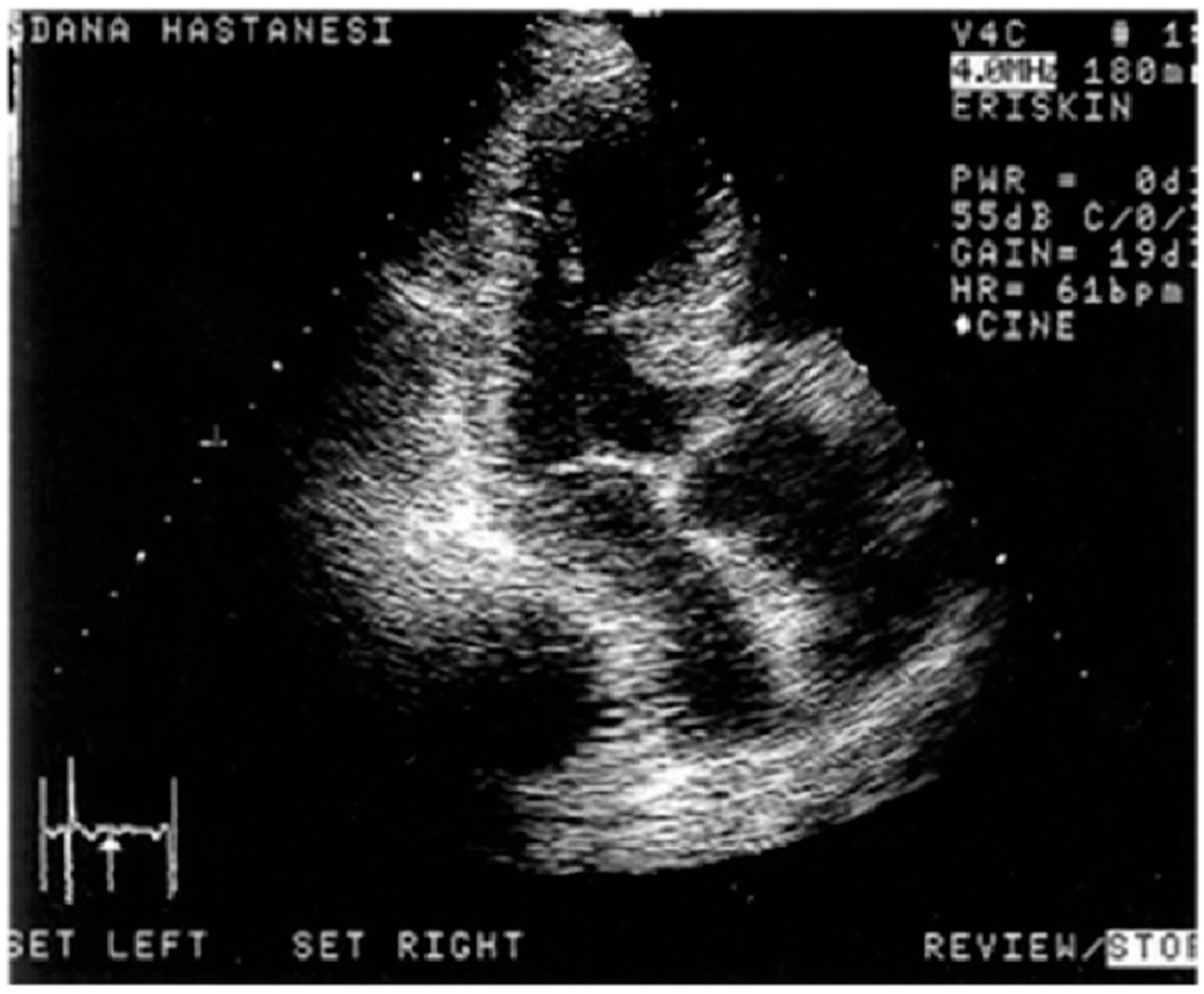
Segmental LV remodeling analysis by real-time three-dimensional echocardiography (RT3DE) showed non-uniform morphology in patients with pressure overload. We previously developed a quantitative volume index to separate long-axis LV cavities into three equal slices as the base, mid, and apex parts using RT3DE to visualize segmental details of LV cavities and realized that secondary LV hypertrophy is associated with diminished basal intracavity volume like ampulla shape (Figure 1) and preserved LV function, while hypertrophic cardiomyopathy had reduced midsegmental volume representing the catenoid geometry and impaired LV function [4,5].

1.2. Stressed Heart Morphology

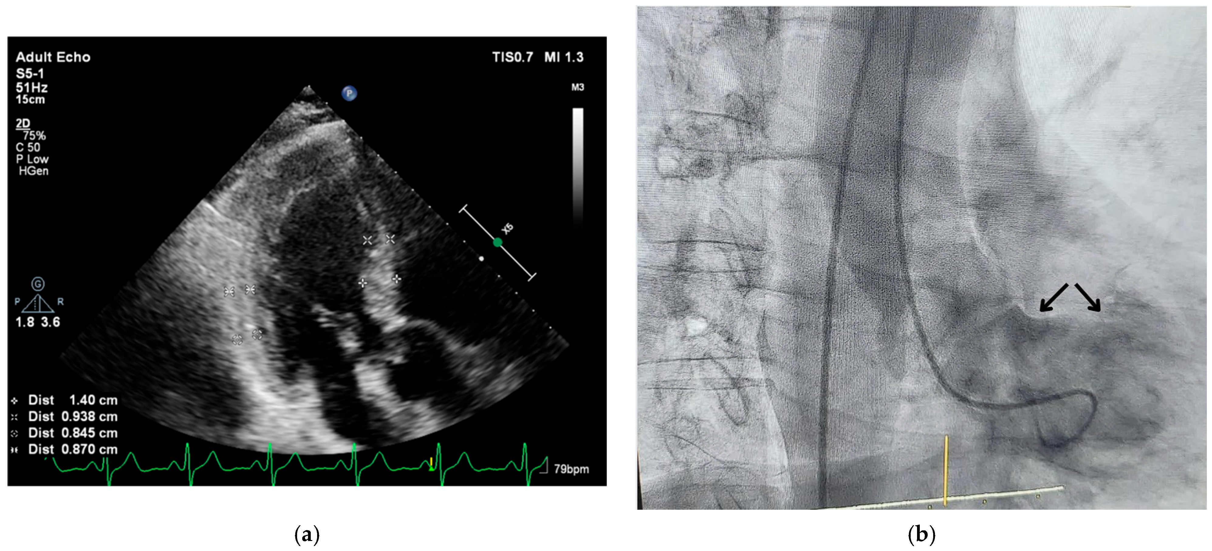
We detected basal septal hypertrophy (BSH) in hypertensives and described predominant septal base as the conjunctive point of determination in both acute and chronic conditions, called “stressed heart morphology (SHM)” [6]. In fact, remarkable midapical enlargement with predominant and hyperdynamic LV base in emotionally developed acute stress cardiomyopathy [7] was a consistent finding with LV basal remodeling (Figure 2a,b), as well as hyperdynamic myocardial function at LV base in hypertensive patients [8].
We validated BSH as the early imaging biomarker in both exercise mice and others with transaortic binding [10,11]. This led us to think that exercise may not always be beneficial in patients with SHM [12] and criticized some cardiac nuclear and MRI studies, ignoring blood pressure monitoring in septal remodeling or hyperdynamic LV function [13,14]. Gottdiener et al. previously showed that exercise hypertension could be detected in healthy individuals [15]. We also realized, years later, after validation studies, that our human heterogeneously distributed septal remodeling data were strikingly different from the regular progression of remodeling in animals [16]. Then, we reported the difficulty in determining the certain prevalence of SHM by multimodality imaging [17] and suggested that scientists should be cautious in the quantification of this morphologic finding [18].

1.3. Third-Generation Microscopic Imaging

Third-generation microscopic ultrasonographic data in animal validation studies showed that hemodynamic stress independent from etiology is associated with the microscopic remodeling in the early time points prior to LV dysfunction [19]. Beyond the emotional component in acute stress cardiomyopathy and the functional component due to increased afterload in patients with hypertensive heart disease, we have recently mentioned the mechanical component of SHM in aortic stenosis patients [20]. According to these observations, we believe that LV segmental remodeling as detected in hypertensive heart disease could also be clinically important in patients with aortic stenosis [21].
In the recent interesting article reported by Tsuda et al., the authors showed that the analyses of septal morphology leading to a dynamic bulge into the LV outflow tract and dynamic obstruction are extremely important since segmental involvement is the case in a variety of stress stimuli [22]. However, despite 55% of the study population being associated with hypertension and segmental involvement of the septum, for the validated finding as an early imaging biomarker in an animal model with pressure overload stress, the description of cardiac morphology was a normal heart in the conclusion, which seems a limitation of the study [22]. The clinical importance of the early imaging biomarker is primarily related to its potential role in the early treatment of early LV remodeling [16]. Therefore, recent microimaging data are consistent with the clinical reports supporting the paradigm that LV segmental remodeling is possibly accepted as the initiation of hypertensive heart disease [1,2]. In fact, since blood pressure monitoring, especially under pharmacologic stress or during exercise, may be ignored even in trials [12,13,14] and previously undiagnosed hypertension seems not rare even in developed countries [23], the prevalence of hypertensive disease may be underestimated in the population.

1.4. Segmental Tissue Function of LV Base

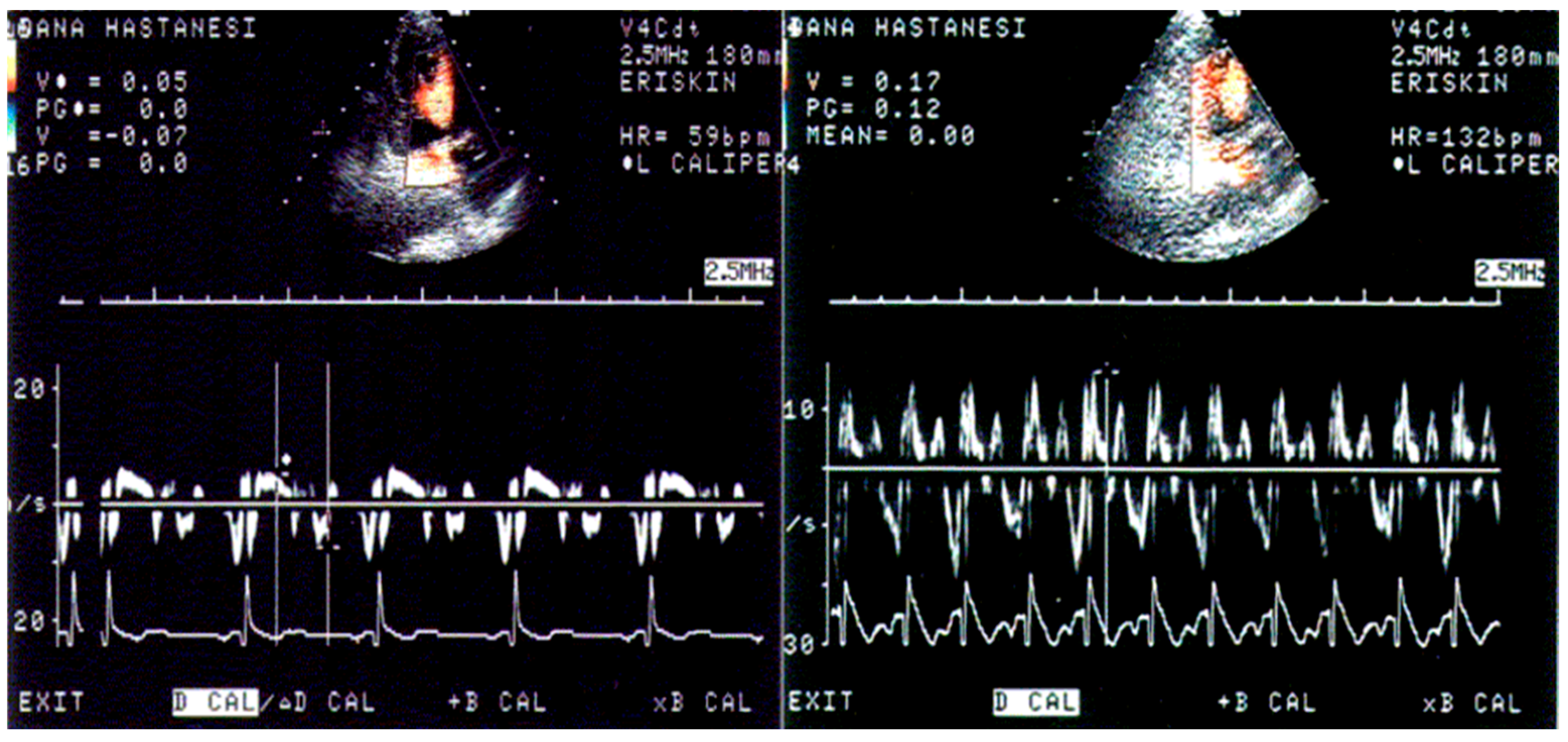
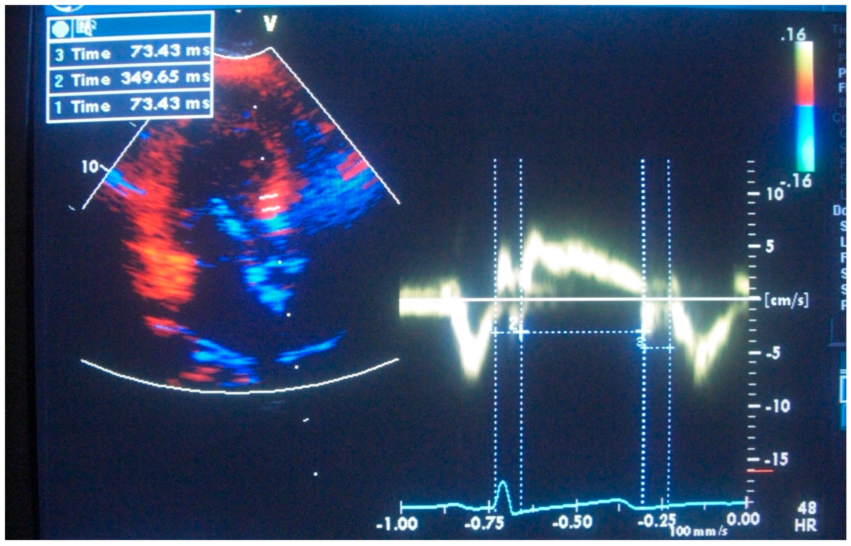
Furthermore, we noted in the animal validation studies that the early increment of systolic velocity up to 4 weeks is related primarily to compensatory hyperfunction in the early time points before the development of LV basal dysfunction [10,11]. Similar to humans with hypertensive basal septal hypertrophy (Figure 3) [8], tissue hyperfunction in the animal validation studies was detected by tissue Doppler imaging, which could obviously provide additional benefit to assess the phases of hypertensive disease [17] and quantitative follow-up of hypertensives (Figure 4) in the LV remodeling process [24]. We also suggested that this early abnormality could progress and result in more severe dysfunction of the septal wall compared to the free wall in global LV hypertrophy (LVH) [25].
In fact, septal ablation can be necessary in advanced cases with LV outflow obstruction due to huge septal hypertrophy [26]. It was shown that the LV base has an increased amount of noradrenaline and is associated with a more intensive sympathetic innervation compared to the apical region in animal and human hearts [27,28,29]. Triple S could be a specific location for a variety of stress stimuli because it is an extremely sensitive region to sympathetic overdrive (Figure 5 and Figure 6), which is independent of the type of stress [18,19].
Moreover, regional remodeling before global LVH development might be accepted as an additional stage of LV remodeling in the future, since early biomarkers for hypertension have gained more importance [31,32]. Despite all of the medical efforts on the importance of the early diagnosis of hypertensive heart disease and the general consensus on the clinical importance of BSH, diagnostic success may not be sufficient since 20% of previously undiagnosed hypertensive cases are associated with target organ damage [23].

1.5. Triple S and Takotsubo Syndrome

Traditionally classified as an acute and self-limiting cardiomyopathy, Takotsubo syndrome has been widely regarded as a transient response to sudden emotional or physical stress, predominantly in postmenopausal women [33]. However, emerging evidence and refined imaging modalities challenge this purely acute paradigm, suggesting that Takotsubo syndrome may in fact represent the acute manifestation of a chronically evolving myocardial susceptibility [34]. While the clinical presentation is abrupt—often mimicking acute coronary syndrome—the underlying myocardial substrate appears to be shaped over time by a constellation of subtle but significant structural, functional, and hemodynamic alterations [35]. This concept, aligned with the Triple S paradigm, proposes that features such as a small left ventricular cavity, modest basal septal hypertrophy, dynamic intraventricular gradients, and altered ventricular compliance may quietly accumulate, especially in the setting of chronic hypertension or age-related remodeling. These changes may remain clinically silent until an acute stressor—emotional, mechanical, or metabolic—acts as the final trigger, unmasking the vulnerable myocardial phenotype and precipitating the characteristic pattern of apical ballooning. In this light, Takotsubo syndrome is reframed not as a purely reactive process but as a predictable expression of a myocardium rendered fragile by chronic maladaptive remodeling. This model better explains cases occurring in the absence of overt emotional stress, as well as the recurrence of the syndrome in predisposed individuals [36,37]. It also underscores the importance of identifying the chronic substrate—through modalities such as stress echocardiography—rather than focusing solely on acute symptomatology. Recognizing Takotsubo syndrome as the acute endpoint of a long-standing structural and physiological vulnerability has meaningful implications for both diagnosis and long-term management, potentially shifting the clinical focus toward preventative strategies and substrate-directed therapy.

2. Why Triple S for Multiple Stressors in Clinical Practice

Triple S—the “stress septal sign” of BSH—often does not correlate in a one-to-one fashion with the severity of pure hemodynamic load. Instead, it appears to arise from the interplay of multiple stress mechanisms. The concept of superposed multiple stressors has been introduced to explain how emotional, mental, physical, and mechanical stress stimuli can converge on the heart to produce focal septal remodeling. In real-world scenarios, acute cardiac events frequently involve combined triggers rather than a single isolated cause.
For example, an episode of extreme emotion may be coupled with sudden exertion, together precipitating a hypertensive surge or stress cardiomyopathy. Accordingly, recent observations emphasize that multiple simultaneous stress inputs (“superposed” stressors) can amplify cardiovascular strain and are likely a major factor in precipitating adverse outcomes.
This paradigm is the rationale behind Triple S: it serves as an early marker of the heart’s maladaptive response to compounded stressors.
In other words, the septal bulge is not merely a marker of long-standing pressure overload but a “conjunctive point of determination” between chronic hypertension (a functional stress) and acute stress cardiomyopathy (an emotional stress).
By recognizing Triple S, clinicians are alerted to an individual’s cumulative stress burden—something that may be missed if one focuses solely on resting blood pressure or a single triggering event.
BSH morphology clinically may not be directly concordant with the severity of hemodynamic overload. Heterogeneity over focal involvement is pretty common in clinical practice and possibly affected by multiple pathogenetic mechanisms [12,17,18,19]. Both long-standing stresses associated with emotional problems and hemodynamic fluctuations under BP variability may not be modulated by parasympathetic balance and restored to the resting values after stress exposure (Figure 7).
We know that early septal remodeling, namely Triple S, can be developed after physiologic exercise in healthy individuals and interpreted to be a physiologically existing phenomenon. However, we clearly detected that blood pressure fluctuations could be neglected even in very precisely conducted National Registries like the LARGE MRI Study, which was supported by the British Heart Foundation [38].
Multiple stressors, at the same time, could be superposed and become clinically dangerous; thus, BSH should be seriously taken into consideration by clinicians as we previously emphasized in the recent books and in scientific exhibitions like the European Society of Hypertension, respectively [39]. Therefore, these reports have given the first messages emphasizing complex SHM developed by superposed multiple stressors, including functional, emotional, or mechanical stress. Both hypertension and anxiety are associated with norepinephrine release and excessive emotional stimulation, even panic attacks. We now know that Triple S is associated with emotional stress in animals, in addition to the pressure overload and physical exercise. These observations may represent a specific location, the septal base, for the nonspecific adaptation of the body to the stress stimuli.
In the validation studies at Johns Hopkins, the morphologic prominence of the LV septal base, which is free tissue from vagal innervation, accounts for half of the total course of global LV remodeling in the small animal model, which supports the underlying chronic hypertension as seen in the predominant septal base in Takotsubo cardiomyopathy. In 2000, we published the ampulla shape using real-time 3D echo at the Cleveland Clinic, which is very similar to LV geometry with that in Takotsubo cardiomyopathy, in which insufficient blood pressure response due to mid-apical microvascular dysfunction, as Messerli described in “Decapitated Hypertension,” may mask the exaggerated hemodynamic response under enormously released neurohormonal blood levels.
As a result, sympathetic overdrive and norepinephrine release are normally associated with hypertension; however, hemodynamic deterioration due to apical ballooning could mask the underlying hypertension clinically in Takotsubo cardiomyopathy. Severity of hypertensive LV hypertrophy is related to norepinephrine release [17], and the predominant involvement of the LV base could possibly be related to the intensive LV basal innervation, which was confirmed histologically [40,41,42]. Consistently, ASC is associated with the predominant LV septal base and excessive emotional stimulation, as well as increased catecholamine levels in the circulation [43].
The role of segmental remodeling involves BSH with dynamic blood pressure fluctuations on exercise [12], which leads to LV cavity dilation [44]. Stress stimuli could be related to a variety of components, including mental, emotional, physical, and mechanical stressors [39]. Superposed stressors take an important role in segmental remodeling, which is the main reason for the existence of “Triple S” in the literature. Incoordination between stress control and hypertension management resulted in an extremely high prevalence of previously undiagnosed hypertension and target organ damage [23,45]. Undiagnosed blood pressure fluctuations due to daily stress lead to high mortality and hemodynamic deterioration in stressful clinical presentations due to emotion-mediated norepinephrine peak levels with acute myocardial decompensation. An interesting part of the clinical picture in Triple S is that both septal bases in hypertensive myocardial tissue and Takotsubo cardiomyopathy give a hyperdynamic response to stress stimuli based on not only visual impression but quantitative analysis of contractility using Doppler tissue imaging [8,46]. Hypercontractile response to sympathetic stimulation is not a rare, common finding in hypertensive patients [8,13,47,48] with LVH, and related hypercontractility may result in diagnostic difficulty in ischemic heart disease [49].
In cross-sectional human data on BSH, the degree of regional hypertrophy over the septal base could be enormous [7,12,17,18,19,39] without any midapical progression compared to regular animal remodeling [10,11]. This discrepancy supports the contribution of the emotional component of stress stimuli and supports the clinical importance of Triple S. In this new paradigm, namely SHM, we have realized that chronic mental, emotional, physical, and mechanical stressors could be superposed on human beings [39] after the determination of heart remodeling findings in animals in microscopic remodeling data. Therefore, years later, we realized that animals that are free of cognitive function have extremely regular segmental remodeling under a variety of stressors, according to Johns Hopkins animal validation studies.
“Scientists themselves don’t always distinguish between the psychological state of stress and the physiological response to stress”, Dr. Ratey JJ. says, adding that chronic stress, which translates emotional stress into physical strain and stress response, can lead to anxiety, depression, as well as high blood pressure [50]. A variety of stressors affect Triple S, the specific location that is free from parasympathetic modulation.

3. The Neuro-Cardiac Anatomical and Clinical Importance of Segmental Examination of the Interventricular Septum

The cardiac neural innervation shows asymmetry and heterogeneity. Compelling evidence from anatomical, functional, and imaging studies identifies the basal region of the interventricular septum as a focal point for sympathetic nervous system activity, rendering it particularly susceptible to the effects of stress. The cardiac parasympathetic innervation by the vagus nerve is shown, showing the innervation of both ventricles; it has more influence on the apex compared to the base [51,52]. This condition of less parasympathetic predominance on the basal segment appears to be a key factor in its early and disproportionate involvement in the hypertrophic remodeling process known as SHM.
The anatomical foundation for this sympathetic dominance is well-documented based on studies that have demonstrated that the base of the left ventricle receives greater sympathetic innervation compared to the apex [53]. Histological analysis of the human heart has confirmed a higher density of autonomic nerves in this basal region [42]. This increased innervation is matched by a higher local concentration of catecholamines; specifically, the basal myocardium contains a greater amount of noradrenaline, the primary neurotransmitter of the sympathetic system. This neurochemical arrangement primes the basal septum for a heightened response to sympathetic signals. This has been further substantiated by advanced imaging techniques. Scintigraphy studies utilizing I-meta-iodobenzylguanidine, which is a norepinephrine analog, have shown increased sympathetic activity specifically within the asymmetrically hypertrophied basal septum of patients with hypertension, directly linking the structural change to a state of localized sympathetic hyperactivity [29].
This dense sympathetic innervation has clear and measurable functional consequences. Under conditions of stress, the basal septum exhibits a uniquely hyperdynamic and hypercontractile response. When challenged with a sympathomimetic agent like dobutamine, hypertensive patients with BSH demonstrate a significantly exaggerated response in the hypertrophied segment [48]. Quantitative tissue Doppler imaging shows that maximum systolic velocities in the basal septum at peak stress are significantly higher in patients with BSH compared to individuals with normal cardiac geometry. This hypercontractility is an early sign of the disease process, often preceding global LV dysfunction [8].
The progression from localized sympathetic overactivity to structural change has been prospectively validated in animal models of pressure overload. Using third-generation microscopic ultrasonography, we have confirmed that BSH is the earliest imaging biomarker to appear. This initial remodeling is characterized as a “compensatory hyperactive stage” where the heart adapts to the increased stress [11]. During this phase, elevated intracavitary gradients are observed, reflecting the hyperdynamic function of the early-remodeling heart. However, this adaptive stage is transient. Animal studies show a sharp transition from this state of compensatory hyperfunction to maladaptation, marked by the onset of tissue dysfunction, blunted intracavitary gradients, and the progression to global, concentric LVH [10,17].
The recognition of BSH as the morphological product of a segmental autonomic imbalance has profound clinical implications. It mandates a shift toward the segmental clinical evaluation of the interventricular septum. Global measurements of septal thickness can easily miss or underestimate the significance of BSH, thereby failing to identify patients in the early, adaptive phase of SHM [21]. The incidental discovery of BSH on a routine echocardiogram should not be dismissed as a benign finding. Instead, it should be recognized as a potential marker of underlying, and possibly undiagnosed, hemodynamic or neurologic stress (emotional with high stress score) [39]. Such a finding warrants a more thorough evaluation, including blood pressure monitoring during exercise, to unmask exercise-induced hypertension, which is strongly associated with septal remodeling.

4. Conclusions

LV geometric and functional features, with precisely processed etiologic assessment using quantitative cardiovascular imaging, could be widely used in the population and contribute to early medical treatment. Diagnosis of hypertension could be underestimated in early septal remodeling, since it could be missed at rest. Missed hypertension may be related to emotional stress and exercise-induced hypertension. Absence of recorded segmental data globally and hemodynamic overload determination during stress may underestimate the evaluation of heart remodeling and failure in antihypertensive medication. In follow-ups of chronic patients, we strongly believe that this preexisting sign (Triple S) should be recalled, and the optimal beta-blocker dosage should be provided. In fact, this group of medications is effective and used worldwide in the control of myocardial tissue O2 consumption.

5. Future Insights

According to the effect of multiple stressors, “stressed heart morphology” may represent an early imaging biomarker in the population with various risk groups [46]. Recently, we have worked on a Special Issue showing imaging and other aspects of the remodeling progression and have summarized the working details of the last two decades in the attached preface of the recent Special Issue (stress septal sign). Animal models using third-generation microscopic ultrasonography have shown that heart remodeling starts on the basal septum. Beyond our description of “microscopic remodeling” in pressure overload and physiologic exercise, animals under emotional stress lead to remodeling of the septum that is free from the vagal nerve. “Superposed multiple stressors”, as the leading killer in the majority of cases, has increased the importance of “segmental remodeling” in cardiac imaging. “Stress septal sign” is the conjunctive point of determination in hypertension as functional and acute stress cardiomyopathy as emotional etiopathologies. Segmental comparative analyses instead of single cross-sections possibly contribute to a neurocardiologic perspective for the combined management of hypertension and stress. These experiences and other published data showed that 60% of chronic diseases are associated with stress. However, only 3% of the population who seek health care find a chance to have specific management for stress. Beyond the clinical aspects of SHM, there is a need to focus on cellular levels of myocardial tissue to investigate whether SHM is the specific location of Selye’s nonspecific general adaptive response to stressors.
However, the industrial design of healthcare is inherently oriented towards costly procedures, potentially resulting in physicians placing excessive emphasis on the device-mediated perspective. In addition, as is the case with advanced phases of diseases, the initiation of treatment, which includes the administration of beta-blockers [54] in conjunction with stress management techniques, could be approached in a more comprehensive manner. This approach is not only cost-effective but also offers a significant public health benefit. Furthermore, there is a strong conviction that SHM could be utilized extensively in the near future from a public health perspective. Combined management of stress and hypertension could control the hazardous effects of superposed multiple stressors, since many cardiac problems can not be explained by classic risk factors; biomarkers could provide additional benefit in clinical practice.

Author Contributions

Conceptualization, F.Y.; methodology, F.Y.; validation, F.Y., M.R.A. and M.J.G.; investigation, F.Y.; writing—original draft preparation, F.Y. and B.C.; writing—review and editing, F.Y. and B.C.; supervision, M.R.A. and M.J.G.; project administration, F.Y. All authors have read and agreed to the published version of the manuscript.

Funding

This research received no external funding.

Institutional Review Board Statement

Not applicable.

Informed Consent Statement

Not applicable.

Data Availability Statement

Not applicable.

Conflicts of Interest

The authors declare no conflict of interest.

Abbreviations and Acronyms

LVleft ventricle
RT3DEreal-time three-dimensional echocardiography
BSHbasal septal hypertrophy
SHMstressed heart morphology
Triple Sstress septal sign
LVLV hypertrophy

References

  1. Maron, B.J.; Edwards, J.E.; Epstein, S.E. Disproportionate Ventricular Septal Thickening in Patients with Systemic Hypertension. Chest 1978, 73, 466–470. [Google Scholar] [CrossRef] [PubMed]
  2. Gaudron, P.D.; Liu, D.; Scholz, F.; Hu, K.; Florescu, C.; Herrmann, S.; Bijnens, B.; Ertl, G.; Störk, S.; Weidemann, F. The Septal Bulge—An Early Echocardiographic Sign in Hypertensive Heart Disease. J. Am. Soc. Hypertens. 2016, 10, 70–80. [Google Scholar] [CrossRef] [PubMed]
  3. Frielingsdorf, J.; Franke, A.; Kühl, H.P.; Hess, O.M.; Flachskampf, F.A. Evaluation of Septal Hypertrophy and Systolic Function in Diseases That Cause Left Ventricular Hypertrophy: A 3-Dimensional Echocardiography Study. J. Am. Soc. Echocardiogr. 2001, 14, 370–377. [Google Scholar] [CrossRef]
  4. Yalçin, F.; Shiota, T.; Odabashian, J.; Agler, D.; Greenberg, N.L.; Garcia, M.J.; Lever, H.M.; Thomas, J.D. Comparison by Real-Time Three-Dimensional Echocardiography of Left Ventricular Geometry in Hypertrophic Cardiomyopathy versus Secondary Left Ventricular Hypertrophy. Am. J. Cardiol. 2000, 85, 1035–1038. [Google Scholar] [CrossRef] [PubMed]
  5. Yalçin, F.; Shiota, M.; Greenberg, N.; Thomas, J.D.; Shiota, T. Real Time Three-Dimensional Echocardiography Evaluation of Mitral Annular Characteristics in Patients with Myocardial Hypertrophy. Echocardiography 2008, 25, 424–428. [Google Scholar] [CrossRef]
  6. Yalçin, F.; Muderrisoǧlu, H. Tako-Tsubo Cardiomyopathy May Be Associated with Cardiac Geometric Features as Observed in Hypertensive Heart Disease. Int. J. Cardiol. 2009, 135, 251–252. [Google Scholar] [CrossRef]
  7. Yalçin, F.; Yalçin, H.; Abraham, T. Stress-Induced Regional Features of Left Ventricle Is Related to Pathogenesis of Clinical Conditions with Both Acute and Chronic Stress. Int. J. Cardiol. 2010, 145, 367–368. [Google Scholar] [CrossRef]
  8. Yalçin, F.; Yiǧit, F.; Erol, T.; Baltali, M.; Korkmaz, M.E.; Müderrisoǧu, H. Effect of Dobutamine Stress on Basal Septal Tissue Dynamics in Hypertensive Patients with Basal Septal Hypertrophy. J. Hum. Hypertens. 2006, 20, 628–630. [Google Scholar] [CrossRef]
  9. Yalçin, F.; Abraham, M.R.; Garcia, M.J. Stress and Heart in Remodeling Process: Multiple Stressors at the Same Time Kill. J. Clin. Med. 2024, 13, 2597. [Google Scholar] [CrossRef]
  10. Yalçin, F.; Kucukler, N.; Cingolani, O.; Mbiyangandu, B.; Sorensen, L.; Pinherio, A.; Abraham, M.R.; Abraham, T.P. Evolution of Ventricular Hypertrophy and Myocardial Mechanics in Physiological and Pathological Hypertrophy. J. Appl. Physiol. 2019, 126, 354–362. [Google Scholar] [CrossRef]
  11. Yalcin, F.; Kucukler, N.; Cingolani, O.; Mbiyangandu, B.; Sorensen, L.L.; Pinheiro, A.C.; Abraham, M.R.; Abraham, T.P. Intracavitary Gradients in Mice With Early Regional Remodeling at the Compensatory Hyperactive Stage Prior To Lv Tissue Dysfunction. J. Am. Coll. Cardiol. 2020, 75, 1585. [Google Scholar] [CrossRef]
  12. Yalçin, F.; Yalçin, H.; Abraham, T.P. Exercise Hypertension Should Be Recalled in Basal Septal Hypertrophy as the Early Imaging Biomarker in Patients with Stressed Heart Morphology. Blood Press. Monit. 2020, 25, 118–119. [Google Scholar] [CrossRef] [PubMed]
  13. Yalçin, F.; Schindler, T.; Abraham, T.P. Hypertension Should Be Ruled out in Patients with Hyperdynamic Left Ventricle on Radionuclide Myocardial Perfusion Imaging, Diastolic Dysfunction and Dyspnea on Exertion. Int. J. Cardiol. Heart Vasc. 2015, 7, 149. [Google Scholar] [CrossRef] [PubMed]
  14. Yalcin, F.; Abraham, T.P.; Gottdiener, J.S. Regarding Article, “Left Ventricular Wall Thickness and the Presence of Asymmetric Hypertrophy in Healthy Young Army Recruits: Data from the LARGE Heart Study. Circ. Cardiovasc. Imaging 2013, 6, 262–267. [Google Scholar] [CrossRef] [PubMed]
  15. Gottdiener, J.S. Left Ventricular Hypertrophy in Men with Normal Blood Pressure: Relation to Exaggerated Blood Pressure Response to Exercise. Ann. Intern. Med. 1990, 112, 161. [Google Scholar] [CrossRef]
  16. Yalçin, F.; Yalçin, H.; Küçükler, N.; Arslan, S.; Akkuş, O.; Kurtul, A.; Abraham, M.R. Basal Septal Hypertrophy as the Early Imaging Biomarker for Adaptive Phase of Remodeling Prior to Heart Failure. J. Clin. Med. 2021, 11, 75. [Google Scholar] [CrossRef]
  17. Yalçin, F.; Yalçin, H.; Abraham, M.R.; Abraham, T.P. Ultimate Phases of Hypertensive Heart Disease and Stressed Heart Morphology by Conventional and Novel Cardiac Imaging. Am. J. Cardiovasc. Dis. 2021, 11, 628. [Google Scholar]
  18. Yalçin, F.; Abraham, R.; Abraham, T.P. Basal Septal Hypertrophy: Extremely Sensitive Region to Variety of Stress Stimuli and Stressed Heart Morphology. J. Hypertens. 2022, 40, 626–627. [Google Scholar] [CrossRef]
  19. Yalçin, F.; Yalçin, H.; Abraham, R.; Abraham, T.P. Hemodynamic Stress and Microscopic Remodeling. Int. J. Cardiol. Cardiovasc. Risk Prev. 2021, 11, 200115. [Google Scholar] [CrossRef]
  20. Yalçin, F.; Abraham, R.; Abraham, T.P. Myocardial Aspects in Aortic Stenosis and Functional Increased Afterload Conditions in Patients with Stressed Heart Morphology. Ann. Thorac. Cardiovasc. Surg. 2021, 27, 332–334. [Google Scholar] [CrossRef]
  21. Yalçin, F.; Abraham, M.R.; Abraham, T.P. It Is Time to Assess Left Ventricular Segmental Remodelling in Aortic Stenosis. Eur. Heart J. Cardiovasc. Imaging 2022, 23, e299–e300. [Google Scholar] [CrossRef]
  22. Tsuda, D.; Mori, S.; Izawa, Y.; Toh, H.; Suzuki, M.; Takahashi, Y.; Toba, T.; Fujiwara, S.; Tanaka, H.; Watanabe, Y.; et al. Diversity and Determinants of the Sigmoid Septum and Its Impact on Morphology of the Outflow Tract as Revealed Using Cardiac Computed Tomography. Echocardiography 2022, 39, 248–259. [Google Scholar] [CrossRef]
  23. Korhonen, P.E.; Kautiainen, H.; Järvenpää, S.; Kantola, I. Target Organ Damage and Cardiovascular Risk Factors among Subjects with Previously Undiagnosed Hypertension. Eur. J. Prev. Cardiol. 2014, 21, 980–988. [Google Scholar] [CrossRef]
  24. Yalçin, F.; Yalçin, H.; Küçükler, N.; Abraham, T.P. Quantitative Left Ventricular Contractility Analysis under Stress: A New Practical Approach in Follow-up of Hypertensive Patients. J. Hum. Hypertens. 2011, 25, 578–584. [Google Scholar] [CrossRef]
  25. Yalçin, F.; Topaloglu, C.; Kuçukler, N.; Ofgeli, M.; Abraham, T.P. Could Early Septal Involvement in the Remodeling Process Be Related to the Advance Hypertensive Heart Disease? IJC Heart Vasc. 2015, 7, 141–145. [Google Scholar] [CrossRef] [PubMed]
  26. Vieira, M.L.C.; Silva Filho, R.M.; Brito Filho, F.S.; Leal, S.B.; Lira Filho, E.B.; Fischer, C.H.; De Souza, J.A.; Perin, M.A. Selective Contrast Echocardiography in Percutaneous Transluminal Septal Myocardial Ablation in an Elderly Patient with Left Ventricular Concentric Hypertrophy. Echocardiography 2003, 20, 563–566. [Google Scholar] [CrossRef] [PubMed]
  27. ANGELAKOS, E.T.; Millard, R.W. Regional Distribution of Catecholamines in the Dog Heart. Circ. Res. 1965, 16, 39–44. [Google Scholar] [CrossRef] [PubMed]
  28. Kimata, S.-I. Chemical Evaluation of the Distribution of Catecholamines in the Heart. Jpn. Circ. J. 1965, 29, 7–10. [Google Scholar] [CrossRef] [PubMed]
  29. Shimizu, M.; Ino, H.; Okeie, K.; Emoto, Y.; Yamaguchi, M.; Yasuda, T.; Fujino, N.; Fujii, H.; Fujita, S.; Mabuchi, H.; et al. Cardiac Sympathetic Activity in the Asymmetrically Hypertrophied Septum in Patients with Hypertension or Hypertrophic Cardiomyopathy. Clin. Cardiol. 2000, 23, 365–370. [Google Scholar] [CrossRef]
  30. Yalcin, F.; Topaloglu, C.; Kucukler, N.; Yeral, N.; Vefali, H.; Abraham, T.P.; Garcia, M.J. Abstract 11796: Determination of Regional Myocardial Performance in Hypertensive Patients with/without Left Ventricular Hypertrophy. Circulation 2011, 124, A11796. [Google Scholar]
  31. Narayanan, A.; Aurigemma, G.P.; Chinali, M.; Hill, J.C.; Meyer, T.E.; Tighe, D.A. Cardiac Mechanics in Mild Hypertensive Heart Disease: A Speckle-Strain Imaging Study. Circ. Cardiovasc. Imaging 2009, 2, 382–390. [Google Scholar] [CrossRef]
  32. Plante, G.E. Predisease Biological Markers: Early Diagnosis and Prevention of Arterial Hypertension. Metabolism 2008, 57, S36–S39. [Google Scholar] [CrossRef]
  33. Huseynov, A.; El-Battrawy, I.; Ansari, U.; Schramm, K.; Zhou, X.; Lang, S.; Borggrefe, M.; Akin, I. Age Related Differences and Outcome of Patients with Takotsubo Syndrome. J. Geriatr. Cardiol. JGC 2017, 14, 632–638. [Google Scholar] [CrossRef] [PubMed]
  34. Singh, T.; Khan, H.; Gamble, D.T.; Scally, C.; Newby, D.E.; Dawson, D. Takotsubo Syndrome: Pathophysiology, Emerging Concepts, and Clinical Implications. Circulation 2022, 145, 1002–1019. [Google Scholar] [CrossRef]
  35. Wilkhoo, H.S.; Visvanathan, H.; Chatterjee, S.; Hussain, S.; Gundaniya, P.; Francis, V.; Singh, B.; Pillai, A.A. Current Clinical Perspectives on Takotsubo Syndrome: Comprehensive Analysis of Diagnosis, Management, and Pathophysiology. Int. J. Cardiovasc. Acad. 2024, 10, 82–90. [Google Scholar] [CrossRef]
  36. Schlossbauer, S.A.; Ghadri, J.-R.; Scherff, F.; Templin, C. The Challenge of Takotsubo Syndrome: Heterogeneity of Clinical Features. Swiss Med. Wkly. 2017, 147, w14490. [Google Scholar] [CrossRef] [PubMed]
  37. Prokudina, E.S.; Kurbatov, B.K.; Zavadovsky, K.V.; Vrublevsky, A.V.; Naryzhnaya, N.V.; Lishmanov, Y.B.; Maslov, L.N.; Oeltgen, P.R. Takotsubo Syndrome: Clinical Manifestations, Etiology and Pathogenesis. Curr. Cardiol. Rev. 2021, 17, 188–203. [Google Scholar] [CrossRef]
  38. Lee, P.T.; Dweck, M.R.; Prasher, S.; Shah, A.; Humphries, S.E.; Pennell, D.J.; Montgomery, H.E.; Payne, J.R. Left Ventricular Wall Thickness and the Presence of Asymmetric Hypertrophy in Healthy Young Army Recruits. Circ. Cardiovasc. Imaging 2013, 6, 262–267. [Google Scholar] [CrossRef] [PubMed]
  39. Yalcin, F.; Melek, I.; Mutlu, T. Stressed Heart Morphology and Neurologic Stress Score Effect Beyond Hemodynamic Stress on Focal Geometry. J. Hypertens. 2022, 40, e79. [Google Scholar] [CrossRef]
  40. Schlaich, M.P.; Kaye, D.M.; Lambert, E.; Sommerville, M.; Socratous, F.; Esler, M.D. Relation Between Cardiac Sympathetic Activity and Hypertensive Left Ventricular Hypertrophy. Circulation 2003, 108, 560–565. [Google Scholar] [CrossRef]
  41. Holmgren, S.; Abrahamsson, T.; Almgren, O. Adrenergic Innervation of Coronary Arteries and Ventricular Myocardium in the Pig: Fluorescence Microscopic Appearance in the Normal State and after Ischemia. Basic Res. Cardiol. 1985, 80, 18–26. [Google Scholar] [CrossRef]
  42. Kawano, H.; Okada, R.; Yano, K. Histological Study on the Distribution of Autonomic Nerves in the Human Heart. Heart Vessels 2003, 18, 32–39. [Google Scholar] [CrossRef]
  43. Wittstein, I.S.; Thiemann, D.R.; Lima, J.A.C.; Baughman, K.L.; Schulman, S.P.; Gerstenblith, G.; Wu, K.C.; Rade, J.J.; Bivalacqua, T.J.; Champion, H.C. Neurohumoral Features of Myocardial Stunning Due to Sudden Emotional Stress. N. Engl. J. Med. 2005, 352, 539–548. [Google Scholar] [CrossRef]
  44. Smelley, M.P.; Virnich, D.E.; Williams, K.A.; Ward, R.P. A Hypertensive Response to Exercise Is Associated with Transient Ischemic Dilation on Myocardial Perfusion SPECT Imaging. J. Nucl. Cardiol. 2007, 14, 537–543. [Google Scholar] [CrossRef]
  45. Semánová, C. NCD Risk Factor Collaboration (NCD-RisC) Worldwide Trends in Hypertension Prevalence and Progress in Treatment and Control from 1990 to 2019: A Pooled Analysis of 1201 Population-Representative Studies with 104 Million Participants. Lancet Lond. Engl. 2021, 398, 957–980. [Google Scholar] [CrossRef]
  46. Azzarelli, S.; Galassi, A.R.; Amico, F.; Giacoppo, M.; Argentino, V.; Fiscella, A. Intraventricular Obstruction in a Patient with Tako-Tsubo Cardiomyopathy. Int. J. Cardiol. 2007, 121, e22–e24. [Google Scholar] [CrossRef] [PubMed]
  47. Krasnow, N. Subaortic Septal Bulge Simulates Hypertrophic Cardiomyopathy by Angulation of the Septum with Age, Independent of Focal Hypertrophy. An Echocardiographic Study. J. Am. Soc. Echocardiogr. 1997, 10, 545–555. [Google Scholar] [CrossRef]
  48. Yalçin, F.; Muderrisoglu, H.; Korkmaz, M.E.; Ozin, B.; Baltali, M.; Yigit, F. The Effect of Dobutamine Stress on Left Ventricular Outflow Tract Gradients in Hypertensive Patients with Basal Septal Hypertrophy. Angiology 2004, 55, 295–301. [Google Scholar] [CrossRef] [PubMed]
  49. Smart, S.C.; Knickelbine, T.; Malik, F.; Sagar, K.B. Dobutamine-Atropine Stress Echocardiography for the Detection of Coronary Artery Disease in Patients With Left Ventricular Hypertrophy. Circulation 2000, 101, 258–263. [Google Scholar] [CrossRef]
  50. Ratey, J.; Hagerman, E. Spark: The Revolutionary New Science of Exercise and the Brain. Psychiatr. Serv. Psychiatr. Serv. 2008, 59, 304. [Google Scholar]
  51. Zandstra, T.E.; Notenboom, R.G.E.; Wink, J.; Kiès, P.; Vliegen, H.W.; Egorova, A.D.; Schalij, M.J.; De Ruiter, M.C.; Jongbloed, M.R.M. Asymmetry and Heterogeneity: Part and Parcel in Cardiac Autonomic Innervation and Function. Front. Physiol. 2021, 12, 665298. [Google Scholar] [CrossRef] [PubMed]
  52. Yamakawa, K.; So, E.L.; Rajendran, P.S.; Hoang, J.D.; Makkar, N.; Mahajan, A.; Shivkumar, K.; Vaseghi, M. Electrophysiological Effects of Right and Left Vagal Nerve Stimulation on the Ventricular Myocardium. Am. J. Physiol. Heart Circ. Physiol. 2014, 307, H722–H731. [Google Scholar] [CrossRef]
  53. Brack, K.E.; Coote, J.H.; Ng, G.A. Interaction between Direct Sympathetic and Vagus Nerve Stimulation on Heart Rate in the Isolated Rabbit Heart. Exp. Physiol. 2004, 89, 128–139. [Google Scholar] [CrossRef] [PubMed]
  54. Xing, F.; Chen, J.; Zhao, B.; Jiang, J.; Tang, A.; Chen, Y. Real Role of β-Blockers in Regression of Left Ventricular Mass in Hypertension Patients: Bayesian Network Meta-Analysis. Medicine 2017, 96, e6290. [Google Scholar] [CrossRef] [PubMed]
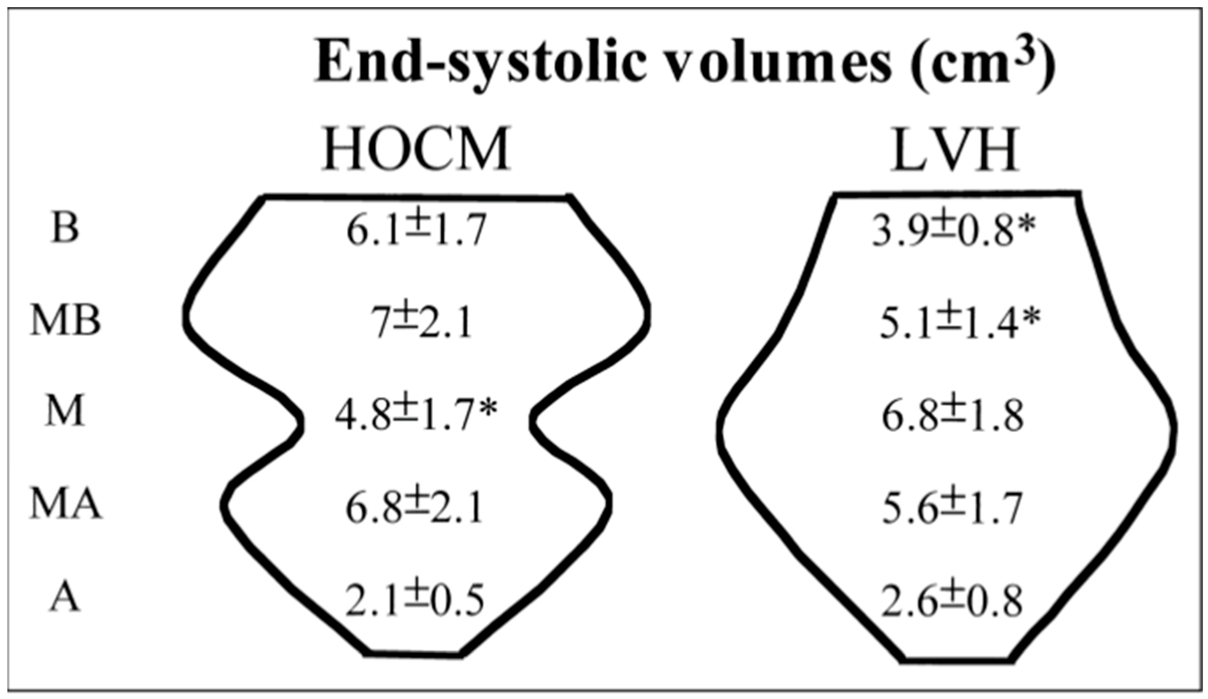
Figure 1. The ampulla shape with a narrowed basal cavity volume analyzed by real-time 3-dimensional segmental cavity volume analyses in LV hypertrophy due to aortic stenosis or hypertension. Reprinted from ref [4]. HOCM: Hypertrophic Obstructive Cardiomyopathy. Values marked with * indicate statistically significant differences.
Jcm 14 06143 g001
Figure 2. (a) A two-dimensional echocardiographic image of a hypertensive patient with a sigmoid septum and ampulla-shaped LV cavity geometry during diastole. Reprinted from ref [9]. (b) Preexisting Triple S in the same patient, similar to Takotsubo geometry. Reprinted from ref [9].
Jcm 14 06143 g002
Figure 3. Diastolic BSH and systolic predominately of BSH in a hypertensive by RT3DE. Reprinted from ref [4,19].
Jcm 14 06143 g003
Figure 4. Measurements of maximum basal septal tissue systolic velocities at rest and peak stress in a hypertensive patient with basal septal hypertrophy. Reprinted from ref [4].
Jcm 14 06143 g004
Figure 5. Quantitative segmental LV functional analysis of a hypertensive patient with basal septal hypertrophy using tissue Doppler echocardiography. Reprinted from ref [30].
Jcm 14 06143 g005
Figure 6. Triple S, which can not be explained by pure hemodynamic overload, is free from activity and related to a high stress score. Reprinted from ref [18].
Jcm 14 06143 g006
Figure 7. Vascular hazards due to blood pressure variability in a variety of stressors, such as exercise, hypertension, emotional sympathetic overdrive, and a combination of multiple stressors. Reprinted from ref [9].
Jcm 14 06143 g007
Disclaimer/Publisher’s Note: The statements, opinions and data contained in all publications are solely those of the individual author(s) and contributor(s) and not of MDPI and/or the editor(s). MDPI and/or the editor(s) disclaim responsibility for any injury to people or property resulting from any ideas, methods, instructions or products referred to in the content.