Article

Left Ventricular Ejection Fraction Predicts Outcomes in Different Subgroups of Patients Undergoing Coronary Angiography

1
Cardiology, Haemostasis, and Medical Intensive Care, Medical Faculty Mannheim, University Medical Centre Mannheim, Heidelberg University, 68167 Mannheim, Germany
2
European Center for AngioScience (ECAS), German Centre for Cardiovascular Research (DZHK), Partner Site Heidelberg/Mannheim, and Centre for Cardiovascular Acute Medicine Mannheim (ZKAM), Medical Centre Mannheim and Medical Faculty Mannheim, Heidelberg University, 68167 Mannheim, Germany
3
Department of Cardiology, Angiology and Pneumology, University Hospital Heidelberg, 69120 Heidelberg, Germany
4
Department of Biomedical Informatics, Center for Preventive Medicine and Digital Health (CPD), Medical Faculty Mannheim, Heidelberg University, 68167 Mannheim, Germany
5
Helmholtz-Institute for Translational AngioCardioScience (HI-TAC), Max Delbrück Center for Molecular Medicine, Helmholtz Association (MDC), Heidelberg University, 10115 Berlin, Germany
*
Author to whom correspondence should be addressed.
These authors contributed equally to this work.
J. Clin. Med. 2025, 14(15), 5219; https://doi.org/10.3390/jcm14155219
Submission received: 16 June 2025 / Revised: 14 July 2025 / Accepted: 17 July 2025 / Published: 23 July 2025
(This article belongs to the Section Cardiology)

Abstract

Objectives: To evaluate the long-term prognostic value of left ventricular ejection fraction (LVEF) in consecutive patients undergoing invasive coronary angiography (CA). Background: LVEF is a key prognostic marker in cardiovascular disease, but its value across different clinical indications for CA remains insufficiently characterized. Methods: Consecutive patients undergoing CA between January 2016 and August 2022 were retrospectively included at one institution. Patients were stratified into four LVEF groups: ≥ 55%, 45–54%, 35–44%, and <35%. The primary endpoint was rehospitalization for heart failure (HF) at 36 months. Secondary endpoints were acute myocardial infarction (AMI) and coronary revascularization. Kaplan–Meier and multivariable Cox regression analyses were conducted within the entire study cohort and pre-defined subgroups. Results: A total of 6888 patients were included (median age: 71 years; 65.2% males). LVEF < 35% was associated with a higher comorbidity burden and more extensive coronary artery disease (e.g., three-vessel CAD: 38.6% vs. 20.7%, p < 0.001). Event rates for HF rehospitalization and AMI increased progressively with declining LVEF, while revascularization rates varied across categories. Statistically significant differences across LVEF groups were observed for all three endpoints in unadjusted analyses (log-rank p < 0.001). In multivariable models, LVEF < 35% independently predicted HF rehospitalization (HR = 3.731, p < 0.001) and AMI (HR = 4.184, p < 0.001), but not revascularization (HR = 0.867, p = 0.378). The prognostic association was demonstrated across all subgroups stratified by age, sex, subtype of acute coronary syndrome, and CAD severity. Conclusions: Reduced LVEF is an independent predictor of HF rehospitalization and AMI in patients undergoing coronary angiography, irrespective of its indication, whereas no independent association was observed with coronary revascularization.

1. Introduction

Coronary artery disease (CAD), the most prevalent form of cardiovascular disease (CVD), affects more than 126 million individuals worldwide and is a leading contributor to the more than 9 million annual deaths attributed to CVD [1,2]. Invasive coronary angiography (CA) remains the cornerstone diagnostic tool for assessing CAD severity and treatment, particularly in patients with acute coronary syndromes or unexplained symptoms [3]. However, the long-term prognostic determinants in consecutive patients undergoing CA remain unclear, as previous studies predominantly investigated outcomes in pre-selected subgroups, such as acute myocardial infarction (AMI) or heart failure (HF) [4].
In patients undergoing CA, left ventricular ejection fraction (LVEF) is a key parameter for both clinical decision-making and risk stratification. As a measure of systolic function, LVEF indicates therapy in patients with HF with reduced ejection fraction (HFrEF) and is strongly associated with mortality, HF progression, and adverse cardiovascular events [5,6,7]. However, the prognostic implications of LVEF categories across a wider clinical spectrum, especially in patients without acute HF at presentation, remain unclear. In particular, the risk associated with moderately reduced (35–45%) and preserved (>45%) LVEF has not been fully explored in real-world angiographic settings. Growing evidence challenges the binary classification of LVEF, suggesting a continuum of risk across LVEF strata [8,9]. Even patients with mildly reduced LVEF may face increased risks of HF rehospitalization and major adverse events. This paradigm shift is clinically relevant, as HF with preserved ejection fraction (HFpEF) now constitutes the majority of HF presentations, despite the lack of disease-modifying therapies [8,10].
Therefore, this study investigates the association between LVEF strata and the risk of HF-related rehospitalization, AMI, and coronary revascularization at 36 months in consecutive patients undergoing CA. Subgroup analyses were performed, stratified by age, sex, and clinical indications for CA, as well as the extent of CAD.

2. Methods

2.1. Study Population, Design, and Data Collection

This retrospective observational study included all consecutive patients of at least 18 years of age who underwent invasive CA at the University Medical Centre Mannheim (UMM), Germany, between January 2016 and August 2022. Patients were identified using German Operation and Procedure Classification System (OPS) codes. Relevant clinical data were retrieved from the institutional electronic hospital information system (SAP®, Walldorf, Germany), including demographic characteristics, cardiovascular risk factors, comorbidities, clinical presentation, angiographic findings, and discharge medications. Patients who underwent multiple CA procedures during the study period were only included once. This study was approved by the Ethics Committee II of the Medical Faculty Mannheim, Heidelberg University (reference: 2022-829), and conducted in accordance with the principles of the Declaration of Helsinki. The registry was registered with the German Clinical Trials Register (DRKS00034765).
All CA procedures were performed by board-certified interventional cardiologists during routine clinical practice in accordance with the current guidelines of the European Society of Cardiology (ESC) and the European Association for Cardio-Thoracic Surgery (EACTS) on myocardial revascularization [3].

2.2. Inclusion and Exclusion Criteria

All patients aged ≥18 years undergoing invasive CA during the study period were eligible. To ensure a reliable LVEF-based subgroup analysis, only patients with documented LVEF at the baseline were included. LVEF was assessed during index hospitalization, primarily via transthoracic echocardiography; if unavailable, values from left ventriculography were used. Patients without documented LVEF during index hospitalization were excluded. No additional exclusion criteria based on specific comorbidities (e.g., recent acute coronary syndrome or renal dysfunction) were applied. Potential confounding from such conditions was addressed through multivariable adjustment, as detailed in the Section 2.5.

2.3. LVEF Stratification

Patients were stratified into four groups based on LVEF measured at index hospitalization in accordance with previously published studies using comparable cut-offs [11,12,13]: ≥55% (normal/preserved systolic function), 45–54% (mild systolic dysfunction), 35–44% (moderate systolic dysfunction) and <35% (severe systolic dysfunction). LVEF measurements followed established guidelines from the American Society of Echocardiography (ASE) and the European Association of Cardiovascular Imaging (EACVI) [14].

2.4. Study Endpoints

The primary endpoint was rehospitalization for HF within 36 months of the follow-up. Secondary endpoints included acute myocardial infarction (AMI) at 36 months and coronary revascularization at 36 months. All clinical endpoints were identified based on International Classification of Diseases (ICD-10) and OPS codes from discharge summaries, ensuring standardized and validated endpoint classification.

2.5. Statistical Methods

Continuous variables are expressed as the median and interquartile range (IQR) or the mean ± standard deviation (SD), as appropriate. Group comparisons were performed using the Kruskal–Wallis test or one-way ANOVA for continuous variables, and the Chi-square test or Fisher’s exact test for categorical variables. Time-to-event data for HF-related rehospitalization, AMI, and coronary revascularization were analyzed using Kaplan–Meier curves, with group comparisons made using the log-rank test. Cox proportional hazard models were used to evaluate the association between LVEF categories and clinical endpoints.
Multivariable models were adjusted for relevant baseline covariates, including age, sex, diabetes mellitus, prior CAD, MI, prior coronary artery bypass grafting (CABG), chronic kidney disease (CKD), type of acute coronary syndrome (STEMI (ST-elevation myocardial infarction) or NSTEMI (non-ST-elevation myocardial infarction)), atrial fibrillation (AF), decompensated HF at index hospitalization, and the use of evidence-based pharmacotherapies at discharge, including ACE (angiotensin-converting enzyme) inhibitors or ARBs (angiotensin II receptor blockers), beta-blockers, aldosterone antagonists, and SGLT2 (sodium–glucose cotransporter 2) inhibitors.
All covariates were assessed for missing values. Patients with incomplete data for any of the included variables were excluded from the regression analyses via listwise deletion (complete case analysis). No imputation was performed. Of the 7691 patients initially identified, 803 were excluded due to missing LVEF and a further 308 (4.0%) due to missing values in one or more covariates, resulting in a final cohort of 6581 patients included in the multivariable analyses.
Subgroup analyses were stratified by age (<70 vs. ≥70 years), sex, extent of CAD, and ACS subtype with regard to the primary endpoint. Hazard ratios (HRs) with 95% confidence intervals (CIs) are reported. Statistical significance was defined as a two-sided p-value ≤ 0.05. Statistical analyses were performed using IBM SPSS Statistics (Version 25, IBM Corp., Armonk, NY, USA).

3. Results

3.1. Study Population

Between January 2016 and August 2022, a total of 7691 patients underwent invasive CA at the University Medical Centre Mannheim. After excluding 803 patients with missing LVEF data during index hospitalization, 6888 patients were included in the final analysis. The median age was 71 years and 65.2% of the patients were males. Patients were stratified into four LVEF groups: LVEF ≥ 55% (n = 3343; 48.5%), LVEF of 45–54% (n = 1541; 22.4%), LVEF of 35–44% (n = 966; 14.0%), and LVEF < 35% (n = 1038; 15.1%). Baseline characteristics differed significantly across the LVEF strata (Table 1). Median age differed significantly across groups, with patients in the LVEF ≥ 55% group being younger (median of 68 years) compared to those with LVEF of 45–54% (median of 71 years), 35–44% (median of 72 years), and <35% (median of 70 years; p = 0.001). The proportion of males increased with decreasing LVEF (i.e., LVEF ≥ 55%: 58.7%; LVEF of 45–54%: 69.0%, LVEF of 35–44%: 69.8%; and LVEF < 35%: 75.6%; p = 0.001). The prevalence of diabetes mellitus (i.e., LVEF ≥ 55%: 23.7%; LVEF of 45–54%: 27.4%; LVEF of 35–44%: 29.7%; and LVEF < 35%: 26.8%; p = 0.001) and CKD (i.e., LVEF ≥ 55%: 4.2%; LVEF of 45–54%: 5.6%; LVEF of 35–44%: 6.8%; and LVEF < 35%: 8.5%; p = 0.001) increased with decreasing LVEF. A similar trend was observed regarding the rates of congestive HF (i.e., LVEF ≥ 55%: 5.0%; LVEF of 45–54%: 10.8%; LVEF of 35–44%: 13.8%; and LVEF < 35%: 20.0%; p = 0.001). Notably, acute decompensated HF on admission was more frequently observed in patients with lower LVEF (i.e., LVEF ≥ 55%: 6.2%; LVEF of 45–54%: 10.0%; LVEF of 35–44%: 21.7%; and LVEF < 35%: 69.5%; p = 0.001) (Table 1 and Figure 1).
The severity and extent of CAD increased progressively with decreasing LVEF. The prevalence of three-vessel disease was highest in patients with reduced LVEF (i.e., LVEF ≥ 55%: 20.7%; LVEF of 45–54%: 33.5%; LVEF of 35–44%: 39.3%; and LVEF < 35%: 38.6%; p = 0.001), and patients with lower LVEF were more frequently sent to CABG (LVEF ≥ 55%: 3.6%; LVEF of 45–54%: 4.7%; LVEF of 35–44%: 7.1%; and LVEF < 35%: 5.1%; p = 0.001). Left main coronary artery involvement and coronary chronic total occlusions were also more frequently observed in patients with reduced LVEF. Correspondingly, PCI rates varied across the groups (i.e., LVEF ≥ 55%: 36.6%; LVEF of 45–54%: 52.0%; LVEF of 35–44%: 48.0%; and LVEF < 35%: 42.6%; p = 0.001). Furthermore, in-hospital all-cause mortality increased markedly with decreasing LVEF (LVEF ≥ 55%: 0.9%; LVEF of 45–54%: 2.5%; LVEF of 35–44%: 4.8%; and LVEF < 35%: 16.6%; p = 0.001) (Table 2).

3.2. Prognostic Impact of LVEF

At 36 months, the primary endpoint of HF-related rehospitalization occurred significantly more often in patients with lower LVEF values (LVEF ≥ 55%: 12.0%; LVEF of 45–54%: 21.7%; LVEF of 35–44%: 37.4%; and LVEF < 35%: 48.5%; p = 0.001) (Table 2, Figure 2A). A similar pattern was observed for the secondary endpoint of AMI (LVEF ≥ 55%: 4.3%; LVEF of 45–54%: 7.4%; LVEF of 35–44%: 8.7%; and LVEF < 35%: 21.4%; p = 0.001) (Table 2, Figure 2C). In contrast, the frequency of coronary revascularization was highest in patients with LVEF of 45–54% (LVEF ≥ 55%: 7.6%; LVEF of 45–54%: 10.1%; LVEF of 35–44%: 9.9%; and LVEF < 35%: 7.7%; p = 0.008) (Table 2, Figure 2B).

3.3. Multivariable Cox Regression Analyses

After multivariable adjustment, lower LVEF remained an independent predictor of adverse clinical outcomes at 36 months (Table 3). Compared to patients with preserved LVEF (≥55%), the risk of HF-related rehospitalization increased progressively across LVEF strata (LVEF of 45–54%: HR = 1.826; 95% CI: 1.573–2.121; p = 0.001; LVEF of 35–44%: HR = 2.948; 95% CI: 2.523–3.446; p = 0.001; and LVEF < 35%: HR = 3.731; 95% CI: 3.168–4.394; p = 0.001). Beyond LVEF, several clinical covariates were independently associated with increased risk of HF-related rehospitalization, including prior CAD (HR = 1.631; p = 0.001), decompensated HF at admission (HR = 1.607; p = 0.001), AF (HR = 1.223; p = 0.004), and diabetes mellitus (HR = 1.151; p = 0.044).
Similarly, lower LVEF values were associated with a higher risk of AMI (LVEF of 45–54%: HR = 1.667; p = 0.001; LVEF of 35–44%: HR = 1.988; p = 0.001; and LVEF < 35%: HR = 4.184; p = 0.001) whereas no statistically significant association was observed between LVEF and the risk of coronary revascularization (LVEF of 45–54%: HR = 1.172; p = 0.136; LVEF of 35–44%: HR = 1.084; p = 0.543; and LVEF < 35%: HR = 0.867; p = 0.378) compared to patients with LVEF ≥ 55%.

3.4. Subgroup Analyses

Subgroup analyses demonstrated that the association between reduced LVEF and HF-related rehospitalization remained consistent across clinically relevant strata, with especially pronounced effects in younger patients and those with acute coronary syndromes (Table 4). Stratified analyses by age, sex, ACS subtype, and CAD extent confirmed the prognostic significance of reduced LVEF across all clinical settings. The effect was particularly pronounced in younger patients; those aged < 70 years with LVEF < 35% had a more than three-fold increased risk (HR = 3.874; 95% CI: 2.783–5.393), compared to an HR of 2.338 in those aged ≥ 70 years (95% CI: 1.793–3.050). Both sexes exhibited significant associations, with HRs of 3.363 (95% CI: 2.459–4.599) in males and 3.061 (95% CI: 2.048–4.579) in females. The impact of reduced LVEF was also evident across ACS presentations, including NSTEMI (HR = 3.420; 95% CI: 2.349–4.979), STEMI (HR = 4.028; 95% CI: 2.351–6.892), and unstable angina (HR = 4.214; 95% CI: 2.800–6.340) (p = 0.001 for all comparisons).

4. Discussion

This study provides robust evidence that LVEF is a strong and independent predictor of adverse cardiovascular outcomes, including HF rehospitalization and AMI in patients undergoing CA during a 36-month follow-up period. Importantly, we demonstrated a continuous relationship between decreasing LVEF and increasing event rate, with patients in the lowest LVEF category (<35%) experiencing the highest risk of adverse outcomes. These findings extend the prognostic relevance of LVEF beyond traditional HF cohorts in a large, consecutive population undergoing invasive CA. Adverse outcomes in patients with impaired LVEF were demonstrated across all analyzed subgroups, suggesting an independent association of HF with adverse long-term prognosis, irrespective of the indication of invasive CA.
The measurement of LVEF represents a well-established cornerstone for risk stratification in patients with HF and AMI [5]. However, most of the existing literature evaluates highly selected populations, often excluding patients with preserved or mildly reduced ejection fraction [8,9]. In contrast, our study included an all-comer cohort undergoing CA, which enabled a more representative and comprehensive understanding of the prognostic role of LVEF in real-world clinical settings. Our findings support the paradigm that LVEF should not be viewed through a binary lens (i.e., reduced vs. preserved), but rather as a continuous variable that reflects gradations of cardiovascular risk, demonstrating increased risk of HF-related rehospitalization with decreasing LVEF across all analyzed subgroups [5,15].
Notably, the association between reduced LVEF and adverse outcomes was particularly pronounced in patients aged < 70 years. This aligns with previous observations from our group [16], where younger patients exhibited a steeper risk trajectory, despite a lower absolute burden of comorbidities. One possible explanation is the greater hemodynamic and structural myocardial response to ischemia in younger patients, which may exacerbate systolic dysfunction over time, if not addressed early. Sex-specific analyses showed similar trends, although male patients were over-represented in the reduced LVEF strata [17]. These differences likely reflect underlying disparities in coronary disease burden and ventricular remodeling; however, further sex-disaggregated analyses are needed to clarify whether treatment effects differ across LVEF categories in men and women.
Our data demonstrated a correlation between lower LVEF and more advanced CAD, including a higher prevalence of three-vessel CAD and chronic total occlusions. This supports the notion that the cumulative ischemic burden contributes to progressive left ventricular dysfunction and adverse remodeling [18,19]. Conversely, patients with preserved LVEF were more likely to have non-obstructive or normal coronary arteries, further underscoring the interaction between anatomical disease and functional impairment [20]. Interestingly, patients with reduced LVEF were less likely to undergo complete revascularization or PCI, despite having more extensive CAD. This may reflect perceived procedural risk, reduced myocardial viability, or a preference for conservative therapy. However, in our multivariable models, LVEF was not independently associated with revascularization, indicating that other factors—such as anatomical disease burden—likely influenced revascularization decisions. Emerging data suggest that incomplete revascularization may contribute to adverse outcomes in these patients and warrants further prospective study [21,22].
Our findings have substantial therapeutic implications. Current European and American guidelines emphasize the role of LVEF in selecting patients for therapies such as beta-blockers, mineralocorticoid receptor antagonists, angiotensin receptor neprilysin inhibitors (ARNis), and implantable cardioverter-defibrillators [5]. However, most recommendations focus on patients with LVEF < 40%, leaving an evidence gap for patients within the 40–55% range. Importantly, the observed stepwise risk increase across adjacent LVEF groups supports clinical vigilance not only in patients with severely reduced LVEF, but also in those with mid-range or mildly reduced values, who have traditionally received less aggressive follow-ups or therapies. Recent randomized trials, such as EMPEROR-Preserved and DELIVER, have shown that sodium–glucose cotransporter 2 (SGLT2) inhibitors reduce HF hospitalization in patients with LVEF > 40% [23,24]. While these data have led to updates in guideline recommendations, their implementation in routine practice remains inconsistent. Our findings support the broader application of risk-guided therapy, independent of categorical LVEF cutoffs, to reduce adverse cardiovascular outcomes in intermediate-risk patients. Furthermore, while treatment with evidence-based therapies such as beta-blockers, SGLT2 inhibitors, and RAAS (Renin–Angiotensin–Aldosterone System) blockade was more common in patients with reduced LVEF, our multivariable models adjusted for these variables to mitigate potential confounding. Thus, the prognostic associations of LVEF were independent of discharge pharmacotherapy. Nonetheless, residual confounding related to unmeasured treatment factors (e.g., dosing, medication adherence, or the specific use of ARNis) cannot be fully excluded.
This is especially relevant given the increasing burden of HFpEF, which now accounts for more than 50% of all HF cases and is associated with significant morbidity, hospitalization, and reduced quality of life despite a relatively preserved LVEF [8,10]. Therefore, earlier identification of at-risk patients using echocardiographic and angiographic data, including borderline LVEF values, may offer an opportunity for earlier intervention and more effective disease modification. Our findings are consistent with the 2023 ESC guidelines for acute coronary syndromes, which emphasize biomarker- and LVEF-guided risk stratification in addition to early invasive strategies for high-risk patients following PCI [25]. Furthermore, recent data from the PRAISE Registry [26] on prognoses in ACS patients support our observed gradient of risk across LVEF categories, underscoring the broader applicability of our findings.
From a clinical perspective, our results support the routine assessment and interpretation of LVEF in all patients undergoing coronary angiography, not only for therapeutic decision-making but also for long-term risk stratification based on its continuous association with outcome risk even across narrow functional differences. This is particularly relevant in the context of aging populations and the rising prevalence of multimorbidity, in which traditional risk markers may be insufficiently sensitive. Beyond its diagnostic role, LVEF can aid in determining the intensity and frequency of post-discharge monitoring, guide referrals for cardiac rehabilitation, and provide patient education regarding symptoms and lifestyle modifications. The integration of LVEF with additional biomarkers, such as N-terminal pro–B-type natriuretic peptide (NT-proBNP), coronary flow reserve, and myocardial strain imaging, may further enhance its predictive value and merits future investigation. Together, these findings highlight the central role of LVEF in guiding both clinical decision-making and long-term monitoring strategies for patients undergoing coronary angiography.

5. Limitations

This retrospective study was subject to residual confounding from unmeasured variables, such as frailty, medication adherence, and socioeconomic status. LVEF was assessed at the baseline, primarily via transthoracic echocardiography, following guideline standards, although variability in technique may have affected classification. The follow-up relied on electronic records and diagnostic coding, which may be prone to misclassification. We did not perform formal multicollinearity testing, such as variance inflation factor analysis, which could further validate our multivariable model. This represents an area for future methodological refinement. Serial LVEF measurements and treatment adjustments during the follow-up period were unavailable. Additionally, the exclusion of patients with missing LVEF (803 patients) or missing data in any of the covariates used in the regression models (308 patients) may introduce a degree of selection bias. However, the overall rate of complete cases was high (85.6%), and the proportion of missingness per variable was low (<15%). No imputation was performed due to the observational nature of the study and the robustness of the sample size. Potential selection bias cannot be excluded given the observational nature of our study. Additionally, although we adjusted for major classes of discharge medication (beta-blockers, ACEi/ARB, aldosterone antagonists, and SGLT2 inhibitors) in our multivariable models, the possibility of residual confounding due to unmeasured differences in treatment intensity (e.g., drug dosages, ARNI use, and adherence) remains. Furthermore, no adjustment for cardiac troponin I levels was performed according to its variable measurement during index hospitalization.

6. Conclusions

Reduced LVEF was independently associated with a higher risk of HF rehospitalization and AMI at 36 months across all subgroups of patients undergoing invasive coronary angiography. This relationship followed a stepwise gradient across LVEF categories, including mid-range and preserved ejection fractions. No independent association was found between LVEF and coronary revascularization in adjusted analyses. These findings support the integration of LVEF into prognostic models for all patients undergoing CA irrespective of clinical presentation. Stratification by LVEF may identify higher-risk individuals beyond those with guideline-defined HFrEF, and highlight patients who may benefit from intensified follow-ups and the earlier initiation of risk-modifying strategies.

Author Contributions

Conceptualization: H.J.S., T.S., I.A. and M.B.; Methodology: H.J.S., L.K., T.S. and P.S.; Software: L.K., J.D., J.R., M.J. and F.S.; Validation: H.J.S., K.W., T.S., M.A. and M.B.; Formal Analysis: H.J.S., L.K., T.S., P.S., M.J. and F.S.; Investigation: H.J.S., L.K., T.S. and M.B.; Resources: T.S., M.B. and K.W.; Data Curation: T.S. and K.W.; Writing—Original Draft: H.J.S. and L.K.; Writing—Review and Editing: H.J.S., T.S. and I.A.; Visualization: T.S. and M.B.; Supervision: T.S., I.A., M.B. and D.D.; Project Administration: H.J.S., T.S., M.B. and I.A. All authors have read and agreed to the published version of the manuscript.

Funding

This research received no external funding.

Institutional Review Board Statement

The study was conducted in accordance with the Declaration of Helsinki and approved by the Medical Ethics Committee II of the Medical Faculty Mannheim (approval code: 2022-829) (19 May 2022).

Informed Consent Statement

Individual informed consent was not required, as no interventions were performed and no personal identifiers were used or disclosed. The study complies with local regulations and the Declaration of Helsinki.

Data Availability Statement

The datasets used and/or analyzed during the current study are available from the corresponding author upon reasonable request.

Acknowledgments

D.D. was supported by the Deutsche Forschungsgemeinschaft (DFG), SFB1366, project number 394046768.

Conflicts of Interest

The authors declare that they do not have any conflicts of interest.

References

  1. Roth, G.A.; Mensah, G.A.; Johnson, C.O.; Addolorato, G.; Ammirati, E.; Baddour, L.M.; Barengo, N.C.; Beaton, A.Z.; Benjamin, E.J.; Benziger, C.P.; et al. Global Burden of Cardiovascular Diseases and Risk Factors, 1990–2019: Update from the GBD 2019 Study. J. Am. Coll. Cardiol. 2020, 76, 2982–3021. [Google Scholar] [CrossRef] [PubMed]
  2. Timmis, A.; Vardas, P.; Townsend, N.; Torbica, A.; Katus, H.; De Smedt, D.; Gale, C.P.; Maggioni, A.P.; Petersen, S.E.; Huculeci, R.; et al. European Society of Cardiology: Cardiovascular disease statistics 2021. Eur. Heart J. 2022, 43, 716–799. [Google Scholar] [CrossRef] [PubMed]
  3. Neumann, F.J.; Sousa-Uva, M.; Ahlsson, A.; Alfonso, F.; Banning, A.P.; Benedetto, U.; Byrne, R.A.; Collet, J.P.; Falk, V.; Head, S.J.; et al. 2018 ESC/EACTS Guidelines on myocardial revascularization. Eur. Heart J. 2019, 40, 87–165. [Google Scholar] [CrossRef] [PubMed]
  4. Ferreira, J.P.; Rossignol, P.; Demissei, B.; Sharma, A.; Girerd, N.; Anker, S.D.; Cleland, J.G.; Dickstein, K.; Filippatos, G.; Hillege, H.L.; et al. Coronary angiography in worsening heart failure: Determinants, findings and prognostic implications. Heart 2018, 104, 606–613. [Google Scholar] [CrossRef] [PubMed]
  5. McDonagh, T.A.; Metra, M.; Adamo, M.; Gardner, R.S.; Baumbach, A.; Böhm, M.; Burri, H.; Butler, J.; Čelutkienė, J.; Chioncel, O.; et al. 2021 ESC Guidelines for the diagnosis and treatment of acute and chronic heart failure. Eur. Heart J. 2021, 42, 3599–3726. [Google Scholar] [CrossRef] [PubMed]
  6. Heidenreich, P.A.; Bozkurt, B.; Aguilar, D.; Allen, L.A.; Byun, J.J.; Colvin, M.M.; Deswal, A.; Drazner, M.H.; Dunlay, S.M.; Evers, L.R.; et al. 2022 AHA/ACC/HFSA Guideline for the Management of Heart Failure: Executive Summary: A Report of the American College of Cardiology/American Heart Association Joint Committee on Clinical Practice Guidelines. J. Am. Coll. Cardiol. 2022, 79, 1757–1780. [Google Scholar] [CrossRef] [PubMed]
  7. Solomon, S.D.; Anavekar, N.; Skali, H.; McMurray, J.J.V.; Swedberg, K.; Yusuf, S.; Granger, C.B.; Michelson, E.L.; Wang, D.; Pocock, S.; et al. Influence of ejection fraction on cardiovascular outcomes in a broad spectrum of heart failure patients. Circulation 2005, 112, 3738–3744. [Google Scholar] [CrossRef] [PubMed]
  8. Shah, K.S.; Xu, H.; Matsouaka, R.A.; Bhatt, D.L.; Heidenreich, P.A.; Hernandez, A.F.; Devore, A.D.; Yancy, C.W.; Fonarow, G.C. Heart Failure with Preserved, Borderline, and Reduced Ejection Fraction: 5-Year Outcomes. J. Am. Coll. Cardiol. 2017, 70, 2476–2486. [Google Scholar] [CrossRef] [PubMed]
  9. Lam, C.S.P.; Solomon, S.D. The middle child in heart failure: Heart failure with mid-range ejection fraction (40–50%). Eur. J. Heart Fail. 2014, 16, 1049–1055. [Google Scholar] [CrossRef] [PubMed]
  10. Borlaug, B.A. Evaluation and management of heart failure with preserved ejection fraction. Nat. Rev. Cardiol. 2020, 17, 559–573. [Google Scholar] [CrossRef] [PubMed]
  11. Shah, S.J.; Heitner, J.F.; Sweitzer, N.K.; Anand, I.S.; Kim, H.Y.; Harty, B.; Boineau, R.; Clausell, N.; Desai, A.S.; Diaz, R.; et al. Baseline characteristics of patients in the Treatment of Preserved Cardiac Function Heart Failure with an Aldosterone Antagonist trial. Circ. Heart Fail. 2013, 6, 184–192. [Google Scholar] [CrossRef] [PubMed]
  12. Kristensen, S.L.; Kober, L.; Jhund, P.S.; Solomon, S.D.; Kjekshus, J.; McKelvie, R.S.; Zile, M.R.; Granger, C.B.; Wikstrand, J.; Komajda, M.; et al. International geographic variation in event rates in trials of heart failure with preserved and reduced ejection fraction. Circulation 2015, 131, 43–53. [Google Scholar] [CrossRef] [PubMed]
  13. Ho, J.E.; Enserro, D.; Brouwers, F.P.; Kizer, J.R.; Shah, S.J.; Psaty, B.M.; Bartz, T.M.; Santhanakrishnan, R.; Lee, D.S.; Chan, C.; et al. Predicting heart failure with preserved and reduced ejection fraction: The International Collaboration on Heart Failure Subtypes. Circ. Heart Fail. 2016, 9, e003116. [Google Scholar] [CrossRef] [PubMed]
  14. Lang, R.M.; Badano, L.P.; Mor-Avi, V.; Afilalo, J.; Armstrong, A.; Ernande, L.; Flachskampf, F.A.; Foster, E.; Goldstein, S.A.; Kuznetsova, T.; et al. Recommendations for cardiac chamber quantification by echocardiography in adults: An update from the American Society of Echocardiography and the European Association of Cardiovascular Imaging. J. Am. Soc. Echocardiogr. 2015, 28, 1–39.e14. [Google Scholar] [CrossRef] [PubMed]
  15. Chan, N.I.; Atherton, J.J.; Krishnan, A.; Hammett, C.; Stewart, P.; Mallouhi, M.; Vollbon, W.; Thomas, L.; Prasad, S.B. Diastolic Dysfunction and Survival in Patients with Preserved or Mildly Reduced Left Ventricular Ejection Fraction Following Myocardial Infarction. J. Am. Soc. Echocardiogr. 2025, 38, 380–391. [Google Scholar] [CrossRef] [PubMed]
  16. Kuhn, L.; Schupp, T.; Steinke, P.; Dudda, J.; Abumayyaleh, M.; Weidner, K.; Bertsch, T.; Rusnak, J.; Akin, I.; Behnes, M. Age-Related Outcomes in Patients Undergoing Coronary Angiography: In Which Subgroups Does Age Matter? Results from a Large-Scale Retrospective Registry. J. Clin. Med. 2025, 14, 928. [Google Scholar] [CrossRef] [PubMed]
  17. Kuhn, L.; Schupp, T.; Steinke, P.; Weidner, K.; Bertsch, T.; Rusnak, J.; Jannesari, M.; Siegel, F.; Duerschmied, D.; Behnes, M.; et al. Sex-Based Differences and Outcomes in Unselected Patients Undergoing Coronary Angiography. J. Clin. Med. 2025, 14, 224. [Google Scholar] [CrossRef] [PubMed]
  18. De Vita, A.; Covino, M.; Pontecorvo, S.; Buonamassa, G.; Marino, A.G.; Marano, R.; Natale, L.; Liuzzo, G.; Burzotta, F.; Franceschi, F. Coronary CT Angiography in the Emergency Department: State of the Art and Future Perspectives. J. Cardiovasc. Dev. Dis. 2025, 12, 48. [Google Scholar] [CrossRef] [PubMed]
  19. Bershtein, L.L.; Sumin, A.N.; Kutina, A.V.; Lunina, M.D.; Evdokimov, D.S.; Nayden, T.V.; Evdokimova, E.D.; Zbyshevskaya, E.V.; Evtushenko, A.E.; Piltakyan, V.K.; et al. The Value of Clinical Variables and the Potential of Longitudinal Ultrasound Carotid Plaque Assessment in Major Adverse Cardiovascular Event Prediction After Uncomplicated Acute Coronary Syndrome. Life 2025, 15, 431. [Google Scholar] [CrossRef] [PubMed]
  20. Kim, S.R.; Cho, D.H.; Kim, M.N.; Park, S.M. Rationale and Study Design of Differences in Cardiopulmonary Exercise Capacity According to Coronary Microvascular Dysfunction and Body Composition in Patients with Suspected Heart Failure with Preserved Ejection Fraction. Int. J. Heart Fail. 2021, 3, 237–243. [Google Scholar] [CrossRef] [PubMed]
  21. Currie, G.; Kiat, H. Beyond the Lumen: Molecular Imaging to Unmask Vulnerable Coronary Plaques. J. Cardiovasc. Dev. Dis. 2025, 12, 51. [Google Scholar] [CrossRef] [PubMed]
  22. Zhang, S.; Meng, J.; Zhou, Y.; Lv, L.; Zhang, X. Prognostic value of the left ventricular ejection fraction reserve acquired by gated myocardial perfusion SPECT in patients with CAD and reduced stress LVEF. Front Cardiovasc. Med. 2024, 11, 1480501. [Google Scholar] [CrossRef] [PubMed]
  23. Packer, M.; Anker, S.D.; Butler, J.; Filippatos, G.; Pocock, S.J.; Carson, P.; Januzzi, J.; Verma, S.; Tsutsui, H.; Brueckmann, M.; et al. Cardiovascular and Renal Outcomes with Empagliflozin in Heart Failure. N. Engl. J. Med. 2020, 383, 1413–1424. [Google Scholar] [CrossRef] [PubMed]
  24. Solomon, S.D.; McMurray, J.J.V.; Claggett, B.; de Boer, R.A.; DeMets, D.; Hernandez, A.F.; Inzucchi, S.E.; Kosiborod, M.N.; Lam, C.S.; Martinez, F.; et al. Dapagliflozin in heart failure with mildly reduced or preserved ejection fraction. N. Engl. J. Med. 2022, 387, 1089–1098. [Google Scholar] [CrossRef] [PubMed]
  25. Byrne, R.A.; Rossello, X.; Coughlan, J.J.; Barbato, E.; Berry, C.; Chieffo, A.; Claeys, M.J.; Dan, G.-A.; Dweck, M.R.; Galbraith, M.; et al. 2023 ESC Guidelines for the management of acute coronary syndromes. Eur. Heart J. 2023, 44, 3720–3826. [Google Scholar] [CrossRef] [PubMed]
  26. Spadafora, L.; Pastena, P.; Cacciatore, S.; Betti, M.; Biondi-Zoccai, G.; D’Ascenzo, F.; De Ferrari, G.M.; De Filippo, O.; Versaci, F.; Sciarretta, S.; et al. One-year prognostic differences and management strategies between ST-elevation and non-ST-elevation myocardial infarction: Insights from the PRAISE Registry. Am. J. Cardiovasc. Drugs, 2025; Online ahead of print. [Google Scholar]
Figure 1. Study flowchart illustrating the inclusion and stratification of patients according to baseline left ventricular ejection fraction (LVEF).
Jcm 14 05219 g001
Figure 2. Prognostic impact of left ventricular ejection fraction (LVEF) in consecutive patients on the risk of HF-related rehospitalization at 36 months (A), coronary revascularization at 36 months (B), and AMI at 36 months (C). AMI, acute myocardial infarction; HF, heart failure; and LVEF, left ventricular ejection fraction.
Jcm 14 05219 g002
Table 1. Baseline characteristics.
LVEF ≥ 55%
(n = 3343)
LVEF of 45–54%
(n = 1541)
LVEF of 35–44%
(n = 966)
LVEF < 35%
(n = 1038)
p-Value
Age, median (IQR)68(57–78)
(n = 3343)
71(59–79)
(n = 1541)
72(63–80)
(n = 966)
70(61–79)
(n = 1038)
0.001
Male sex, n (%)1964(58.7)1063(69.0)674(69.8)785(75.6)0.001
Body mass index, kg/m2, median (IQR)27.6(24.6–31.1) (n = 3343)27.8(24.8–31.3)
(n = 1541)
26.8(23.9–30.8)
(n = 966)
26.7(24.1–30.4)
(n = 1038)
0.001
Cardiovascular risk factors, n (%)
 Arterial hypertension2882(86.2)1428(92.7)879(91.0)861(82.9)0.001
 Diabetes mellitus793(23.7)422(27.4)287(29.7)278(26.8)0.001
 Hyperlipidemia1286(38.5)613(39.8)316(32.7)318(30.6)0.001
Prior medical history, n (%)
 Congestive heart failure167(5.0)131(8.5)126(13.0)208(20.0)0.001
 Pacemaker8(0.2)17(1.1)23(2.4)52(5.0)0.001
 COPD125(3.7)57(3.7)43(4.5)43(4.1)0.718
 Chronic kidney disease142(4.2)87(5.6)66(6.8)88(8.5)0.001
 Liver cirrhosis39(1.2)10(0.6)13(1.3)10(1.0)0.293
 Malignancy162(4.8)85(5.5)81(8.4)54(5.2)0.001
 Stroke31(0.9)15(1.0)6(0.6)4(0.4)0.281
Comorbidities at index hospitalization,
n (%)
Acute coronary syndrome
 Unstable angina1210(36.2)370(24.0)140(14.5)141(13.6)0.001
 STEMI210(6.3)271(17.6)206(21.3)144(13.9)0.001
 NSTEMI564(16.9)339(22.0)217(22.5)160(15.4)0.001
Atrial fibrillation764(22.9)419(27.2)308(31.9)359(34.6)0.001
Atrial flutter63(1.9)30(1.9)24(2.5)30(2.9)0.198
Acute decompensated heart failure207(6.2)154(10.0)210(21.7)720(69.5)0.001
Cardiogenic shock18(0.5)24(1.6)32(3.3)152(14.6)0.001
Atrioventricular block85(2.5)37(2.4)28(2.9)22(2.1)0.721
Cardiopulmonary resuscitation83(2.5)64(4.2)76(7.9)173(16.7)0.001
 Out of hospital62(1.9)43(2.8)55(5.7)112(10.8)0.001
 In hospital21(0.6)21(1.4)21(2.2)61(5.9)0.001
Valvular heart disease498(14.9)258(16.7)213(22.0)301(29.0)0.001
Stroke98(2.9)64(4.2)38(3.9)57(5.5)0.001
COPD, chronic obstructive pulmonary disease; LVEF, left ventricular ejection fraction; IQR, interquartile range; and (N)STEMI, non-ST-segment elevation myocardial infarction. Level of significance: p ≤ 0.05. Bold type indicates statistical significance.
Table 2. Procedural, laboratory and follow-up data.
LVEF ≥ 55%
(n = 3343)
LVEF of 45–54%
(n = 1541)
LVEF of 35–44%
(n = 966)
LVEF < 35%
(n = 1038)
p-Value
Coronary angiography, n (%)
 No evidence of coronary artery disease1331(39.8)359(23.3)196(20.3)251(24.2)0.001
 One-vessel disease703(21.0)312(20.2)199(20.6)167(16.1)
 Two-vessel disease617(18.5)353(22.9)201(20.8)219(21.1)
 Three-vessel disease692(20.7)517(33.5)370(39.3)401(38.6)
 CABG32(1.0)46(3.0)41(4.2)78(7.5)0.001
 Chronic total occlusion147(4.4)138(9.0)125(12.9)133(12.8)0.001
Diseased vessels, n (%)
 Right coronary artery1250(37.4)825(53.5)527(54.6)570(54.9)0.001
 Left main trunk246(7.4)168(10.9)153(15.8)171(16.5)0.001
 Left anterior descending1498(44.8)907(58.9)640(66.3)653(62.9)0.001
 Left circumflex1092(32.7)745(48.3)475(49.2)524(50.5)0.001
 Ramus intermedius266(8.0)194(12.6)129(13.4)159(15.3)0.001
PCI, n (%)1223(36.6)801(52.0)464(48.0)442(42.6)0.001
 Right coronary artery484(14.5)360(23.4)162(16.8)143(13.8)0.001
 Left main trunk86(2.6)49(3.2)52(5.4)58(5.6)0.001
 Left anterior descending624(18.7)376(24.4)268(27.7)240(23.1)0.001
 Left circumflex394(11.8)290(18.8)152(15.7)139(13.4)0.001
 Ramus intermedius48(1.4)28(1.8)19(2.0)23(2.2)0.314
 CABG8(0.2)12(0.8)7(0.7)22(2.1)0.001
Sent to CABG, n (%)121(3.6)73(4.7)69(7.1)53(5.1)0.001
Procedural data
 Number of stents, n (%)2(1–3)
(n = 1210)
2(1–3)
(n = 794)
2(1–4)
(n = 454)
2(1–4)
(n = 437)
0.001
 Stent length, mm, median (IQR)40(23–68)
(n = 1073)
44(24–79)
(n = 710)
50(28–83)
(n = 398)
48(24–86)
(n = 362)
0.001
Baseline laboratory values, median (IQR)
 Sodium, mmol/L139(138–141)
(n = 3315)
139(138–141)
(n = 1532)
139(138–141)
(n = 960)
139(137–141)
(n = 1030)
0.379
 Potassium, mmol/L3.90(3.70–4.13)
(n = 3286)
3.95(3.74–4.18)
(n = 1525)
3.97(3.75–4.21)
(n = 956)
4.03(3.78–4.30)
(n = 1022)
0.001
 Creatinine, mg/dL0.960(0.815–1.160)
(n = 3318)
1.020(0.865–1.251)
(n = 1534)
1.11(0.90–1.46)
(n = 960)
1.253(0.990–1.800)
(n = 1030)
0.001
 eGFR, mL/min/1.73 m272.64(57.18–87.11)
(n = 3318)
69.71(52.10–84.52)
(n = 1534)
63.83(45.27–80.74)
(n = 960)
57.35(39.74–76.20)
(n = 1030)
0.001
 Urea, mg/dL34.50(27.80–44.75)
(n = 3279)
36.78(29.46–49.34)
(n = 1520)
43.15(31.78–62.15)
(n = 957)
49.75(36.96–74.41)
(n = 1026)
0.001
 Hemoglobin, g/dL13.45(12.10–14.60)
(n = 3322)
13.30(11.70–14.55)
(n = 1532)
12.78(11.10–14.20)
(n = 964)
12.96(11.10–14.40)
(n = 1033)
0.001
 WBC count, ×109/L8.41(6.87–10.45)
(n = 3322)
8.93(7.20–11.15)
(n = 1532)
9.57(7.36–12.12)
(n = 964)
9.76(7.75–12.86)
(n = 1033)
0.001
 Platelet count, ×109/L238(196–286)
(n = 3321)
234(191–279)
(n = 1532)
234(192–287)
(n = 964)
228(182–285)
(n = 1033)
0.001
 HbA1c, %5.8(5.4–6.4)
(n = 1644)
5.8(5.4–6.6)
(n = 881)
5.9(5.5–6.9)
(n = 524)
6.0(5.5–6.9)
(n = 566)
0.001
 LDL-cholesterol, mg/dL 110(83–140)
(n = 2271)
109(81–140)
(n = 1118)
98(74–126)
(n = 678)
94(71–123)
(n = 712)
0.001
 C-reactive protein, mg/L15.35(6.70–53.55)
(n = 2012)
24.15(9.50–70.00)
(n = 1117)
34.72(11.23–96.25)
(n = 792)
45.25(14.30–109.10)
(n = 863)
0.001
 Albumin, g/L35.40(32.48–38.00)
(n = 3265)
34.45(31.21–37.05)
(n = 1516)
33.00(29.35–36.15)
(n = 954)
31.70(27.50–35.15)
(n = 1024)
0.001
 INR1.04(0.99–1.10)
(n = 3264)
1.05(1.01–1.12)
(n = 1509)
1.08(1.02–1.18)
(n = 940)
1.13(1.05–1.31)
(n = 1012)
0.001
 NT-pro BNP, pg/mL670(193–2214)
(n = 1003)
1623(499–3635)
(n = 586)
3410(1602–8554)
(n = 446)
5208(2375–11,869)
(n = 603)
0.001
All-cause mortality, in-hospital31(0.9)38(2.5)46(4.8)172(16.6)0.001
Patients discharged alive3312(99.1)1503(97.5)920(95.2)866(83.4)
Medication at discharge, n (%)
 ACE inhibitor1542(46.6)844(56.3)542(59.2)494(57.5)0.001
 ARB889(26.9)384(25.6)195(21.3)123(14.3)0.001
 Beta-blocker2049(61.9)1162(77.5)774(84.6)755(87.9)0.001
 Aldosterone antagonist149(4.5)113(7.5)266(29.1)502(58.4)0.001
 ARNI1(0.0)4(0.3)11(1.2)59(6.9)0.001
 SGLT2 inhibitor91(2.8)71(4.7)76(8.3)88(10.2)0.001
 Statin2385(72.1)1189(79.3)711(77.7)628(73.1)0.001
 ASA2096(63.4)1041(69.4)613(67.0)500(58.2)0.001
 P2Y12 inhibitor1376(41.6)866(57.8)510(55.7)374(43.5)0.001
 OAK784(23.7)436(29.1)304(33.2)345(40.2)0.001
Follow-up data, median (IQR)
 Hospitalization time6 (4–11)
(n = 3343)
7(4–11)
(n = 1541)
9(5–15)
(n = 966)
11(6–19)
(n = 1038)
0.001
 ICU time0 (0-0)
(n = 3343)
0(0-0)
(n = 1541)
0(0.0)
(n = 966)
0(0-0)
(n = 1038)
0.001
Primary endpoint, n (%)
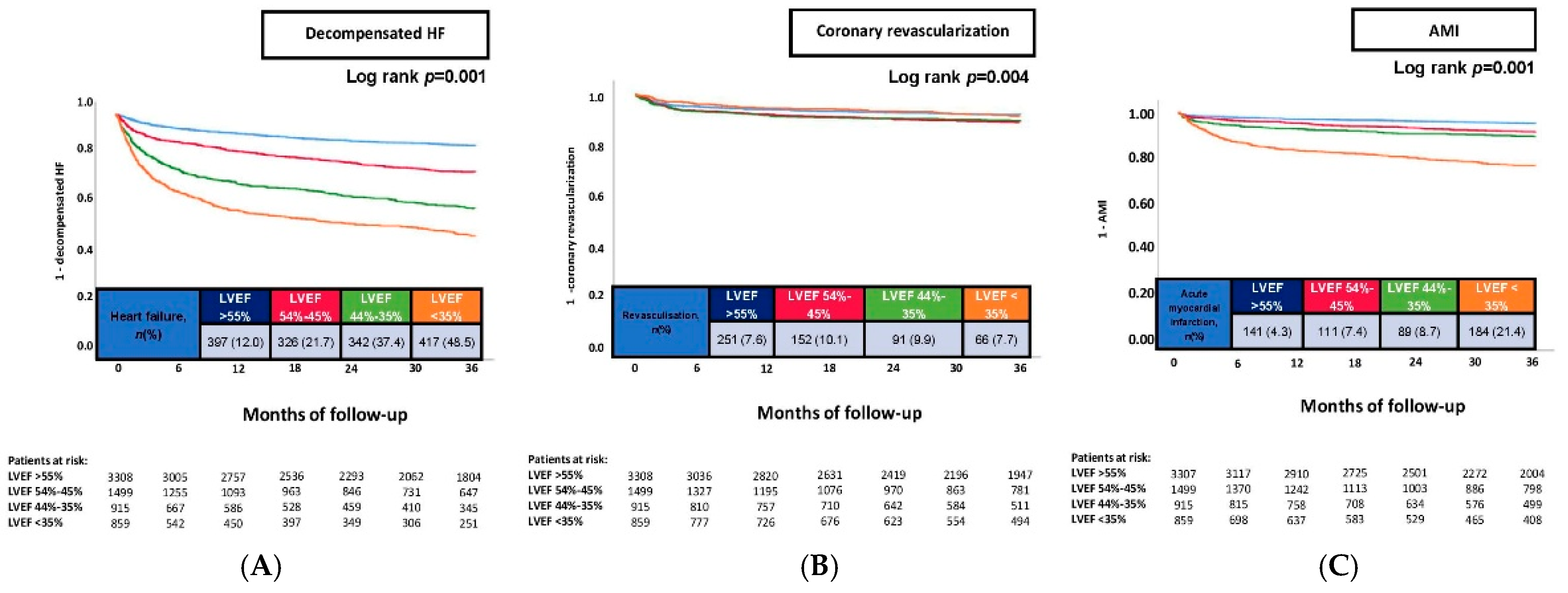
Heart failure, at 36 months397(12.0)326(21.7)342(37.4)417(48.5)0.001
Secondary endpoints, n (%)
 Acute myocardial infarction, at 36 months141(4.3)111(7.4)89(8.7)184(21.4)0.001
 Coronary revascularization, at 36 months251(7.6)152(10.1)91(9.9)66(7.7)0.008
ACE, angiotensin-converting enzyme; ARB, angiotensin receptor blocker; ARNI, angiotensin receptor neprilysin inhibitor; ASA, acetylsalicylic acid; CABG, coronary artery bypass grafting; eGFR, estimated glomerular filtration rate; HbA1c, glycated hemoglobin; ICU, intensive care unit; IQR, interquartile range; LDL, low-density lipoprotein; NT-pro BNP, aminoterminal pro-B-type natriuretic peptide; PCI, percutaneous coronary intervention; SGLT2, sodium–glucose linked transporter 2; and WBC, white blood cell. Level of significance: p ≤ 0.05. Bold type indicates statistical significance.
Table 3. Multivariable Cox regression analyses with regard to risk of heart-failure-related rehospitalization, myocardial infarction, and coronary revascularization at 36 months.
HF-Related RehospitalizationCoronary RevascularizationAMI
VariablesHR95% CIp-ValueHR95% CIp-ValueHR95% CIp-Value
Age (per year increase)1.0121.008–1.0170.0011.0140.903–1.1390.8080.9900.983–0.9970.005
Male sex1.0200.887–1.1730.7781.0250.906–1.1610.6950.9230.764–1.1160.408
Diabetes mellitus1.1511.004–1.3190.0441.3321.186–1.4960.0011.2341.031–1.4770.022
Prior coronary artery disease1.6311.350–1.9710.0011.1750.883–1.5640.2681.2150.919–1.6060.171
Prior myocardial infarction0.9890.672–1.4560.9561.6221.250–2.1050.0011.6481.043–2.6050.032
Prior CABG1.2781.022–1.5970.0321.2340.882–1.7280.2201.2480.923–1.6880.150
Chronic kidney disease1.3191.066–1.6310.0111.5881.328–1.8990.0010.9590.676–1.3610.816
STEMI0.9240.693–1.2320.5880.8030.545–1.1820.2660.7980.621–1.0270.079
NSTEMI0.9450.796–1.1230.5200.9590.758–1.2130.7270.7510.595–0.9480.016
Atrial fibrillation1.2231.066–1.4030.0041.1851.055–1.3310.0040.9740.796–1.1930.801
Decompensated heart failure1.6071.374–1.8800.0011.4901.287–1.7250.0011.4511.161–1.8120.001
LVEF of 54–45%1.8261.573-2.1210.0011.1720.952-1.4430.1361.6671.292-2.1500.001
LVEF of 44–35%2.9482.523-3.4460.0011.0840.836-1.4050.5431.9881.497-2.6400.001
LVEF < 35%3.7313.168-4.3940.0010.8670.632-1.1900.3784.1843.200-5.4710.001
LVEF ≥ 55%(Reference group)(Reference group)(Reference group)
CABG, coronary artery bypass grafting; CI, confidence interval; HR, hazard ratio; HF, heart failure; AMI, acute myocardial infarction; and LVEF, left ventricular ejection fraction. Level of significance: p ≤ 0.05. Bold type indicates statistical significance.
Table 4. Multivariable Cox regression analyses with regard to the risk of heart-failure-related rehospitalization at 36 months in pre-specified subgroup.
Heart-Failure-Related Rehospitalization
Variable HR95% CIp-Value
Age ≥ 70 yearsLVEF < 35%2.3381.793–3.0500.001
LVEF of 44–35%2.0071.558–2.5860.001
LVEF of 54–45%1.5121.182–1.9350.001
LVEF ≥ 55%(Reference group)
Age < 70 years LVEF < 35%3.8742.783–5.3930.001
LVEF of 44–35%2.9252.043–4.1890.001
LVEF of 54–45%1.8021.256–2.5850.002
LVEF ≥ 55%(Reference group)
Male sexLVEF < 35%3.3632.459–4.5990.001
LVEF of 44–35%2.5991.900–3.5550.001
LVEF of 54–45%1.7861.317–2.4220.001
LVEF ≥ 55%(Reference group)
Female sexLVEF < 35%3.0612.048–4.5790.001
LVEF of 44–35%2.2171.515–3.2460.001
LVEF of 54–45%1.4551.021–2.0740.038
LVEF ≥ 55%(Reference group)
Unstable anginaLVEF < 35%4.2142.800–6.3400.001
LVEF of 44–35%3.5312.247–5.5500.001
LVEF of 54–45%1.9791.234–3.1740.005
LVEF ≥ 55%(Reference group)
STEMILVEF < 35%4.0282.351–6.8920.001
LVEF of 44–35%2.9731.682–5.2550.001
LVEF of 54–45%1.7781.001–3.1590.050
LVEF ≥ 55%(Reference group)
NSTEMILVEF < 35%3.4202.349–4.9790.001
LVEF of 44–35%2.3461.572–3.5010.001
LVEF of 54–45%1.6241.123–2.3480.010
LVEF ≥ 55%(Reference group)
Decompensated heart failureLVEF < 35%5.6123.950–7.9720.001
LVEF of 44–35%3.6942.525–5.4020.001
LVEF of 54–45%1.9381.291–2.9100.001
LVEF ≥ 55%(Reference group)
No/–one-vessel diseaseLVEF < 35%3.1452.326–4.2540.001
LVEF of 44–35%2.2551.630–3.1210.001
LVEF of 54–45%1.4051.035–1.9070.029
LVEF ≥ 55%(Reference group)
Two/three-vessel diseaseLVEF < 35%4.8473.049–7.7080.001
LVEF of 44–35%3.2772.047–5.2430.001
LVEF of 54–45%1.7201.041–2.8410.034
LVEF ≥ 55%(Reference group)
CI, confidence interval; HR, hazard ratio; LVEF, left ventricular ejection fraction; STEMI, ST-elevation myocardial infarction; and NSTEMI, non-ST-elevation myocardial Infarction. Level of significance: p ≤ 0.05. Bold type indicates statistical significance.
Disclaimer/Publisher’s Note: The statements, opinions and data contained in all publications are solely those of the individual author(s) and contributor(s) and not of MDPI and/or the editor(s). MDPI and/or the editor(s) disclaim responsibility for any injury to people or property resulting from any ideas, methods, instructions or products referred to in the content.

