

Pellucid Marginal Degeneration: A Comprehensive Review of Pathophysiology, Diagnosis, and Management Strategies

 ,
,
Abstract
1. Introduction
2. Pathophysiology and Epidemiology
3. Clinical Signs
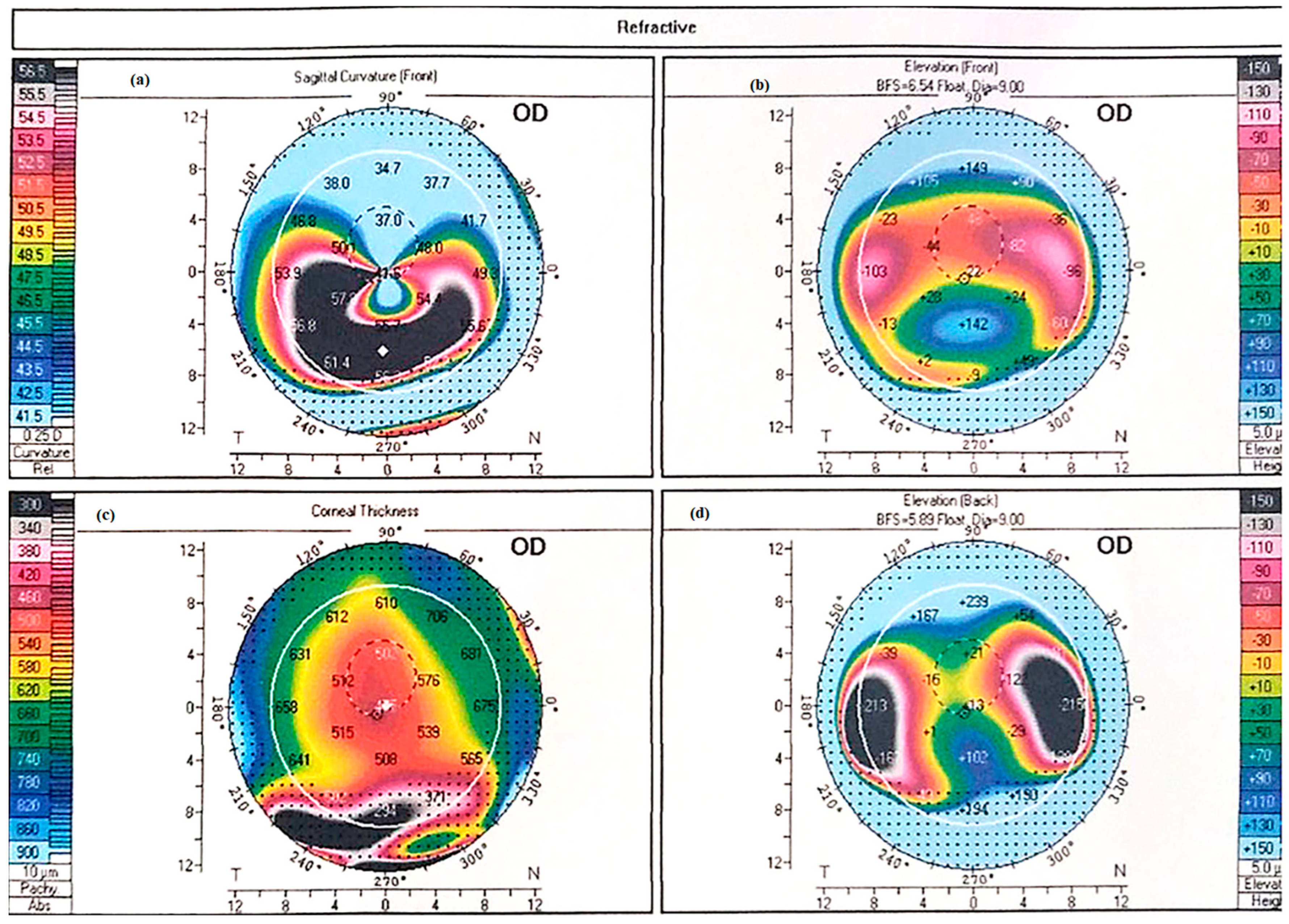
4. Diagnosis
5. Differential Diagnosis
6. Management
7. Conservative Management
8. Surgical Management
- (1)
- Alterations of corneal biomechanics
- (2)
- Toric intraocular lens implantation
- (3)
- Full-thickness surgery
- (4)
- Partial-thickness surgery
8.1. Alterations of Corneal Biomechanics
8.1.1. Collagen Cross Linking
8.1.2. Intrastromal Corneal Rings
8.2. Toric Intraocular Lens Implantation
8.3. Full-Thickness Surgery
8.3.1. Penetrating Keratoplasty
Penetrating Keratoplasty (PK) in PMD
8.3.2. PK Surgical Alternatives
Full-Thickness Crescentic Wedge Resection
8.4. Partial-Thickness Procedures
8.4.1. Crescentic Lamellar Keratoplasty (CLK)
8.4.2. Deep Anterior Lamellar Keratoplasty (DALK)
8.4.3. Historical Techniques
9. Conclusions
Pellucid Marginal Degeneration: Diagnostic and Therapeutic Challenges
Funding
Conflicts of Interest
References
- Sridhar, M.; Mahesh, S.; Bansal, A.K.; Nutheti, R.; Rao, G. Pellucid marginal corneal degeneration. Ophthalmology 2004, 111, 1102–1107. [Google Scholar] [CrossRef]
- Kompella, V.B.; Aasuri, M.K.; Rao, G.N. Management of pellucid marginal corneal degeneration with rigid gas permeable contact lenses. CLAO J. 2002, 28, 140–145. [Google Scholar] [PubMed]
- Sahu, J.; Raizada, K. Pellucid marginal corneal degeneration. In StatPearls [Internet]; StatPearls Publishing: Treasure Island, FL, USA, 2023. [Google Scholar]
- Cameron, J.A.; Mahmood, M.A. Superior corneal thinning with pellucid marginal degeneration. Am. J. Ophthalmol. 1990, 109, 486–487. [Google Scholar] [CrossRef] [PubMed]
- Karabatsas, C.H.; Cook, S.D. Topographic analysis in pellucid marginal corneal degeneration and keratoglobus. Eye 1996, 10, 451–455. [Google Scholar] [CrossRef] [PubMed]
- Gomes, J.A.P.; Rodrigues, P.F.; Lamazales, L.L. Keratoconus epidemiology: A review. Saudi J. Ophthalmol. 2022, 36, 3–6. [Google Scholar] [CrossRef] [PubMed] [PubMed Central]
- Das, A.V.; Pillutla, L.N.; Chaurasia, S. Clinical profile and demographic distribution of pellucid marginal corneal degeneration in India: A study of 559 patients. Indian J. Ophthalmol. 2021, 69, 3488–3493. [Google Scholar] [CrossRef] [PubMed]
- Krachmer, J.H.; Feder, R.S.; Belin, M.W. Keratoconus and related noninflammatory corneal thinning disorders. Surv. Ophthalmol. 1984, 28, 232–293. [Google Scholar] [CrossRef] [PubMed]
- Robin, J.B.; Schanzlin, D.J.; Verity, S.M.; Barron, B.A.; Arffa, R.C.; Suarez, E.; Kaufman, H.E. Peripheral corneal disorders. Surv. Ophthalmol. 1986, 31, 1–36. [Google Scholar] [CrossRef] [PubMed]
- Jinabhai, A.; Radhakrishnan, H.; O’Donnell, C. Pellucid corneal marginal degeneration: A review. Contact Lens Anterior Eye 2011, 34, 56–63. [Google Scholar] [CrossRef] [PubMed]
- Rao, S.K.; Fogla, R.; Padmanabhan, P.; Prema, M.; Sitalakshmi, G. Corneal topography in atypical pellucid marginal degeneration. Cornea 1999, 18, 265–272. [Google Scholar] [CrossRef] [PubMed]
- Wagenhorst, B.B. Unilateral pellucid marginal degeneration in an elderly patient. Br. J. Ophthalmol. 1996, 80, 927–928. [Google Scholar] [CrossRef] [PubMed]
- Taglia, D.P.; Sugar, J. Superior pellucid marginal corneal degeneration with hydrops. Arch. Ophthalmol. 1997, 115, 274–275. [Google Scholar] [CrossRef] [PubMed]
- Varley, G.A.; Macsai, M.S.; Krachmer, J.H. The results of penetrating keratoplasty for pellucid marginal corneal degeneration. Am. J. Ophthalmol. 1990, 110, 149–152. [Google Scholar] [CrossRef] [PubMed]
- Kayazawa, F.; Nishimura, K.; Kodama, Y.; Tsuji, T.; Itoi, M. Keratoconus with pellucid marginal corneal degeneration. Arch. Ophthalmol. 1984, 102, 895–896. [Google Scholar] [CrossRef] [PubMed]
- Jain, A.; Paulus, Y.M.; Cockerham, G.C.; Kenyon, K.R. Keratoconus and Other Non-inflammatory Thinning Conditions. In Duane’s Foundations of Clinical Ophthalmology; Tasman, W., Jaeger, E.A., Eds.; Lippincott Williams and Wilkins: Philadelphia, PA, USA, 2008; Volume 4, Chapter 16C. [Google Scholar]
- McKay, T.B.; Hjortdal, J.; Sejersen, H.; Asara, J.M.; Wu, J.; Karamichos, D. Endocrine and metabolic pathways linked to keratoconus: Implications for the role of hormones in the stromal microenvironment. Sci. Rep. 2016, 6, 25534. [Google Scholar] [CrossRef] [PubMed]
- Zhao, X.; Yuan, Y.; Sun, T.; Zhang, Y.; Chen, Y. Associations between keratoconus and the level of sex hormones: A cross-sectional study. Front. Med. 2022, 9, 828233. [Google Scholar] [CrossRef] [PubMed]
- Krachmer, J.H. Pellucid marginal corneal degeneration. Arch. Ophthalmol. 1978, 96, 1217–1221. [Google Scholar] [CrossRef] [PubMed]
- Ueji, N.; Kato, K.; Yonekawa, Y.; Takeuchi, M.; Takashima, Y.; Hirano, K.; Kondo, M. Case of unilateral pellucid marginal corneal degeneration progressing to corneal perforation with keratoconus in contralateral eye. Am. J. Ophthalmol Case Rep. 2022, 25, 101293. [Google Scholar] [CrossRef] [PubMed]
- Mounir, A.; Abdellah, M.M.; Awny, I.; Aldghaimy, A.H.; Mostafa, E.M. Demographic, clinical and tomographic characteristics of pellucid marginal degeneration patients in South Egyptian population Keratoconus with pellucid marginal corneal degeneration. Int. Ophthalmol. 2022, 42, 3237–3242. [Google Scholar] [CrossRef] [PubMed]
- Iselin, K.C.; Mrochen, M. Evaluation of ectatic corneal disorders using swept-source anterior segment OCT: Current role and limitations. Clin. Ophthalmol. 2021, 15, 1499–1509. [Google Scholar]
- Moshirfar, M.; Edmonds, N.J.; Behunin, L.N.; Christiansen, M.S. Current options in the management of Pellucid Marginal Degeneration. J. Refract. Surg. 2014, 30, 474–485. [Google Scholar] [CrossRef] [PubMed]
- Lee, B.W.; Jurkunas, U.V.; Harissi-Dagher, M.; Poothullil, A.M.; Tobaigy, F.M.; Azar, D.T. Ectatic disorders associated with a claw-shaped pattern on corneal topography. Am. J. Ophthalmol. 2007, 144, 154–156. [Google Scholar] [CrossRef] [PubMed]
- Ding, Y.; Murri, M.S.; Birdsong, C.O.; Ronquillo, Y.; Morshifar, M. Terrien marginal degeneration. Surv. Ophthalmol. 2019, 64, 162–174. [Google Scholar] [CrossRef] [PubMed]
- Chan, T.A.; Ulate, R.; Goldich, Y.; Rootman, S.D.; Chan, C.C. Terrien marginal degeneration: Clinical characteristics and outcomes. Am. J. Ophthalmol. 2015, 160, 867–872. [Google Scholar] [CrossRef] [PubMed]
- Carter, J.B.; Jones, D.B.; Wilhelmus, K.R. Acute hydrops in pellucid marginal corneal degeneration. Am. J. Ophthalmol. 1989, 107, 167–170. [Google Scholar] [CrossRef] [PubMed]
- Tzelikis, P.; Cohen, E.J.; Rapuano, C.; Hammersmith, K.; Laibson, P.R. Management of pellucid marginal corneal degeneration. Cornea 2005, 24, 895–899. [Google Scholar] [CrossRef] [PubMed]
- Jinabhai, A.; Radhakrishnan, H.; Tromans, C.; O’Donnell, C. Visual performance and optical quality with soft lenses in keratoconus patients. Ophthalmic Physiol. Opt. 2012, 32, 100–116. [Google Scholar] [CrossRef] [PubMed]
- Dominguez, C.E.; Shah, A.; Weissman, B.A. Bitoric gas-permeable contact lens application in pellucid marginal corneal degeneration. Eye Contact Lens 2005, 31, 241–243. [Google Scholar] [CrossRef] [PubMed]
- Kastl, P.R.; Kirby, R.G. Bitoric rigid gas permeable lens fitting in highly astigmatic patients. Eye Contact Lens 1987, 13, 215–216. [Google Scholar]
- Visser, E.S.; Visser, R.; van Lier, H.J.; Otten, H.M. Modern scleral lenses part I: Clinical features. Eye Contact Lens 2007, 33, 13–20. [Google Scholar] [CrossRef] [PubMed]
- Tan, D.T.; Pullum, K.W.; Buckley, R.J. Medical applications of scleral contact lenses: 2. Gas-permeable scleral contact lenses. Cornea 1995, 14, 130–137. [Google Scholar] [CrossRef] [PubMed]
- Levit, A.; Benwell, M.; Evans, B.J.W. Randomised controlled trial of corneal vs. scleral rigid gas permeable contact lenses for keratoconus and other ectatic corneal disorders. Contact Lens Anterior Eye 2020, 43, 237–243. [Google Scholar] [CrossRef] [PubMed]
- Hashemi, H.; Shaygan, N.; Asgari, S.; Rezvan, F.; Asgari, S. ClearKone-Synergeyes or rigid gas-permeable contact lens in keratoconic patients. Eye Contact Lens 2014, 40, 95–98. [Google Scholar] [CrossRef] [PubMed]
- Akcay, B.I.S.; Kockar, A.; Limon, U.; Kardes, E.; Dursun, A.D. Comparison of Clinical and Topographic Outcomes of Hybrid and Scleral Lenses in Advanced Keratoconus. Beyoglu Eye J. 2021, 7, 59–65. [Google Scholar] [CrossRef] [PubMed]
- Snibson, G.R. Collagen cross-linking: A new treatment paradigm in corneal disease—A review. Clin. Exp. Ophthalmol. 2010, 38, 141–153. [Google Scholar] [CrossRef] [PubMed]
- Kymionis, G.D.; Karavitaki, A.E.; Kounis, G.A.; Portaliou, D.M.; Yoo, S.H.; Pallikaris, I.G. Management of pellucid marginal corneal degeneration with simultaneous customized photorefractive keratectomy and collagen crosslinking. J. Cataract Refract. Surg. 2009, 35, 1298–1301. [Google Scholar] [CrossRef] [PubMed]
- Irajpour, M.; Noorshargh, P.; Peyman, A. Corneal Cross-Linking in Pellucid Marginal Degeneration: Evaluation after Five Years. J. Curr. Ophthalmol. 2022, 34, 229–233. [Google Scholar] [CrossRef] [PubMed]
- Mamoosa, B.; Razmjoo, H.; Peyman, A.; Ashtari, A.; Ghafouri, I.; Moghaddam, A.G. Short-term result of collagen crosslinking in pellucid marginal degeneration. Adv. Biomed. Res. 2016, 5, 194. [Google Scholar] [CrossRef] [PubMed]
- Kymionis, G.D.; Grentzelos, M.A.; Portaliou, D.M.; Karavitaki, A.E.; Krasia, M.S.; Dranidis, G.K.; Kozobolis, V.P. Photorefractive keratectomy followed by same-day corneal collagen crosslinking after intrastromal corneal ring segment implantation for pellucid marginal degeneration. J. Cataract Refract. Surg. 2010, 36, 1783–1785. [Google Scholar] [CrossRef] [PubMed]
- Kymionis, G.D.; Kontadakis, G.A.; Kounis, G.A.; Portaliou, D.M.; Karavitaki, A.E.; Pallikaris, I.G. Corneal collagen cross-linking with riboflavin and ultraviolet-A irradiation in patients with thin corneas. J. Cataract Refract. Surg. 2010, 36, 28–37. [Google Scholar] [CrossRef] [PubMed]
- Cagil, N.; Sarac, O.; Kocaturk, T.; Can, I. Combined transepithelial phototherapeutic keratectomy and corneal collagen cross-linking for pellucid marginal degeneration. J. Cataract Refract. Surg. 2015, 41, 1215–1222. [Google Scholar]
- Hashemian, S.J.; Abdolalizadeh, P.; Ghiasian, L.; Aghaei, H.; Hadavandkhani, A.; Semnani, F.N.; Jafari, M.E.; Hashemian, S.M.; Hashemian, M.S. Outcomes of a Single-Segment Intrastromal Corneal Ring in Early Keratoconus and Early Pellucid Marginal Degeneration. Int. Ophthalmol. 2022, 42, 2987–2996. [Google Scholar] [CrossRef] [PubMed]
- Jacob, S.; Kumar, D.A.; Agarwal, A.; Rajaraman, R.; Saijimol, A.I. Corneal allogenic intrastromal ring segments (CAIRS) combined with corneal crosslinking for keratoconus: Initial clinical results. J. Refract. Surg. 2018, 34, 269–276. [Google Scholar] [CrossRef] [PubMed]
- Kubaloglu, A.; Sari, E.S.; Cinar, Y.; Koytak, A.; Kurnaz, E.; Piñero, D.P.; Ozerturk, Y. A single 210-degree arc length intrastromal corneal ring implantation for the management of pellucid marginal corneal degeneration. Am. J. Ophthalmol. 2010, 150, 185–192.e1. [Google Scholar] [CrossRef] [PubMed]
- Mularoni, A.; Torreggiani, A.; di Biase, A.; Laffi, G.L.; Tassinari, G. Conservative treatment of early and moderate pellucid marginal degeneration: A new refractive approach with intracorneal rings. Ophthalmology 2005, 112, 660–666. [Google Scholar] [CrossRef] [PubMed]
- Kugler, L.J.; Hill, S.; Sztipanovits, D.; Boerman, H.; Swartz, T.S.; Wang, M.X. Corneal melt of incisions overlying corneal ring segments: Case series and literature review. Cornea 2011, 30, 968–971. [Google Scholar] [CrossRef] [PubMed]
- Balestrazzi, A.; Baiocchi, S.; Cartocci, G.; Tosi, G.M.; Martone, G.; Michieletto, P. Mini-incision cataract surgery and toric lens implantation for the reduction of high myopic astigmatism in patients with pellucid marginal degeneration. Eye 2015, 29, 637–642. [Google Scholar] [CrossRef] [PubMed]
- Matalia, H.; Nandini, C.; Matalia, J. Long-term outcome of custom toric intraocular lens for treating high astigmatism in case of cataract associated with pellucid marginal corneal degeneration. Indian J. Ophthalmol. 2020, 68, 3082–3084. [Google Scholar] [CrossRef] [PubMed]
- Bahar, I.; Bialer, O. Cataract extraction and toric intraocular lens implantation for the management of pellucid marginal degeneration and cataract. Int. J. Keratoconus Ectatic Corneal Dis. 2012, 1, 66–67. [Google Scholar] [CrossRef]
- Han, D.C.Y.; Lim, L. Implantation of toric intraocular lens in pellucid marginal degeneration: A case report on ocular aberrometry outcome. J. Clin. Exp. Ophthalmol. 2012, 3, 204. [Google Scholar]
- Luck, J. Customized ultra-high-power toric intraocular lens implantation for pellucid marginal degeneration and cataract. J. Cataract Refract. Surg. 2010, 36, 1235–1238. [Google Scholar] [CrossRef] [PubMed]
- Camoriano, G.D.; Aman, S.; Gimbel, H.V. Toric collagen copolymer phakic intraocular lens to correct myopic astigmatism in eyes with pellucid marginal degeneration. J. Cataract Refract. Surg. 2012, 38, 256–262. [Google Scholar] [CrossRef] [PubMed]
- de Vries, E.; de Vries, D.; de Vries, L. Use of Verisyse/Artisan phakic intraocular lens for the correction of high myopia in a patient with early-stage pellucid marginal degeneration. J. Cataract Refract. Surg. 2008, 34, 1700–1702. [Google Scholar] [CrossRef] [PubMed]
- Zhu, X.; He, W.; Zhang, K.; Lu, Y.; Zhu, Y. Toric intraocular lens implantation in eyes with pellucid marginal degeneration. BMC Ophthalmol. 2017, 17, 22. [Google Scholar]
- Cherry, P.M.; Diggory, P.; Ficker, L.A. Corneal graft size and endothelial cell loss. Br. J. Ophthalmol. 1979, 63, 815–820. [Google Scholar]
- Tuberville, A.W.; Wood, T.O.; McLaughlin, B.J. The effects of eccentric donor recipient grafts on the survival of corneal allografts. Am. J. Ophthalmol. 1983, 95, 177–182. [Google Scholar]
- Tzelikis, P.F.; Cohen, E.J.; Rapuano, C.J. Long-term visual outcome of penetrating keratoplasty for pellucid marginal degeneration. Ophthalmology 1999, 106, 776–781. [Google Scholar]
- Speaker, M.G.; Guerriero, P.N.; Met, J.; Freilich, D.B. A study of penetrating keratoplasty using frozen and fresh donor tissue. Am. J. Ophthalmol. 1983, 96, 729–735. [Google Scholar]
- Varley, G.A.; Meisler, D.M.; Parks, M.M. Penetrating keratoplasty in pellucid marginal corneal degeneration. Ophthalmology 1995, 102, 590–594. [Google Scholar] [CrossRef] [PubMed]
- Durand, M.; Hersh, P.S. Corneal wedge resection for pellucid marginal corneal degeneration. Ophthalmic Surg. Lasers Imaging Retin. 1991, 22, 442–447. [Google Scholar]
- MacLean, H.; Khan, S.; Muhtaseb, M. The results of femtosecond laser-assisted wedge resection for the treatment of pellucid marginal degeneration. J. Refract. Surg. 2012, 28, 884–888. [Google Scholar]
- Busin, M.; Scorcia, V.; Zambianchi, L.; Ponzin, D. Combined use of crescentic resection and relaxing incisions for the treatment of pellucid marginal corneal degeneration. Am. J. Ophthalmol. 2008, 145, 675–681. [Google Scholar]
- Genç, S.; Çakir, H.; Güler, E.; Çalli, Ü. Refractive and corneal aberrometric changes after crescentic lamellar wedge resection in pellucid marginal degeneration. Eye Contact Lens 2018, 44 (Suppl. S2), S76–S80. [Google Scholar] [CrossRef] [PubMed]
- Michael, T.; Ioannis, G.; Ferdinardo, M.; Ioannis, A. Endothelium sparing—Air assisted wedge resection for the treatment of pellucid marginal degeneration. Indian J. Ophthalmol. 2024, 72 (Suppl. S2), S314–S318. [Google Scholar] [CrossRef] [PubMed]
- Tsatsos, M.; Giachos, I.; Martini, F. Femto second laser assisted wedge resection for the treatment of PMD. Indian J. Ophthalmol. 2024, 72 (Suppl. S5), S923–S924. [Google Scholar] [CrossRef] [PubMed]
- Kymionis, G.; Voulgari, N.; Samutelela, E.; Kontadakis, G.; Tabibian, D. Combined corneal wedge resection and corneal cross-linking for pellucid marginal degeneration: A first report. Ther. Clin. Risk Manag. 2019, 15, 1319–1324. [Google Scholar] [CrossRef] [PubMed] [PubMed Central]
- Schanzlin, D.J.; Sarno, E.M.; Robin, J.B. Crescentic lamellar keratoplasty for pellucid marginal degeneration. Am. J. Ophthalmol. 1983, 96, 253–254. [Google Scholar] [CrossRef] [PubMed]
- Schnitzer, J.I. Crescentic lamellar keratoplasty for pellucid marginal degeneration. Am. J. Ophthalmol. 1984, 97, 250–252. [Google Scholar] [CrossRef] [PubMed]
- Al-Torbak, A.A.; Al-Motowa, S.; Al-Assiri, A.; Al-Kharashi, S.; Al-Shahwan, S.; Al-Mezaine, H.; Teichmann, K. Deep anterior lamellar keratoplasty for management of keratoconus in Saudi Arabia. Br. J. Ophthalmol. 2007, 91, 1212–1216. [Google Scholar]
- Kodavoor, K.S.; Deb, B.; Ramamurthy, D. Outcome of deep anterior lamellar keratoplasty patients with intraoperative Descemet's membrane perforation: A retrospective cross-sectional study. Indian J. Ophthalmol. 2018, 66, 1574–1579. [Google Scholar] [CrossRef] [PubMed]
- Millar, M.J.; Maloof, A. Deep lamellar keratoplasty for pellucid marginal degeneration: Review of management options for corneal perforation. Cornea 2008, 27, 953–956. [Google Scholar] [CrossRef] [PubMed]
- Biswas, S.; Brahma, A.; Tromans, C.; Ridgway, A. Management of pellucid marginal corneal degeneration. Eye 2000, 14, 629–634. [Google Scholar] [CrossRef] [PubMed]


| Disease | Key Characteristics | Differentiating Features |
|---|---|---|
| Pellucid Marginal Degeneration (PMD) | - Inferior peripheral corneal thinning (crescent-shaped). - Νon-inflammatory. - Irregular astigmatism. - “Beer-belly” appearance on tomography. | - Thinning is localized to the inferior periphery. - No lipid deposits or vascularization. - Crab-claw pattern on tomography. |
| Keratoconus (KC) | - Central or paracentral corneal thinning. - Cone-shaped protrusion. - Irregular astigmatism. | - Thinning and protrusion are central or paracentral. - No inferior crescent-shaped thinning. - Different tomography pattern (not usually crab-claw). |
| Keratoglobus | - Congenital, diffuse corneal thinning. - Globular protrusion of the entire cornea. | - Thinning and protrusion are global, not localized. - Onset is congenital, unlike PMD. |
| Terrien’s Marginal Degeneration | - Peripheral circumferential thinning. - Lipid deposits and pseudopterygium. - Non-inflammatory. | - Thinning is circumferential, not inferior. - Lipid deposits and pseudopterygium are common. - No crab-claw tomography. |
| Peripheral Ulcerative Keratitis (PUK) | - Inflammatory peripheral corneal thinning. - Associated with systemic autoimmune diseases. | - Painful, inflammatory, and often associated with systemic disease. - Epithelial defect and vascularization. |
| Mooren’s Ulcer | - Painful, inflammatory peripheral ulcer. - Epithelial defect. - No systemic association. | - Severe pain and inflammation. - Epithelial defect present. - No lipid deposits or pseudopterygium. |
| Acute Hydrops | - Fluid accumulation in the cornea due to Descemet’s membrane rupture. - Associated with advanced ectatic diseases [28]. | - More common in keratoconus but higher risk in PMD. - Sudden onset of corneal edema and vision loss. |
| Treatment Modality | Indications | Benefits | Limitations |
|---|---|---|---|
| Glasses | Early stages of PMD with mild refractive errors | - Simple and non-invasive - First-line option for mild astigmatism | - Limited effectiveness in advanced stages - Cannot correct irregular astigmatism |
| Soft Contact Lenses | Early stages of PMD with regular astigmatism | - Easy to use and comfortable - Effective for regular astigmatism | - Less effective as irregular astigmatism progresses - Limited in advanced PMD |
| Corneal Rigid Gas Permeable (RGP) Lenses | Moderate to advanced PMD with irregular astigmatism | - Provide better visual acuity in irregular astigmatism - Improved stability | - Fitting can be challenging - Require gradual adjustment of lens diameter |
| Limbal/Mini-Scleral RGP Lenses | Advanced PMD with significant corneal irregularity | - Better centration and stability compared to corneal RGP lenses | - Require expertise in fitting - May still dislodge in severe cases |
| Scleral RGP Lenses | Advanced PMD with inferior corneal protrusion and severe irregularity | - Excellent stability and centration - Vault over the cornea, reducing irregularity impact | - Limited availability of trained practitioners - Reduced tear turnover and debris buildup |
| Hybrid Contact Lenses | Patient intolerance to RGP lenses or needing combined comfort and visual acuity | - Combines RGP vision correction with soft lens comfort | - Prone to tearing at the RGP-soft lens junction - Risk of oxygen permeability issues |
| Third-Generation Hybrid Lenses | Patients with sensitivity to RGP lenses | - Improved visual acuity (up to 20/30) - Better comfort for sensitive patients | - Higher complication rate (e.g., conjunctivitis, edema, allergic reactions) |
| Emerging Hybrid Lenses (e.g., AirFlex) | Emerging option for PMD management | - Promising early results - Combines comfort and visual correction | - Limited long-term data and research |
| Procedure | Mechanism | Indications | Key Benefits | Limitations/Risks |
|---|---|---|---|---|
| Collagen Cross-Linking (CXL) | UV-A-induced photochemical bonding of collagen fibrils to increase corneal rigidity | Progressive PMD with sufficient corneal thickness | - May halt progression of ectasia - Improved corneal biomechanics - Minimally invasive | - Variable visual outcomes - Limited efficacy in highly irregular astigmatism - Risk to limbal stem cells in inferiorly thinned areas |
| CXL-Plus (PRK + CXL) | Surface ablation followed by CXL for refractive and structural improvement | Select PMD cases with central astigmatism and adequate stromal thickness | - May reduce irregular astigmatism - Potential for better uncorrected visual acuity | - Higher biomechanical risk, especially in peripherally thinned corneas - Risk of ectasia progression if poorly selected |
| Intrastromal Corneal Ring Segments (ICRS) | Synthetic PMMA ring segments reshaping the corneal curvature | Mild to moderate PMD with contact lens intolerance | - Reduces astigmatism (2–4 D) - Improves BCVA - Reversible and minimally invasive | - Unpredictable long-term outcomes - Possible complications (e.g., extrusion, infection) - Limited effect in advanced PMD |
| Toric Intraocular Lenses (IOLs) | Astigmatism correction via toric optics during cataract or refractive lens surgery | PMD with coexisting cataract or stable ectasia | - Reduces refractive astigmatism - Good BCVA in selected cases - Single-step solution in cataract surgery | - Does not treat underlying ectasia - Risk of misalignment - IOL power calculation can be inaccurate in irregular corneas |
| Procedure | Benefits | Negatives (Limitations) |
|---|---|---|
| 1. Penetrating Keratoplasty | - Removes all irregular corneal tissue, providing a clear optical surface. - Improves visual acuity significantly (e.g., 20/153 to 20/43 in Tzelikis et al. [60]). | - High risk of graft rejection (up to 50%). - Postoperative astigmatism is common and challenging to manage. - Requires large, eccentric grafts, increasing surgical complexity. - Risk of complications like neovascularization, glaucoma, and infection. |
| 2. Full-Thickness Crescentic Wedge Resection | - Does not require donor tissue. - Shorter recovery time compared to PK. - Reduces astigmatism in some cases. | - Limited evidence and small sample sizes in studies. - Risk of astigmatic drift. - Intraoperative challenges and potential for complications like microperforation. |
| 3. Partial-Thickness Crescentic Wedge Resection | - Preserves endothelial layer, reducing rejection risk. - Improves BCVA (e.g., 20/125 to 20/32 in Genc et al. [65]). - No donor tissue required. | - Risk of intraoperative microperforation (14% in some studies). - Suture-related complications (e.g., infiltration in 34% of cases). - Limited long-term data. |
| 4. Crescentic Lamellar Keratoplasty | - Preserves endothelium, reducing rejection risk. - Improves visual outcomes with contact lenses or spectacles. - Maintains structural integrity of the eye. | - Postoperative astigmatism is common. - Risk of graft–host interface opacification. - Technically challenging with a steep learning curve. |
| 5. Deep Anterior Lamellar Keratoplasty | - Preserves recipient endothelium, reducing rejection risk. - Lower postoperative astigmatism compared to PK. - Shorter steroid therapy duration and fewer complications. | - Technically demanding and time-consuming. - High risk of intraoperative perforation, especially in corneas with hydrops. - Limited improvement in BCVA compared to PK. |
| 6. Lamellar Thermokeratoplasty and Epikeratoplasty | - Historical procedures with limited use today. | - High complication rates. - Outdated and replaced by safer, more effective techniques. |
Disclaimer/Publisher’s Note: The statements, opinions and data contained in all publications are solely those of the individual author(s) and contributor(s) and not of MDPI and/or the editor(s). MDPI and/or the editor(s) disclaim responsibility for any injury to people or property resulting from any ideas, methods, instructions or products referred to in the content. |
© 2025 by the authors. Licensee MDPI, Basel, Switzerland. This article is an open access article distributed under the terms and conditions of the Creative Commons Attribution (CC BY) license (https://creativecommons.org/licenses/by/4.0/).
Share and Cite
Tsatsos, M.; Koulotsiou, K.; Giachos, I.; Tsinopoulos, I.; Ziakas, N. Pellucid Marginal Degeneration: A Comprehensive Review of Pathophysiology, Diagnosis, and Management Strategies. J. Clin. Med. 2025, 14, 5178. https://doi.org/10.3390/jcm14155178
Tsatsos M, Koulotsiou K, Giachos I, Tsinopoulos I, Ziakas N. Pellucid Marginal Degeneration: A Comprehensive Review of Pathophysiology, Diagnosis, and Management Strategies. Journal of Clinical Medicine. 2025; 14(15):5178. https://doi.org/10.3390/jcm14155178
Chicago/Turabian StyleTsatsos, Michael, Konstantina Koulotsiou, Ioannis Giachos, Ioannis Tsinopoulos, and Nikolaos Ziakas. 2025. "Pellucid Marginal Degeneration: A Comprehensive Review of Pathophysiology, Diagnosis, and Management Strategies" Journal of Clinical Medicine 14, no. 15: 5178. https://doi.org/10.3390/jcm14155178
APA StyleTsatsos, M., Koulotsiou, K., Giachos, I., Tsinopoulos, I., & Ziakas, N. (2025). Pellucid Marginal Degeneration: A Comprehensive Review of Pathophysiology, Diagnosis, and Management Strategies. Journal of Clinical Medicine, 14(15), 5178. https://doi.org/10.3390/jcm14155178