Article

Expanding the Spectrum of CSF3R-Mutated Myeloid Neoplasm Beyond Chronic Neutrophilic Leukemia and Atypical Chronic Myeloid Leukemia: A Comprehensive Analysis of 13 Cases

1
Department of Pathology and Laboratory Medicine, Donald and Barbara Zucker School of Medicine at Hofstra/Northwell, Greenvale, NY 11548, USA
2
Department of Medicine, Northwell Cancer Institute, Donald and Barbara Zucker School of Medicine at Hofstra/Northwell, Manhasset, NY 11030, USA
*
Authors to whom correspondence should be addressed.
J. Clin. Med. 2025, 14(15), 5174; https://doi.org/10.3390/jcm14155174
Submission received: 27 June 2025 / Revised: 17 July 2025 / Accepted: 18 July 2025 / Published: 22 July 2025
(This article belongs to the Special Issue Novel Therapeutic Strategies for Acute Myeloid Leukemia)

Abstract

Background: Genetic alterations in CSF3R, typically associated with chronic neutrophilic leukemia (CNL) and atypical chronic myeloid leukemia (aCML), rarely occur in other myeloid neoplasms. Methods: This study characterized the clinical, morphologic, cytogenetic, and molecular features of 13 patients with non-CNL non-aCML myeloid neoplasms with CSF3R alterations. Patients (median age, 77 years) were categorized into groups with a myelodysplastic/myeloproliferative neoplasm (MDS/MPN) (n = 5), acute leukemia (n = 4), and other myeloid neoplasms (n = 4) based on the WHO 2022 and ICC criteria. Results: The CSF3R p.Thr618Ile mutation was most frequent (11/13), with additional pathogenic variants including p.Gln743Ter and frameshift mutations affecting the cytoplasmic tail. Variant allele frequencies (VAFs) ranged from 2% to 49%, with the highest median VAF in the MDS/MPN group. Co-mutations varied by subtype; MDS/MPN, NOS, and CMML cases frequently harbored mutations in epigenetic regulators (ASXL1, TET2) and splicing factors (SF3B1, SRSF2, ZRSR2), while acute leukemia cases showed alterations in JAK3, STAT3, and NRAS. Survival analysis revealed distinct patterns across the three diagnostic groups, with MDS/MPN having the poorest prognosis. Conclusion: This study expands the recognized spectrum of CSF3R-related myeloid neoplasms and highlights the clinical and molecular heterogeneity associated with these mutations, emphasizing the need for comprehensive molecular profiling and the potential for targeted therapies.

1. Introduction

The colony-stimulating factor 3 receptor (CSF3R) is a key member of the hematopoietic receptor super-family. The protein CSF3R, encoded by the eponymous gene (CSF3R), plays a crucial role in the proliferation, differentiation, and survival of granulocytes [1,2]. Upon binding its ligand granulocyte colony stimulating factor (G-CSF), CSF3R triggers various downstream pathways, including the JAK-STAT pathway, leading to its biological effects [3]. Due to this key biological function in hematopoiesis, genetic alterations in CSF3R are strongly linked to granulocytic dysfunction. The T618I mutation in CSF3R, in particular, is the most frequent genetic alteration that leads to constitutive receptor activation, resulting in aberrant JAK-STAT, SRC family kinase, and RAS-MAPK signaling, driving granulocytic proliferation and cytokine-independent cell survival [4,5].
Somatic activating CSF3R mutations are highly associated with chronic neutrophilic leukemia (CNL) and present with neutrophilia, hypercellular bone marrow with granulocytic proliferation, and the absence of dysplasia or increased blasts. This rare myeloproliferative neoplasm (MPN) usually shows an indolent clinical course, although the outcome may vary [6,7]. CNL may acquire additional mutations and progress to blast crisis with or without the acquisition of dysplasia; however, the specific alteration in CSF3R persists in the progressing disease, often with increased allele frequency, suggesting a linear molecular progression of this disease [8,9]. In addition to CNL, CSF3R alterations are frequently seen in atypical chronic myeloid leukemia (aCML), also known as myelodysplastic/myeloproliferative neoplasm with neutrophilia (MDS/MPN-N), and severe congenital neutropenia (SCN) [10,11]. Alterations in CSF3R are also infrequently associated with other myeloid neoplasms, such as chronic myelomonocytic leukemia (CMML), myelodysplastic neoplasms (MDS), or acute myeloid leukemia (AML) [12].
Although CSF3R genetic alterations may belong to three different classes, the point mutation T618I, affecting the transmembrane proximal domain, is the most common pathogenic alteration in myeloid neoplasms. This mutation often acts as the driver mutation in CNL, persisting throughout the disease progression [8]. Importantly, CNL patients with the T618I mutation show a significantly worse prognosis compared to those with other CSF3R mutations [13]. While this mutation may not independently drive leukemogenesis, it may play a synergistic role in disease pathogenesis. Its ability to constitutively activate the JAK-STAT pathway has made it a rational therapeutic target, with JAK inhibitors such as ruxolitinib having been successfully used to treat neoplasms driven by this alteration [11,14,15]. All these factors make the identification of myeloid neoplasms with CSF3R alteration clinically relevant.
In this study, we examined the clinical, hematological, histomorphological cytogenetic, and molecular characteristics of 13 cases of non-CNL, non-aCML myeloid, and non-SCN myeloid neoplasms. We further documented the mainstay therapy that patients received and their outcome. By investigating these rare cases, we have explored whether there are any underlying unifying characteristics in these neoplasms.

2. Materials and Methods

2.1. Patient Selection and Data Collection

We searched our institutional database for all pathology diagnostic reports for “CSF3R”. All retrieved cases were manually reviewed independently by two board-certified hematopathologists. Patients were included if they harbored a CSF3R mutation confirmed by next-generation sequencing (NGS), had a clinical and hematopathology diagnosis that did not meet the criteria for CNL, aCML, or SCN, and had comprehensive molecular, cytogenetic, and morphologic data available. Of all such cases, only newly diagnosed myeloid neoplasms were included in this study. Demographic and clinical data, including age, sex, clinical presentation, and laboratory findings, were extracted from the institutional electronic medical record system manually.

2.2. Hematologic and Morphologic Analysis

Complete blood counts, peripheral blood smears, and bone marrow aspirates/core biopsies were reviewed. Dysplasia across erythroid, myeloid, and megakaryocytic lineages was documented.

2.3. Cytogenetic and FISH Analysis

Conventional karyotyping and fluorescence in situ hybridization (FISH) were performed for all cases as part of standard diagnostic evaluation.

2.4. Molecular Studies

Targeted NGS was performed through an OnkoSight myeloid panel (BioReference HealthTM) comprising up to 50 genes, which included ABL1, ANKRD26, ASXL1, ATRX, BCOR, BCORL1, BRAF, CALR, CBL, CCND2, CDKN2A, CEBPA, CSF3R, CUX1, DDX41, DNMT3A, ETNK1, ETV6, EZH2, FBXW7, FLT3, GATA2, HRAS, IDH1, IDH2, JAK2, KDM6A, KIT, KMT2A, KRAS, MAP2K1, MPL, MYD88, NF1, NPM1, NRAS, PDGFRA, PHF6, PTEN, PTPN11, RUNX1, SETBP1, SF3B1, SRSF2, STAG2, TET2, TP53, U2AF1, WT1, and ZRSR2. The OnkoSight Myeloid panel used has a validated analytical sensitivity of 1–2% VAF for SNVs and small indels, with a minimum read depth > 500×. Only variants that passed internal quality control filters were included in the final analysis.

2.5. Statistical Analysis

Descriptive analysis was employed to summarize clinical and laboratory data. All statistical analyses including Kaplan–Meier survival analysis and data visualizations were performed using Python 3.14TM. Tables, and all visualizations with histograms, box plots, bar charts, heatmaps, and lollipop plots were generated with libraries such as Matplotlib, Seaborn, and Pandas.

2.6. Ethical Considerations

This study was conducted in accordance with institutional review board (IRB) policies regarding the use of patient data in research. Patient identifiers were anonymized to ensure confidentiality.

3. Results

3.1. Clinical Findings

We examined the molecular profiles of 1400 patients with myeloid neoplasms diagnosed at our institute over a seven-year period. We identified thirteen (0.9%) with CSF3R-mutated myeloid neoplasms, which were categorized into three diagnostic groups. The MDS/MPN group includes three cases of MDS/MPN-unclassified (MDS/MPN-U) and two cases of CMML; the acute leukemia group includes two cases of AML, one case of mixed phenotype acute leukemia (MPAL)-M/T, and one case of myeloid sarcoma (MS). The third group includes three cases of de novo MDS and one case of MPN with progression to MDS. The median age for the whole cohort was 77 years (range, 22 to 89 years), with a male predominance (nine males, four females). Patients in the MDS/MPN group were predominantly elderly (median age, 84 years; range, 78 to 89 years). The acute leukemia cases spanned a wide age range (22 to 69 years; median, 56 years). Patients in the other myeloid neoplasm group had a median age of 68 years (range, 57 to 79 years). An elevated lactate dehydrogenase (LDH) level (≥242 U/L) was present in 11 out of 13 patients, and splenomegaly was seen in 2 patients. Clinical history was significant for previous malignancy (four cases), autoimmune disease (three cases), and hepatitis (one case) (Table 1).

3.2. Peripheral Blood Findings

For the whole cohort, the median white blood cell (WBC) count was 14.6 × 10⁹/L (range, 1.4 × 10⁹/L to 58.5 × 10⁹/L), with both leukopenia and leukocytosis observed. Hemoglobin (Hb) levels ranged from 7.3 g/dL to 12.0 g/dL, (median, 9.4 g/dL), with severe anemia (Hb < 8.0 g/dL) noted in four patients. Platelet counts ranged from 27 × 109/L to 216 × 109/L (median, 95 × 109/L). Patients in the MDS/MPN group presented with leukocytosis and varying degrees of anemia, and thrombocytopenia (3/5 cases). Peripheral blood smears revealed neutrophilia (>80% of leukocytes), with immature granulocytes comprising <10% of the WBC count in all cases. Absolute monocytosis (>1 × 10⁹/L and ≥10% of cells) were noted only in the CMML patients. There was no increase in basophils or blasts in any case. Peripheral smears in the acute leukemia group exhibited varying degrees of leukocytosis and cytopenia, with circulating blasts ranging from 1% to 77%. Peripheral smears in the other myeloid neoplasm group revealed normocytic anemia, anisopoikilocytosis, thrombocytopenia (3/4 cases), and dysgranulopoiesis.

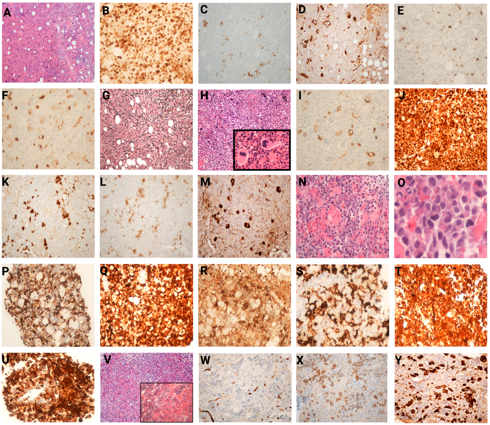
3.3. Histopathological Findings in Bone Marrow

Bone marrow biopsies in the MDS/MPN group consistently revealed marked hypercellularity (70 to 100% cellularity) with myeloid predominance in three cases and erythroid predominance or a normal M/E ratio in each of the remaining two cases. Dysplasia is noted in all cases. All but one case (patient 4) had less than 5% blasts. Ring sideroblasts exceeding 15% were observed in two cases (Case 1 and Case 4). Moderate to marked reticulin fibrosis (grade 2–3) was seen in two patients (Case 2 and Case 3).
Bone marrow aspirates and biopsies in the acute leukemia patients revealed hypercellularity (70% to 85% cellularity) with extensive blast infiltration and decreased normal hematopoiesis in three cases. The bone marrow in the myeloid sarcoma case showed myeloid hyperplasia, left-shifted granulopoiesis, and dyserythropoiesis and the extramedullary site involved (the lymph node) showed the effacement of the architecture by a diffuse proliferation of immature myeloid cells, consistent with MS.
The bone marrow findings in the other myeloid neoplasm group ranged from normocellular to markedly hypercellular marrow (30 to 100% cellularity), with myeloid predominance in 3 out of 4 patients, and dysplasia in at least one lineage in all patients. Increased blasts (5 to 8%) were noted in the bone marrow in all patients.
The histomorphological and immunophenotypic findings from the bone marrow biopsies of representative cases are depicted in Figure 1.

3.4. Cytogenetic and FISH Analysis

Among the 13 cases, normal karyotypes were observed in 8 cases. Abnormalities included monosomy 7 (Case 2), deletion 7q (Case 4), a complex karyotype in Case 9 (44-45,X,-Y,der(4)t(4;6)(p12;p11.2)der(4)(q31),-8,-9,+mar1,+mar2[6]/46,XY [cp14]), and a derivative chromosome involving chromosomes 13 and 14 in Case 12. Case 13 demonstrated a 45,-X,-Y karyotype. FISH studies identified monosomy 7 in Case 2 and deletion 7q in Case 4, confirming karyotype findings. The amplification of the RUNX1 gene was detected in Case 6 (three copies), and a RUNX1T1/RUNX1 rearrangement was identified in Case 9. FISH was normal in seven cases; results were unavailable in two cases. One patient with MDS/MPN-U had an isolated deletion of the Y chromosome, likely an age-related finding.

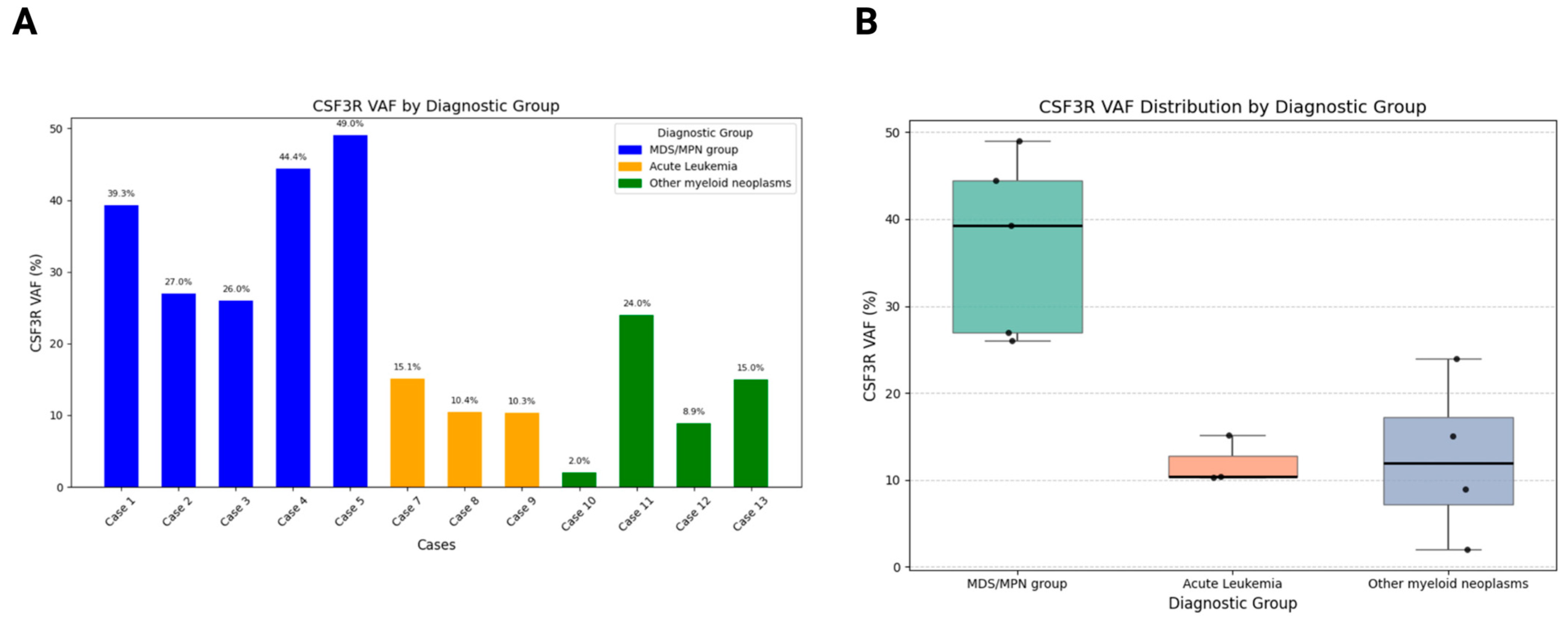
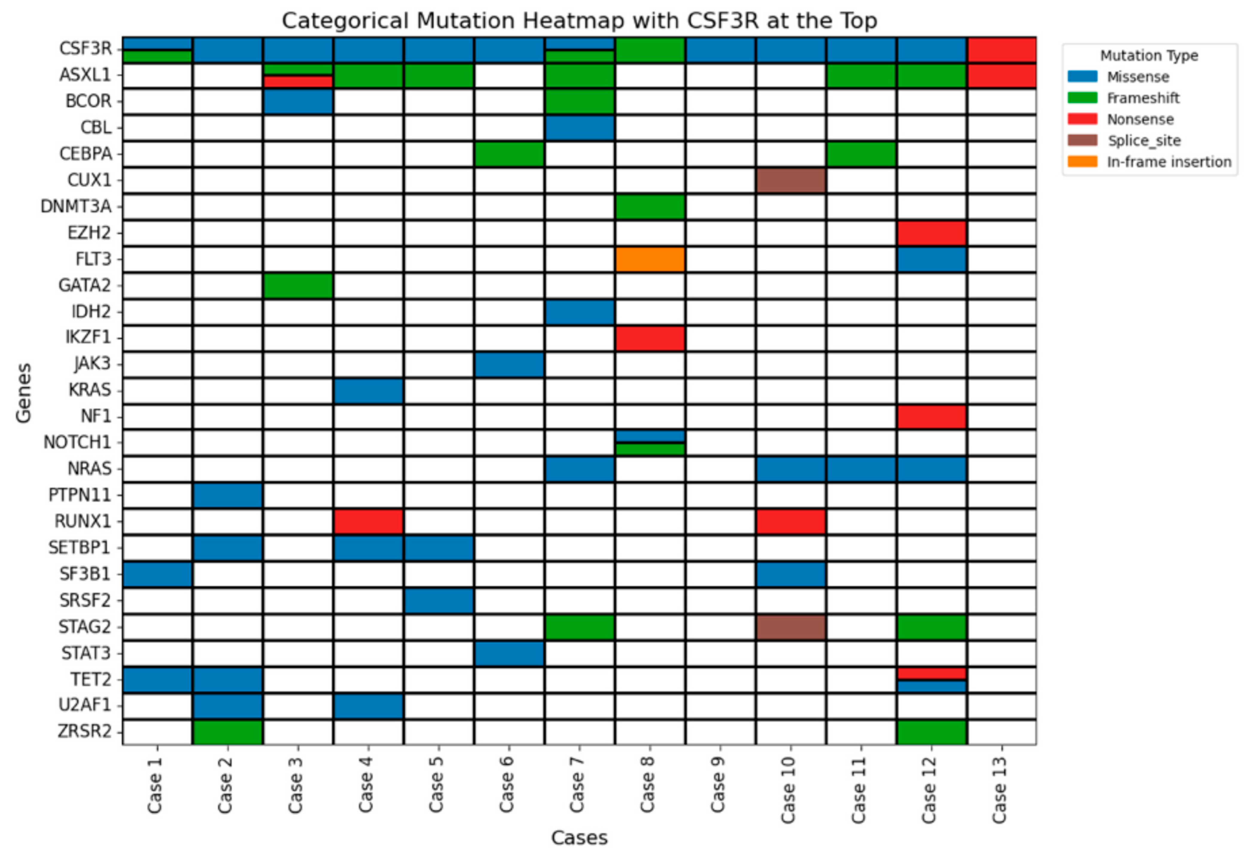
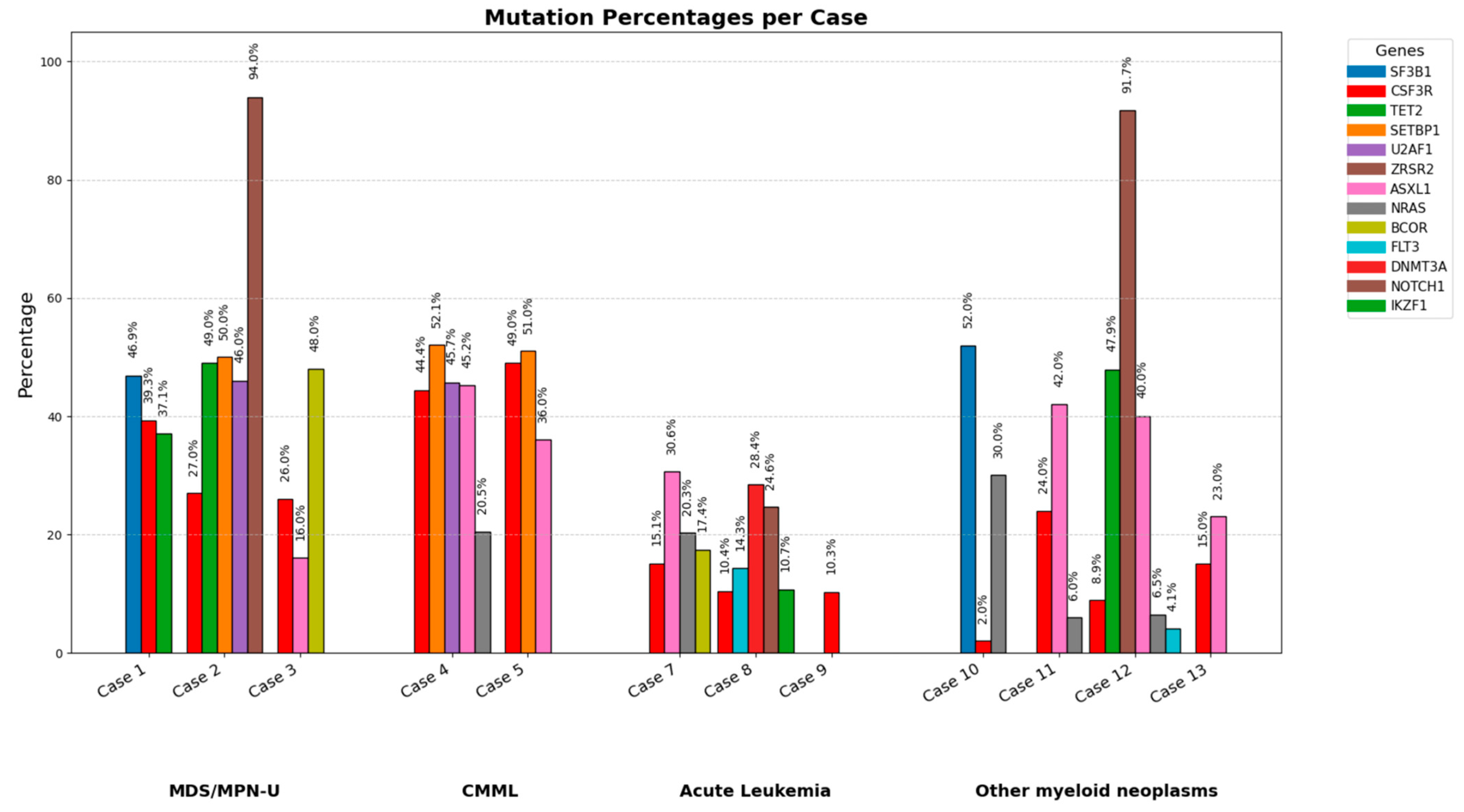
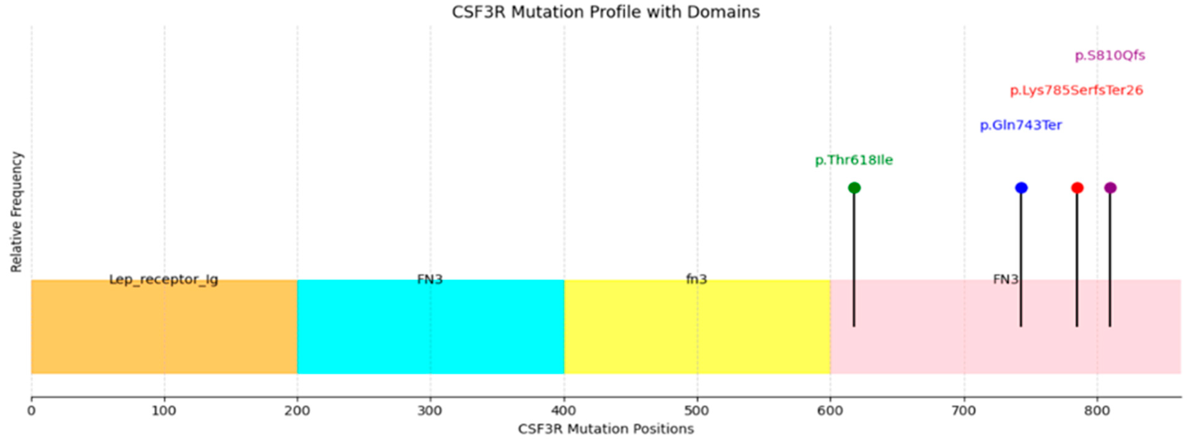
3.5. Molecular Findings

The mutational landscape across the 13 patients with CSF3R-mutated myeloid neoplasms was highly heterogeneous. All patients harbored CSF3R alterations, with variant allele frequencies (VAFs) ranging from 2% to 49%, indicating varying degrees of clonal involvement.
Grouped analysis revealed that patients with MDS/MPN-U and CMML demonstrated the highest CSF3R VAFs (median, 39%; range, 26–49%), consistent with a predominant clonal driver role. In contrast, cases with acute leukemia exhibited the lowest VAFs (median, 10%; range, 10–15%), suggesting a possible subclonal or secondary role of CSF3R in leukemogenesis. Other myeloid neoplasms displayed intermediate VAFs (median, 12%; range, 2–24%), indicating a smaller contribution of CSF3R to disease pathogenesis. These differences are visualized in Figure 2A,B, which display VAF distributions across diagnostic groups using bars and box plots, respectively.
Co-mutations varied by disease subtype and are depicted in a categorical mutation heatmap (Figure 3). The most common co-mutations were ASXL1 and mutations in the RAS signaling pathway (NRAS, KRAS, and PTPN11). MDS/MPN-U and CMML patients frequently carried mutations in epigenetic regulators (ASXL1, TET2) and RNA splicing genes (SF3B1, SRSF2, ZRSR2). A SETBP1 mutation was identified in one patient with CMML. In the acute leukemia group, CSF3R mutations co-occurred with alterations in signaling pathway genes, including JAK3, STAT3, and NRAS. For instance, Case 6 (AML with biallelic CEBPA mutations) also harbored the STAT3 mutation. Other myeloid neoplasms, including MDS, showed low-frequency CSF3R mutations (≤10%), suggesting a smaller subclonal role. Interestingly, KRAS mutations were observed in two of three MDS cases, possibly indicating an underlying MDS/MPN-like biology.
The most frequent CSF3R mutation was the canonical p.Thr618Ile missense mutation in exon 14, identified in 11 cases. Three additional pathogenic mutations were detected: a nonsense mutation (p.Gln743Ter) in two cases and two frameshift mutations (p.Lys785SerfsTer26 and p.S810Qfs) affecting the cytoplasmic tail of the protein in three cases. Case 1 carried both p.Thr618Ile and p.S810Qfs mutations. Only two patients had an isolated CSF3R mutation; one of these was of AML with a RUNX1::RUNX1T1 translocation, suggesting that CSF3R was not the driver mutation in these two cases.
The mutation landscape was visualized using a stacked bar chart showing mutation percentages per case (Figure 4). Across the cohort, the most frequently mutated genes included ASXL1, NRAS, SETBP1, and TET2. Cases 4 and 5 demonstrated the highest mutation burden, with CSF3R mutations comprising 44.4% and 49% of total mutations, respectively. In contrast, Cases 6–9 (acute leukemia) exhibited lower overall mutation percentages, with no single gene exceeding 30% VAF. The stacked bars also reveal patterns of co-mutations in individual cases, such as the presence of multiple gene mutations in Cases 1, 4, and 5 (MDS/MPN-U and CMML), compared to a more restricted mutational profile in some acute leukemia cases. This diversity in mutation percentages highlights the complex clonal architecture of CSF3R-mutated myeloid neoplasms and emphasizes the importance of considering the broader mutational context when interpreting the role of CSF3R in disease pathogenesis and therapeutic decision-making.

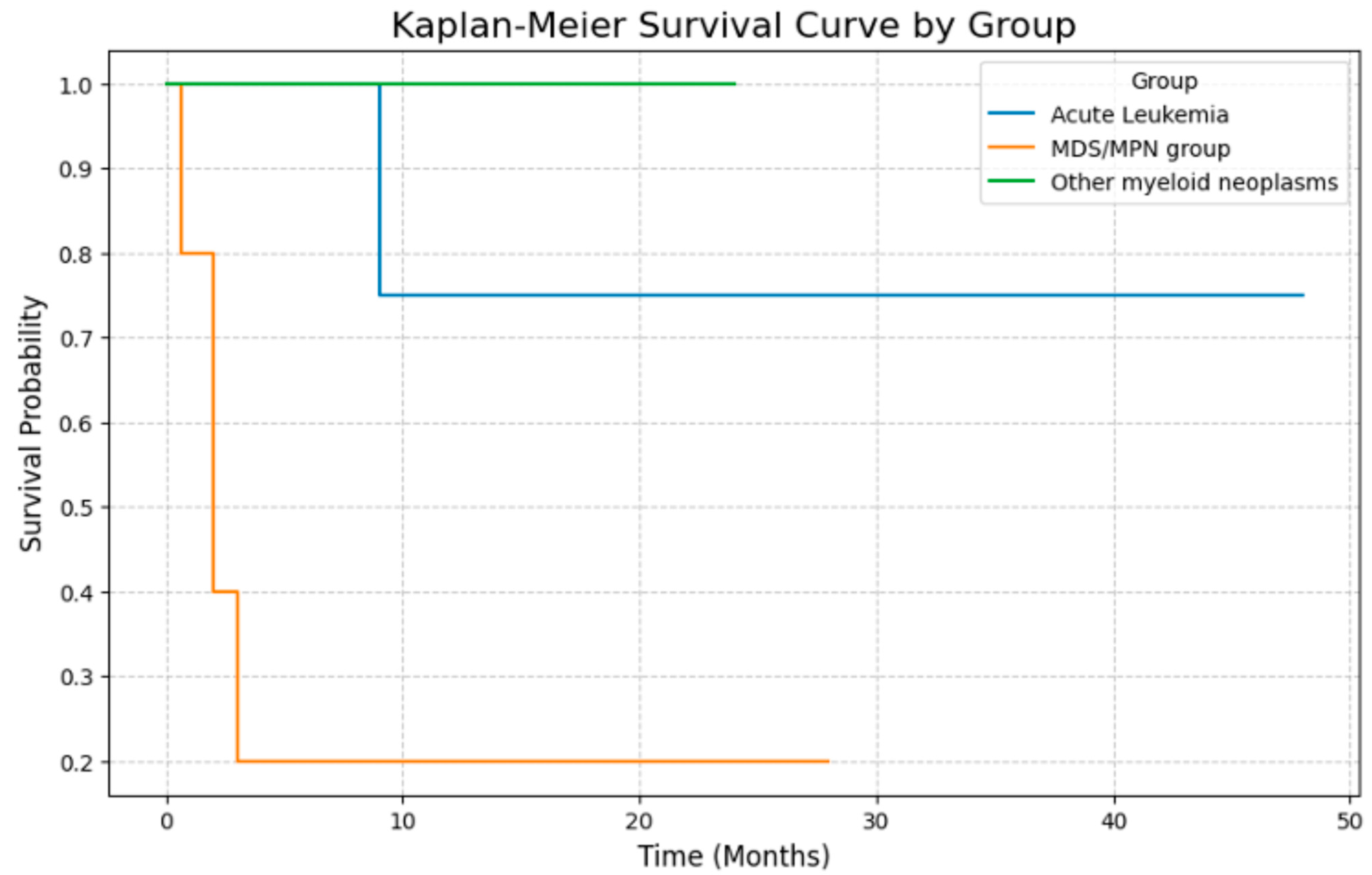
3.6. Clinical Outcomes

Survival data were available for all patients across the three diagnostic categories: MDS/MPN, acute leukemia, and other myeloid neoplasms. Kaplan–Meier analysis demonstrated distinct survival patterns among the groups. The median survival for the entire cohort was approximately 12 months. The MDS/MPN group exhibited the steepest decline in survival, with most patients succumbing to the disease early in its course. The acute leukemia group had a more gradual decline, reflecting variable clinical trajectories. In contrast, the other myeloid neoplasms group, comprising mainly MDS-IB1, had a more stable survival curve, consistent with slower disease progression. Survival curves for each group are shown in Figure 5.

4. Discussion

Our study highlights the significant presence of the CSF3R mutation across a diverse range of myeloid neoplasms, expanding their known spectrum beyond the traditional associations with CNL, aCML, and severe congenital neutropenia (SCN) [16]. In our cohort of thirteen patients, we identified CSF3R alterations across a variety of myeloid neoplasms, including MDS/MPN-U, AML, MPN with disease progression, MPAL, CMML, and MDS-IB1, suggesting a broader role for CSF3R in pathogenesis of myeloid neoplasms and clonal evolution. Given the rarity of CSF3R-mutated myeloid neoplasms outside of CNL and aCML, our cohort represents one of the largest institutional case series described to date. However, the small sample size inherently limits the statistical power of subgroup comparisons and survival analyses. Therefore, our findings should be interpreted as exploratory and hypothesis-generating, requiring validation in larger, multi-institutional cohorts.
Functionally, these mutations affect distinct receptor domains. The membrane-proximal missense mutation p.Thr618Ile impairs self O-glycosylation, resulting in ligand-independent receptor dimerization and continuous signaling in the JAK-STAT signaling pathway [17]. In contrast, truncation mutations (p.Gln743Ter, p.Lys785SerfsTer26) and frameshift mutations (p.S810Qfs) cluster within the cytoplasmic tail and disrupt negative regulatory motifs responsible for receptor internalization and degradation, thereby prolonging receptor activity and preferentially activating the SRC/TNK2 signaling pathway and enhanced cell proliferation [11,18,19]. The lollipop plot shown in Figure 6 illustrates their distribution across different functional domains.
While the CSF3R T618I mutation remains a hallmark of CNL (reported in up to 83% of cases) and is also present in aCML [17], our findings reinstate that this mutation is not restricted to these entities. This functional divergence emphasizes its distinct roles in myeloid pathogenesis and aligns with prior studies showing that CSF3R truncation mutations are frequently associated with severe disease phenotypes and resistance to therapy [5,20]. While their location in the cytoplasmic tail suggests the disruption of key regulatory domains, these effects remain inferential. Future studies utilizing predictive modeling or cellular assays will be necessary to clarify their true biological and clinical impact.
We also found frequent co-occurrences of CSF3R mutations with alterations involving other genes including, but not limited to, ASXL1, NRAS, KRAS, and SETBP1 in the MDS/MPN-U and CMML patients. These co-mutations likely contribute to the phenotypic heterogeneity and clinical variability observed in these CSF3R-driven neoplasms [21]. Among these, ASXL1 mutations were the most frequent co-mutations in our cohort, consistent with their known association with poor prognosis and disease progression in myeloid malignancies [13,22]. This suggests that ASXL1 co-mutations might exacerbate the effects of CSF3R mutations by promoting epigenetic dysregulation. The ASXL1 mutation, in combination with proliferative mutations such as ETNK1 and SETBP1, bring about the MDS/MPN phenotype in aCML. It is likely that the combination of ASXL1 and CSF3R creates a similar phenotype with the same mechanism. NRAS and KRAS mutations, observed in a few cases, highlight the role of aberrant RAS-MAPK signaling in driving clonal proliferation and leukemic transformation in these neoplasms [23].
In their study of CSF3R alteration in CNL and CMML, Ouyang and colleagues found that SRSF2 and SETBP1 were associated with a worse prognosis, whereas CSF3R alteration did not affect outcomes [24]. Other studies have suggested a role of SETBP1 alteration as secondary drivers of leukemic transformation and resistance to therapy [25]. In our dataset, both SETBP1 (Case 2, 3, and 5) and SRSF2 (Case 5) were noted predominantly in the MDS/MPN subcategory and were associated with poorer outcomes.
Amongst acute leukemia patients in our cohort, one patient had RUNX1::RUNX1T1 translocation, while another had a biallelic CEBPA mutation, both of which are known to co-occur with CSF3R alterations [26,27]. This pattern implies that in these settings, CSF3R mutations may contribute to disease progression. It is also important to note that none of the patients in our cohort received JAK inhibitor therapy, primarily due to advanced age, comorbidities, or limited clinical access at the time of diagnosis. As a result, while the therapeutic potential of CSF3R-targeted therapies such as ruxolitinib is discussed, actual treatment response data could not be assessed in this study.
Clinically, the identification of CSF3R mutations has profound therapeutic implications, emphasizing the need for routine molecular profiling in myeloid malignancies to identify CSF3R mutations and associated co-mutations. This is particularly relevant in patients without clearly defined category (MDS/MPN-U), where the presence of p.Thr618Ile or truncation mutations could provide a therapeutic target. The association of ASXL1, SRSF2, and SETBP1 mutations with poor prognosis, as evidenced by other studies, underscores the need for comprehensive molecular profiling [28]. As previously discussed, the constitutive activation of signaling pathways like JAK-STAT and SRC/TNK2 in CSF3R-altered neoplasms presents opportunities for targeted interventions with documented efficacy using JAK inhibitors such as ruxolitinib in these neoplasms [11,29]. Additionally, inhibitors targeting downstream pathways, such as SRC family kinases, could complement JAK inhibitors in cases where alternative signaling predominates. These therapeutic approaches could potentially be combined with other emerging therapeutic approaches such as MEK inhibitors to target RAS pathway or DNA methyltransferase inhibitors in ASXL1-mutated neoplasms.
From a practical standpoint, while CSF3R mutations are rare in myeloid neoplasms outside of CNL and aCML (~0.9% in our series), they remain clinically actionable. In resource-limited settings where broad NGS testing may not be available, focused hotspot testing for canonical mutations like p.Thr618Ile in patients presenting with neutrophilic leukocytosis or MDS/MPN-like features could offer a cost-effective approach. Developing such context-specific testing algorithms may optimize molecular diagnostics and facilitate access to targeted therapies, even in settings with limited resources.

5. Conclusions

Our study provides valuable insights into the genetic landscape of CSF3R-mutated myeloid neoplasm and adds to the growing body of evidence that CSF3R mutations, especially T618I, are a critical component of the molecular landscape beyond CNL, aCML, and SCN. We identified these mutations across a spectrum of myeloid disorders, including MDS/MPN, AML, and other myeloid disorders, reinforcing their relevance across diagnostic categories.
While our findings suggest that CSF3R mutations may assist in disease stratification and represent potential therapeutic targets, these conclusions are limited by the small sample size and lack of statistically significant outcome associations in this study. Therefore, we propose that CSF3R mutations be considered as potential markers of interest rather than definitive stratifiers at this stage. Further multicenter studies with larger cohorts and integrated clinical, genomic, and treatment data will be essential in validating their prognostic utility and defining their role in personalized therapeutic strategies.

Author Contributions

N.S.; writing—original draft preparation, review and editing, and data curation; J.B., P.H. and J.K.; writing—review and editing; P.Q.D.; writing—review and editing, visualization, conceptualization, X.Z.; writing—review and editing, visualization, conceptualization, and supervision. All authors have read and agreed to the published version of the manuscript.

Funding

This research did not receive any specific grant from funding agencies in the public, commercial, or not-for-profit sectors.

Institutional Review Board Statement

The study was conducted in accordance with the Declaration of Helsinki and approved by the Institutional Review Board of Northwell Health (approval number 13-273B and approval date on 5 June 2019).

Informed Consent Statement

The patients were not directly under our care, and the Institutional Review Board determined that written informed consent was not required for this retrospective analysis of anonymized data.

Data Availability Statement

All data and information concerning this study will be made available from the corresponding authors upon reasonable request.

Conflicts of Interest

The authors declare no conflicts of interest.

References

  1. Beekman, R.; Touw, I.P. G-CSF and its receptor in myeloid malignancy. Blood 2010, 115, 5131–5136. [Google Scholar] [CrossRef] [PubMed]
  2. Liu, F.; Wu, H.Y.; Wesselschmidt, R.; Kornaga, T.; Link, D.C. Impaired production and increased apoptosis of neutrophils in granulocyte colony-stimulating factor receptor-deficient mice. Immunity 1996, 5, 491–501. [Google Scholar] [CrossRef] [PubMed]
  3. Zhu, Q.S.; Xia, L.; Mills, G.B.; Lowell, C.A.; Touw, I.P.; Corey, S.J. G-CSF induced reactive oxygen species involves Lyn-PI3-kinase-Akt and contributes to myeloid cell growth. Blood 2006, 107, 1847–1856. [Google Scholar] [CrossRef] [PubMed]
  4. Price, A.; Druhan, L.J.; Lance, A.; Clark, G.; Vestal, C.G.; Zhang, Q.; Foureau, D.; Parsons, J.; Hamilton, A.; Steuerwald, N.M.; et al. T618I CSF3R mutations in chronic neutrophilic leukemia induce oncogenic signals through aberrant trafficking and constitutive phosphorylation of the O-glycosylated receptor form. Biochem. Biophys. Res. Commun. 2020, 523, 208–213. [Google Scholar] [CrossRef] [PubMed]
  5. Zhang, H.; Coblentz, C.; Watanabe-Smith, K.; Means, S.; Means, J.; Maxson, J.E.; Tyner, J.W. Gain-of-function mutations in granulocyte colony-stimulating factor receptor (CSF3R) reveal distinct mechanisms of CSF3R activation. J. Biol. Chem. 2018, 293, 7387–7396. [Google Scholar] [CrossRef] [PubMed]
  6. Elliott, M.A. Chronic neutrophilic leukemia: A contemporary review. Curr. Hematol. Rep. 2004, 3, 210–217. [Google Scholar] [PubMed]
  7. Reilly, J.T. Chronic neutrophilic leukaemia: A distinct clinical entity? Br. J. Haematol. 2002, 116, 10–18. [Google Scholar] [CrossRef] [PubMed]
  8. Zhang, H.; Wilmot, B.; Bottomly, D.; Dao, K.-H.T.; Stevens, E.; Eide, C.A.; Khanna, V.; Rofelty, A.; Savage, S.; Schultz, A.R.; et al. Genomic landscape of neutrophilic leukemias of ambiguous diagnosis. Blood 2019, 134, 867–879. [Google Scholar] [CrossRef] [PubMed]
  9. Langabeer, S.E.; Haslam, K.; Kelly, J.; Quinn, J.; Morrell, R.; Conneally, E. Targeted next-generation sequencing identifies clinically relevant mutations in patients with chronic neutrophilic leukemia at diagnosis and blast crisis. Clin. Transl. Oncol. 2018, 20, 420–423. [Google Scholar] [CrossRef] [PubMed]
  10. Faisal, M.; Stark, H.; Büsche, G.; Schlue, J.; Teiken, K.; Kreipe, H.H.; Lehmann, U.; Bartels, S. Comprehensive mutation profiling and mRNA expression analysis in atypical chronic myeloid leukemia in comparison with chronic myelomonocytic leukemia. Cancer Med. 2019, 8, 742–750. [Google Scholar] [CrossRef] [PubMed]
  11. Maxson, J.E.; Gotlib, J.; Pollyea, D.A.; Fleischman, A.G.; Agarwal, A.; Eide, C.A.; Bottomly, D.; Wilmot, B.; McWeeney, S.K.; Tognon, C.E.; et al. Oncogenic CSF3R mutations in chronic neutrophilic leukemia and atypical CML. N. Engl. J. Med. 2013, 368, 1781–1790. [Google Scholar] [CrossRef] [PubMed]
  12. Meggendorfer, M.; Haferlach, T.; Alpermann, T.; Jeromin, S.; Haferlach, C.; Kern, W.; Schnittger, S. Specific molecular mutation patterns delineate chronic neutrophilic leukemia, atypical chronic myeloid leukemia, and chronic myelomonocytic leukemia. Haematologica 2014, 99, e244–e246. [Google Scholar] [CrossRef] [PubMed]
  13. Szuber, N.; Finke, C.M.; Lasho, T.L.; Elliott, M.A.; Hanson, C.A.; Pardanani, A.; Tefferi, A. CSF3R-mutated chronic neutrophilic leukemia: Long-term outcome in 19 consecutive patients and risk model for survival. Blood Cancer J. 2018, 8, 21. [Google Scholar] [CrossRef] [PubMed]
  14. Dao, K.H.; Solti, M.B.; Maxson, J.E.; Winton, E.F.; Press, R.D.; Druker, B.J.; Tyner, J.W. Significant clinical response to JAK1/2 inhibition in a patient with CSF3R-T618I-positive atypical chronic myeloid leukemia. Leuk. Res. Rep. 2014, 3, 67–69. [Google Scholar] [CrossRef] [PubMed]
  15. Dao, K.T.; Gotlib, J.; Deininger, M.M.; Oh, S.T.; Cortes, J.E.; Collins, R.H.; Winton, E.F.; Parker, D.R.; Lee, H.; Reister, A.; et al. Efficacy of Ruxolitinib in Patients With Chronic Neutrophilic Leukemia and Atypical Chronic Myeloid Leukemia. J. Clin. Oncol. 2020, 38, 1006–1018. [Google Scholar] [CrossRef] [PubMed]
  16. Mohamed, A.; Gao, J.; Chen, Y.-H.; Abaza, Y.; Altman, J.; Jennings, L.; Vormittag-Nocito, E.; Sukhanova, M.; Lu, X.; Chen, Q. CSF3R mutated myeloid neoplasms: Beyond chronic neutrophilic leukemia. Hum. Pathol. 2024, 149, 66–74. [Google Scholar] [CrossRef] [PubMed]
  17. Maxson, J.E.; Luty, S.B.; MacManiman, J.D.; Abel, M.L.; Druker, B.J.; Tyner, J.W. Ligand independence of the T618I mutation in the colony-stimulating factor 3 receptor (CSF3R) protein results from loss of O-linked glycosylation and increased receptor dimerization. J. Biol. Chem. 2014, 289, 5820–5827. [Google Scholar] [CrossRef] [PubMed]
  18. Zhang, H.; Schultz, A.R.; Luty, S.; Rofelty, A.; Su, Y.; Means, S.; Bottomly, D.; Wilmot, B.; McWeeney, S.K.; Tyner, J.W. Characterization of the leukemogenic potential of distal cytoplasmic CSF3R truncation and missense mutations. Leukemia 2017, 31, 2752–2760. [Google Scholar] [CrossRef] [PubMed]
  19. Mehta, H.M.; Futami, M.; Glaubach, T.; Lee, D.W.; Andolina, J.R.; Yang, Q.; Whichard, Z.; Quinn, M.; Lu, H.F.; Kao, W.M.; et al. Alternatively spliced, truncated GCSF receptor promotes leukemogenic properties and sensitivity to JAK inhibition. Leukemia 2014, 28, 1041–1051. [Google Scholar] [CrossRef] [PubMed]
  20. Rohrabaugh, S.; Kesarwani, M.; Kincaid, Z.; Huber, E.; Leddonne, J.; Siddiqui, Z.; Khalifa, Y.; Komurov, K.; Grimes, H.L.; Azam, M. Enhanced MAPK signaling is essential for CSF3R-induced leukemia. Leukemia 2017, 31, 1770–1778. [Google Scholar] [CrossRef] [PubMed]
  21. Adam, F.C.; Szybinski, J.; Halter, J.P.; Cantoni, N.; Wenzel, F.; Leonards, K.; Brkic, S.; Passweg, J.R.; Touw, I.; Maxson, J.E.; et al. Co-Occurring CSF3R W791* Germline and Somatic T618I Driver Mutations Induce Early CNL and Clonal Progression to Mixed Phenotype Acute Leukemia. Curr. Oncol. 2022, 29, 805–815. [Google Scholar] [CrossRef] [PubMed]
  22. Elliott, M.A.; Pardanani, A.; Hanson, C.A.; Lasho, T.L.; Finke, C.M.; Belachew, A.A.; Tefferi, A. ASXL1 mutations are frequent and prognostically detrimental in CSF3R-mutated chronic neutrophilic leukemia. Am. J. Hematol. 2015, 90, 653–656. [Google Scholar] [CrossRef] [PubMed]
  23. Alawieh, D.; Cysique-Foinlan, L.; Willekens, C.; Renneville, A. RAS mutations in myeloid malignancies: Revisiting old questions with novel insights and therapeutic perspectives. Blood Cancer J. 2024, 14, 72. [Google Scholar] [CrossRef] [PubMed]
  24. Ouyang, Y.; Qiao, C.; Chen, Y.; Zhang, S.-J. Clinical significance of CSF3R, SRSF2 and SETBP1 mutations in chronic neutrophilic leukemia and chronic myelomonocytic leukemia. Oncotarget 2017, 8, 20834–20841. [Google Scholar] [CrossRef] [PubMed]
  25. Makishima, H.; Yoshida, K.; Nguyen, N.; Przychodzen, B.; Sanada, M.; Okuno, Y.; Ng, K.P.; Gudmundsson, K.O.; Vishwakarma, B.A.; Jerez, A.; et al. Somatic SETBP1 mutations in myeloid malignancies. Nat. Genet. 2013, 45, 942–946. [Google Scholar] [CrossRef] [PubMed]
  26. Swoboda, A.S.; Arfelli, V.C.; Danese, A.; Windisch, R.; Kerbs, P.; Monte, E.R.; Bagnoli, J.W.; Chen-Wichmann, L.; Caroleo, A.; Cusan, M.; et al. CSF3R T618I Collaborates With RUNX1-RUNX1T1 to Expand Hematopoietic Progenitors and Sensitizes to GLI Inhibition. Hemasphere 2023, 7, e958. [Google Scholar] [CrossRef] [PubMed]
  27. Wang, B.; Wen, L.; Wang, Z.; Chen, S.; Qiu, H. Differential Implications of CSF3R Mutations in t(8;21) and CEBPA Double Mutated Acute Myeloid Leukemia. Clin. Lymphoma Myeloma Leuk. 2022, 22, 393–404. [Google Scholar] [CrossRef] [PubMed]
  28. Qian, Y.; Chen, Y.; Li, X. CSF3R T618I, SETBP1 G870S, SRSF2 P95H, and ASXL1 Q780* tetramutation co-contribute to myeloblast transformation in a chronic neutrophilic leukemia. Ann. Hematol. 2021, 100, 1459–1461. [Google Scholar] [CrossRef] [PubMed]
  29. Gunawan, A.S.; McLornan, D.P.; Wilkins, B.; Waghorn, K.; Hoade, Y.; Cross, N.C.P.; Harrison, C.N. Ruxolitinib, a potent JAK1/JAK2 inhibitor, induces temporary reductions in the allelic burden of concurrent CSF3R mutations in chronic neutrophilic leukemia. Haematologica 2017, 102, e238–e240. [Google Scholar] [CrossRef] [PubMed]
Figure 1. Histomorphological and immunohistochemical features of myelodysplastic/myeloproliferative neoplasms. (AG) Photomicrographs showing features of the bone marrow of an MDS/MPN-U patient with CSF3R alteration. (A) Photomicrograph showing bone marrow with marked hypercellularity, myeloid predominance, and decreased erythroid and megakaryocytic populations. (B) Myeloperoxidase and (C) CD71 confirm myeloid predominance. (D) Factor VIII IHC showing megakaryocytes with atypia. (E) CD34 IHC demonstrating <5% CD34-positive blasts. (F) CD14 IHC highlighting an increased monocytic population. (G) Reticulin stain showing grade 2-3 myelofibrosis. (HM) Photomicrographs showing features of the bone marrow of a CMML patient with CSF3R alteration. (H) Photomicrograph of a bone marrow biopsy showing markedly hypercellular marrow with significant granulocytic hyperplasia and a prominent left shift in myeloid maturation (H&E, 40×, inset 100×). (I) CD34 IHC demonstrating less than 5% blasts and megakaryocytes with aberrant CD34 expression. (J) Myeloperoxidase, (K) CD71, and (L) E-cadherin confirm myeloid predominance and left shift in the erythroid compartment. (M) Factor VIII highlights dysplastic megakaryocytes. (NU) Photomicrographs showing features of myeloid sarcoma with CSF3R alteration. Photomicrograph showing an atypical proliferation of pleomorphic hematopoietic cells with necrosis and apoptosis with (N) H&E, 40× and (O) H&E, 400×. Neoplastic cells are positive for (P) CD45, (Q) CD4, (R) CD33, (S) MPO, (T) CD163, and (U) Muramidase (IHC, 40×). (VY) Photomicrographs showing features of bone marrow in patient with essential thrombocythemia gaining a secondary CSF3R alteration. (V) Photomicrograph showing a markedly hypercellular bone marrow (H&E, 20×) with left-shifted myeloid maturation and inset showing increased atypical megakaryocytic forms (H&E, 400×). (W) CD34 immunohistochemical stain highlighting the sinusoids and several megakaryocytic forms indicative of dyspoiesis (IHC, 100×). (X) E-cadherin immunohistochemical stain showing the prominence of pronormoblasts (IHC, 100×). (Y) Factor VIII marking the increased megakaryocytes with atypia and occasional clustering (IHC, 100×, 400×).
Jcm 14 05174 g001
Figure 2. (A) This bar chart illustrates the VAF of CSF3R mutations across individual cases, categorized by diagnostic groups. Blue bars represent cases classified as being in the MDS/MPN group, which demonstrate intermediate-to-high CSF3R VAF values (median, 39%; range, 26–49%). Orange bars represent cases diagnosed as acute leukemia (median, 10%; range, 10–15%), which tend to exhibit lower CSF3R VAF values compared to the MDS/MPN group. Green bars represent cases diagnosed for other myeloid neoplasms, which show varying levels of CSF3R VAF (median, 12%; range, 2–24%). (B) Box plot showing the CSF3R variant allele frequency (VAF%) across three diagnostic groups: MDS/MPN group, acute leukemia, and other myeloid neoplasms.
Jcm 14 05174 g002
Figure 3. The heatmap highlights the mutation patterns and recurrently altered genes seen across 13 cases of myeloid neoplasms. Colored boxes represent mutation types: blue for missense, green for frameshift, red for nonsense, and brown for splice-site mutations, and orange for in-frame insertions. White boxes indicate no mutation.
Jcm 14 05174 g003
Figure 4. Genetic mutation frequencies in CSF3R-driven myeloid neoplasms. This bar chart illustrates the percentage frequencies of key representative genetic mutations identified in various cases with CSF3R-driven myeloid neoplasms showcasing the heterogeneity of these disorders. Case 6, which harbors CSF3R, STAT3, CEBPA, and JAK3 mutations, is excluded from this graph, as percentage data for these mutations are not available.
Jcm 14 05174 g004
Figure 5. The Kaplan–Meier survival curve illustrates the survival probabilities of three diagnostic groups: MDS/MPN group (orange), acute leukemia (blue), and other myeloid neoplasms (green), over time in months. The MDS/MPN group shows a steep decline in survival, reflecting the poor prognosis in disorders like MDS/MPN-U and CMML. The acute leukemia group exhibits a more gradual decrease, indicating variable outcomes in aggressive diseases like AML, MPAL, and myeloid sarcoma. The other myeloid neoplasms demonstrate relatively stable survival, consistent with their indolent nature. Vertical ticks on the curves mark censoring, representing patients still alive or lost to follow-up.
Jcm 14 05174 g005
Figure 6. Lollipop plot conveying CSF3R mutation profile with functional domains and hotspot mutations.
Jcm 14 05174 g006
Table 1. Comprehensive clinicopathologic, cytogenetic, and outcome profile of the 13-patient myeloid neoplasm cohort.
Case 1Case 2Case 3Case 4Case 5Case 6Case 7Case 8Case 9Case 10Case 11Case 12Case 13
Age89818784782269555657776379
GenderFMFMMMFMMFMMM
WBC58.514.64416.251.925.54.26.11.410.42.625.32.5
Hb10.67.37.49.211.711.712117.69.410.67.711.2
MCV99.7100.4103.985.5105.594.68186.292.690.984.398.796.3
Plt21627141321584718495441172807765
Mono(%)36NA1012.109145.2NA2.99
Mono#1.20.92.31.86.300.400.10.54NA0.70.22
PB blasts (%)0000085063140NA00
LDH38522053141888840633731466641612691176ND
Cellularity9085–909570–8510070–85Limited 95–10070959095–10030
M/E10:1MPMP1:12:10.8:13:1MP5.2:16.5:11.6:110.8:1MP
ErythroidDys+Dys+DecDys+DecDys+DecDecDecDys+Dys+IncMild dec
MyeloidNANAIncDys+IncMinimalDecDecDecDys+Dys+RareDys+
MegakaryocytesNADys+DecDys+ Inc, Dys+No RareDecPresentDys+Dys+Dys+Dec
BM blasts (%)<3<3<37166483568885
Fibrosis0Gr 2-3Gr 2-300000000Gr 1-20
Karyotype46, XX 45,XY,-746, XX46,XY del(7)(q22)46, XX46, XY46, XX46,XYComplex46,XX46,XY46,XY,der(13;14)9q10;q10)45, -X,-Y
FISHNormalMonosomy 7NADel(7q)NormalRUNX1 (3 copies)NormalNormalRUNX1T1/RUNX1NormalNormalNANormal
DiagnosisMDS/MPN-UMDS/MPN-UMDS/MPN-UCMMLCMMLAMLMSMPALAMLMDS-EB1MDS-EB1ET/MPN transformed to AMLMDS-EB1
TreatmentHy 500 mg ODAzax1 cycleNASupportive care onlyHy 500 mg OD7+3+GO, Consolidation HiDAC + GO × 47+3; HiDAC + Aza × 2; Ven + Dec × 27+37+3No RxVen + Dec × 4; GO+ LD Ara-CVen + Dec; ASCTNo Rx
PrognosisDied, 33 monthsDied, 2 months2 monthsDied, 19 daysAlive, 28 monthsCR, 4 yearsRelapse, 14 monthsDied, 9 monthsRelapse, 19 monthsAlive, 1 yearTransform into AML, alive, 2 yearsCR, 1 yearAlive, 5 months
Clinical NoneHepatitis, cirrhosisBreast Ca, colon CaProstate CaB-ALL, remissionNone RADVTNoneOA kneeMGNoneProstate Ca
SplenomegalyNoYesYesNoNoNo NoNoNoNoNoNoNo
Abbreviations: MDS/MPN-U, Myelodysplastic syndrome/Myeloproliferative neoplasm—Unclassifiable; HA, hypomethylating agent; Hy, hydroxyurea; Dys, dysplasia; Inc, increased; Dec, decreased; MS, myeloid sarcoma; RA, rheumatoid arthritis; MG, Myasthenia gravis; OA, osteoarthritis; DVT, deep vein thrombosis; MP, myeloid predominant; Rx, treatment; OD, once daily; Aza, Azacytidine; Dec, Decitabine; HiDAC, high-dose cytarabine; 7+3, cytarabine + Daunorubicin; Ven, Venetoclax; GO, Gemtuzumab Ozogamicin; LD, low-dose; Ara-C, Cytarabine; ASCT, allogenic stem cell transplant; NA, not available.
Disclaimer/Publisher’s Note: The statements, opinions and data contained in all publications are solely those of the individual author(s) and contributor(s) and not of MDPI and/or the editor(s). MDPI and/or the editor(s) disclaim responsibility for any injury to people or property resulting from any ideas, methods, instructions or products referred to in the content.

