
Ophthalmic Artery Doppler Indices at 11–13 Weeks of Gestation in Relation to Early and Late Preeclampsia

 ,
,
Abstract
1. Introduction
2. Materials and Methods
2.1. Study Design and Participants
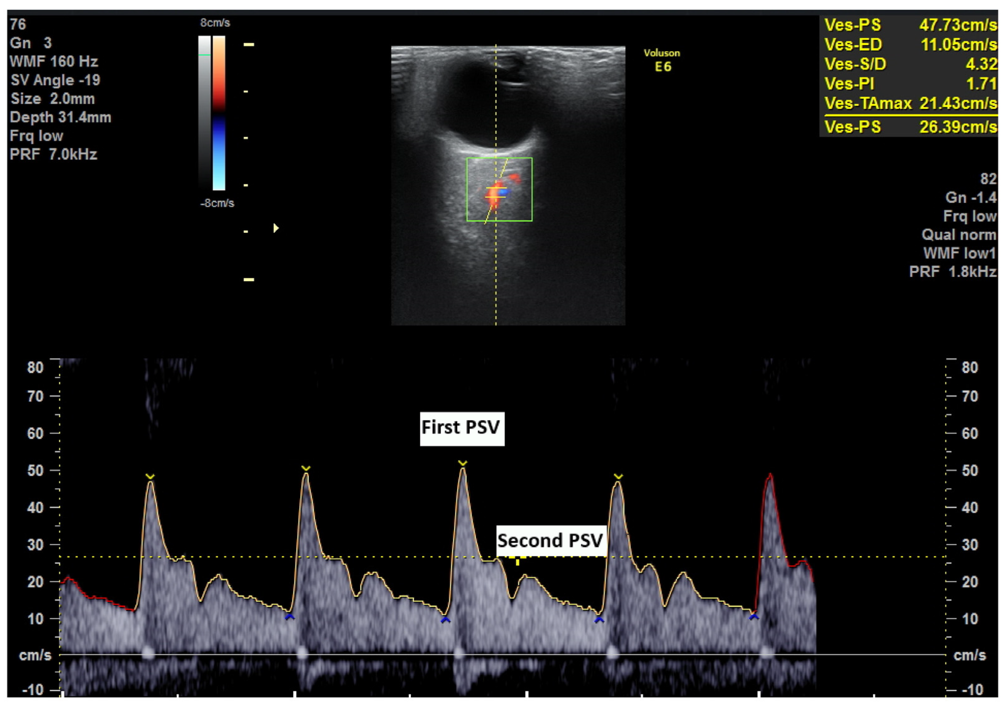
2.2. Ophtalmic Artery Doppler Indices
2.3. Pregnancy Outcome
2.4. Data Analysis
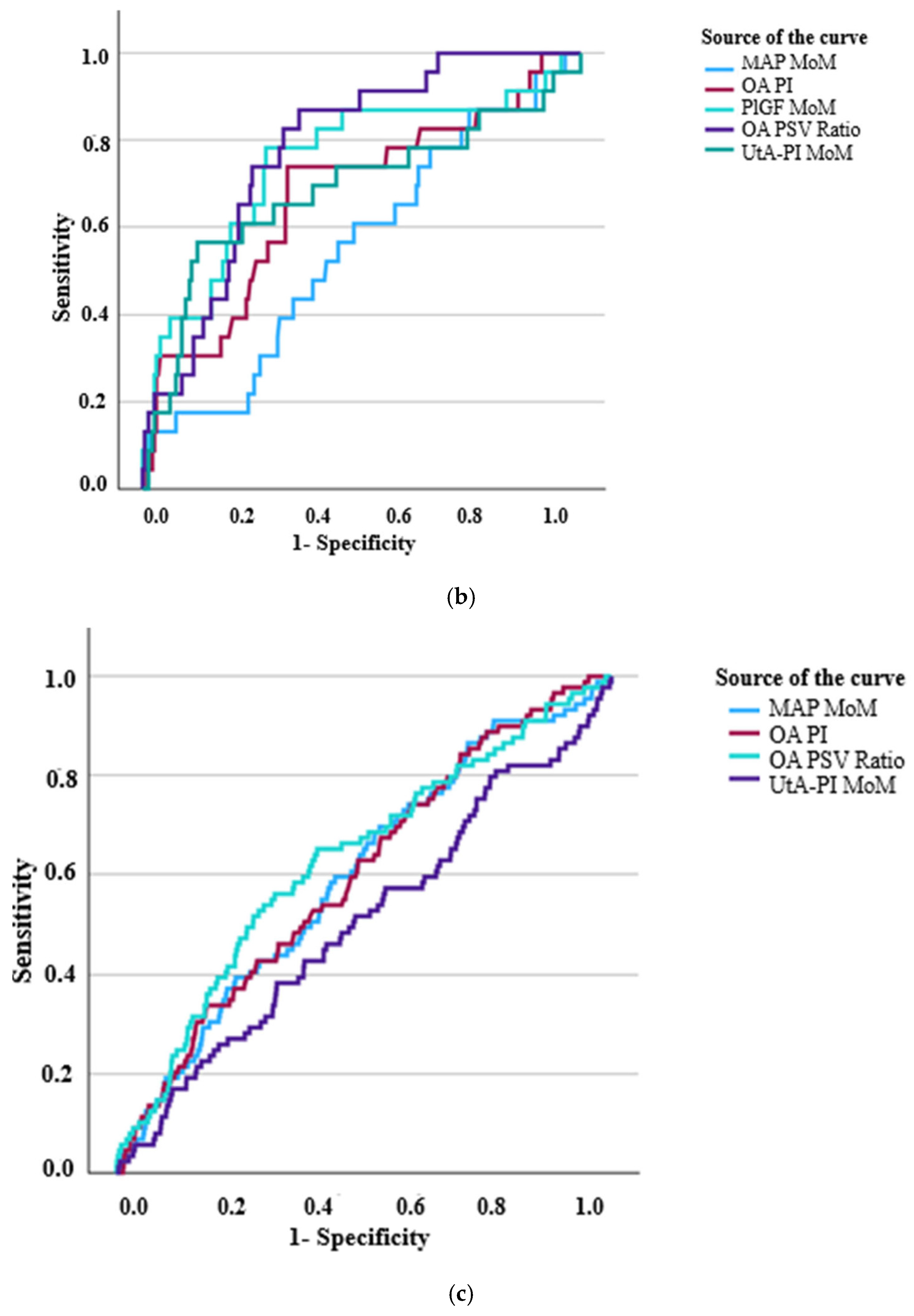
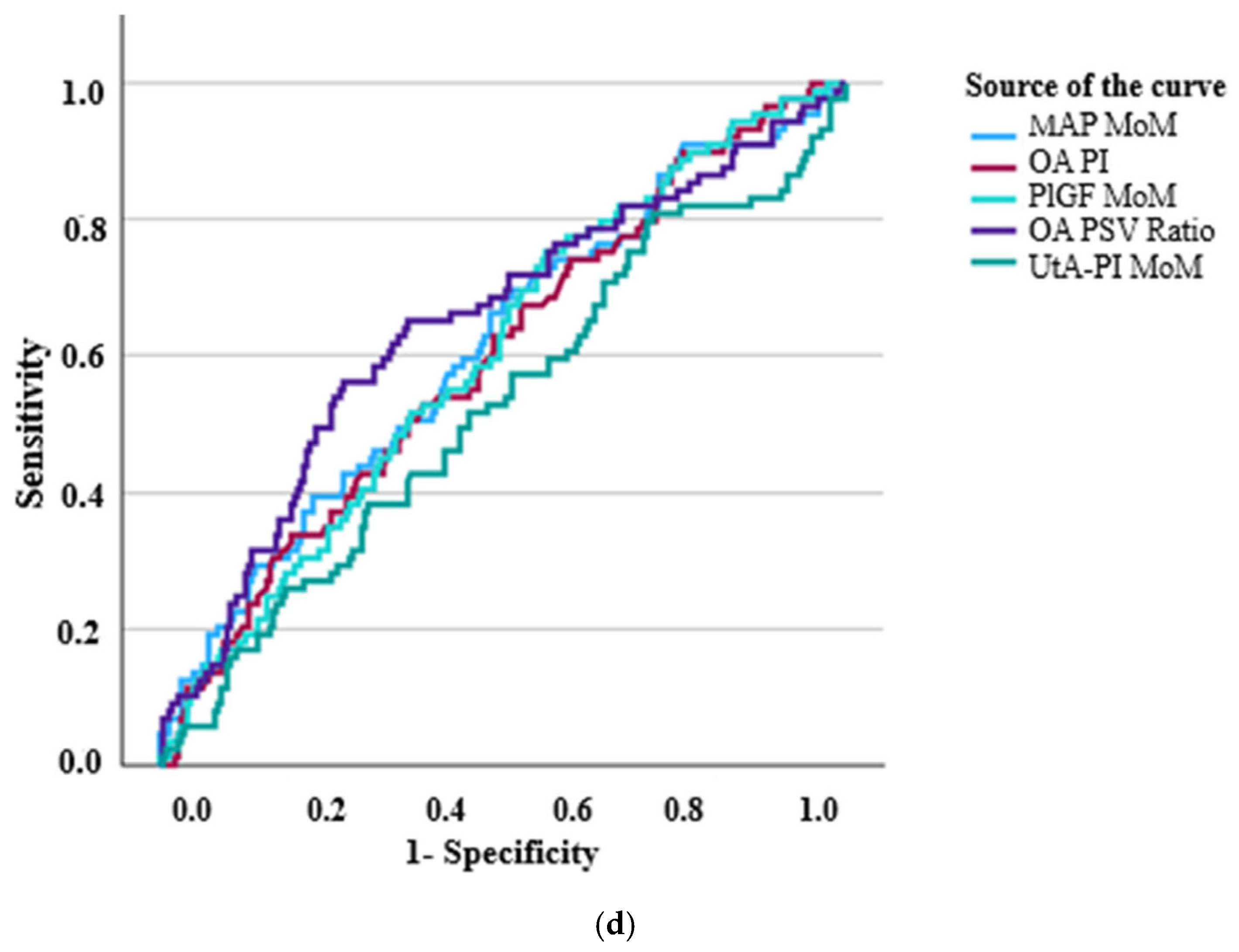
3. Results
4. Discussion
4.1. Hemodynamic Adaptations and Vascular Dysfunction
4.2. Relevance of Ophthalmic Artery Doppler
4.3. Comparisons with Existing Predictive Models
4.4. Clinical Implications and Future Directions
4.5. Limitations and Next Steps
5. Conclusions
Author Contributions
Funding
Institutional Review Board Statement
Informed Consent Statement
Data Availability Statement
Acknowledgments
Conflicts of Interest
Abbreviations
| AUC | Area under the curve |
| BMI | Body mass index |
| CI | Confidence interval |
| IVF | In vitro fertilization |
| MAP | Mean arterial pressure |
| MOM | Multiples of median |
| OA | Ophtalmic artery |
| OR | Odds ratio |
| PE | Preeclampsia |
| PI | Pulsatility index |
| PlGF | Placental growth factor |
| PSV | Peak systolic velocity |
| ROC | Receiver operating characteristic curve |
| SD | Standard deviation |
| Ut-A-PI | Uterine artery pulsatility index |
References
- Roberts, J.M. Preeclampsia epidemiology(ies) and pathophysiology(ies). Best Pract. Res. Clin. Obstet. Gynaecol. 2024, 94, 102480. [Google Scholar] [CrossRef] [PubMed]
- Sánchez-Aranguren, L.C.; Prada, C.E.; Riaño-Medina, C.E.; Lopez, M. Endothelial dysfunction and preeclampsia: Role of oxidative stress. Front. Physiol. 2014, 5, 372. [Google Scholar] [CrossRef] [PubMed] [PubMed Central]
- Nankali, A.; Malek-Khosravi, S.; Zangeneh, M.; Rezaei, M.; Hemati, Z.; Kohzadi, M. Maternal complications associated with severe preeclampsia. J. Obstet. Gynaecol. India 2013, 63, 112–115. [Google Scholar] [CrossRef] [PubMed] [PubMed Central]
- Mikat, B.; Gellhaus, A.; Wagner, N.; Birdir, C.; Kimmig, R.; Köninger, A. Early detection of maternal risk for preeclampsia. ISRN Obstet. Gynecol. 2012, 2012, 172808. [Google Scholar] [CrossRef] [PubMed] [PubMed Central]
- Ramos, J.G.L.; Sass, N.; Costa, S.H.M. Preeclampsia. Rev. Bras. Ginecol. Obstet. 2017, 39, 496–512. [Google Scholar] [CrossRef] [PubMed] [PubMed Central]
- O’Gorman, N.; Wright, D.; Rolnik, D.L.; Nicolaides, K.H.; Poon, L.C. Study protocol for the randomised controlled trial: Combined multimarker screening and randomised patient treatment with ASpirin for evidence-based PREeclampsia prevention (ASPRE). BMJ Open 2016, 6, e011801. [Google Scholar] [CrossRef] [PubMed] [PubMed Central]
- Konijnenberg, A.; Stokkers, E.W.; van der Post, J.A.; Schaap, M.C.; Boer, K.; Bleker, O.P.; Sturk, A. Extensive platelet activation in preeclampsia compared with normal pregnancy: Enhanced expression of cell adhesion molecules. Am. J. Obstet. Gynecol. 1997, 176, 461–469. [Google Scholar] [CrossRef] [PubMed]
- O’Gorman, N.; Wright, D.; Syngelaki, A.; Akolekar, R.; Wright, A.; Poon, L.C.; Nicolaides, K.H. Competing risks model in screening for preeclampsia by maternal factors and biomarkers at 11–13 weeks’ gestation. Am. J. Obstet. Gynecol. 2016, 214, 103.e1–103.e12. [Google Scholar] [CrossRef]
- Reddy, M.; Springhall, E.A.; Rolnik, D.L.; da Silva Costa, F. How to perform first trimester combined screening for pre-eclampsia. Australas. J. Ultrasound Med. 2018, 21, 191–197. [Google Scholar] [CrossRef] [PubMed] [PubMed Central]
- Kane, S.C.; Brennecke, S.P.; da Silva Costa, F. Ophthalmic artery Doppler analysis: A window into the cerebrovasculature of women with pre-eclampsia. Ultrasound Obstet. Gynecol. 2017, 49, 15–21. [Google Scholar] [CrossRef]
- de Oliveira, C.A.; de Sá, R.A.M.; Velarde, L.G.C.; Monteiro, V.N.P.; Netto, H.C. Doppler Velocimetry of the Ophthalmic Artery. J. Ultrasound Med. 2012, 31, 879–884. [Google Scholar] [CrossRef] [PubMed]
- Diniz, A.L.; Moron, A.F.; dos Santos, M.C.; Sass, N.; Pires, C.R.; Debs, C.L. Ophthalmic artery Doppler as a measure of severe pre-eclampsia. Int. J. Gynaecol. Obstet. 2008, 100, 216–220. [Google Scholar] [CrossRef] [PubMed]
- de Oliveira, C.A.; de Sá, R.A.; Velarde, L.G.; da Silva, F.C.; doVale, F.A.; Netto, H.C. Changes in ophthalmic artery Doppler indices in hypertensive disorders during pregnancy. J. Ultrasound Med. 2013, 32, 609–616. [Google Scholar] [CrossRef] [PubMed]
- Takata, M.; Nakatsuka, M.; Kudo, T. Differential blood flow in uterine, ophthalmic, and brachial arteries of preeclamptic women. Obstet. Gynecol. 2002, 100, 931–939. [Google Scholar]
- Tudor, A.; Novac, L.; Camen, I.V.; Manolea, M.M.; Sandulescu, M.S.; Vrabie, S.C.; Serbanescu, M.S.; Boldeanu, M.V.; Istrate-Ofiteru, A.M.; Dijmarescu, A.L. The Role of Uterine Artery Doppler in the Second and Third Trimesters for Prediction of Preeclampsia and Fetal Growth Restriction Developed as a Consequence of Placental-Mediated Diseases. Curr. Health Sci. J. 2023, 49, 251–256. [Google Scholar] [CrossRef] [PubMed] [PubMed Central]
- de Melo, P.F.M.V.; Roever, L.; Mendonça, T.M.S.; da Silva Costa, F.; Rolnik, D.L.; Diniz, A.L.D. Ophthalmic artery Doppler in the complementary diagnosis of preeclampsia: A systematic review and meta-analysis. BMC Pregnancy Childbirth 2023, 23, 343. [Google Scholar] [CrossRef] [PubMed] [PubMed Central]
- Jackson, R.A.; Gibson, K.A.; Wu, Y.W.; Croughan, M.S. Perinatal Outcomes in Singletons Following In Vitro Fertilization: A Meta-Analysis. Obstet. Gynecol. 2004, 103, 551–563. [Google Scholar] [CrossRef]
- Wander, G.S.; McDonagh, S.T.J.; Rao, M.S.; Alagesan, R.; Mohan, J.C.; Bhagwat, A.; Pancholia, A.K.; Viswanathan, M.; Chopda, M.B.; Purnanand, A.; et al. Clinical relevance of double-arm blood pressure measurement and prevalence of clinically important inter-arm blood pressure differences in Indian primary care. J. Clin. Hypertens. 2022, 24, 993–1002. [Google Scholar] [CrossRef] [PubMed] [PubMed Central]
- Pickering, T.G.; Hall, J.E.; Appel, L.J.; Falkner, B.E.; Graves, J.; Hill, M.N.; Jones, D.W.; Kurtz, T.; Sheps, S.G.; Roccella, E.J. Recommendations for blood pressure measurement in humans and experimental animals: Part 1: Blood pressure measurement in humans: A statement for professionals from the Subcommittee of Professional and Public Education of the American Heart Association Council on High Blood Pressure Research. Circulation 2005, 111, 697–716. [Google Scholar] [CrossRef] [PubMed]
- Bhide, A.; Acharya, G.; Bilardo, C.M.; Brezinka, C.; Cafici, D.; Hernandez-Andrade, E.; Kalache, K.; Kingdom, J.; Kiserud, T.; Lee, W.; et al. ISUOG Practice Guidelines: Use of Doppler ultrasonography in obstetrics. Ultrasound Obstet. Gynecol. 2013, 41, 233–239. [Google Scholar] [CrossRef]
- Gana, N.; Sarno, M.; Vieira, N.; Wright, A.; Charakida, M.; Nicolaides, K.H. Ophthalmic artery Doppler at 11–13 weeks’ gestation in prediction of pre-eclampsia. Ultrasound Obstet. Gynecol. 2022, 59, 731–736. [Google Scholar] [CrossRef] [PubMed]
- Erickson, S.J.; Hendrix, L.E.; Massaro, B.M.; Harris, G.J.; Lewandowski, M.F.; Foley, W.D.; Lawson, T.L. Color Doppler flow imaging of the normal and abnormal orbit. Radiology 1989, 173, 511–516. [Google Scholar] [CrossRef] [PubMed]
- Flint, K.; Bottenus, N.; Bradway, D.; McNally, P.; Ellestad, S.; Trahey, G. An Automated ALARA Method for Ultrasound: An Obstetric Ultrasound Feasibility Study. J. Ultrasound Med. 2021, 40, 1863–1877. [Google Scholar] [CrossRef] [PubMed] [PubMed Central]
- Varthaliti, A.; Fasoulakis, Z.; Lygizos, V.; Zolota, V.; Chatziioannou, M.I.; Daskalaki, M.A.; Daskalakis, G.; Antsaklis, P. Safety of Obstetric Ultrasound: Mechanical and Thermal Indexes-A Systematic Review. J. Clin. Med. 2024, 13, 6588. [Google Scholar] [CrossRef] [PubMed] [PubMed Central]
- Microsoft Word—BMUS Safety Guidelines_2009 Revision_Feb 2010.doc. Available online: https://www.bmus.org/static/uploads/resources/BMUS-Safety-Guidelines-2009-revision-FINAL-Nov-2009.pdf (accessed on 1 June 2025).
- American College of Obstetricians and Gynecologists. Task Force on Hypertension in Pregnancy. Gestational hypertension and preeclampsia. ACOG Practice Bulletin No. 202. American College of Obstetricians and Gynecologists. Obstet. Gynecol. 2019, 133, e1–e25. [Google Scholar]
- Kusuma, R.A.; Nurdiati, D.S.; Al Fattah, A.N.; Danukusumo, D.; Abdullah, S.; Sini, I. Ophthalmic artery Doppler for pre-eclampsia prediction at the first trimester: A Bayesian survival-time model. J. Ultrasound 2023, 26, 155–162. [Google Scholar] [CrossRef]
- Saleh, M.; Naemi, M.; Aghajanian, S.; Saleh, M.; Hessami, K.; Bakhtiyari, M. Diagnostic value of ophthalmic artery Doppler indices for prediction of preeclampsia at 28–32 weeks of gestation. Int. J. Gynaecol. Obstet. 2023, 160, 120–130. [Google Scholar] [CrossRef] [PubMed]
- Gyokova, E.; Hristova-Atanasova, E.; Iskrov, G. Preeclampsia Management and Maternal Ophthalmic Artery Doppler Measurements between 19 and 23 Weeks of Gestation. J. Clin. Med. 2024, 13, 950. [Google Scholar] [CrossRef] [PubMed] [PubMed Central]
- Gonser, M.; Vonzun, L.; Ochsenbein-Kölble, N. Ophthalmic artery Doppler as a marker of pre-eclampsia: Why does it work? BJOG 2023, 130, 120–121. [Google Scholar] [CrossRef] [PubMed]
- Dai, X.; Kang, L.; Ge, H. Doppler parameters of ophthalmic artery in women with preeclampsia: A meta-analysis. J. Clin. Hypertens. 2023, 25, 5–12. [Google Scholar] [CrossRef] [PubMed] [PubMed Central]

| Characteristics | Early PE (n = 25) | Late PE (n = 89) | No PE Group (n = 3941) | p-Value |
|---|---|---|---|---|
| Maternal age (years) | 32.08 ± 6.00 | 32.39 ± 5.15 | 32.82 ± 4.82 | 0.545 |
| Weight (kg) | 74.59 ± 17.33 | 76.61 ± 18.67 | 69.92 ± 15.04 | <0.001 |
| Height (cm) | 163.07 ± 8.96 | 166.81 ± 7.25 | 165.84 ± 6.77 | 0.056 |
| BMI (kg/m2) | 28.17 ± 7.00 | 27.62 ± 6.46 | 25.42 ± 5.24 | <0.001 |
| Interval from last birth (years) | 9.08 ± 8.56 | 3.68 ± 2.68 | 3.38 ± 3.01 | <0.001 |
| UtA-PI MoM | 1.28 ± 0.42 | 1.03 ± 0.34 | 1.03 ± 0.31 | <0.001 |
| MAP MoM | 1.03 ± 0.09 | 1.04 ± 0.09 | 1.02 ± 0.08 | 0.015 |
| Conception | 0.591 | |||
| IVF | 2 (8.3%) | 6 (6.7%) | 176 (4.5%) | |
| Ovulation drugs | 0 | 1 (1.1%) | 18 (0.5%) | |
| Spontaneous | 22 (91.7%) | 82 (92.1%) | 3851 (95%) | |
| Ethnicity | <0.001 | |||
| Black | 5 (20.8%) | 16 (18%) | 510 (12.9%) | |
| Mixed | 5 (20.8%) | 7 (7.9%) | 133 (3.4%) | |
| South Asian | 2 (8.3%) | 4 (4.5%) | 268 (6.8%0 | |
| East Asian | 0 | 0 | 93 (2.4%) | |
| White | 12 (50%) | 62 (69.7%) | 2937 (74.5%) | |
| Smoking | 0.289 | |||
| Yes | 1 (4.2%) | 0 | 91 (2.3%) | |
| No | 23 (95.8%) | 89 (100%) | 3850 (97.7%) | |
| Low-dose aspirin | 0.358 | |||
| Yes | 2 (8.3%) | 3 (3.4%) | 125 (3.2%) | |
| No | 22 (91.7%) | 86 (96.6%) | 3816 (96.8%) | |
| Previous PE | <0.001 | |||
| Parous, no previous PE | 4 (16.7%) | 26 (29.2%) | 1850 (46.9%) | |
| Parous, previous PE | 7 (29.2%) | 5 (5.6%) | 77 (2%) | |
| Nulliparous | 13 (54.2%) | 58 (65.2%) | 2014 (51.1%) | |
| Family history of PE | 0.038 | |||
| Mother | 3 (12.5%) | 2 (2.2%) | 116 (2.9%) | |
| Sister | 1 (4.2%) | 1 (1.1%) | 40 (1%) | |
| No history | 20 (83.3%) | 86 (96.6%) | 3785 (96%) |
| (a) | ||||||||
|---|---|---|---|---|---|---|---|---|
| All Patients | Early PE (n = 24) | No PE (n = 3941) | ||||||
| Variable | Mean ± SD | Mean ± SD | p (t-Test) | AUC | p (ROC) | OR | OR 95% CI | p (Logistics) |
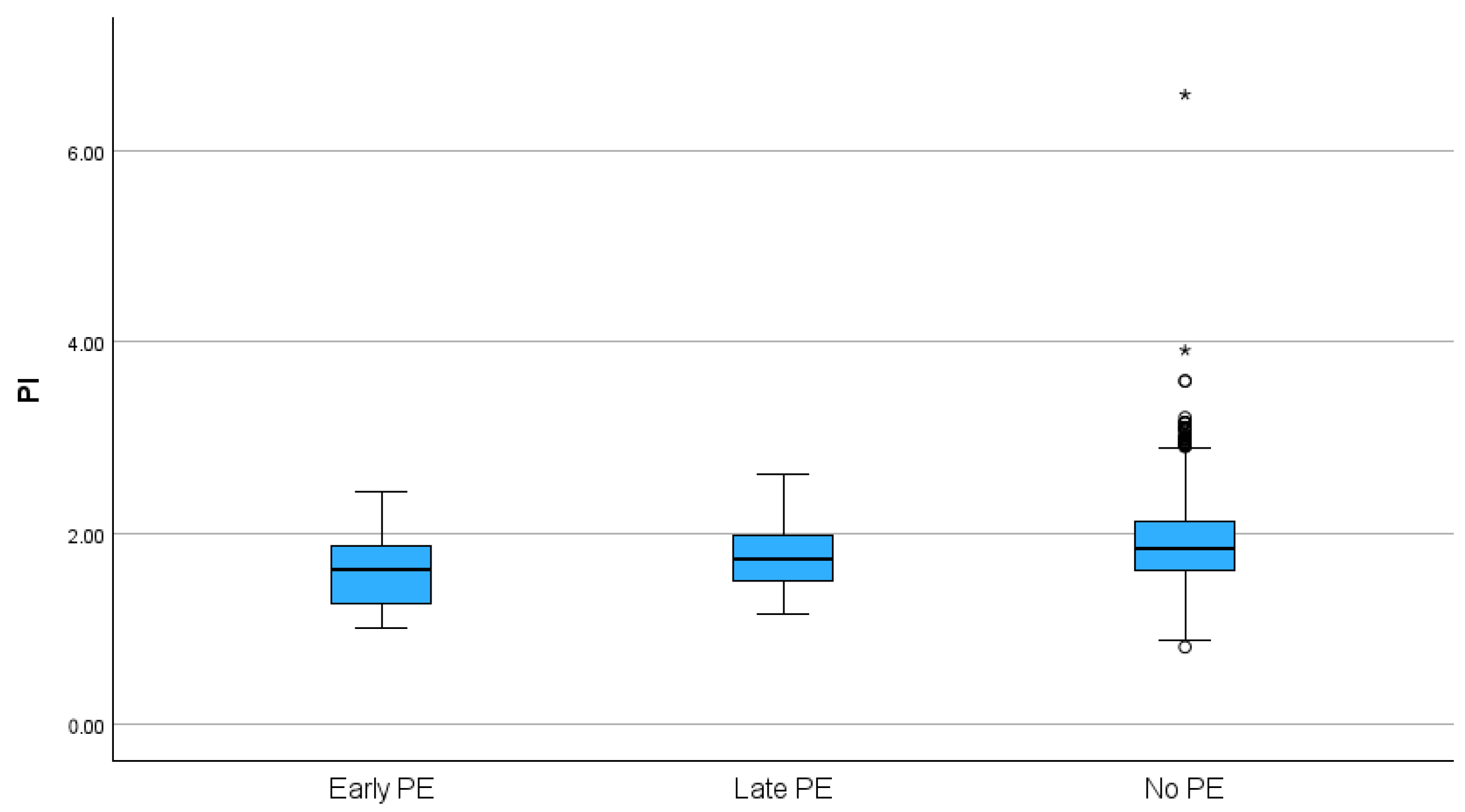
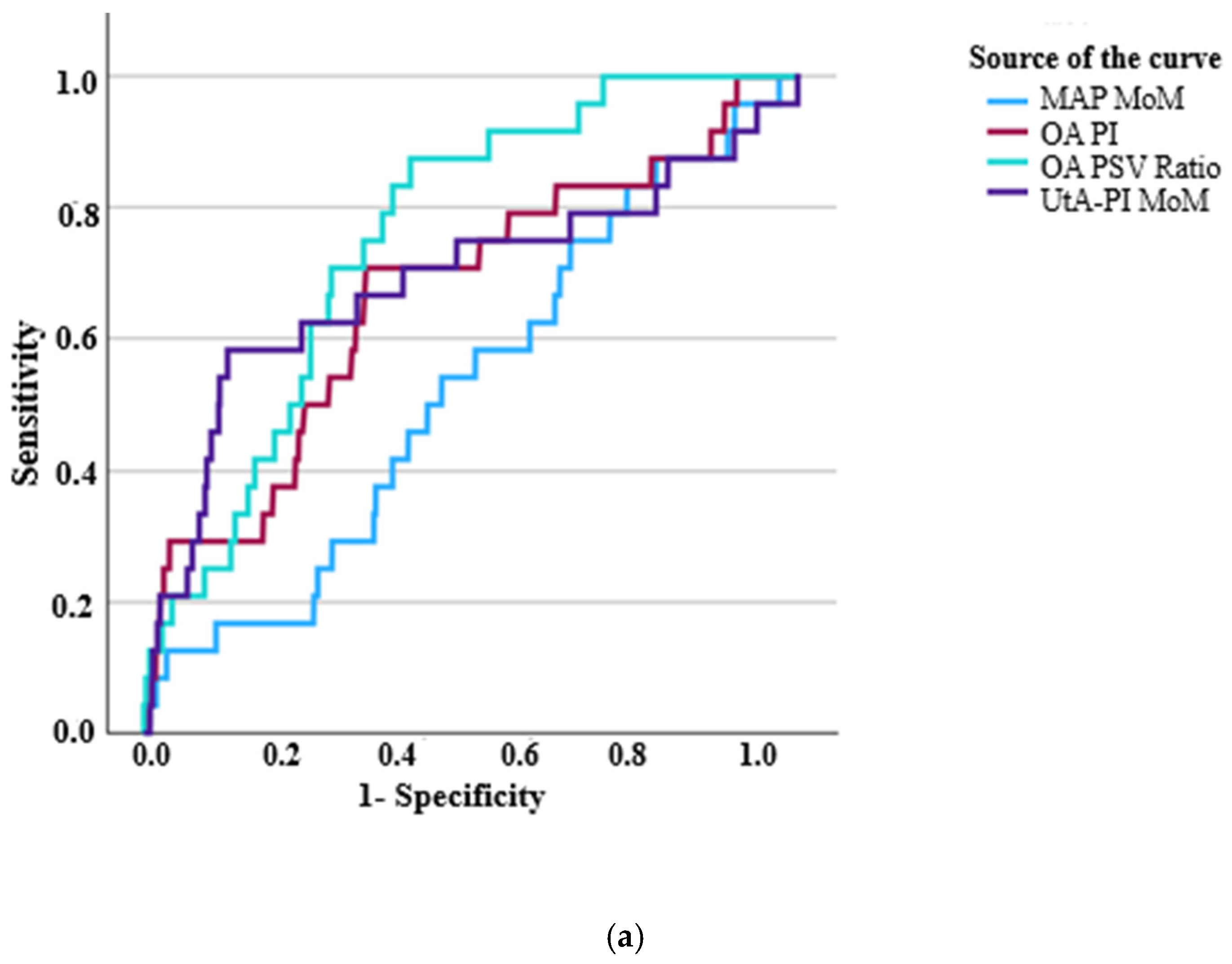
| 1/OA PI | 0.64 ± 0.15 | 0.55 ± 0.15 | <0.001 | 0.671 | 0.004 | 1.82 | 0.029–118 | 0774 |
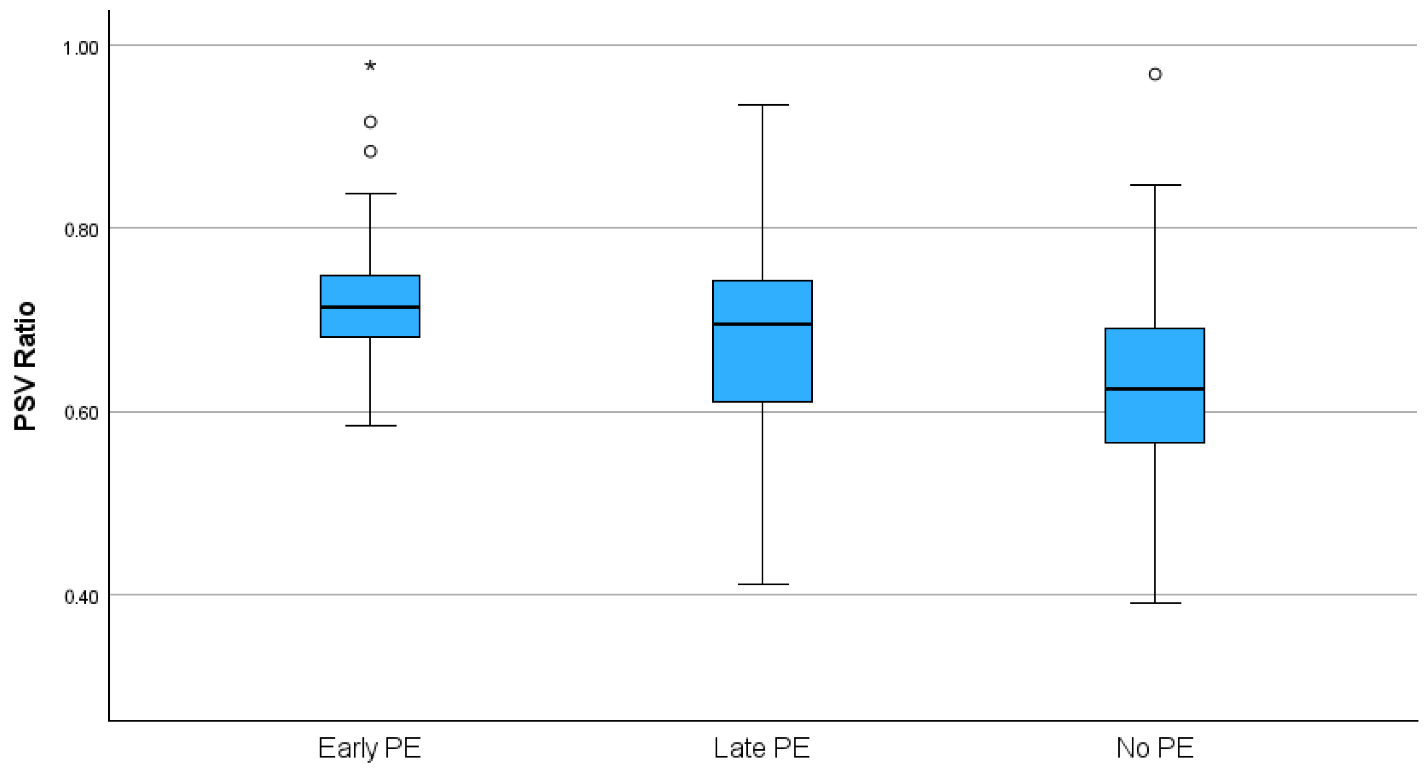
| PSV ratio | 0.73 ± 0.1 | 0.64 ± 0.1 | <0.001 | 0.758 | <0.001 | 5576 | 21–1,475,892 | 0.002 |
| UtA-PI MoM | 1.28 ± 0.42 | 1.03 ± 0.31 | <0.001 | 0.690 | 0.005 | 7.65 | 2.62–22 | <0.001 |
| MAP MoM | 1.03 ± 0.09 | 1.02 ± 0.08 | 0.233 | 0.528 | 0.622 | 2.67 | 0.02–330 | 0.680 |
| PlGF Subset | (n = 23) | (n = 224) | ||||||
| 1/OA PI | 0.64 ± 0.15 | 0.55 ± 0.11 | <0.001 | 0.674 | 0.006 | 0.221 | 0.001–56 | 0.590 |
| PSV ratio | 0.73 ± 0.96 | 0.63 ± 0.93 | <0.001 | 0.782 | <0.001 | 958,732 | 271–3,385,765,152 | 0.001 |
| UtA-PI MoM | 1.25 ± 0.41 | 1.01 ± 0.31 | <0.001 | 0.685 | 0.009 | 8.11 | 1.72–38 | 0.008 |
| MAP MoM | 1.03 ± 0.085 | 1.01 ± 0.074 | 0.204 | 0.554 | 0.390 | 10.5 | 0.015–7475 | 0.480 |
| 1/PlGF MoM | 1.64 ± 0.73 | 1.08 ± 0.43 | <0.001 | 0.750 | <0.001 | 4.16 | 1.68–10 | 0.002 |
| (b) | ||||||||
| All Patients | Late PE (n = 89) | No PE (n = 3941) | ||||||
| Variable | Mean ± SD | Mean ± SD | p (t-Test) | AUC | p (ROC) | OR | OR 95% CI | p (Logistics) |
| 1/OA PI | 0.59 ± 0.12 | 0.55 ± 0.12 | 0.002 | 0.599 | 0.001 | 0.80 | 0.07–9.2 | 0.860 |
| PSV ratio | 0.68 ± 0.11 | 0.64 ± 0.1 | <0.001 | 0.627 | <0.001 | 66.9 | 3.22–1392 | 0.010 |
| UtA-PI MoM | 1.03 ± 0.34 | 1.03 ± 0.31 | 0.964 | 0.504 | 0.895 | 1.09 | 0.56–2.1 | 0.790 |
| MAP MoM | 1.04 ± 0.09 | 1.02 ± 0.08 | 0.005 | 0.596 | 0.001 | 15.96 | 1.36–187 | 0.030 |
| PlGF Subset | (n = 89) | (n = 224) | ||||||
| 1/OA PI | 0.59 ± 0.12 | 0.56 ± 0.12 | 0.011 | 0.596 | 0.006 | 0.25 | 0.009–7.0 | 0.417 |
| PSV ratio | 0.68 ± 0.11 | 0.63 ± 0.09 | <0.001 | 0.644 | <0.001 | 297 | 4.9–17,978 | 0.007 |
| UtA-PI MoM | 1.03 ± 0.34 | 1.01 ± 0.31 | 0.634 | 0.523 | 0.534 | 1.08 | 0.48–2.40 | 0.861 |
| MAP MoM | 1.04 ± 0.09 | 1.01 ± 0.07 | 0.003 | 0.609 | 0.002 | 32 | 1.04–1008 | 0.047 |
| 1/PlGF MoM | 1.24 ± 0.52 | 1.08 ± 0.43 | 0.006 | 0.600 | 0.004 | 1.80 | 1.04–3.1 | 0.036 |
Disclaimer/Publisher’s Note: The statements, opinions and data contained in all publications are solely those of the individual author(s) and contributor(s) and not of MDPI and/or the editor(s). MDPI and/or the editor(s) disclaim responsibility for any injury to people or property resulting from any ideas, methods, instructions or products referred to in the content. |
© 2025 by the authors. Licensee MDPI, Basel, Switzerland. This article is an open access article distributed under the terms and conditions of the Creative Commons Attribution (CC BY) license (https://creativecommons.org/licenses/by/4.0/).
Share and Cite
Gana, N.; Pittokopitou, S.; Solonos, F.; Perdeica, A.; Fitiri, M.; Nicolaides, K.H. Ophthalmic Artery Doppler Indices at 11–13 Weeks of Gestation in Relation to Early and Late Preeclampsia. J. Clin. Med. 2025, 14, 4811. https://doi.org/10.3390/jcm14134811
Gana N, Pittokopitou S, Solonos F, Perdeica A, Fitiri M, Nicolaides KH. Ophthalmic Artery Doppler Indices at 11–13 Weeks of Gestation in Relation to Early and Late Preeclampsia. Journal of Clinical Medicine. 2025; 14(13):4811. https://doi.org/10.3390/jcm14134811
Chicago/Turabian StyleGana, Nicoleta, Savia Pittokopitou, Filippos Solonos, Alina Perdeica, Marina Fitiri, and Kypros H. Nicolaides. 2025. "Ophthalmic Artery Doppler Indices at 11–13 Weeks of Gestation in Relation to Early and Late Preeclampsia" Journal of Clinical Medicine 14, no. 13: 4811. https://doi.org/10.3390/jcm14134811
APA StyleGana, N., Pittokopitou, S., Solonos, F., Perdeica, A., Fitiri, M., & Nicolaides, K. H. (2025). Ophthalmic Artery Doppler Indices at 11–13 Weeks of Gestation in Relation to Early and Late Preeclampsia. Journal of Clinical Medicine, 14(13), 4811. https://doi.org/10.3390/jcm14134811