Case Report

Complicated Diagnosis and Treatment of Rare Painless Acanthamoeba Keratitis

by
Dominika Wróbel-Dudzińska
1,
Marta Ziaja-Sołtys
2,*,
Beata Rymgayłło-Jankowska
1,
Monika Derda
3,
Robert Klepacz
4,
Daniel Zalewski
2,
Tomasz Żarnowski
1 and
Anna Bogucka-Kocka
2
1
Department of Diagnostic and Microsurgery of Glaucoma, Medical University of Lublin, Chmielna 1 Street, 20-079 Lublin, Poland
2
Department of Biology with Genetics, Medical University of Lublin, Witolda Chodźki 4A Street, 20-093 Lublin, Poland
3
Department of Biology and Medical Parasitology, Poznan University of Medical Sciences, H. Swiecicki Street 4, 60-781 Poznan, Poland
4
Department of Clinical Pathology, Medical University of Lublin, Jaczewskiego 8B Street, 20-090 Lublin, Poland
*
Author to whom correspondence should be addressed.
J. Clin. Med. 2025, 14(13), 4763; https://doi.org/10.3390/jcm14134763
Submission received: 27 March 2025 / Revised: 28 May 2025 / Accepted: 3 July 2025 / Published: 5 July 2025
(This article belongs to the Special Issue Influence of the Environment on Ocular Diseases)

Abstract

Objectives: The aim was to present the complicated diagnostic and therapeutic process of atypical, painless keratitis caused by a cosmopolitan protozoan of the genus Acanthamoeba. Methods: This Case Report describes a medical case involving a 48-year-old woman who occasionally wears soft contact lenses and was referred to our hospital for treatment due to deteriorating visual acuity in her left eye. The diagnostic process included the isolation of amoebae from corneal scrapings and the morphological and molecular identification of the etiological agent of the infection. Results: After examination, painless atypical keratitis was diagnosed, initially considered recurrent herpetic keratitis. However, antiviral treatment did not bring about any improvement. Further observation revealed a dense, central, annular infiltrate on the periphery of the cornea. Despite treatment, the corneal infiltrate did not improve and the patient required therapeutic penetrating keratoplasty. Ultimately, the patient underwent combined surgery: corneal transplantation with cataract phacoemulsification and intraocular lens implantation. The postoperative course was uneventful. Conclusions: Acanthamoeba keratitis should be included in the differential diagnosis of keratitis, even in the absence of its characteristic feature of severe ocular pain, especially in contact lens wearers and patients who have had herpetic keratitis. Infection of the cornea with the Herpes simplex type 1 virus causes nerve degeneration, which probably translates into a painless course of Acanthamoeba castellanii infection.

1. Introduction

Acanthamoeba keratitis (AK) is a severe corneal infection caused by pathogenic genotypes of the Acanthamoeba genus, which, according to the current scientific literature, often leads to blindness [1,2,3,4]. Acanthamoeba sp. is a protozoan from the group of free-living amoeba together with other species, such as Balamuthia, Naegleria, and Sappinia sp. All of them live in soil and lakes, but can also occur in swimming pools and drinking water reservoirs [5]. Acanthamoeba sp. exists in two different stages, both of which are infective: the trophozoite, which is able to feed and divide, and the cyst, which is a dormant stage resistant to harsh conditions [5,6,7]. This free-living amoeba is pathogenic, especially to immunocompromised patients [5]. Acanthamoeba amoebas have also been detected in contaminated contact lens cleaning solutions and have been confirmed to grow on lenses cleaned with tap water [8].
The genus Acanthamoeba was classified according to the 18S rRNA (rDNA) sequence (types T1-T22). Acanthamoeba castellanii type T4 was found to be the etiological factor in 94.3% cases of Acanthamoeba keratitis. However, genotypes T2, T3, T5, T6, T11, T13, and T15 have also been related to AK and amoebic encephalitis [9,10,11,12,13]. The other listed species, i.e., A. polyphaga, A. culbertsoni, A. hatchetti, A. rhysodes, A. griffini, A. quina, and A. lugdunensis, are detected less frequently [14]. In recent years, an increase in the number of infections with this parasite has been observed around the world [15]. The annual incidence of AK worldwide is 2.9 cases per million people per year, but there are significant differences between countries [16]. This represents 2% of all corneal inflammations and is usually one-sided, although it can affect both eyes in lens-wearing patients [9,10].
AK is an infection that poses a major challenge for physicians to diagnose and treat [2]. Clinical manifestations are nonspecific and include tearing, unilateral photophobia, and redness [17,18]. At the beginning of the disease, dendritic epitheliopathy resembling Herpes simplex keratitis (HSK) often develops, with or without stroma and endotheliitis [19]. Therefore, the initially nonspecific acute presentation of AK is often misdiagnosed as adenoviral herpetic, bacterial, mycotic keratitis, or corneal epithelial erosion [16,20,21].
The hallmark symptom of Acanthamoeba keratitis is exquisite pain, disproportionate to the clinical picture [22]. This pronounced pain is attributed to the exceptional sensitivity of the cornea due to its dense sensory, sympathetic and parasympathetic nerve fiber network, which makes it the best innervated structure in the human body. Most of the sensory innervation of the cornea consists of long ciliary and nasociliary neurons originating from the ophthalmic division of the trigeminal ganglion. These fibers do not contain myelin, making the tissue transparent [23,24,25,26].
It is believed that any disruption to the proper state of corneal innervation, as a result of infection or surgery, can lead to transient or chronic neurotrophic keratopathy (NK) [24,27]. The disease begins spontaneously with the breakdown of epithelial cells, and the impaired healing mechanisms of the corneal surface increase susceptibility to soreness. A similar pathophysiological process may underlie the pain associated with AK. However, some studies have documented cases of ocular acanthamoebiasis presenting without pain [28,29,30]. Decreased corneal sensation has previously been reported in the context of ocular infection with the Herpes simplex type 1 virus and as a consequence of iatrogenic injury during neurosurgical procedures [26,31]. The underlying mechanisms of this phenomenon may also play a role in the atypical, painless clinical presentation of AK; nevertheless, the underlying cause of the absence of pain in these cases remains inconclusive.
Our aim is to present the diagnostic and therapeutic process of painless keratitis caused by Acanthamoeba and to summarize the current literature data on this type of ocular infection.

2. Detailed Case Description

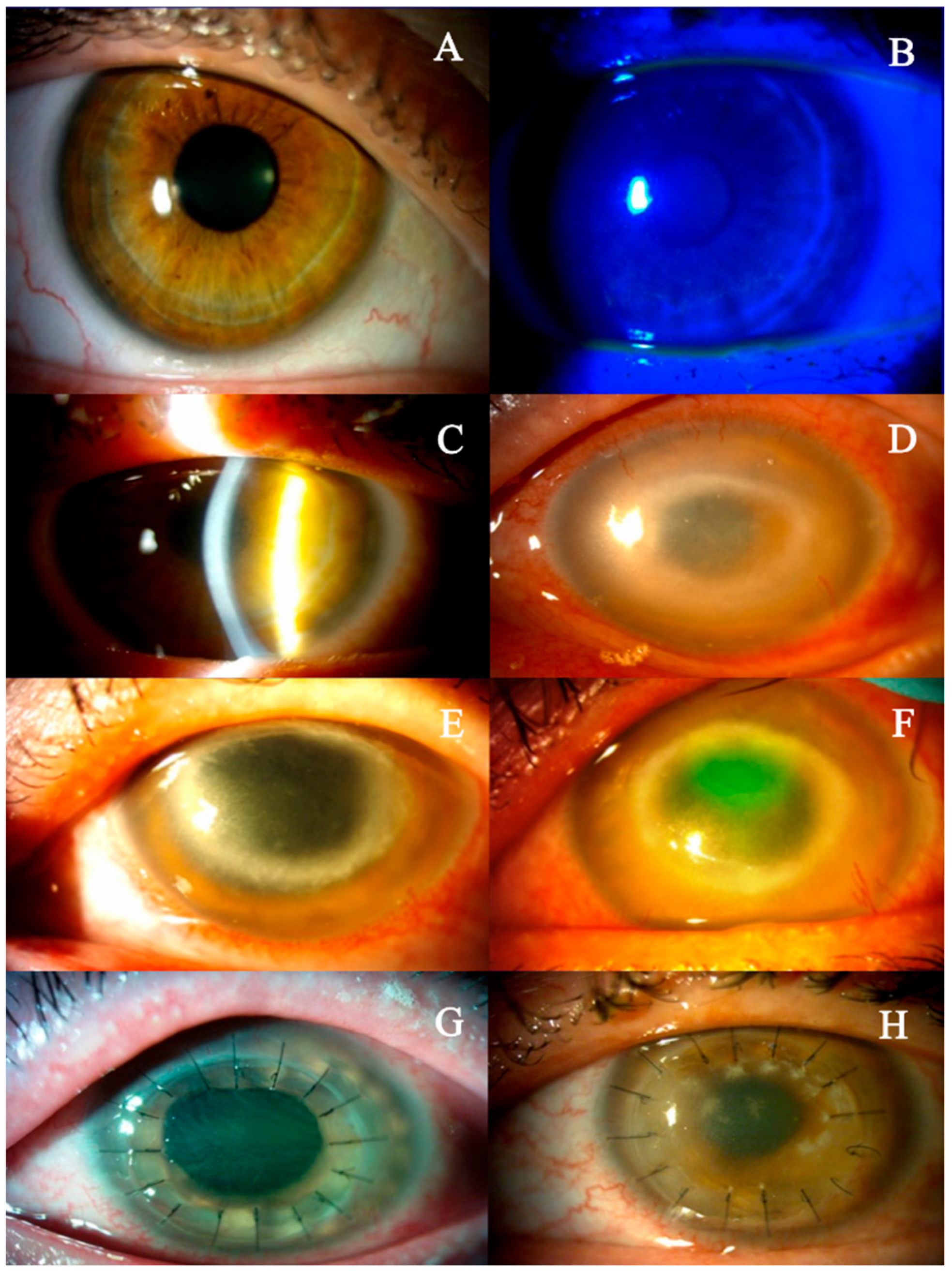
In March 2017, a 48-year-old female patient with myopia presented to our hospital with unilateral keratitis and a two-week history of decreased visual acuity in the left eye (Figure 1). Her overall health was good. According to the medical history, she occasionally wore contact lenses and had been treated with acyclovir (locally four times a day, and orally four times a day at 400 mg) five months earlier due to herpetic keratitis of the left eye in another medical center. Upon the examination, the best corrected visual acuity (BCVA) was 0.7 in the left eye and 1.0 in the right eye, and the intraocular pressure (IOP) in the left eye was 14 mmHg, in the right eye 13 mmHg. Ocular examination of the left eye revealed mild conjunctival hyperemia and subtle epithelial changes resembling a dendritic-like ulcer located in the inferior corneal hemisphere (Figure 2A,B). The corneal sensation in the left eye was decreased. Recurrent herpetic keratitis was diagnosed and local (four times a day) and oral (four times at 400 mg) acyclovir treatment was initiated. After three weeks (April 2017), despite the slight improvement, that is, no epitheliopathy, stromal swelling was observed in the lower corneal hemisphere. Furthermore, visual acuity in the left eye had declined to 0.3 (Figure 2C). In May 2017, a deeper corneal ulcer was detected in the superior corneal hemisphere. Upon inquiry, the patient reported no ocular pain. There was no abnormal finding in the left eye. During further observations (June 2017), a dense mid-peripheral ring-like stromal infiltrate developed (Figure 2D).
The visual acuity of the affected eye deteriorated to the counting fingers stage. Deep corneal scrapings were collected and labeled as AC55. The scraping sample was inoculated into a non-nutrient agar (BD Difco Ltd., Detroit, MI, USA), poured on a Petri dish and covered with a suspension of the bacterium Enterobacter aerogenes. The amoebae were grown in axenic liquid cultures containing 2% Bacto Casiton (Gibco, Life Technologies, Carlsbad, CA, USA) and 10% normal horse serum according to the procedure described by Červa [32]. The plates were incubated at a temperature of 28 °C. After 1–5 days, an increase in the number of amoebae was observed and examined with an inverted microscope at ×200. The amoebae isolated from the cornea were identified according to morphological criteria, measurements of the size of cysts and tests for flagellation [33]. The culture was positive for Acanthamoeba sp.
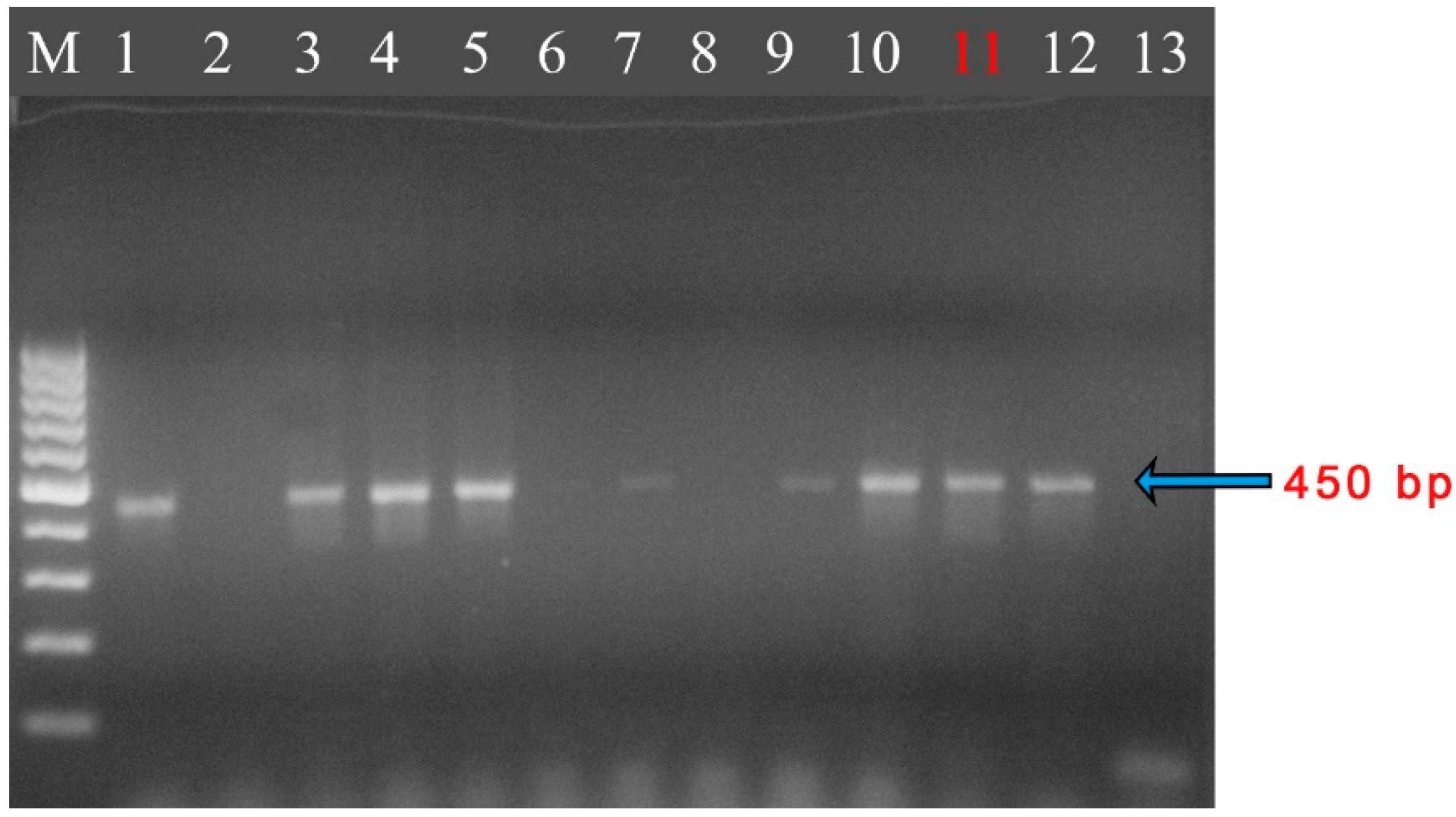
The polymerase chain reaction (PCR) for Acanthamoeba was performed using genus-specific primers previously described by Schroeder et al. [34] (see Supplementary Material for details). DNA was isolated from the corneal scrapings (AC55 sample), and amplicons of the 18S rRNA gene fragment were obtained. The results of the PCR confirmed that the sequences obtained from the AC55 isolate were identical to the Acanthamoeba sequences deposited in GenBank (Figure 3). The PCR products were subsequently submitted for sequencing analysis (see Supplementary Material for details). The analysis revealed that the sequences were 100% identical to the parasite’s 18S rRNA gene isolated from the liver of an infected pheasant Tragopan temminckii (GQ889265), corneal scrapings (KF318460, DQ087297), a contact lens (DQ087296), and an environmental sample (EU377583) (Table 1) [35]. The Acanthamoeba sequences from the isolates obtained from corneal scrapings (AC55) were deposited in GenBank (NCBI) under accession number KP120880. The isolated amoeba strain belonged to genotype T4 of the genus Acanthamoeba.
Intensive treatment was initiated, consisting of topical 0.1% propamidine isethionate, 0.2% polyhexamethylene biguanide and neomycin, in combination with systemic antifungal therapy using fluconazole and/or ketoconazole. In July 2017, a large epithelial defect was identified at the border of the stromal ring. After three months of amoebicidal therapy, there was no significant improvement in the stromal infiltrate, necessitating therapeutic penetrating keratoplasty (September 2017) (Figure 2E,F). The patient did not report any pain at any time before the surgery.
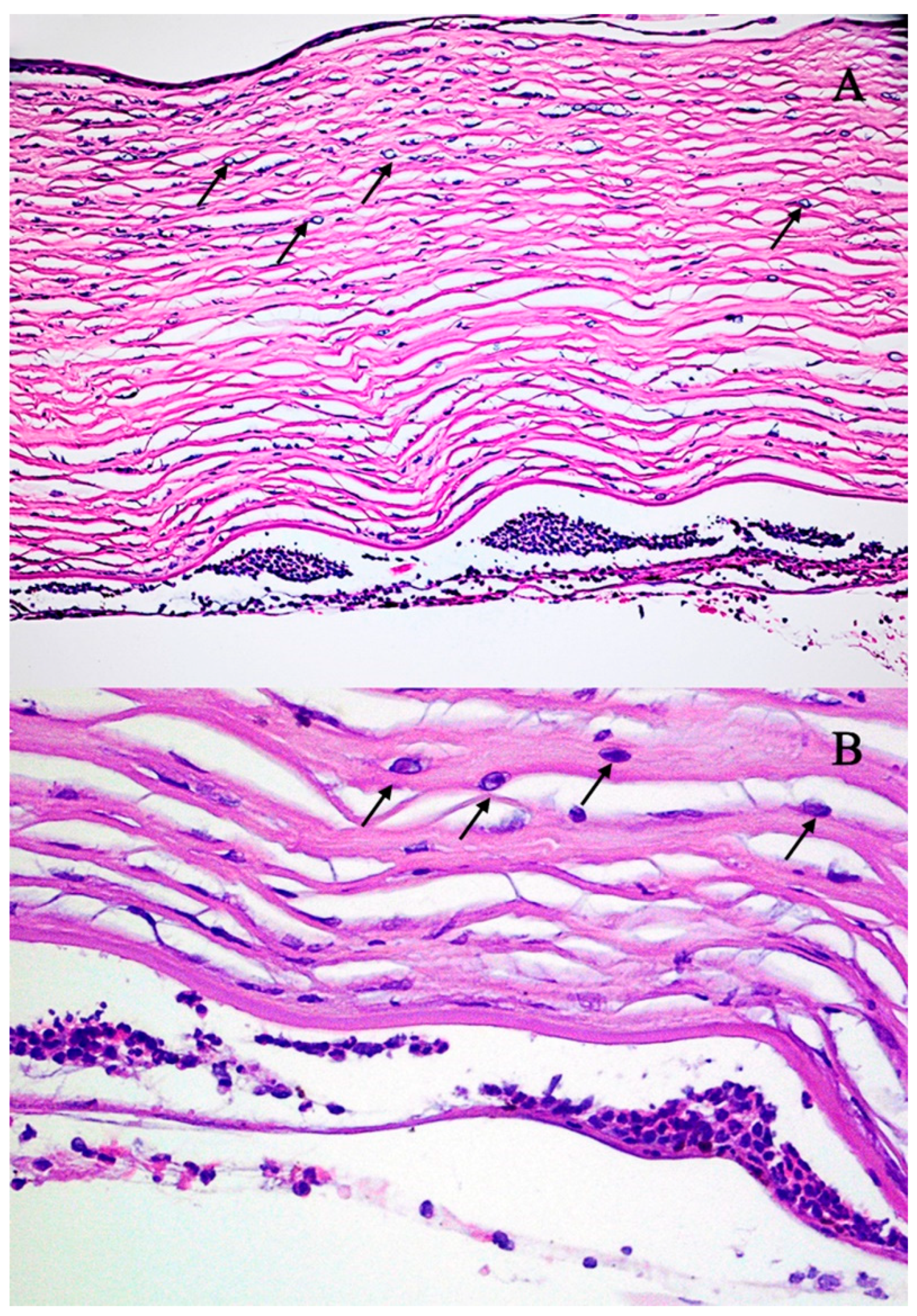
The tissue was transferred to the eye pathology laboratory. Macroscopic histological examination of the corneal tissue revealed that the specimen consisted of an opaque corneal nodule measuring 8 mm in diameter. Microscopic examination revealed marked attenuation and focal detachment of the corneal epithelium. The stroma exhibited pronounced edema accompanied by focal neutrophilic infiltration. Numerous Acanthamoeba cysts and trophozoites were widely distributed throughout the stromal tissue. The descemet membrane appeared structurally intact but was extensively covered with inflammatory cells, predominantly neutrophils. Only a sparse population of corneal endothelial cells was identified (Figure 4A,B). At the initial follow-up visit in October 2017, the ocular examination revealed a transparent corneal transplant secured with 16 Nylon 10/0 sutures (Figure 2G). After three months (December 2017), the transplant lost transparency, and a few sutures were loosened (Figure 2H). Moreover, a cataract was diagnosed in the left eye. At the final follow-up, our patient’s visual acuity was at the hand movement stage. Three months later (April 2018), the patient underwent combined surgery: corneal transplantation along with cataract phacoemulsification and intraocular lens implantation. The postoperative course was without complications, and on the last follow-up visit the best corrected visual acuity was 0.5 (Figure 5).

3. Discussion and Review

Acanthamoeba keratitis is a potentially vision-threatening corneal infection. Diagnostic challenges primarily stem from the nonspecific clinical signs and symptoms observed during the initial stages of the disease; moreover, this pathology may mimic other infections [17]. The German Acanthamoeba Keratitis Registry has revealed that Acanthamoeba keratitis has been misdiagnosed as herpetic (in 47.6% of cases), mycotic (in 25.2%), and bacterial (in 3.9%) keratitis in patients [41]. It is noteworthy that a mixed infection with viruses, bacteria, or fungi is present in approximately 23% of keratitis cases [42]. According to other sources, Acanthamoeba infections are polymicrobial, with 12.5% of co-infections with bacteria, 40% of co-infections with fungi, and 5% of triple infections. In addition, co-infections account for 55% of all Acanthamoeba infections [43].
So far, 54 cases of painless AK have been reported in the literature. Some studies have shown the tendency of Acanthamoeba to co-exist with other microorganisms, which increases their pathogenicity and survival, thus making their treatment more difficult. In total, 16/54 (29.6%) patients wore contact lenses. Infection only with Acanthamoeba castellanii was diagnosed in 38/54 (70.37%) patients without concomitant bacterial, viral, or fungal infection. Bacterial co-infection was detected in 13 (81.25%) of 16 patients, and co-infection was caused solely by viruses in 4 of 16 (25%) patients. Co-infection with bacteria and Herpes simplex virus was confirmed in one patient. These data are shown in Table 2.
In the literature, the pain in Acanthamoeba keratitis is described as severe and disproportionate to the clinical findings during the slit lamp examination [44,45]. Other initial symptoms are often nonspecific [29], which means that Acanthamoeba keratitis may often be misdiagnosed as herpetic keratitis or epithelial erosions early in the course of the disease, resulting from the presence of an epithelial defect. Therefore, high clinical suspicion should be maintained for Acanthamoeba keratitis in patients presenting with relevant risk factors, even in the absence of ocular pain [46,47].
This case illustrates an atypical presentation of Acanthamoeba keratitis, characterized by the complete absence of ocular pain throughout the disease course. The patient experienced only mild ocular discomfort without any reported pain. The mechanism of painless Acanthamoeba keratitis is not completely understood, but it is suggested to be the result of perineuritis, preexisting neurotrophic cornea (such as Herpes keratitis), or pretreatment with topical steroids, which mask the clinical signs of this keratitis [30,48,49,50].
Table 2. Cases of painless AK reported in the literature; ”+”—yes, “−“—no.
ReferenceNumber of CasesContact Lens UseAcanthamoeba castellanii OnlyPrevious Infections or Coinfections
BacterialViralFungal
Perry et al., 1995 [51]6++
++
++
++
+
+
Sharma et al., 2000 [52]3434−25+9+34−34−
Roters et al., 2001 [53]1++
Tabin et al., 2001 [29]2+
Herpes
simplex
Georgakopoulos et al., 2006 [54]1++
Stemberger et al., 2007 [55]1++
Elabjer et al., 2009 [56]1++
Shukla et al., 2012 [30]5+Herpes
simplex
++
++
+Herpes
simplex
++
Kwok et al., 2017 [46]1++
Sun et al., 2020 [57]1++Herpes
simplex
Lin et al., 2023 [58]1++
5416381340
Differences in the immune response and virulence might have an impact on the variable clinical presentation. In their work, Kurbanyan et al. showed that corneal innervation density, nerve length, and branching are significantly lower in cases of active fungal infection or acanthamoebiasis [27]. Moreover, it is suggested that the loss of corneal nerves in AK and fungal keratitis (FK) seems to be greater compared to the changes observed in herpetic keratitis. However, the cause of these changes must be investigated even more thoroughly to confirm the involvement of pathogens in their formation. As a result of eye infection with the Herpes simplex type 1 virus, corneal hypoesthesia is observed, while patients with AK often experience severe pain. Such symptoms can be explained by abnormalities and hypersensitivity in the corneal nerves’ survival or regeneration. This pain may also be triggered by cytokines (such as interleukin-1) or nociceptors [59,60].
Five months before the visit to our hospital, the patient was diagnosed with Herpes keratitis, which was treated. This prior treatment may have contributed to a reduction in corneal sensitivity. These circumstances can explain the painless course of the disease that caused a considerable delay in the proper diagnosis and treatment in our patient. In addition, it led to the avoidable loss of visual acuity before transplantation and, finally, the transplant failure. The corneal graft was performed in our patient due to the lack of response to the treatment and worsening of the local condition.
Perry et al. draws attention to the symptom of reduced corneal sensation in patients with keratitis caused by Acanthamoeba, which very often results in an incorrect diagnosis, usually herpetic keratitis, a delay in treatment and increased morbidity. In the described cases (six patients), a marked decrease in corneal sensation values was observed in all patients using a Cachet–Bonnet esthesiometer (Luneau, Paris, France) [51].
Until now, there have been no standard guidelines for treating acanthamoebiasis or treatment strategies discussed in case reports. According to the literature, to date, several randomized, controlled clinical trials have been conducted on the treatment of AK. There were no differences in the effectiveness of monotherapy with polyhexamethylene biguanide and chlorhexidine. However, combined therapy with biguanides, diamidine derivatives and antibiotic ointments is recommended [59,60]. Moreover, therapeutic penetrating keratoplasty might be considered when the infection escalates to the paracentral corneal stroma, despite maximum antiamoebic therapy [61]. The rate of corneal transplantation performed to control Acanthamoeba keratitis ranges from 5% to 68% [62]. The surgical treatment should be delayed until the eye is not inflamed and after completion of the antiacanthamoebic treatment due to the poor prognosis and the high risk of graft failure [63]. The post-operative complications after keratoplasty, in addition to rejection of the graft, include recurrence of the Acanthamoeba infection, other infections, glaucoma, cataract, wound leak and irregular astigmatism. Roozbahani et al. observed 75% of graft failures, 50% of cataract, 17% of uncontrolled glaucoma, and 8% of Acanthamoeba reactivation in a study group of therapeutic penetrating keratoplasty for Acanthamoeba keratitis [64]. Unfortunately, graft failure and cataract development were observed in our patient. The next stage of therapy, a combined surgery including corneal transplantation with cataract phacoemulsification and intraocular lens implantation, was carried out without complications. It was found that diagnosis made within 18 days of symptom onset and initiation of antiamoebic therapy resulted in a better final BCVA after the completion of treatment and eliminated the need for urgent and elective penetrating keratoplasty [65].
It has been suggested that in vivo corneal confocal microscopy (IVCM), which allows a detailed examination of the corneal subbasal nerve plexus and the determination of the degree of corneal damage resulting from infectious keratitis, may not be sensitive enough to detect deep corneal lesions and confirm Acanthamoeba infection when diagnosing keratitis [24,57]. In contrast, scleral scattering slit-lamp microscopy, an advanced diagnostic modality that enhances the detection of early and subtle keratitic changes, may offer complementary diagnostic information that facilitates the identification of features characteristic of AK.
Kent et al., describing a painless case of AK, suggest that the reason for the absence of a pain symptom is related to the severity of the accompanying scleritis [30]. In the case of suspected Acanthamoeba infection, including mixed infections, taking into account typical symptoms and causes, but also the atypical painless course of the disease, Sun et al. recommended considering and, if necessary, implementing appropriate treatment, even before confirmation by laboratory test results [57].

4. Conclusions

In conclusion, Acanthamoeba keratitis should be included in the differential diagnosis of keratitis, even in the absence of the classical symptom of severe ocular pain, especially in contact lens wearers and patients who have had herpetic keratitis. Data from the literature and our observations confirm that cornea infection with the H. simplex type 1 virus causes nerve degeneration, which translates into a painless course of Acanthamoeba castellanii infection. This may be the reason for an incorrect diagnosis and treatment that may further change the clinical picture of the disease and delay the proper therapy [30,48,49,50]. As a consequence, the patient’s quality of life is significantly reduced due to prolonged poor visual acuity and illness, which generates additional costs in terms of medical care.

Supplementary Materials

The following supporting information can be downloaded at https://www.mdpi.com/article/10.3390/jcm14134763/s1.

Author Contributions

Conceptualization, D.W.-D., B.R.-J. and M.Z.-S.; Methodology, D.W.-D., B.R.-J., M.D. and R.K.; Writing—original draft preparation, M.Z.-S., D.W.-D. and B.R.-J.; Writing—review and editing, M.Z.-S., D.W.-D., B.R.-J., M.D. and D.Z.; Supervision, T.Ż. and A.B.-K. All authors have read and agreed to the published version of the manuscript.

Funding

This research was financed from the funds allocated to the statutory activities of the Medical University of Lublin (number DS43; DS179) and the statutory funds of the Department of Biology and Medical Parasitology, Poznań University of Medical Sciences.

Institutional Review Board Statement

Ethical review and approval were waived for this study due to the fact that case did not involve an experimental procedure, in accordance with the Regulations of the Polish Bioethics Committee.

Informed Consent Statement

Patient consent was waived as the article did not contain any information that could identify the patient.

Data Availability Statement

The raw data supporting the conclusions of this article will be made available by the authors on request.

Conflicts of Interest

The authors declare no conflicts of interest.

Abbreviations

The following abbreviations are used in this manuscript:
AKAcanthamoeba keratitis
BCVABest corrected visual acuity
FKFungal keratitis
HSKHerpes simplex keratitis
IOPIntraocular pressure
IVCMIn vivo corneal confocal microscopy
NKNeurotrophic keratopathy

References

  1. Peguda, H.K.; Lakshminarayanan, R.; Carnt, N.A.; Gu, Z.; Willcox, M.D.P. The Activity of Polyhomoarginine against Acanthamoeba Castellanii. Biology 2022, 11, 1726. [Google Scholar] [CrossRef] [PubMed]
  2. Chomicz, L.; Szaflik, J.P.; Szostakowska, B.; Izdebska, J.; Baltaza, W.; Łazicka-Gałecka, M.; Kuligowska, A.; Machalińska, A.; Zawadzki, P.J.; Szaflik, J. Successive Acanthamoeba Corneal Isolates Identified in Poland Monitored in Terms of In Vitro Dynamics. Microorganisms 2023, 11, 1174. [Google Scholar] [CrossRef] [PubMed]
  3. Randag, A.C.; van Rooij, J.; van Goor, A.T.; Verkerk, S.; Wisse, R.P.L.; Saelens, I.E.Y.; Stoutenbeek, R.; van Dooren, B.T.H.; Cheng, Y.Y.Y.; Eggink, C.A. The Rising Incidence of Acanthamoeba Keratitis: A 7-Year Nationwide Survey and Clinical Assessment of Risk Factors and Functional Outcomes. PLoS ONE 2019, 14, e0222092. [Google Scholar] [CrossRef]
  4. Carnt, N.; Hoffman, J.J.; Verma, S.; Hau, S.; Radford, C.F.; Minassian, D.C.; Dart, J.K.G. Acanthamoeba Keratitis: Confirmation of the UK Outbreak and a Prospective Case-Control Study Identifying Contributing Risk Factors. Br. J. Ophthalmol. 2018, 102, 1621–1628. [Google Scholar] [CrossRef] [PubMed]
  5. Gonçalves, D.d.S.; Ferreira, M.d.S.; Guimarães, A.J. Extracellular Vesicles from the Protozoa Acanthamoeba Castellanii: Their Role in Pathogenesis, Environmental Adaptation and Potential Applications. Bioengineering 2019, 6, 13. [Google Scholar] [CrossRef]
  6. Siddiqui, R.; Khan, N.A. Biology and Pathogenesis of Acanthamoeba. Parasit. Vectors 2012, 5, 6. [Google Scholar] [CrossRef]
  7. Khan, N.A. Acanthamoeba: Biology and Increasing Importance in Human Health. FEMS Microbiol. Rev. 2006, 30, 564–595. [Google Scholar] [CrossRef]
  8. Hassan, F.A.M.; Tolba, M.E.M.; Abed, G.H.; Omar, H.M.; Abdel-Hakeem, S.S. Contact Lenses Contamination by Acanthamoeba spp. in Upper Egypt. PLoS ONE 2021, 16, e0259847. [Google Scholar] [CrossRef]
  9. Alkharashi, M.; Lindsley, K.; Law, H.A.; Sikder, S. Medical Interventions for Acanthamoeba Keratitis. Cochrane Database Syst. Rev. 2015, 2015, CD010792. [Google Scholar] [CrossRef]
  10. Szentmáry, N.; Daas, L.; Shi, L.; Laurik, K.L.; Lepper, S.; Milioti, G.; Seitz, B. Acanthamoeba Keratitis—Clinical Signs, Differential Diagnosis and Treatment. J. Curr. Ophthalmol. 2019, 31, 16–23. [Google Scholar] [CrossRef]
  11. Sifaoui, I.; Capote Yanes, E.; Reyes-Batlle, M.; Rodríguez-Expósito, R.L.; Piñero, J.E.; Lorenzo-Morales, J. Combined Amoebicidal Effect of Atorvastatin and Commercial Eye Drops against Acanthamoeba Castellanii Neff: In Vitro Assay Based on Mixture Design. Pathogens 2020, 9, 219. [Google Scholar] [CrossRef] [PubMed]
  12. Magnet, A.; Galván, A.L.; Fenoy, S.; Izquierdo, F.; Rueda, C.; Fernandez Vadillo, C.; Pérez-Irezábal, J.; Bandyopadhyay, K.; Visvesvara, G.S.; da Silva, A.J.; et al. Molecular Characterization of Acanthamoeba Isolated in Water Treatment Plants and Comparison with Clinical Isolates. Parasitol. Res. 2012, 111, 383–392. [Google Scholar] [CrossRef]
  13. Guimaraes, A.J.; Gomes, K.X.; Cortines, J.R.; Peralta, J.M.; Peralta, R.H.S. Acanthamoeba spp. as a Universal Host for Pathogenic Microorganisms: One Bridge from Environment to Host Virulence. Microbiol. Res. 2016, 193, 30–38. [Google Scholar] [CrossRef]
  14. Somani, S.N.; Ronquillo, Y.; Moshirfar, M. Acanthamoeba Keratitis. In StatPearls; StatPearls Publishing: Treasure Island, FL, USA, 2025. [Google Scholar]
  15. Zhang, Y.; Xu, X.; Wei, Z.; Cao, K.; Zhang, Z.; Liang, Q. The Global Epidemiology and Clinical Diagnosis of Acanthamoeba Keratitis. J. Infect. Public Health 2023, 16, 841–852. [Google Scholar] [CrossRef]
  16. Azzopardi, M.; Chong, Y.J.; Ng, B.; Recchioni, A.; Logeswaran, A.; Ting, D.S.J. Diagnosis of Acanthamoeba Keratitis: Past, Present and Future. Diagnostics 2023, 13, 2655. [Google Scholar] [CrossRef]
  17. Chin, J.; Young, A.L.; Hui, M.; Jhanji, V. Acanthamoeba Keratitis: 10-Year Study at a Tertiary Eye Care Center in Hong Kong. Cont. Lens Anterior Eye 2015, 38, 99–103. [Google Scholar] [CrossRef]
  18. Lorenzo-Morales, J.; Khan, N.A.; Walochnik, J. An Update on Acanthamoeba Keratitis: Diagnosis, Pathogenesis and Treatment. Parasite 2015, 22, 10. [Google Scholar] [CrossRef] [PubMed]
  19. Illingworth, C.D.; Cook, S.D. Acanthamoeba Keratitis. Surv. Ophthalmol. 1998, 42, 493–508. [Google Scholar] [CrossRef] [PubMed]
  20. Rosińska, J.; Kozubski, W. Peripheral nervous system involvement in the course of herpes virus infections. Neurol. Neurochir. Pol. 2013, 47, 170–178. [Google Scholar] [CrossRef]
  21. Amponin, D.E.; Przybek-Skrzypecka, J.; Zyablitskaya, M.; Takaoka, A.; Suh, L.H.; Nagasaki, T.; Trokel, S.L.; Paik, D.C. Ex Vivo Anti-Microbial Efficacy of Various Formaldehyde Releasers against Antibiotic Resistant and Antibiotic Sensitive Microorganisms Involved in Infectious Keratitis. BMC Ophthalmol. 2020, 20, 28. [Google Scholar] [CrossRef]
  22. Marciano-Cabral, F.; Cabral, G. Acanthamoeba spp. as Agents of Disease in Humans. Clin. Microbiol. Rev. 2003, 16, 273–307. [Google Scholar] [CrossRef] [PubMed]
  23. Mastropasqua, L.; Massaro-Giordano, G.; Nubile, M.; Sacchetti, M. Understanding the Pathogenesis of Neurotrophic Keratitis: The Role of Corneal Nerves. J. Cell Physiol. 2017, 232, 717–724. [Google Scholar] [CrossRef] [PubMed]
  24. Müller, L.J.; Marfurt, C.F.; Kruse, F.; Tervo, T.M.T. Corneal Nerves: Structure, Contents and Function. Exp. Eye Res. 2003, 76, 521–542. [Google Scholar] [CrossRef] [PubMed]
  25. Meng, I.D.; Kurose, M. The Role of Corneal Afferent Neurons in Regulating Tears under Normal and Dry Eye Conditions. Exp. Eye Res. 2013, 117, 79–87. [Google Scholar] [CrossRef]
  26. Duncan, N.W.; Houser, K.H.; Khandelwal, S.S. Neurotrophic Keratitis: A Review. Touchreviews Ophthalmol. 2022, 16, 67–71. [Google Scholar] [CrossRef]
  27. Kurbanyan, K.; Hoesl, L.M.; Schrems, W.A.; Hamrah, P. Corneal Nerve Alterations in Acute Acanthamoeba and Fungal Keratitis: An in Vivo Confocal Microscopy Study. Eye 2012, 26, 126–132. [Google Scholar] [CrossRef]
  28. Dart, J.K.G.; Saw, V.P.J.; Kilvington, S. Acanthamoeba Keratitis: Diagnosis and Treatment Update 2009. Am. J. Ophthalmol. 2009, 148, 487–499.e2. [Google Scholar] [CrossRef]
  29. Tabin, G.; Taylor, H.; Snibson, G.; Murchison, A.; Gushchin, A.; Rogers, S. Atypical Presentation of Acanthamoeba Keratitis. Cornea 2001, 20, 757–759. [Google Scholar] [CrossRef]
  30. Shukla Kent, S.; Robert, M.-C.; Tokarewicz, A.C.; Mather, R. Painless Acanthamoeba Keratitis. Can. J. Ophthalmol. 2012, 47, 383–384. [Google Scholar] [CrossRef]
  31. Bonini, S.; Rama, P.; Olzi, D.; Lambiase, A. Neurotrophic Keratitis. Eye 2003, 17, 989–995. [Google Scholar] [CrossRef]
  32. Cerva, L. Amoebic Meningoencephalitis: Axenic Culture of Naegleria. Science 1969, 163, 576. [Google Scholar] [CrossRef] [PubMed]
  33. Page, F. An Illustrated Key to Freshwater and Soil Amoebae; Freshwater Biological Association: Ambleside, UK, 1967; Volume 34. [Google Scholar]
  34. Schroeder, J.M.; Booton, G.C.; Hay, J.; Niszl, I.A.; Seal, D.V.; Markus, M.B.; Fuerst, P.A.; Byers, T.J. Use of Subgenic 18S Ribosomal DNA PCR and Sequencing for Genus and Genotype Identification of Acanthamoebae from Humans with Keratitis and from Sewage Sludge. J. Clin. Microbiol. 2001, 39, 1903–1911. [Google Scholar] [CrossRef] [PubMed]
  35. Derda, M.; Solarczyk, P.; Cholewiński, M.; Hadaś, E. Genotypic Characterization of Amoeba Isolated from Acanthamoeba Keratitis in Poland. Parasitol. Res. 2015, 114, 1233–1237. [Google Scholar] [CrossRef]
  36. Visvesvara, G.S.; Shoff, M.E.; Sriram, R.; Booton, G.C.; Crary, M.; Fuerst, P.A.; Hanley, C.S.; Garner, M.M. Isolation, Morphologic, Serologic and Molecular Identification of Acanthamoeba T4 Genotype from the Liver of a Temminck’s Tragopan (Tragopan Temminckii). Vet. Parasitol. 2010, 170, 197–200. [Google Scholar] [CrossRef] [PubMed]
  37. Mafra, C.S.P.; Carrijo-Carvalho, L.C.; Chudzinski-Tavassi, A.M.; Taguchi, F.M.d.C.; Foronda, A.S.; Carvalho, F.R.d.S.; de Freitas, D. Antimicrobial Action of Biguanides on the Viability of Acanthamoeba Cysts and Assessment of Cell Toxicity. Investig. Ophthalmol. Vis. Sci. 2013, 54, 6363–6372. [Google Scholar] [CrossRef]
  38. Corsaro, D.; Feroldi, V.; Saucedo, G.; Ribas, F.; Loret, J.-F.; Greub, G. Novel Chlamydiales Strains Isolated from a Water Treatment Plant. Environ. Microbiol. 2009, 11, 188–200. [Google Scholar] [CrossRef]
  39. Yera, H.; Zamfir, O.; Bourcier, T.; Viscogliosi, E.; Noël, C.; Dupouy-Camet, J.; Chaumeil, C. The Genotypic Characterisation of Acanthamoeba Isolates from Human Ocular Samples. Br. J. Ophthalmol. 2008, 92, 1139–1141. [Google Scholar] [CrossRef]
  40. Yera, H.; Zamfir, O.; Bourcier, T.; Ancelle, T.; Batellier, L.; Dupouy-Camet, J.; Chaumeil, C. Comparison of PCR, Microscopic Examination and Culture for the Early Diagnosis and Characterization of Acanthamoeba Isolates from Ocular Infections. Eur. J. Clin. Microbiol. Infect. Dis. 2007, 26, 221–224. [Google Scholar] [CrossRef]
  41. Daas, L.; Szentmáry, N.; Eppig, T.; Langenbucher, A.; Hasenfus, A.; Roth, M.; Saeger, M.; Nölle, B.; Lippmann, B.; Böhringer, D.; et al. The German Acanthamoeba keratitis register: Initial results of a multicenter study. Ophthalmologe 2015, 112, 752–763. [Google Scholar] [CrossRef]
  42. Szentmáry, N.; Goebels, S.; Matoula, P.; Schirra, F.; Seitz, B. Acanthamoeba keratitis--a rare and often late diagnosed disease. Klin. Monbl Augenheilkd. 2012, 229, 521–528. [Google Scholar] [CrossRef]
  43. Raghavan, A.; Baidwal, S.; Venkatapathy, N.; Rammohan, R. The Acanthamoeba-Fungal Keratitis Study. Am. J. Ophthalmol. 2019, 201, 31–36. [Google Scholar] [CrossRef] [PubMed]
  44. Osato, M.; Wilhalmus, K. Infectious Keratitis. In Cornea; Little, Brown: Boston, MA, USA, 1994; pp. 202–266. [Google Scholar]
  45. Bacon, A.S.; Frazer, D.G.; Dart, J.K.; Matheson, M.; Ficker, L.A.; Wright, P. A Review of 72 Consecutive Cases of Acanthamoeba Keratitis, 1984–1992. Eye 1993, 7 Pt 6, 719–725. [Google Scholar] [CrossRef] [PubMed]
  46. Kwok, P.W.R.; Kam, K.W.; Jhanji, V.; Young, A.L. Painless Acanthamoeba Keratitis with Normal Vision. Optom. Vis. Sci. 2017, 94, 432–435. [Google Scholar] [CrossRef] [PubMed]
  47. Johns, K.J.; O’Day, D.M.; Head, W.S.; Neff, R.J.; Elliott, J.H. Herpes Simplex Masquerade Syndrome: Acanthamoeba Keratitis. Curr. Eye Res. 1987, 6, 207–212. [Google Scholar] [CrossRef]
  48. Maycock, N.J.R.; Jayaswal, R. Update on Acanthamoeba Keratitis: Diagnosis, Treatment, and Outcomes. Cornea 2016, 35, 713–720. [Google Scholar] [CrossRef]
  49. Pettit, D.A.; Williamson, J.; Cabral, G.A.; Marciano-Cabral, F. In Vitro Destruction of Nerve Cell Cultures by Acanthamoeba spp.: A Transmission and Scanning Electron Microscopy Study. J. Parasitol. 1996, 82, 769–777. [Google Scholar] [CrossRef]
  50. Kaiserman, I.; Bahar, I.; McAllum, P.; Srinivasan, S.; Elbaz, U.; Slomovic, A.R.; Rootman, D.S. Prognostic Factors in Acanthamoeba Keratitis. Can. J. Ophthalmol. 2012, 47, 312–317. [Google Scholar] [CrossRef]
  51. Perry, H.D.; Donnenfeld, E.D.; Foulks, G.N.; Moadel, K.; Kanellopoulos, A.J. Decreased Corneal Sensation as an Initial Feature of Acanthamoeba Keratitis. Ophthalmology 1995, 102, 1565–1568. [Google Scholar] [CrossRef]
  52. Sharma, S.; Garg, P.; Rao, G.N. Patient Characteristics, Diagnosis, and Treatment of Non-Contact Lens Related Acanthamoeba Keratitis. Br. J. Ophthalmol. 2000, 84, 1103–1108. [Google Scholar] [CrossRef]
  53. Roters, S.; Aisenbrey, S.; Severin, M.; Konen, W.; Seitz, H.M.; Krieglstein, G.K. Painless acanthamoeba keratitis. Klin. Monbl Augenheilkd. 2001, 218, 570–573. [Google Scholar] [CrossRef]
  54. Georgakopoulos, C.D.; Exarchou, A.M.; Gartaganis, S.P. Unusual Case of Acanthamoeba Keratitis in a Contact Lens Wearer. Eye Contact Lens 2006, 32, 166–167. [Google Scholar] [CrossRef] [PubMed]
  55. Stemberger, K.; Dick, B.; Kramann, C.; Thieme, H.; Weber, A.; Petry, F.; Pfeiffer, N. Painless acanthamoeba keratitis. Ophthalmologe 2007, 104, 415–417. [Google Scholar] [CrossRef]
  56. Elabjer, B.K.; Busić, M.; Sviben, M.; Elabjer, E.; Predović, J. Painless Acanthamoeba Keratitis in a Soft Contact Lens Wearer—Case Report. Coll. Antropol. 2009, 33, 951–954. [Google Scholar]
  57. Sun, Y.; Li, W.; Wang, M.; Xing, Q.; Sun, X. Clinical Diagnosis and Treatment of Rare Painless Keratitis Caused by Three Pathogens: Clinical Practice and Experiential Discussion. J. Int. Med. Res. 2020, 48, 300060519895671. [Google Scholar] [CrossRef] [PubMed]
  58. Lin, H.; Khaliddin, N. Different Outcomes of Acanthamoeba Keratitis: A Case Series. Cureus 2023, 15, e48129. [Google Scholar] [CrossRef] [PubMed]
  59. List, W.; Glatz, W.; Riedl, R.; Mossboeck, G.; Steinwender, G.; Wedrich, A. Evaluation of Acanthamoeba Keratitis Cases in a Tertiary Medical Care Centre over 21 Years. Sci. Rep. 2021, 11, 1036. [Google Scholar] [CrossRef]
  60. Lim, N.; Goh, D.; Bunce, C.; Xing, W.; Fraenkel, G.; Poole, T.R.G.; Ficker, L. Comparison of Polyhexamethylene Biguanide and Chlorhexidine as Monotherapy Agents in the Treatment of Acanthamoeba Keratitis. Am. J. Ophthalmol. 2008, 145, 130–135. [Google Scholar] [CrossRef]
  61. Cohen, E.J.; Parlato, C.J.; Arentsen, J.J.; Genvert, G.I.; Eagle, R.C.; Wieland, M.R.; Laibson, P.R. Medical and Surgical Treatment of Acanthamoeba Keratitis. Am. J. Ophthalmol. 1987, 103, 615–625. [Google Scholar] [CrossRef]
  62. Bouheraoua, N.; Gaujoux, T.; Goldschmidt, P.; Chaumeil, C.; Laroche, L.; Borderie, V.M. Prognostic Factors Associated with the Need for Surgical Treatments in Acanthamoeba Keratitis. Cornea 2013, 32, 130–136. [Google Scholar] [CrossRef]
  63. Robaei, D.; Carnt, N.; Minassian, D.C.; Dart, J.K.G. Therapeutic and Optical Keratoplasty in the Management of Acanthamoeba Keratitis: Risk Factors, Outcomes, and Summary of the Literature. Ophthalmology 2015, 122, 17–24. [Google Scholar] [CrossRef]
  64. Roozbahani, M.; Hammersmith, K.M.; Rapuano, C.J.; Nagra, P.K.; Zhang, Q. Therapeutic Penetrating Keratoplasty for Acanthamoeba Keratitis: A Review of Cases, Complications and Predictive Factors. Int. Ophthalmol. 2019, 39, 2889–2896. [Google Scholar] [CrossRef] [PubMed]
  65. Claerhout, I.; Goegebuer, A.; Van Den Broecke, C.; Kestelyn, P. Delay in Diagnosis and Outcome of Acanthamoeba Keratitis. Graefes Arch. Clin. Exp. Ophthalmol. 2004, 242, 648–653. [Google Scholar] [CrossRef] [PubMed]
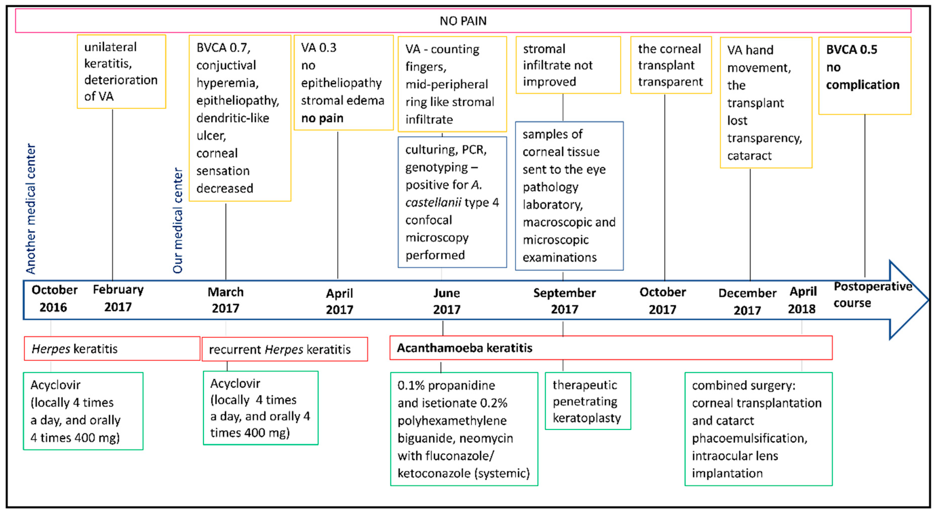
Figure 1. Timeline of the diagnostic procedures and treatment decisions in the patient’s case.
Jcm 14 04763 g001
Figure 2. (A) Conjunctival hyperemia. (B) Subtle epithelial irregularities resembling a dendritic-like ulcer located in the lower corneal hemisphere. (C) Stromal edema. (D) Dense mid-peripheral ring-like stromal infiltrate. (E) Ring infiltration. (F) Extensive stromal infiltrate and epithelial defect. (G) Clear corneal transplant. (H) Graft lost transparency and developed cataracts.
Jcm 14 04763 g002
Figure 3. Gel showing the separation of DNA obtained in a PCR using primers JDP1 and JDP2 (450 bp product). Samples: M—standard, 1—control (A. castellanii), 2—negative control, 3—A. castellanii, 4—A. rhysodes, 5—A. polyphaga, 6—Hartmannella vermiformis, 7—strain IC1, 8–9—Naegleria sp., 10—sample AC54, 11—sample AC55, 12—sample AC60, 13—sample AC66. Obtained results confirmed the presence of Acanthamoeba sp. in sample AC55. Samples AC54, AC60, and AC66 belong to an independent study.
Jcm 14 04763 g003
Figure 4. Ocular pathology examination of the patient’s cornea, demonstrating multiple intrastromal Acanthamoeba cysts ((A), arrows) and trophozoites ((B), arrows). Hematoxylin and eosin staining: panel (A)—magnification 40×; panel (B)—magnification 400×.
Jcm 14 04763 g004
Figure 5. Clear corneal transplant with IOL (intraocular lens) following cataract phacoemulsification.
Jcm 14 04763 g005
Table 1. Results of the genotyping of Acanthamoeba sp. from water and clinical samples.
SamplingIsolate, Accession No.Published Sequences in GeneBank
Accession No.Sampling, IsolateRegion of OriginReferences
Corneal scrapingAc55, KP120880GQ889265Liver of a Temminck’s tragopan, Acanthamoeba sp., genotype: T4
CDCV600
USAVisvesvara et al., 2010 [36]
KF318460Corneal surface tissue,
Acanthamoeba sp.,
1 FRC-2013
BrazilMafra et al., 2013 [37]
EU377583Biofilm, Acanthamoeba sp.,
CRIB53
SwitzerlandCorsaro et al., 2009 [38]
DQ087296Contact lenses and contact lens case, Acanthamoeba sp.,
S6
FranceYera et al., 2008 [39]
DQ087297Corneal scraping,
Acanthamoeba sp.,
222BAL
FranceYera et al., 2007 [40]
Disclaimer/Publisher’s Note: The statements, opinions and data contained in all publications are solely those of the individual author(s) and contributor(s) and not of MDPI and/or the editor(s). MDPI and/or the editor(s) disclaim responsibility for any injury to people or property resulting from any ideas, methods, instructions or products referred to in the content.