Article

Stereotactically Guided Microsurgical Approach for Deep-Seated Eloquently Located Lesions

by
Jun Thorsteinsdottir
1,*,
Sebastian Siller
1,2,
Biyan Nathanael Harapan
1,
Robert Forbrig
3,
Jörg-Christian Tonn
1,
Tobias Greve
1,
Stefanie Quach
1,4 and
Christian Schichor
1
1
Department of Neurosurgery, LMU University Hospital, LMU Munich, 81377 Munich, Germany
2
Department of Neurosurgery, University Hospital Regensburg, 93053 Regensburg, Germany
3
Institute of Neuroradiology, LMU University Hospital, LMU Munich, 81377 Munich, Germany
4
Department of Neurosurgery, Evangelisches Klinikum Bethel, Bielefeld University, 33617 Bielefeld, Germany
*
Author to whom correspondence should be addressed.
J. Clin. Med. 2025, 14(12), 4175; https://doi.org/10.3390/jcm14124175
Submission received: 25 April 2025 / Revised: 8 June 2025 / Accepted: 10 June 2025 / Published: 12 June 2025

Abstract

:
Background/Objectives: Advancements in neuronavigation and intraoperative imaging have made gross-total resection of deep-seated lesions more feasible. However, in eloquently located regions, brain shift can lead to unintentional damage of functionally critical tissue during the approach. This study analyzes the feasibility and outcomes of a stereotactically guided microsurgical approach supported by intraoperative CT (iCT) for such lesions. Methods: Patients with deep-seated, eloquently located lesions treated between 03/2017 and 04/2023 at the Department of Neurosurgery, Ludwig-Maximilians-University (LMU) Munich, Germany, were included. Frame-based, image-guided stereotaxy was used for trajectory planning and catheter placement, verified by iCT. Microsurgical resection was conducted along the catheter trajectory using 2 mm conical blade retractors and continuous neurophysiological monitoring. Postoperative MRI assessed the extent of resection. Neurological outcomes were evaluated postoperatively, at 6 weeks, and at long-term follow-up in 12/2023. Results: A total of 12 patients were treated using the stereotactically guided microsurgical approach described in this study. In all cases, the implanted catheter precisely matched the preoperative trajectory, as confirmed by fused iCT data. Median durations were 23 min for stereotaxy and 3 h 7 min for microsurgery. Complete resection was achieved in all cases. One patient experienced transient hemiparesis and aphasia, both of which were fully resolved. All other patients showed neurological improvement or remained seizure-free at long-term follow-up. Conclusions: In selected cases, a stereotactically guided microsurgical approach with iCT enabled intraoperative localization of the target with high spatial accuracy and without immediate procedure-related complications in this limited cohort. Our findings support the feasibility of the technique; however, conclusions regarding clinical efficacy or broader applicability are limited by the small sample size and non-comparative study design.

1. Introduction

Modern neurosurgical technical armamentarium, including neuronavigation, ultrasound and intraoperative MRI or CT, allows a safe resection of deep-seated lesions in the majority of cases. However, in selected cases, especially small lesions located in the basal ganglia, thalamus or mesencephalon, microsurgical approaches can be challenging. The pitfalls are (1) parenchymal damage caused by localized pressure of retractor systems, (2) white matter tract dissection in functionally relevant locations and (3) aberrancies of the trajectory in the depth [1,2,3,4]. Localized pressure of retractor systems could lead to perioperative morbidity, including venous infarction and cerebral edema, resulting in focal neurological deficits or seizures [5,6]. Additionally, in cases of small lesions, spatial disorientation—particularly in depth—can increase the risk of inadvertently affecting functionally important brain areas during surgery. Therefore, technical advances in minimally invasive approaches, including tubular brain retraction and endoscopy-assisted surgeries, have been introduced to create a safer and smaller trajectory [7,8,9,10,11]. Advanced multimodal neuroimaging has been established to be incorporated into neuronavigation for minimizing trajectory aberrancies in depth [12,13,14]. Diffusion-tensor-imaging (DTI) fibertracking has been proven to be useful for planning the surgical approach for deep-seated lesions adjacent to the corticospinal tract [12,13]. Furthermore, intraoperative image guidance by intraoperative ultrasound navigation has improved depth orientation, enhancing surgical precision [15]. However, these technical features are not available in every neurosurgical center. A stereotactic approach offers a precise alternative for targeting deep-seated eloquent lesions while minimizing functional risk. In our center, we have developed significant expertise in stereotactic procedures, characterized by a very low rate of perioperative morbidity and reduced surgical time [16]. Stereotaxy is of advantage compared to neuronavigation in providing a trajectory with very high accuracy, avoiding brain shift caused by craniotomy and subsequent loss of cerebrospinal fluid (CSF). Especially in deep-seated eloquent regions, minor errors in the angulation of the approach could induce severe neurological deficits [17]. While stereotactic procedures are commonly used to obtain small tissue samples, they typically do not facilitate the resection of lesions.
In consideration of the benefits and limitations of the mentioned techniques, we sought to optimize the surgical strategy for resection of deeply-seated eloquently located lesions by combining frame-based stereotaxy—to define the approach by positioning a stereotactically placed catheter with its tip pointing at the lesion—and frameless microsurgery for removal of the lesion. This study aims to investigate the surgical and neurological outcome in a series of selected patients harboring deep-seated lesions located in functionally relevant areas using this combined stereotactically catheter-guided microsurgical approach.

2. Materials and Methods

2.1. Patient Selection and Study Design

All procedures involving human participants adhered to the ethical standards set by the institutional and/or national research ethics committees, as well as the 1964 Declaration of Helsinki and its subsequent revisions or equivalent ethical guidelines. Informed consent was obtained from all participants and/or their legal representatives. The study received approval from the Institutional Review Board of Ludwig-Maximilians-University Munich (Project #18-098) and complied fully with its regulations concerning informed consent.
We conducted a single-center retrospective study to investigate the clinical and surgical outcomes in all consecutive cases operated upon with this technique between March 2017 and April 2023 (Table 1).
Demographic information, case records, radiological images and operative outcomes were analyzed. All patients received pre-operative MRI for stereotaxy and neuronavigation (GE signa HDx 3T scanner), including T1, T2, T2 *, FIESTA images (1 mm slice thickness), contrast-enhanced T1, MR angiography and diffusion-tensor-imaging. Postoperative CT scan was performed on day 1 after surgery. Extent of resection was assessed by MRI during in-hospital stay (except for one patient who suffered from a pulmonary embolism and received a CT). Deviation of intraoperative catheter position was matched with postoperative MRI using neuronavigation, and deviation was calculated in 3 points along the catheter. Craniotomy size, trajectory length and trajectory diameter were measured using iPlan 3.0 Cranial (Brainlab, Munich, Bavaria, Germany). Craniotomy size was assessed on postoperative CT, trajectory length was determined based on the implanted catheter and trajectory diameter was measured on postoperative MRI in axial, coronal and sagittal slices. Postoperative clinical outcome was assessed daily during hospitalization and after six weeks. Clinical outcome was assigned by modified Rankin score (mRS) and Glasgow Outcome Scale (GOS). Long-term follow-up with a particular focus on seizure outcomes was performed in 12/2023.

2.2. Surgical Technique

The protocol for surgical technique is illustrated in Figure 1. Frame-based multimodal image-guided stereotactic technique was used for trajectory planning connecting the cortical entry point and the surface of the lesion. For each patient, an optimal trajectory to the lesion was generated, avoiding contact with arterial or venous vessels and maintaining a minimal distance of 2 mm between the trajectory and any vessel. Subsequently, a catheter targeting the lesion was stereotactically implanted using this trajectory. Stereotactically localized CT angiography (0.6 mm slice-spacing, 1.46 mSv) was fused with preoperative MRA, T1- and T2-weighted MRI data. Trajectories were planned using Target@1.19 software (Brainlab, Munich, Bavaria, Germany). The catheter (1.3 mm ventricular catheter, Medtronic, Minneapolis, MN, USA) was implanted through a 5 mm skin incision, a 3 mm diameter burr hole, and secured with a microclip at dura level, and skin was closed using 4.0 prolene suture. After removal of the stereotactic frame, the patient was positioned on a carbon-made radiolucent surgical table (TruSystem 7500, Trumpf Medical, Saalfeld, Thüringen, Germany). The patient’s head was fixed in radiolucent head clamp (Mayfield, A-2002, Integra, Princeton, NJ, USA) [18], and a high-resolution unenhanced cranial CT (0.6 mm slice spacing, 1.05 mSv) was acquired using iCT to confirm the correct catheter position. ICT (Siemens SOMATOM Definition AS+, Siemens Healthineers, Munich, Bavaria, Germany) was obtained by a ”double room CT scanner“, as described earlier [19,20,21]. ICT data set was fused with preoperative multimodal imaging data, including diffusion-tensor-imaging using iPlan 3.0 Cranial (Brainlab, Munich, Bavaria, Germany). After an approximately 3 × 3 cm craniotomy, a 1.5 × 1.5 cm durotomy around the catheter and corticotomy of 5 mm, the minimally invasive approach was continued microscopically along the surface of the catheter. Retractors with conical blades with a 2 mm diameter (DORO®, Black Forest Medical group, Freiburg, Baden-Württemberg, Germany) were used for preparation. The trajectory was followed by standard navigation system during the approach to visualize potential brain shift. During resection, continuous intraoperative monitoring (IOM), including somatosensory (SEP) and motor evoked potentials (MEPs), was recorded in all cases as described previously [22]. Once the lesion was located, the stereotactically implanted catheter was removed and the lesion was resected. In case of cavernous malformations, we first bluntly entered the lesion with small, fine-tip bipolar forceps to evacuate blood and decompress the lesion. The cavernoma was then carefully separated from the surrounding tissue using suction and microdissectors, and in most cases, it was removed in a piecemeal manner. Hemostasis was performed with a long bayonet-style fine bipolar (SilverGlide Keyhole Maximum Access, Stryker, Fremont, CA, USA) and oxidized regenerated cellulose (Tabotamp, Ethicon Co. for Johnson & Johnson Medical, Piscataway, NJ, USA). Dural and craniotomy closure were performed as usual.

3. Results

3.1. Patient Characteristics

Among 1927 patients harboring supratentorial tumors and 93 patients harboring supratentorial cavernomas, a total of 0.6% of patients (2 male, 10 female) with deep-seated and/or highly eloquent located cavernous malformations were operated using this stereotactically guided microsurgical approach. Characteristics of patients and lesions are given in Table 2. Median age was 36.5 years (range: 17.5–66.5 years). Lesions were located on the right/left hemisphere in 7/5 patients, and in the basal ganglia in three patients (25%), cella media in two patients (16.7%), thalamus in one patient (8.3%), insula in one patient (8.3%), superior temporal gyrus in two patients (16.7%), cerebral peduncle in one patient (8.3%), trigonum in one patient (8.3%) and splenium in one patient (8.3%). Initial symptoms were focal seizures in six patients (50.0%), hypesthesia in five patients (41.6%), aphasia in three patients (25%) and hemiparesis in two patients (16.7%). The median size of the lesion was 20 mm (range: 11–31 mm), and the median depth of the lesion from the cortical surface was 42 mm (range: 10–62 mm). Histology revealed cavernous malformation in ten patients (83.3%), pilocytic astrocytoma (8.3%) in one patient and meningioma in one patient (8.3%).

3.2. Surgical Results Using the Stereotactically Guided Neuronavigated Microsurgical Approach

Lesion- and surgery-associated parameters are presented in Table 3. Median craniotomy size was 35 mm (range: 27–45 mm), median trajectory length was 62.7 mm (range: 43.2–72.1 mm) and mean trajectory diameter was 6.0 mm ± 1.2 mm (range: 4.2–9.4 mm) in postoperative MRI. The median duration of stereotaxy was 23 min, and the mean duration of microsurgery was 3 h 7 min. Median blood loss was 100 mL (range: 30–300 mL), and median hospital stay was 7 days (range: 6–15 days). After stereotactic target achievement by catheter implantation, the skin was closed, and iCT confirmed the correct placement of the catheter targeting the lesion. A standard microsurgical approach was then initiated using intraoperative neuronavigation with ultrasound as a backup system to confirm the expected brain shift. However, the implanted catheter pointed with its tip at the lesion in all cases and was not affected by brain shift. Complete removal of the lesion was achieved in all patients verified by postoperative MRI. In one case, a patient with a cavernoma located in the splenium was first operated by a standard microsurgical approach. As the lesion was not entirely removed in the postoperative MRI, the patient was successfully operated on using the stereotactic approach (Supplementary Figure S1). Postoperatively, no cases of epidural, subdural or intraparenchymal bleeding were observed. There were no cases in which this method was unexpectedly deemed unsuitable during the operation. Moreover, there were no instances where conversion to a standard microsurgical approach involving a larger craniotomy was necessary. One adverse event occurred in one patient suffering from pulmonary embolism on the first postoperative day, which was treated with anticoagulation.

3.3. Neurological Outcome

Neurological outcome in our patient cohort is shown in Table 1. Immediately after surgery, eight symptomatic patients (mRS score = 1) presented without any neurological deficit (mRS score = 0). One asymptomatic patient with a cavernoma in the basal ganglia and one asymptomatic patient with a meningioma in the trigonum remained without any neurological deficit. One patient with a cavernous malformation in the left thalamus suffered from transient right hemiparesis (2/5) and hypesthesia in the right arm (mRS score = 3). Postoperative CT did not reveal any haemorrhage or infarction. The symptoms were attributed to perioperative manipulation in close vicinity to the corticospinal tract. Until discharge, symptoms resolved completely and were not detectable in neurological examination at the last FU (mRS score = 0). One patient with a partially cystic pilocytic astrocytoma located in the thalamus, cerebral crus and internal capsule, who was pretreated with interstitial brachytherapy, suffered preoperatively from left hemiparesis (mRS score = 3). Immediately after surgery and until the last FU, hemiparesis improved from initially 3/5 proximal and 1/5 distal to proximal 5/5 and distal 3/5 (mRS score = 2).
In total, after a median follow-up time of 34.5 months (range: 4.5–52.0 months), 11 patients did not show any neurological deficit and one patient with preoperative hemiparesis improved neurologically. One patient had one focal seizure after cessation of anticonvulsive therapy but remained asymptomatic until the last FU. None of the patients showed any new deficit, and all patients were seizure-free at long-term follow-up.

3.4. Case Report

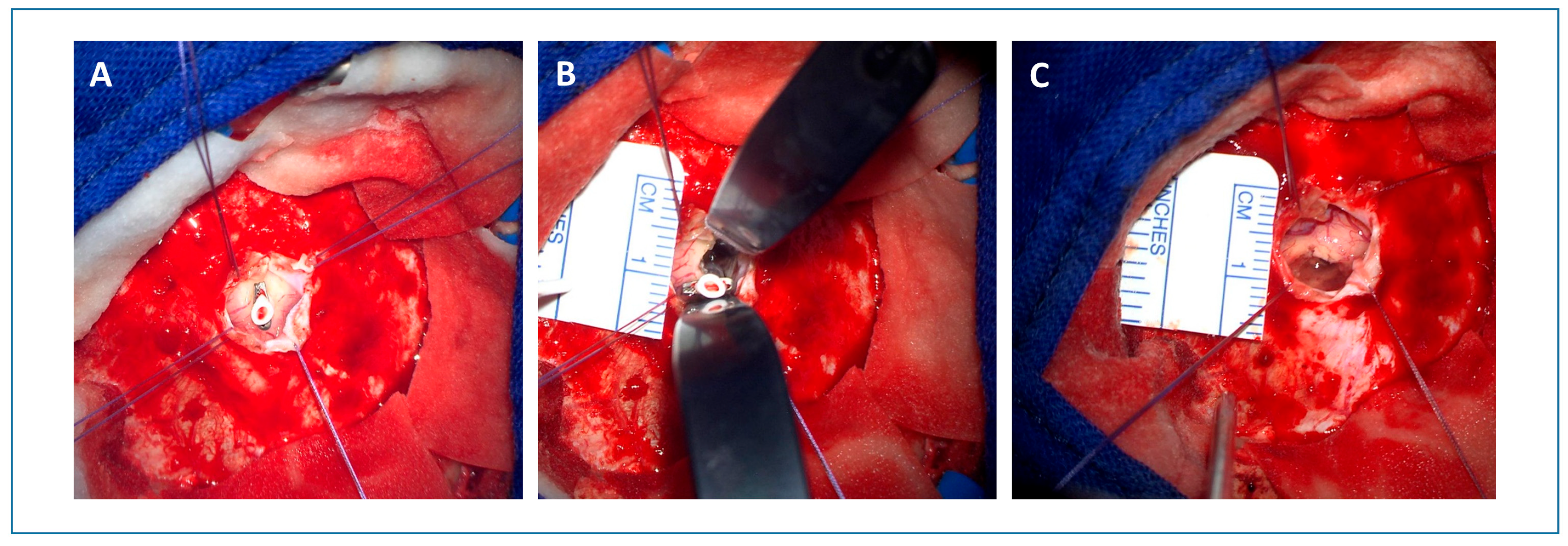
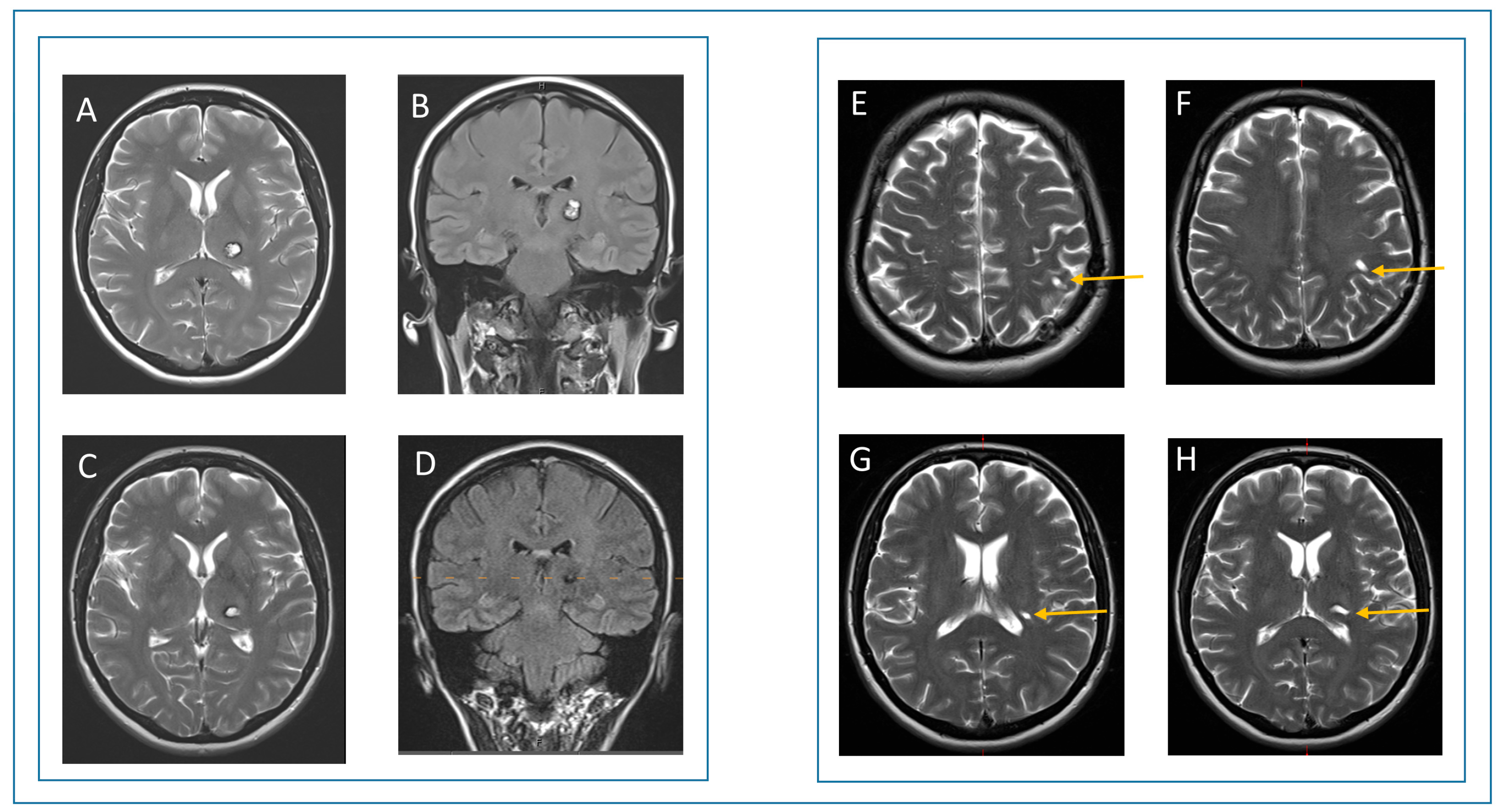
A 52-year-old female presented with acute hemihypesthesia in the right face and arm accompanied by a motor deficit of the right leg and a decrease in her fine motor skills in the right hand (patient #11, see Table 1 and Table 2). The symptoms resolved partially within a month. The first MRI revealed a partially hemorrhagic lesion suspicious of cavernous malformation in the left thalamus (Supplementary Figure S2). The patient was referred to our department for a second opinion after six months. Based on the evidence of space-occupying hemorrhage and the symptoms, the indication for surgery was set. Preoperative MRI showed a cystic, slightly contrast-enhancing lesion with previous hemorrhages in the left thalamus (Figure 2). A catheter targeting the lesion was stereotactically implanted. Post-implantation intraoperative CT confirmed a correct catheter position. Thereafter, a microsurgical approach was performed along the surface of the catheter (Figure 3). Intraoperative monitoring, including MEP and SEP, showed no abnormalities. Postoperatively, the patient presented with a transient hemiparesis on the right, which resolved completely after 7 days, including the pre-operative deficit. Postoperative CT showed neither new nor remaining hemorrhage. Control MRI after three months showed a complete removal of the cavernous malformation (Figure 4). At the last follow-up after 2 years, the patient did not show any neurological deficit.

4. Discussion

Recent technical innovations in microneurosurgery have significantly improved access to deep-seated cerebral lesions by enabling more effective and less invasive surgical corridors. Techniques such as transcortical approaches, cortical/subcortical electrical stimulation and awake surgeries have enhanced surgical safety and efficacy by tailoring the surgical corridor to the functional topography [23,24]. However, the approach to deep-seated, eloquently located lesions remains a considerable challenge.
Intraoperative brain deformations during surgery can lead to severe neurological deficits, especially in eloquent regions, where deviations of just a few millimeters can profoundly affect the patient’s neurological outcome. This precision is particularly crucial in procedures such as deep brain stimulation for Parkinson’s disease or stereoelectroencephalography for identifying epileptogenic foci, where therapeutic success depends on the accurate placement of electrodes in the correct anatomical targets [25,26]. It has been demonstrated that opening the dura mater can cause displacement of neural structure especially due to the loss of the physiological negative intracranial pressure, air inflow, and loss of CSF [25]. Reports show substantial variability in deformation errors ranging from 15 to 24 mm for cortical displacements [27,28] and 8 to 31 mm for tumor regions using neuronavigation-assisted microsurgery [29].
To address these challenges, we employed a stereotactically guided microsurgical approach that combines precise lesion localization within eloquent areas and allows bimanual resection of the lesion under intraoperative monitoring to preserve functional outcomes.
As early as the 1990s, Hassenbusch et al. used stereotactic markers to visualize the margins of brain tumors, primarily to delineate tumor borders in an era where intraoperative imaging was not available [30]. In the past decade, intraoperative imaging systems have received significant attention. In particular, intraoperative ultrasound (iUS) integrated with neuronavigation data has proven useful for real-time intraoperative representation of large, eloquently located tumors, assessing the extent of resection [31,32,33]. However, iUS is highly operator-dependent, challenging to standardize and may require repeated examinations to detect small deep-seated lesions [31,32,33]. Alternatively, intraoperative MRI (iMRI) provides valuable information on the extent of tumor resection, which is crucial for postoperative outcomes [27,34]. However, iMRI is time-consuming and not available in every neurosurgical center [27,34]. iUS, iMRI, and fluorophores such as 5-ALA or Indocyanine green (ICG) are among the most advanced tools for real-time intraoperative visualization. While these technologies are crucial for large, infiltrating lesions to assess the extent of resection, our stereotactic technique may be advantageous for maintaining the correct trajectory during dissection to small, well-circumscribed lesions without significant edema.
In our study, iCT was used for stereotactic catheter implantation and verification of the correct catheter position. To minimize radiation exposure, our neuroradiologists developed a new iCT protocol using an automated tube current modulation and voltage selection, reducing the radiation dose from 2.97 mSv to 1.46 mSv [35]. In the future, MRI-based stereotaxy may be a viable alternative to avoid radiation exposure.
We intentionally separated the stereotactic catheter implantation from microsurgical resection in order to minimize the risk of catheter dislocation during patient repositioning or craniotomy by employing the following individual measures: (1) stereotactic technique to maximize accuracy in targeting the lesion; (2) verification of correct catheter placement using intraoperative CT after removal of the stereotactic frame; (3) facilitating bimanual manipulation for lesion resection under intraoperative stimulation and monitoring and (4) adaptation of the aperture diameter/location in depth during resection. Importantly, we did not observe any catheter dislocation, as it was sufficiently secured.
In minimally invasive neurosurgery, several techniques have been developed to enhance surgical precision while reducing patient morbidity. Each approach offers distinct advantages and presents specific challenges. Awake surgery approaches allow for continuous monitoring of neurological functions during surgery with immediate adjustments based on patients’ responses, which can enhance neurological outcomes. Although awake surgeries do not demand specialized equipment, the necessity for a highly skilled multidisciplinary team can negate potential cost savings. Endoscopic or tube systems, combined with conventional microscopy, enable minimally invasive approaches to deep-seated lesions with improved illumination and magnification [7,8,9,11,36,37,38]. On the downside, many endoscopic systems may restrict instrument maneuverability, potentially complicating surgical procedures [7]. Therefore, surgeons may require specialized training to effectively use endoscopic equipment. Endoscopic systems and specialized instruments represent a financial investment, though they may be more cost-effective compared to the acquisition and maintenance of neuronavigation systems and intraoperative CT. Tubular retractor systems provide increased visualization of deep intra-axial sites, with minimal disruption to the functional tissue compared to traditional plate retraction systems [11,36,37]. Also, self-retaining retractors create a stable operative field, potentially reducing operative time. Depending solely on microscopic visualization through the tube may not provide comprehensive views of the surgical field. Also, similarly to endoscopic systems, the confined space within the tube can limit instrument movement and complicate lesion resection [9,11]. Due to low costs and simple technology, tubular retractor systems may be more accessible in various surgical settings.
In our study, we used a retractor system with conical blades (2 mm diameter), which simultaneously minimized the risk of iatrogenic tissue and vascular damage from brain retraction while allowing bimanual manipulation and adjustment of the aperture diameter in depth. With a mean size of 6.0 ± 1.2 mm, the trajectory diameter in postoperative MRI was smaller than endoscopic or tubular approaches with a mean cannulation diameter of 9 mm [7,36,37]. Although our procedure, including stereotaxy planning, intraoperative CT and microsurgical resection, required slightly more surgery time than former studies, no deviations from the planned trajectory or uncertainties of the intraoperative approach were encountered. A recent study involving five patients demonstrated the feasibility of resecting deep-seated arteriovenous malformations (AVM) using stereotactically guided tubular retractor systems [39]. However, the mean maximum AVM nidal diameter was 8.2 mm, limiting the procedures to smaller AVMs but with favorable neurological and surgical outcomes [39]. A comparison with other surgical techniques is illustrated in Table 4. In recent studies, augmented reality (AR) was able to improve surgical planning, refine neuronavigation and reduce operation time by overlaying computer-generated images onto the user’s real-world visual field. Especially with regard to the extent of resection, AR offers a better identification of tumor boarders. Also, AR enhances surgical planning for skull-base lesions by intraoperative navigation-integrated projections onto the surface of the skull [40]. However, studies for stereotactic procedures showed that AR-guided or ARN-assisted operations may be feasible for large targets as the accuracy error is still 1–3 mm, and most AR systems do not prevent accuracy loss due to brain shift [41].
Limitations of the study are the retrospective design and the small sample size as this was a practicality and feasibility study. Our study included 10 patients with cavernomas, 1 patient with pilocytic astrocytoma and 1 patient with a meningioma. Resection of the cavernomas and meningioma was more straightforward due to clear visualization and tactile differentiation from normal brain tissue compared to the pilocytic astrocytoma. Nonetheless, we acknowledge that lesion consistency, vascularity and border definition—which vary significantly between, for example, cavernomas and gliomas—could affect technical reproducibility and feasibility. The haptic differences among these lesions emphasize that while the technical access (trajectory planning, stereotactic guidance and microsurgical technique) remains constant, intraoperative adaptability based on tactile feedback is essential. We now note that reproducibility of this technique is likely highest in well-circumscribed lesions such as cavernomas or deep-seated, encapsulated tumors and may require further refinement or adaptation in other histologies.
Evidently, this technique necessitates specialized neurosurgical equipment for the operative workflow, including stereotaxy, intraoperative CT and microsurgery. Nevertheless, in experienced centers, this approach may be reasonable in selected cases. Given the potential for severe neurological deficits resulting from millimeter-scale deviation caused by brain shift, we believe that this approach combining frame-based stereotaxy and frameless catheter-guided microsurgery is justified. Importantly, this study should be understood as a feasibility study. The selected cohort represents a highly specific and narrowly defined subset of patients for whom the stereotactically guided microsurgical technique was judged to be particularly beneficial. We acknowledge the limitations that arise from this selective approach, including its impact on the generalizability of our findings. It is also important to clarify that this feasibility study does not demonstrate the superiority of the technique over other approaches, and no such claims should be inferred from the results. We believe that demonstrating safe and effective outcomes in this highly selected cohort provides valuable insights for other centers managing similarly challenging cases. These findings may also serve as a foundation for future prospective or comparative studies aimed at further evaluating the utility and generalizability of this approach. Further research with larger, more diverse cohorts is necessary to validate the efficacy and safety of this approach in a multicenter prospective study.

5. Conclusions

In summary, we implemented a frame-based multimodal imaging-guided stereotactic technique for trajectory planning and catheter implantation targeting the lesion. Simultaneously, intraoperative monitoring, including motor- and sensory-evoked potentials, enabled real-time assessment of neurological function during microsurgical resection of the lesion. In this small cohort, the technique enabled accurate trajectory planning with no intraoperative deviations documented and parenchymal disruption appeared limited based on postoperative imaging and clinical course. Neurological outcomes were favorable, and seizure status remained stable or improved in those with a history of epilepsy. This study demonstrates the feasibility of this stereotactically guided microsurgical approach in a highly selected patient cohort. While initial outcomes appear encouraging, conclusions regarding clinical efficacy are limited by the small, heterogeneous cohort and the non-comparative study design.

Supplementary Materials

The following supporting information can be downloaded at: https://www.mdpi.com/article/10.3390/jcm14124175/s1, Figure S1: Preoperative MRI, MRI after first surgery and MRI after second surgery (stereotactically guided approach); Figure S2: Initial MRI at diagnosis (case report).

Author Contributions

Conceptualization, J.T., J.-C.T. and C.S.; formal analysis, J.T., R.F., T.G. and S.Q.; investigation, J.T., S.S., B.N.H., R.F., T.G. and S.Q.; methodology, J.T., S.S., J.-C.T. and C.S.; supervision, J.-C.T. and C.S.; visualization, J.T., B.N.H. and R.F.; writing—original draft, J.T., B.N.H. and R.F.; writing—review and editing, J.T. and B.N.H. All authors have read and agreed to the published version of the manuscript.

Funding

This research received no external funding.

Institutional Review Board Statement

The study was conducted in accordance with the Declaration of Helsinki and approved by the Institutional Review Board of the Ludwig-Maximilans-University Munich, Munich (Ethikkommission der Medizinischen Fakultät der Ludwig-Maximilians-Universität München; project # 18-098; 8 March 2018).

Informed Consent Statement

This retrospective study was approved by the Institutional Review Board of the Ludwig-Maximilians-University Hospital, Munich (project #18-098). The requirement for informed consent was waived due to the use of anonymized data and the study’s retrospective design.

Data Availability Statement

Authors can confirm that all relevant data are included in the article. Dataset(s) derived from public resources and made available with the article (references).

Conflicts of Interest

The authors declare no conflicts of interest.

Abbreviations

The following abbreviations are used in this manuscript:
iCTIntraoperative computer tomography
DTIDiffusion-tensor-imaging
mRSmodified Rankin score
GOSGlasgow Outcome Scale
CSFCerebrospinal fluid
iUSIntraoperative ultrasound
iMRIIntraoperative magnetic resonance imaging
GTRgross total resection
STRsubtotal resection
NTRnear total resection
DVTdeep vein thrombosis

References

  1. Gassie, K.; Wijesekera, O.; Chaichana, K.L. Minimally invasive tubular retractor-assisted biopsy and resection of subcortical intra-axial gliomas and other neoplasms. J. Neurosurg. Sci. 2018, 62, 682–689. [Google Scholar] [CrossRef] [PubMed]
  2. Iyer, R.; Chaichana, K.L. Minimally Invasive Resection of Deep-seated High-grade Gliomas Using Tubular Retractors and Exoscopic Visualization. J. Neurol. Surg. A Cent. Eur. Neurosurg. 2018, 79, 330–336. [Google Scholar] [CrossRef]
  3. Jackson, C.; Gallia, G.L.; Chaichana, K.L. Minimally Invasive Biopsies of Deep-Seated Brain Lesions Using Tubular Retractors Under Exoscopic Visualization. J. Neurol. Surg. A Cent. Eur. Neurosurg. 2017, 78, 588–594. [Google Scholar] [CrossRef] [PubMed]
  4. Chaichana, K.L.; Vivas-Buitrago, T.; Jackson, C.; Ehresman, J.; Olivi, A.; Bettegowda, C.; Quinones-Hinojosa, A. The Radiographic Effects of Surgical Approach and Use of Retractors on the Brain After Anterior Cranial Fossa Meningioma Resection. World Neurosurg. 2018, 112, e505–e513. [Google Scholar] [CrossRef]
  5. Rosenorn, J.; Diemer, N.H. Reduction of regional cerebral blood flow during brain retraction pressure in the rat. J. Neurosurg. 1982, 56, 826–829. [Google Scholar] [CrossRef] [PubMed]
  6. Zhong, J.; Dujovny, M.; Perlin, A.R.; Perez-Arjona, E.; Park, H.K.; Diaz, F.G. Brain retraction injury. Neurol. Res. 2003, 25, 831–838. [Google Scholar] [CrossRef]
  7. Newman, W.C.; Engh, J.A. Stereotactic-Guided Dilatable Endoscopic Port Surgery for Deep-Seated Brain Tumors: Technical Report with Comparative Case Series Analysis. World Neurosurg. 2019, 125, e812–e819. [Google Scholar] [CrossRef]
  8. Bander, E.D.; Jones, S.H.; Kovanlikaya, I.; Schwartz, T.H. Utility of tubular retractors to minimize surgical brain injury in the removal of deep intraparenchymal lesions: A quantitative analysis of FLAIR hyperintensity and apparent diffusion coefficient maps. J. Neurosurg. 2016, 124, 1053–1060. [Google Scholar] [CrossRef]
  9. Cohen-Gadol, A.A. Minitubular transcortical microsurgical approach for gross total resection of third ventricular colloid cysts: Technique and assessment. World Neurosurg. 2013, 79, 207.e7–207.e10. [Google Scholar] [CrossRef]
  10. Marenco-Hillembrand, L.; Alvarado-Estrada, K.; Chaichana, K.L. Contemporary Surgical Management of Deep-Seated Metastatic Brain Tumors Using Minimally Invasive Approaches. Front. Oncol. 2018, 8, 558. [Google Scholar] [CrossRef]
  11. Shapiro, S.Z.; Sabacinski, K.A.; Mansour, S.A.; Echeverry, N.B.; Shah, S.S.; Stein, A.A.; Snelling, B.M. Use of Vycor Tubular Retractors in the Management of Deep Brain Lesions: A Review of Current Studies. World Neurosurg. 2020, 133, 283–290. [Google Scholar] [CrossRef] [PubMed]
  12. Flores, B.C.; Whittemore, A.R.; Samson, D.S.; Barnett, S.L. The utility of preoperative diffusion tensor imaging in the surgical management of brainstem cavernous malformations. J. Neurosurg. 2015, 122, 653–662. [Google Scholar] [CrossRef] [PubMed]
  13. Holodny, A.I.; Ollenschleger, M.D.; Liu, W.C.; Schulder, M.; Kalnin, A.J. Identification of the corticospinal tracts achieved using blood-oxygen-level-dependent and diffusion functional MR imaging in patients with brain tumors. AJNR Am. J. Neuroradiol. 2001, 22, 83–88. [Google Scholar] [PubMed]
  14. Holodny, A.I.; Schwartz, T.H.; Ollenschleger, M.; Liu, W.C.; Schulder, M. Tumor involvement of the corticospinal tract: Diffusion magnetic resonance tractography with intraoperative correlation. J. Neurosurg. 2001, 95, 1082. [Google Scholar] [CrossRef] [PubMed]
  15. Bastos, D.C.A.; Juvekar, P.; Tie, Y.; Jowkar, N.; Pieper, S.; Wells, W.M.; Bi, W.L.; Golby, A.; Frisken, S.; Kapur, T. Challenges and Opportunities of Intraoperative 3D Ultrasound With Neuronavigation in Relation to Intraoperative MRI. Front. Oncol. 2021, 11, 656519. [Google Scholar] [CrossRef]
  16. Katzendobler, S.; Do, A.; Weller, J.; Dorostkar, M.M.; Albert, N.L.; Forbrig, R.; Niyazi, M.; Egensperger, R.; Thon, N.; Tonn, J.C.; et al. Diagnostic Yield and Complication Rate of Stereotactic Biopsies in Precision Medicine of Gliomas. Front. Neurol. 2022, 13, 822362. [Google Scholar] [CrossRef]
  17. Mohammadi, A.; Ahmadian, A.; Azar, A.D.; Sheykh, A.D.; Amiri, F.; Alirezaie, J. Estimation of intraoperative brain shift by combination of stereovision and doppler ultrasound: Phantom and animal model study. Int. J. Comput. Assist. Radiol. Surg. 2015, 10, 1753–1764. [Google Scholar] [CrossRef]
  18. Ardeshiri, A.; Radina, C.; Edlauer, M.; Ardeshiri, A.; Riepertinger, A.; Nerlich, A.; Tonn, J.C.; Winkler, P.A. Evaluation of new radiolucent polymer headholder pins for use in intraoperative computed tomography. J. Neurosurg. 2009, 111, 1168–1174. [Google Scholar] [CrossRef]
  19. Schichor, C.; Rachinger, W.; Morhard, D.; Zausinger, S.; Heigl, T.J.; Reiser, M.; Tonn, J.C. Intraoperative computed tomography angiography with computed tomography perfusion imaging in vascular neurosurgery: Feasibility of a new concept. J. Neurosurg. 2010, 112, 722–728. [Google Scholar] [CrossRef]
  20. Schichor, C.; Terpolilli, N.; Thorsteinsdottir, J.; Tonn, J.C. Intraoperative Computed Tomography in Cranial Neurosurgery. Neurosurg. Clin. N. Am. 2017, 28, 595–602. [Google Scholar] [CrossRef]
  21. Schnell, O.; Morhard, D.; Holtmannspotter, M.; Reiser, M.; Tonn, J.C.; Schichor, C. Near-infrared indocyanine green videoangiography (ICGVA) and intraoperative computed tomography (iCT): Are they complementary or competitive imaging techniques in aneurysm surgery? Acta Neurochir. 2012, 154, 1861–1868. [Google Scholar] [CrossRef] [PubMed]
  22. Szelenyi, A.; Langer, D.; Kothbauer, K.; De Camargo, A.B.; Flamm, E.S.; Deletis, V. Monitoring of muscle motor evoked potentials during cerebral aneurysm surgery: Intraoperative changes and postoperative outcome. J. Neurosurg. 2006, 105, 675–681. [Google Scholar] [CrossRef]
  23. Spena, G.; Garbossa, D.; Panciani, P.P.; Griva, F.; Fontanella, M.M. Purely subcortical tumors in eloquent areas: Awake surgery and cortical and subcortical electrical stimulation (CSES) ensure safe and effective surgery. Clin. Neurol. Neurosurg. 2013, 115, 1595–1601. [Google Scholar] [CrossRef]
  24. Zanello, M.; Meyer, B.; Still, M.; Goodden, J.R.; Colle, H.; Schichor, C.; Bello, L.; Wager, M.; Smits, A.; Rydenhag, B.; et al. Surgical resection of cavernous angioma located within eloquent brain areas: International survey of the practical management among 19 specialized centers. Seizure 2019, 69, 31–40. [Google Scholar] [CrossRef]
  25. Beggio, G.; Raneri, F.; Rustemi, O.; Scerrati, A.; Zambon, G.; Piacentino, M. Techniques for pneumocephalus and brain shift reduction in DBS surgery: A review of the literature. Neurosurg. Rev. 2020, 43, 95–99. [Google Scholar] [CrossRef] [PubMed]
  26. Brimley, C.; Shimony, N. Accuracy and Utility of Frameless Stereotactic Placement of Stereoelectroencephalography Electrodes. World Neurosurg. 2023, 180, e226–e232. [Google Scholar] [CrossRef]
  27. Nimsky, C.; Ganslandt, O.; Cerny, S.; Hastreiter, P.; Greiner, G.; Fahlbusch, R. Quantification of, visualization of, and compensation for brain shift using intraoperative magnetic resonance imaging. Neurosurgery 2000, 47, 1070–1079; discussion 1079–1080. [Google Scholar] [CrossRef] [PubMed]
  28. Reinges, M.H.; Nguyen, H.H.; Krings, T.; Hutter, B.O.; Rohde, V.; Gilsbach, J.M. Course of brain shift during microsurgical resection of supratentorial cerebral lesions: Limits of conventional neuronavigation. Acta Neurochir. 2004, 146, 369–377; discussion 377. [Google Scholar] [CrossRef]
  29. Trantakis, C.; Tittgemeyer, M.; Schneider, J.P.; Lindner, D.; Winkler, D.; Strauss, G.; Meixensberger, J. Investigation of time-dependency of intracranial brain shift and its relation to the extent of tumor removal using intra-operative MRI. Neurol. Res. 2003, 25, 9–12. [Google Scholar] [CrossRef]
  30. Hassenbusch, S.J.; Anderson, J.S.; Pillay, P.K. Brain tumor resection aided with markers placed using stereotaxis guided by magnetic resonance imaging and computed tomography. Neurosurgery 1991, 28, 801–805; discussion 805–806. [Google Scholar] [CrossRef]
  31. Dixon, L.; Lim, A.; Grech-Sollars, M.; Nandi, D.; Camp, S. Intraoperative ultrasound in brain tumor surgery: A review and implementation guide. Neurosurg. Rev. 2022, 45, 2503–2515. [Google Scholar] [CrossRef]
  32. Mazzucchi, E.; La Rocca, G.; Hiepe, P.; Pignotti, F.; Galieri, G.; Policicchio, D.; Boccaletti, R.; Rinaldi, P.; Gaudino, S.; Ius, T.; et al. Intraoperative Integration of Multimodal Imaging to Improve Neuronavigation: A Technical Note. World Neurosurg. 2022, 164, 330–340. [Google Scholar] [CrossRef] [PubMed]
  33. Sass, B.; Zivkovic, D.; Pojskic, M.; Nimsky, C.; Bopp, M.H.A. Navigated Intraoperative 3D Ultrasound in Glioblastoma Surgery: Analysis of Imaging Features and Impact on Extent of Resection. Front. Neurosci. 2022, 16, 883584. [Google Scholar] [CrossRef] [PubMed]
  34. Sommer, B.; Kasper, B.S.; Coras, R.; Blumcke, I.; Hamer, H.M.; Buchfelder, M.; Roessler, K. Surgical management of epilepsy due to cerebral cavernomas using neuronavigation and intraoperative MR imaging. Neurol. Res. 2013, 35, 1076–1083. [Google Scholar] [CrossRef] [PubMed]
  35. Forbrig, R.; Geyer, L.L.; Stahl, R.; Thorsteinsdottir, J.; Schichor, C.; Kreth, F.W.; Patzig, M.; Herzberg, M.; Liebig, T.; Dorn, F.; et al. Radiation dose and image quality in intraoperative CT (iCT) angiography of the brain with stereotactic head frames. Eur. Radiol. 2019, 29, 2859–2867. [Google Scholar] [CrossRef]
  36. Gassie, K.; Alvarado-Estrada, K.; Bechtle, P.; Chaichana, K.L. Surgical Management of Deep-Seated Metastatic Brain Tumors Using Minimally Invasive Approaches. J. Neurol. Surg. A Cent. Eur. Neurosurg. 2019, 80, 198–204. [Google Scholar] [CrossRef]
  37. Takeuchi, K.; Nagata, Y.; Tanahashi, K.; Araki, Y.; Mizuno, A.; Sasaki, H.; Harada, H.; Ito, K.; Saito, R. Efficacy and safety of the endoscopic “wet-field” technique for removal of supratentorial cavernous malformations. Acta Neurochir. 2022, 164, 2587–2594. [Google Scholar] [CrossRef]
  38. Liu, S.; Wu, S.; Xie, T.; Yeh, Y.Y.; Li, C.; Liu, T.; Sun, C.; Yang, L.; Li, Z.; Yu, Y.; et al. Neuronavigation-Guided Transcortical-Transventricular Endoport-Assisted Endoscopic Resection for Thalamic Lesions: Preliminary Experience. World Neurosurg. 2022, 166, 19–27. [Google Scholar] [CrossRef]
  39. Achey, R.; Kashkoush, A.; Potter, T.; Davison, M.; Moore, N.Z.; Kshettry, V.R.; Bain, M. Surgical Resection of Deep-Seated Arteriovenous Malformations Through Stereotactically Guided Tubular Retractor Systems: A Case Series. Oper Neurosurg. 2023, 24, 499–506. [Google Scholar] [CrossRef]
  40. Hey, G.; Guyot, M.; Carter, A.; Lucke-Wold, B. Augmented Reality in Neurosurgery: A New Paradigm for Training. Medicina 2023, 59, 1721. [Google Scholar] [CrossRef]
  41. Satoh, M.; Nakajima, T.; Watanabe, E.; Kawai, K. Augmented Reality in Stereotactic Neurosurgery: Current Status and Issues. Neurol. Med. Chir. 2023, 63, 137–140. [Google Scholar] [CrossRef] [PubMed]
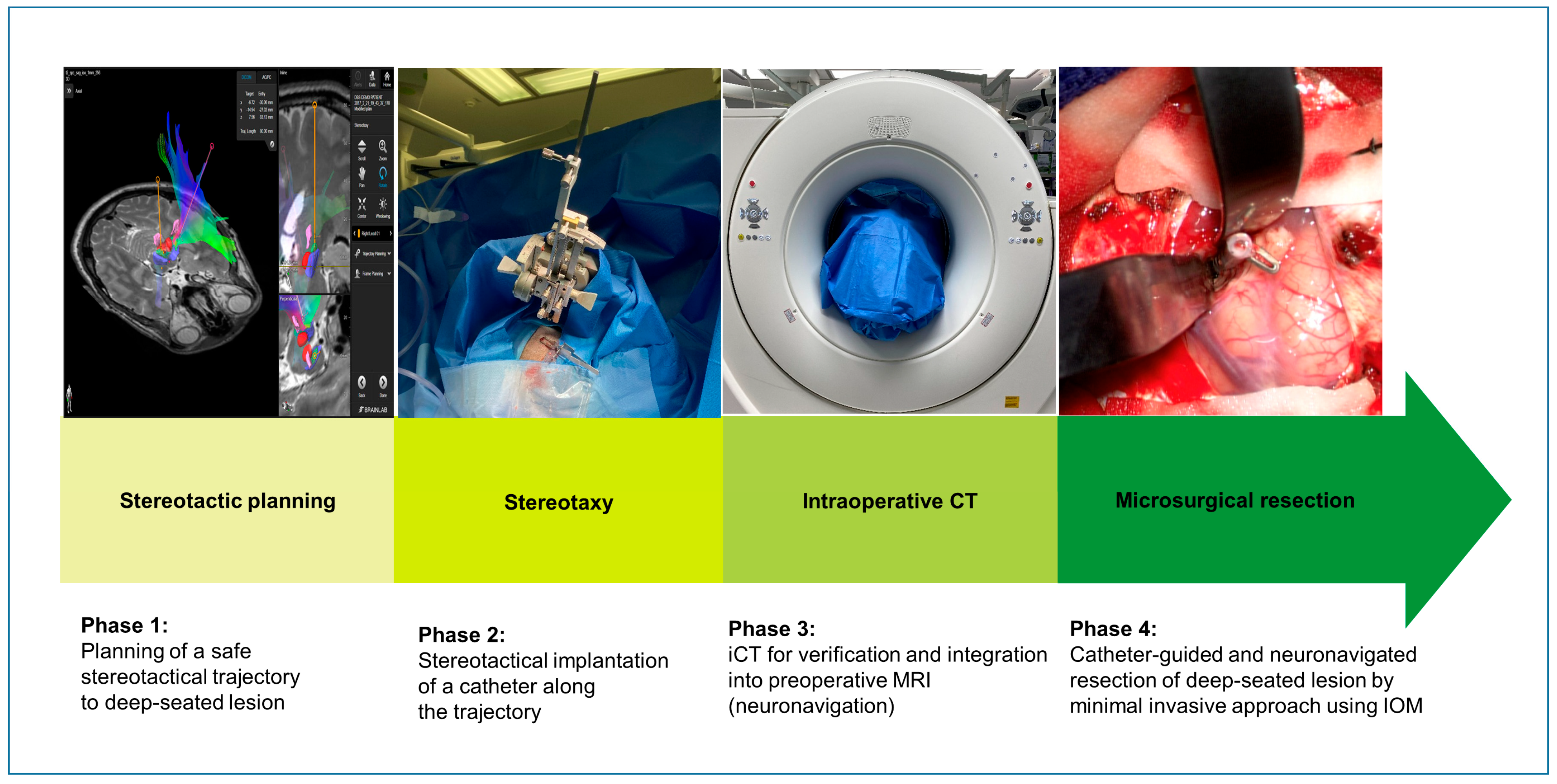
Figure 1. Workflow of the stereotactic catheter-guided neuronavigated approach. Phase 1 shows the stereotactical planning of a safe trajectory to the deep-seated, eloquently located lesion. Phase 2 demonstrates the stereotactical implantation of the catheter targeting the lesion. Phase 3 illustrates intraoperative CT for verification of the correct catheter location. Phase 4 shows the stereotactic guided approach along the implanted catheter to the lesion.
Jcm 14 04175 g001
Figure 2. Fusion of the preoperative T1-weighted MRI and intraoperative CT. The preoperative contrast-enhanced T1-weighted MRI sequence in axial (A), coronal (B), and sagittal (C) reconstruction was fused with intraoperative CT showing the catheter which targets the lesion. Red arrows point to the tip of the stereotactically implanted catheter targeting the lesion.
Jcm 14 04175 g002
Figure 3. Microsurgical approach. A small craniotomy was performed, and the dura was opened around the stereotactically implanted catheter (A). Using a microscope, preparation with brain stem spatulas was performed along the stereotactically implanted catheter, and the trajectory was confirmed by neuronavigation (B). As soon as the lesion was found and confirmed by neuronavigation, the implanted catheter was removed and the lesion was thoroughly resected. The picture illustrates the corticotomy after removal of the lesion (C).
Jcm 14 04175 g003
Figure 4. Pre- and postoperative MRI. On the left, the first row shows a cystic lesion in the left thalamus in T2 weighted sequence (A) and T2-FLAIR sequence (B). The second row shows a complete removal of the lesion. The hemosiderin ring is still slightly visible in T2-weighted sequence (C) and T2-FLAIR sequence (D). On the right, the postoperative course and size of the trajectory (yellow arrows) are shown in axial plane (EH).
Jcm 14 04175 g004
Table 1. Inclusion and exclusion criteria for lesion selection.
Inclusion CriteriaExclusion Criteria
- single lesion- multiple lesions
- intraparenchymatous/intraventricular lesion- no anatomical association to nerval structures
- well circumscribed in T1-weighted imaging with contrast enhancement- infiltrative growth
- without significant perilesional edema (on T2-weighted imaging)- perilesional edema (on T2-weighted imaging)
- 10–30 mm in diameter- >30 mm in diameter
- ≥4 cm from the cortical surface AND/OR location in eloquent regions, e.g., basal ganglia, thalamus, mesencephalon or language-associated cortical areas
- suspicion of a benign lesion- suspicion of a highly aggressive tumor
Table 2. Patient characteristics and neurological outcomes.
Patient No.AgeGenderSideLocationPreoperative Neurological DeficitPreoperative mRSPreoperative GOSPostoperative Neurological DeficitPostoperative mRSPostoperative GOSNeurological Deficit at Last FUmRS, Last FUGOS Last FU
143FLwhite matter of the central regionfocal seizures (facial numbness and spasms)14none05none05
218FLdorsal capsula interna, lentiform nucleus focal seizures, ataxia, spasticity14none05none05
316FRthalamus, cerebral crus, dorsal internal capsuleleft hemiparesis (proximal 3/5, distal 1/5), hemihypesthesia33left hemiparesis (proximal 4/5, distal 2/5), hemihypesthesia improved24left hemiparesis (distal 3/5), hemihypesthesia improved24
466MRfrontal operculumhemihypesthesia14none05none05
559FRbasal gangliano deficit (incidental finding)05none05none05
628FRinsula, external capsule, putamenhemihypesthesia14none05one seizure after cessation of anticonvulsants15
737FLeloquent superior temporal gyrusfocal seizures (expressive aphasia)14none05none05
827FLeloquent superior temporal gyrusfocal seizures (expressive aphasia)14none05none05
936FRanterior part of the insulafocal seizures (hypesthesia right arm, dysarthria)14none05none05
1019MRtrigonumno deficit (incidental finding)05none05none05
1152FLthalamushypesthesia right arm14transient right hemiparesis (2/5), hypesthesia right arm33none05
1246FRspleniumfocal seizures15none00none05
F, female; FU, follow up; L, left; M, male; R, right.
Table 3. Lesion- and surgery-associated parameters.
Patient No.Lesion Size [mm]Lesion Depth [mm]HistologyCraniotomy Size [mm]Trajectory Length [mm]Mean Trajectory Diameter [mm]Std Mean Trajectory Diameter [mm]Duration of Planning and Stereotaxy [min]Duration of Surgery [min]Extent of Resection (in MRI)
11843Cavernoma4361.45.91.600:2303:43GTR
22246Cavernoma4562.74.91.000:3404:00GTR
33162pilocytic astrocytoma3464.56.82.500:2305:42GTR
41941Cavernoma3648.66.51.300:2502:44GTR
52752Cavernoma3366.64.71.500:1503:14GTR
62336Cavernoma2743.25.50.400:2203:48GTR
71113Cavernoma3245.77.00.600:2302:16GTR
82214Cavernoma3144.09.43.100:2203:00GTR
92024Cavernoma3668.84.20.600:1902:53GTR
101836Meningioma3066.86.10.400:4603:25GTR
111046Cavernoma4172.15.20.800:2502:55GTR
121957Cavernoma19635.10.300:3201:57GTR (after second surgery)
GTR, gross total resection; MRI, magnet resonance imaging; std, standard deviation.
Table 4. Comparison of our approach with recent neurosurgical techniques for deep-seated lesions.
Study (Year)ApproachNo. of PatientsDiameter
[mm]
Median Size of Lesion [mm]Median
Craniotomy
Size
[mm]
Operative TimeMedian Blood Loss [mL]Complication RateNeurological OutcomeExtent of Resection
Liu et al. [38]
(2022)
Neuronavigation-Guided Transcortical-Transventricular Endoport-Assisted Endoscopic Resection for Thalamic Lesions: Preliminary Experience8NA31NANANAn = 2 (electrolyte disturbance)
n = 1 (subdural hematoma)
n = 1 (severe pneumonia)
n = 1 (DVT)
Improved (n = 3)
Stable (n = 3)
Worsened (n = 2)
GTR (n = 4)
NTR (n = 3)
STR (n = 1)
Achey et al. [39] (2023)Surgical Resection of Deep-Seated Arteriovenous Malformations Through Stereotactically Guided Tubular Retractor Systems: A Case Series.5NA8.2NANANANANAGTR (n = 5)
Takeuchi et al. [37] (2022)Efficacy and safety of the endoscopic “wet-field” technique for removal of supratentorial cavernous malformations136 (n = 8)
10 (n = 4)
17 (n = 1)
22.955
(range: 40–70)
NANANAImproved (n = 12)
Stable
(n = 1)
GTR (n = 12)
STR (n = 1)
Our studyStereotactically guided microsurgical resection of deep-seated or eloquently located lesions using intraoperative computed tomography126 ± 1.219.535
(range: 27–45)
23 min/3 h 7 min100 (range: 30–300)NAImproved (n = 1)
No deficit (n = 11)
GTR (n = 12)
Disclaimer/Publisher’s Note: The statements, opinions and data contained in all publications are solely those of the individual author(s) and contributor(s) and not of MDPI and/or the editor(s). MDPI and/or the editor(s) disclaim responsibility for any injury to people or property resulting from any ideas, methods, instructions or products referred to in the content.