Article

Soluble Urokinase Plasminogen Activator Receptor (suPAR) as a Biomarker of Erectile Dysfunction in Diabetic Patients

1
Department of Internal Medicine, Kanuni Sultan Suleyman Training and Research Hospital, Istanbul 34303, Turkey
2
Department of Urology, Kanuni Sultan Suleyman Training and Research Hospital, Istanbul 34303, Turkey
3
Department of Clinical Biochemistry, Kanuni Sultan Suleyman Training and Research Hospital, Istanbul 34303, Turkey
*
Author to whom correspondence should be addressed.
J. Clin. Med. 2025, 14(12), 4029; https://doi.org/10.3390/jcm14124029
Submission received: 17 May 2025 / Revised: 31 May 2025 / Accepted: 5 June 2025 / Published: 6 June 2025
(This article belongs to the Section Endocrinology & Metabolism)

Abstract

Background/Objectives: Erectile dysfunction (ED) is a common complication of diabetes mellitus (DM), largely attributable to vascular, neurological and metabolic dysfunctions. Soluble urokinase plasminogen activator receptor (suPAR) is a biomarker of systemic inflammation and endothelial dysfunction, both of which play key roles in ED pathophysiology. This study aimed to evaluate the relationship between serum suPAR levels and ED in patients with type 2 DM, assessing its potential as a biomarker for early detection. Methods: This prospective, cross-sectional study included 127 male patients with type 2 DM and 46 healthy controls. Erectile function was assessed using the International Index of Erectile Function-5. Patients were divided into three groups: controls, diabetic patients without ED (DM-NoED) and diabetic patients with ED (DMED). Serum suPAR levels were measured via ELISA. Statistical analyses included Kruskal–Wallis tests, Dunn’s post hoc comparisons and ROC curve analysis to evaluate diagnostic performance. Results: Serum suPAR levels were significantly elevated in the DMED group compared to both the DM-NoED and control groups (p < 0.001). The median suPAR levels were 107.9 pg/mL (controls), 130.3 pg/mL (DM-NoED) and 218.7 pg/mL (DMED). ROC analysis revealed an AUC of 0.836 in distinguishing DMED from DM-NoED with 87.5% sensitivity and 79.2% specificity. Conclusions: Elevated serum suPAR levels are significantly associated with ED in men with type 2 DM, independent of glycemic control and conventional cardiovascular risk factors. These findings suggest that suPAR may be a promising biomarker for the early detection and risk assessment of ED in diabetic patients. Future prospective studies are needed to confirm its clinical utility.

1. Introduction

Erectile dysfunction (ED) is a common condition in diabetes mellitus (DM), defined as a man’s inability to attain or sustain a sufficient erection during sexual intercourse [1,2]. ED is a multifaceted disorder arising from a complex interaction of vascular, neurological, hormonal and psychological elements.
During sexual arousal, activation of the parasympathetic nervous system stimulates the release of nitric oxide (NO) from both endothelial cells and non-adrenergic, non-cholinergic neurons. NO activates guanylate cyclase, increasing intracellular cyclic guanosine monophosphate (cGMP) levels. Elevated cGMP reduces intracellular calcium concentrations, leading to relaxation of the cavernosal smooth muscles. This relaxation compresses the subtunical venules, thereby occluding venous return and resulting in an erection. The phosphodiesterase 5 (PDE5) enzyme regulates cGMP-dependent penile erection by stimulating the hydrolysis of cGMP [3]. Disruptions at any point—whether resulting from vascular, neurologic and hormonal imbalances or psychological stress—may cause ED [4,5,6,7].
The onset of ED in diabetic patients occurs earlier and more severely, attributable to a combination of vascular, neurological and metabolic dysfunctions [8]. Persistent hyperglycemia plays a pivotal role that leads to endothelial dysfunction, oxidative damage, autonomous neuropathy and smooth muscle abnormalities [9]. Reduced NO bioavailability due to endothelial dysfunction, a hallmark of diabetes, hinders the relaxation of cavernous smooth muscle and vasodilation [10,11,12]. Furthermore, hyperglycemia encourages the formation of advanced glycation end-products, which worsens penile artery atherosclerosis, inflammation and vascular stiffness [13,14]. Diabetic neuropathy exacerbates ED by impairing autonomic and somatic neurons associated with the erectile response [15,16]. Also, diabetes is related to hypogonadism, which causes problems with libido and erectile function [17]. In diabetics, multifactorial aspects aggravate ED, rendering it more severe and less susceptible to standard therapies such as PDE5 inhibitors [18].
Soluble urokinase plasminogen activator receptor (suPAR) represents the soluble variant of the membrane-bound urokinase plasminogen activator receptor (uPAR), which is a glycosylphosphatidylinositol-anchored protein found on immune, endothelial and smooth muscle cells. uPAR regulates plasminogen activation and extracellular matrix degradation, which are vital for tissue remodeling, immune response and cell migration. SuPAR is released into the circulation upon cell activation and acts as a systemic biomarker for immune system activation, prolonged inflammation and endothelial dysfunction. In addition to its involvement in fibrinolysis and angiogenesis, suPAR impacts integrin signaling and plays a role in immune cell trafficking, particularly upon infection or chronic inflammation [19]. SuPAR has emerged as a promising biomarker reflecting systemic inflammation, immune activation and vascular pathology across a broad spectrum of clinical conditions. Raggam et al. conducted a prospective study involving 902 patients with systemic inflammatory response syndrome SIRS and demonstrated that early measurement of serum suPAR levels is predictive of mortality. Moreover, suPAR outperformed traditional biomarkers such as C-reactive protein (CRP), interleukin-6 and procalcitonin in reflecting systemic inflammation [20]. SuPAR has also gained attention in infectious diseases, emphasizing its utility in acute inflammatory syndromes [21]. In pediatric endocrinology, Sherif et al. demonstrated elevated suPAR levels in children with type 1 diabetes, correlating with the presence of microvascular complications, thereby highlighting its relevance in diabetic vasculopathy [22]. Similarly, Wu et al. showed that suPAR levels were significantly associated with the severity of diabetic nephropathy and other biopsy-proven kidney diseases, reinforcing its role as a marker of renal involvement and disease progression [23]. Collectively, these findings underscore suPAR’s clinical potential as a dynamic indicator of systemic inflammatory burden and organ damage.
SuPAR has been associated with endothelial dysfunction, increased vascular permeability and oxidative stress in cardiovascular and metabolic disorders, which are all key factors in the development of diabetic vascular complications, such as nephropathy, retinopathy and ED. SuPAR levels correlate with the severity of coronary artery disease and cardiovascular mortality, highlighting its significance in vascular health [24,25,26,27,28]. In a study conducted by Corban MT. et al., it was shown that patients with early coronary atherosclerosis exhibited elevated local coronary production of suPAR, which could be associated with both epicardial and microvascular endothelial dysfunction [29]. Furthermore, suPAR has been linked to glomerular and podocyte damage, leading to impaired renal function, and is associated with diabetes duration and complications, independent of other risk factors [30].
Given the close interplay between inflammation, endothelial dysfunction and erectile physiology, investigating suPAR’s utility as a biomarker in diabetic patients with ED could provide novel insights. To the best of our knowledge, no studies have directly examined the association between suPAR and ED. This article aims to evaluate the relationship between suPAR levels and erectile dysfunction in patients with diabetes mellitus, examining whether suPAR could be a potential biomarker for ED.

2. Materials and Methods

2.1. Study Design and Setting

This prospective, cross-sectional study included 127 male patients with type 2 DM and 46 male participants without any known disease or complaint, aged between 18 and 80 years, who were referred to internal medicine outpatient clinics of our hospital. A detailed medical history was obtained and a physical examination (weight, height, blood pressure), including an endocrinological evaluation, was performed. After explaining the purpose and details of the study and taking written consent, they were referred to a urologist with over 10 years of experience. All participants underwent urologic examination, and their sexual health was assessed via the clinician-administered International Index of Erectile Function-5 (IIEF-5) questionnaire. Patients with IIEF-5 scores <22 were included in the erectile dysfunction group, while those with scores ≥22 were classified as having no ED.
Participants were split into three groups: Group 1 (n = 46) was a control group that consisted of healthy individuals, Group 2 (n = 54) consisted of diabetic patients without ED and Group 3 (n = 73) consisted of diabetic patients with ED. Patients with type 1 DM, hyperprolactinemia, hypogonadism or thyroid disorders, or who had undergone major pelvic surgery; patients with a past history of total or transurethral prostatectomy, prostate malignancy or benign prostate hyperplasia; patients with severe renal, hepatic, cardiovascular or neurologic disease; patients with acute or chronic infection; patients who were using medications that may cause erectile dysfunction—such as certain antihypertensive agents (e.g., beta-blockers, thiazide diuretics), antidepressants (e.g., selective serotonin reuptake inhibitors), antipsychotics or hormonal therapies (e.g., antiandrogens); and patients with a history of psychiatric disorders were excluded from the study. These strict exclusion criteria aimed to obtain a homogeneous study population.

2.2. Erectile Function Assessment

The IIEF-5 questionnaire includes five questions, each with a maximum score of five, yielding a total maximum score of 25, which evaluates an individual’s ability to attain and sustain an erection. An IIEF-5 score <22 indicates ED, while an IIEF-5 score ≥22 indicates normal erectile function. ED severity was categorized as severe (5–7), moderate (8–11), mild to moderate (12–16) or mild (17–21) [31].

2.3. Blood Sampling and Biochemical Analysis

Blood was taken from the subjects’ cubital vein and collected in serum and plasma containers. The staff of the laboratory were functionally blinded to the aims of the study. The samples were centrifuged at 2000 rpm for 20 min at 4 °C. Subsequently, the supernatant was collected and kept at −80 °C until analyses were conducted. Fasting plasma glucose, creatinine, alanine aminotransferase, albumin, lipid profile (enzymatic colorimetric method) and CRP (immunoturbidimetric method) were measured using a Roche Cobas 8000 c 702 analyzer (Roche Diagnostics, Mannheim, Germany). Glycated hemoglobin (HbA1c) was assessed via high-performance liquid chromatography using an ARKRAY/ADAMS HA-8180V (ARKRAY Inc., Kyoto, Japan). Total testosterone, total prostate-specific antigen (PSA), luteinizing hormone (LH), follicle-stimulating hormone (FSH), prolactin, thyroid-stimulating hormone (TSH) and insulin were analyzed by electrochemiluminescence on a Roche Cobas E801 analyzer (Roche Diagnostics, Mannheim, Germany).

2.4. Serum suPAR Measurement

Venous blood samples were collected for suPAR and were centrifuged, and the sera were separated into Eppendorf tubes and stored at −80 °C until analyses. A commercially available double-antibody sandwich technique enzyme-linked immunosorbent assay kit (Human suPAR, Sunred Biological Technology Co., Baoshan, Shanghai, China), validated for 5 pg/mL and 1000 pg/mL, was used according to the manufacturer’s protocol. Measurements were performed using an ELX800DA device (Diagnostic Automation Inc., Woodland Hills, CA, USA) and analyzed via KC Junior software version 1.41 (Agilent Technologies, Santa Clara, CA, USA). A single-blinded technician conducted all of the measurements.

2.5. Outcomes

The primary outcome of this study was to evaluate the association between serum suPAR levels and the presence of ED in diabetic patients. The secondary outcome included evaluating the diagnostic accuracy of suPAR for ED through receiver operating characteristic (ROC) analysis.

2.6. Statistical Analyses

Statistical analyses were performed using SPSS version 27.0 (IBM Corp., Armonk, NY, USA). The data distribution was checked using the Kolmogorov–Smirnov test, and the results indicated that they did not have a normal distribution. Due to this distribution, the values are expressed as the median and interquartile range (IQR). Categorical parameters are indicated as percentages. Therefore, the Kruskal–Wallis test was used for group comparisons with a Dunn’s post hoc test. The Chi-square test was used in the analysis of qualitative independent data. ROC analysis was performed, and the optimal cut-off value was calculated using the Youden index. A p-value below 0.05 was deemed statistically significant.

3. Results

A total of 173 male participants were analyzed, including 46 healthy controls, 54 diabetic patients without ED (DM-NoED) and 73 diabetic patients with ED (DMED). While age showed an increasing trend across groups, this did not reach statistical significance (p = 0.055). Body mass index was significantly elevated in both diabetic groups compared to the controls (p < 0.001). Diabetes duration was significantly longer in the DMED group (14 (6–19.5) years) than in the DM-NoED group (7 (3–14) years, p < 0.001). Systolic and diastolic blood pressure did not differ significantly across the groups. Table 1 summarizes the demographic, anthropometric and clinical data across the study groups.
As shown in Table 2, serum suPAR levels differed significantly among all groups (p < 0.001). The median levels were 107.9 pg/mL in the controls, 130.3 pg/mL in DM-NoED and 218.7 pg/mL in DMED. Post hoc analyses confirmed that suPAR levels in the DMED group were significantly higher than both those in DM-NoED and the controls (p < 0.001).
The IIEF-5 scores were significantly lower in the DMED group (11 (6–15)) than in the DM-NoED (24 (23–25)) and control groups (23 (22–24)) (p < 0.001). ED severity in the DMED group was distributed as follows: 20.5% mild, 26.0% mild to moderate, 22.0% moderate and 31.5% severe. Despite these findings, comorbidities including ischemic heart disease, hypertension and hyperlipidemia did not differ significantly between the DM-NoED and DMED groups.
Fasting blood glucose, HbA1c and HOMA-IR values were significantly elevated in both diabetic groups compared to the controls (all p < 0.001); however, there were no statistically significant differences between the DM-NoED and DMED groups for these parameters.
Lipid profiles, testosterone, PSA, prolactin, FSH, LH, TSH, renal and hepatic function parameters and inflammatory markers, including CRP and erythrocyte sedimentation rate (ESR), did not differ significantly among the three groups (all p > 0.05).
ROC curve analysis revealed that the distinction between diabetic patients with and without erectile dysfunction (DMED vs. DM-NoED) based on serum suPAR levels was statistically significant, with a cut-off value of 167.5 pg/mL, yielding an area under the curve (AUC) of 0.836 (95% CI: 0.753–0.911), sensitivity of 87.5%, specificity of 79.2%, a positive predictive value of 85.1% and a negative predictive value of 82.4% (Figure 1).
Furthermore, suPAR also successfully distinguished DMED patients from healthy controls with an AUC of 0.948 (95% CI: 0.899–0.996) at a cut-off of 147.5 pg/mL (sensitivity: 95.9%; specificity: 93.6%) (Figure 2).

4. Discussion

This study demonstrates a significant association between elevated serum suPAR levels and the presence of ED in male patients with type 2 diabetes mellitus. The findings show that suPAR concentrations were markedly higher in the DMED group than in both the DM-NoED group and healthy controls. Importantly, this difference was observed despite no significant intergroup variation in glycemic indices (HbA1c, fasting glucose, HOMA-IR), hormonal parameters (testosterone, LH, FSH), lipid profiles or conventional inflammatory markers such as CRP and ESR.
In this study, no significant differences were detected between the DMED and DM-NoED groups regarding glycemic control parameters such as HbA1c, fasting plasma glucose and HOMA-IR. However, the fact that serum suPAR levels were found to be markedly higher in the DMED group despite this is noteworthy. This finding may reveal the potential of suPAR to more sensitively reflect the underlying pathophysiological processes—particularly chronic inflammation and subclinical endothelial dysfunction—behind erectile dysfunction in diabetic men, independent of glycemic control status. In this context, suPAR may be considered in clinical practice as a complementary biomarker to glycemic parameters for the early diagnosis of ED and vascular risk assessment in diabetic individuals.
A link between suPAR and ED in diabetes may be biologically plausible, given suPAR’s known roles that could potentially support this association. suPAR is shed into the circulation upon immune cell activation and may reflect immune system engagement and low-grade inflammation [32,33]. In a recent review, Rasmussen et al. emphasized that suPAR may represent a potential gold-standard biomarker of systemic chronic inflammation due to its stable association with immune activation and inflammatory burden [19]. Moreover, Hindy et al. reported that elevated suPAR levels modulate monocyte activation and trafficking, thereby promoting vascular inflammation and atherosclerosis [34]. Consistent with this perspective, our study found significantly higher suPAR concentrations in the DMED group compared to the DM-NoED group, despite the absence of significant differences in lipid, hormonal and conventional inflammatory markers. This suggests that suPAR may reflect chronic inflammation and subclinical endothelial dysfunction not captured by routine laboratory indices.
The strong performance of suPAR in ROC analysis underscores its potential as a predictive biomarker. An AUC of 0.873 demonstrates discriminatory power, aligning with previous literature linking suPAR to cardiovascular and renal complications in diabetes [35]. According to Velissaris et al.’s review, elevated suPAR levels are independently associated with adverse outcomes and mortality in clinical scenarios such as acute coronary syndromes and congestive heart failure. Among various biomarkers used for clinical entities with underlying inflammatory pathophysiology, including cardiac diseases, suPAR stands out as a novel and promising indicator for prognostic risk stratification in cardiac patients [36]. A study conducted by Curovic et al. linking suPAR to diabetic complications revealed that elevated suPAR levels were significantly correlated with cardiovascular events and a decline in the estimated glomerular filtration rate [37]. Similarly, Mortberg et al. showed that suPAR was independently associated with the composite outcome of myocardial infarction, ischemic stroke, heart failure or death [38].
This study has some limitations. First, its cross-sectional design prevents assessment of temporal or causal relationships. Longitudinal studies are required to determine whether elevated suPAR levels precede ED onset or track with disease progression. Second, although rigorous exclusion criteria were applied, residual confounding unmeasured variables (e.g., psychosocial stress, physical activity) cannot be excluded. Third, the relatively small sample size and the single-center nature of the study may limit the generalizability of our findings and necessitate confirmation in larger, multicenter cohorts.
In conclusion, serum suPAR levels are significantly elevated in diabetic patients with ED. suPAR showed significant diagnostic performance in identifying diabetic men with ED, independent of glycemic control or classical cardiovascular risk factors. Given its strong predictive value and biological relevance, suPAR may serve as a promising biomarker for the early detection and risk stratification of ED in diabetes mellitus. Future prospective studies are warranted to validate these findings in larger and more diverse populations and to explore the utility of suPAR-guided interventions in clinical practice.

Author Contributions

Conceptualization, O.E.; methodology, O.E. and O.Y.; software, O.E.; validation, M.A. (Murat Akarsu) and M.A. (Murvet Algemi); formal analysis, O.E.; investigation, O.E., O.Y., M.A. (Murvet Algemi) and T.Y.K.; resources, O.E. and T.Y.K.; data curation, O.E., O.Y., M.A. (Murvet Algemi), T.Y.K. and M.A. (Murat Akarsu); writing—original draft preparation, O.E.; writing—review and editing, O.E.; visualization, O.E.; supervision, M.A. (Murat Akarsu); project administration, O.E., M.A. (Murat Akarsu) and O.Y.; funding acquisition, O.E., O.Y., M.A. (Murvet Algemi) and T.Y.K. All authors have read and agreed to the published version of the manuscript.

Funding

This research received no external funding.

Institutional Review Board Statement

This study was conducted in accordance with the ethical principles outlined in the Declaration of Helsinki. The Local Ethics Committee of Kanuni Sultan Suleyman Training and Research Hospital approved the study (decision date: 23 January 2025; number: E-80929729-000-266352047).

Informed Consent Statement

All subjects provided written informed consent to participate in this study after being given a comprehensive explanation of its aims.

Data Availability Statement

The data that support the findings of this study are available from the corresponding author upon reasonable request.

Conflicts of Interest

The authors declare no conflicts of interest.

Abbreviations

EDErectile dysfunction
DMDiabetes mellitus
suPARSoluble urokinase plasminogen activator receptor
uPARUrokinase plasminogen activator receptor
NO Nitric oxide
PDE5 Phosphodiesterase type 5
IIEF-5International Index of Erectile Function-5
CRPC-reactive protein
HbA1cGlycated hemoglobin
PSAProstate-specific antigen
LHLuteinizing hormone
FSHFollicle-stimulating hormone
TSHThyroid-stimulating hormone
ROCReceiver operating characteristic
IQRInterquartile range
DM-NoEDDiabetic patients without erectile dysfunction
DMEDDiabetic patients with erectile dysfunction
ESRErythrocyte sedimentation rate
AUCArea under the curve

References

  1. Nutalapati, S.; Ghagane, S.C.; Nerli, R.B.; Jali, M.V.; Dixit, N.S. Association of erectile dysfunction and type II diabetes mellitus at a tertiary care centre of South India. Diabetes Metab. Syndr. 2020, 14, 649–653. [Google Scholar] [CrossRef] [PubMed]
  2. NIH Consensus Conference. Impotence. NIH Consensus Development Panel on Impotence. JAMA 1993, 270, 83–90. [Google Scholar]
  3. MacDonald, S.M.; Burnett, A.L. Physiology of Erection and Pathophysiology of Erectile Dysfunction. Urol. Clin. North. Am. 2021, 48, 513–525. [Google Scholar] [CrossRef]
  4. Thomas, C.; Konstantinidis, C. Neurogenic Erectile Dysfunction. Where Do We Stand? Medicines 2021, 8, 3. [Google Scholar] [CrossRef]
  5. Sangiorgi, G.; Cereda, A.; Benedetto, D.; Bonanni, M.; Chiricolo, G.; Cota, L.; Martuscelli, E.; Greco, F. Anatomy, Pathophysiology, Molecular Mechanisms and Clinical Management of Erectile Dysfunction in Patients Affected by Coronary Artery Disease: A Review. Biomedicines 2021, 9, 432. [Google Scholar] [CrossRef]
  6. Roychoudhury, S.; Chakraborty, S.; Choudhury, A.P.; Das, A.; Jha, N.K.; Slama, P.; Nath, M.; Massanyi, P.; Ruokolainen, J.; Kesari, K.K. Environmental Factors-Induced Oxidative Stress: Hormonal and Molecular Pathway Disruptions in Hypogonadism and Erectile Dysfunction. Antioxidants 2021, 10, 837. [Google Scholar] [CrossRef]
  7. Zhang, H.; Wang, S.; Ma, S.; Zhang, C.; Wang, Z.; Yan, P. Causal relationship between worry, tension, insomnia, sensitivity to environmental stress and adversity and erectile dysfunction: A study using Mendelian randomization. Andrology 2024, 12, 1272–1279. [Google Scholar] [CrossRef]
  8. Peng, J.; Li, D.; Liu, L.; Xiang, Y.; Tang, Y. Comparison of characteristics between Chinese diabetes mellitus-induced erectile dysfunction populations and non-diabetes mellitus-induced erectile dysfunction populations: A cross-sectional study. Front. Endocrinol. 2022, 13, 1096045. [Google Scholar] [CrossRef]
  9. Xu, W.; Jiang, H.; Liu, J.; Li, H. Non-Coding RNAs: New Dawn for Diabetes Mellitus Induced Erectile Dysfunction. Front. Mol. Biosci. 2022, 9, 888624. [Google Scholar] [CrossRef]
  10. Castela, Â.; Costa, C. Molecular mechanisms associated with diabetic endothelial-erectile dysfunction. Nat. Rev. Urol. 2016, 13, 266–274. [Google Scholar] [CrossRef]
  11. Zhou, B.; Chen, Y.; Yuan, H.; Wang, T.; Feng, J.; Li, M.; Liu, J. NOX1/4 Inhibitor GKT-137831 Improves Erectile Function in Diabetic Rats by ROS Reduction and Endothelial Nitric Oxide Synthase Reconstitution. J. Sex. Med. 2021, 18, 1970–1983. [Google Scholar] [CrossRef] [PubMed]
  12. Musicki, B.; Hannan, J.L.; Lagoda, G.; Bivalacqua, T.J.; Burnett, A.L. Mechanistic link between erectile dysfunction and systemic endothelial dysfunction in type 2 diabetic rats. Andrology 2016, 4, 977–983. [Google Scholar] [CrossRef] [PubMed]
  13. Neves, D. Advanced glycation end-products: A common pathway in diabetes and age-related erectile dysfunction. Free Radic. Res. 2013, 47 (Suppl. S1), 49–69. [Google Scholar] [CrossRef] [PubMed]
  14. Seftel, A.D.; Vaziri, N.D.; Ni, Z.; Razmjouei, K.; Fogarty, J.; Hampel, N.; Polak, J.; Wang, R.-Z.; Ferguson, K.; Block, C.; et al. Advanced glycation end products in human penis: Elevation in diabetic tissue, site of deposition and possible effect through iNOS or eNOS. Urology 1997, 50, 1016–1026. [Google Scholar] [CrossRef] [PubMed]
  15. Goldney, J.; Sargeant, J.A.; Davies, M.J. Incretins and microvascular complications of diabetes: Neuropathy, nephropathy, retinopathy and microangiopathy. Diabetologia 2023, 66, 1832–1845. [Google Scholar] [CrossRef]
  16. Pop-Busui, R.; Hotaling, J.; Braffett, B.H.; Cleary, P.A.; Dunn, R.L.; Martin, C.L.; Jacobson, A.M.; Wessells, H.; Sarma, A.V.; DCCT/EDIC Research Group. Cardiovascular autonomic neuropathy, erectile dysfunction and lower urinary tract symptoms in men with type 1 diabetes: Findings from the DCCT/EDIC. J. Urol. 2015, 193, 2045–2051, Erratum in: J. Urol. 2015, 194, 855. [Google Scholar] [CrossRef]
  17. Raghuraman, R.; Bhuyan, A.K.; Baro, A.; Saikia, U.K. Male Sexual Dysfunction and Hypogonadism in Young Adults with Type 2 Diabetes Mellitus: A Cross Sectional Study. J. Hum. Reprod. Sci. 2024, 17, 170–177. [Google Scholar] [CrossRef]
  18. Swiecicka, A. The efficacy of PDE5 inhibitors in diabetic patients. Andrology 2023, 11, 245–256. [Google Scholar] [CrossRef]
  19. Rasmusssen, L.J.H.; Petersen, J.E.V.; Eugen-Olsen, J. Soluble Urokinase Plasminogen Activator Receptor (suPAR) as a Biomarker of Systemic Chronic Inflammation. Front. Immunol. 2021, 12, 780641. [Google Scholar] [CrossRef]
  20. Raggam, R.B.; Wagner, J.; Prüller, F.; Grisold, A.; Leitner, E.; Zollner-Schwetz, I.; Valentin, T.; Krause, R.; Hoenigl, M. Soluble urokinase plasminogen activator receptor predicts mortality in patients with systemic inflammatory response syndrome. J. Intern. Med. 2014, 276, 651–658. [Google Scholar] [CrossRef]
  21. Padelli, M.; Gueye, P.; Guilloux, D.; Banydeen, R.; Campana, V.; Cabie, A.; Neviere, R. Soluble urokinase plasminogen activator receptor levels are predictive of COVID-19 severity in Afro-Caribbean patients. Biomark. Med. 2022, 16, 169–177. [Google Scholar] [CrossRef] [PubMed]
  22. Sherif, E.M.; El Maksood, A.A.A.; Youssef, O.I.; Salah El-Din, N.Y.; Khater, O.K.M. Soluble urokinase plasminogen activator receptor in type 1 diabetic children, relation to vascular complications. J. Diabetes Complicat. 2019, 33, 628–633. [Google Scholar] [CrossRef] [PubMed]
  23. Wu, C.-Z.; Chang, L.-C.; Lin, Y.-F.; Hung, Y.-J.; Pei, D.; Chu, N.-F.; Chen, J.-S. Urokinase plasminogen activator receptor and its soluble form in common biopsy-proven kidney diseases and in staging of diabetic nephropathy. Clin. Biochem. 2015, 48, 1324–1329. [Google Scholar] [CrossRef]
  24. Goodchild, T.T.; Li, Z.; Lefer, D.J. Soluble urokinase plasminogen activator receptor: From biomarker to active participant in atherosclerosis and cardiovascular disease. J. Clin. Investig. 2022, 132, e165868. [Google Scholar] [CrossRef]
  25. Ismail, A.; Hayek, S.S. Role of Soluble Urokinase-Type Plasminogen Activator Receptor in Cardiovascular Disease. Curr. Cardiol. Rep. 2023, 25, 1797–1810. [Google Scholar] [CrossRef]
  26. Yang, D.R.; Wang, M.Y.; Zhang, C.L.; Wang, Y. Endothelial dysfunction in vascular complications of diabetes: A comprehensive review of mechanisms and implications. Front. Endocrinol. 2024, 15, 1359255. [Google Scholar] [CrossRef]
  27. Kostopoulou, E.; Kalavrizioti, D.; Davoulou, P.; Sinopidis, X.; Papachristou, E.; Goumenos, D.S.; Dimitriou, G.; Spiliotis, B.E.; Papasotiriou, M. Soluble urokinase plasminogen activator receptor (suPAR) in children with obesity or type 1 diabetes as a marker of endothelial dysfunction: A cross-sectional study. Eur. J. Pediatr. 2024, 183, 2383–2389. [Google Scholar] [CrossRef]
  28. Olson, N.C.; Raffield, L.M.; Moxley, A.H.; Miller-Fleming, T.W.; Auer, P.L.; Franceschini, N.; Ngo, D.; Thornton, T.A.; Lange, E.M.; Li, Y.; et al. Soluble Urokinase Plasminogen Activator Receptor: Genetic Variation and Cardiovascular Disease Risk in Black Adults. Circ. Genom. Precis. Med. 2021, 14, e003421. [Google Scholar] [CrossRef]
  29. Corban, M.T.; Prasad, A.; Nesbitt, L.; Loeffler, D.; Herrmann, J.; Lerman, L.O.; Lerman, A. Local Production of Soluble Urokinase Plasminogen Activator Receptor and Plasminogen Activator Inhibitor-1 in the Coronary Circulation Is Associated with Coronary Endothelial Dysfunction in Humans. J. Am. Heart Assoc. 2018, 7, e009881. [Google Scholar] [CrossRef]
  30. Iversen, E.; Kallemose, T.; Hornum, M.; Bengaard, A.K.; Nehlin, J.O.; Rasmussen, L.J.H.; Sandholdt, H.; Tavenier, J.; Feldt-Rasmussen, B.; Andersen, O.; et al. Soluble urokinase plasminogen activator receptor and decline in kidney function among patients without kidney disease. Clin. Kidney J. 2022, 15, 1534–1541. [Google Scholar] [CrossRef]
  31. Rosen, R.C.; Riley, A.; Wagner, G.; Osterloh, I.H.; Kirkpatrick, J.; Mishra, A. The International Index of Erectile Function (IIEF): A multidimensional scale for assessment of erectile dysfunction. Urology 1997, 49, 822–830. [Google Scholar] [CrossRef] [PubMed]
  32. Thunø, M.; Macho, B.; Eugen-Olsen, J. suPAR: The molecular crystal ball. Dis. Markers 2009, 27, 157–172. [Google Scholar] [CrossRef] [PubMed]
  33. Eugen-Olsen, J.; Andersen, O.; Linneberg, A.; Ladelund, S.; Hansen, T.W.; Langkilde, A.; Petersen, J.; Pielak, T.; Møller, L.N.; Jeppesen, J.; et al. Circulating soluble urokinase plasminogen activator receptor predicts cancer, cardiovascular disease, diabetes and mortality in the general population. J. Intern. Med. 2010, 268, 296–308. [Google Scholar] [CrossRef] [PubMed]
  34. Hindy, G.; Tyrrell, D.J.; Vasbinder, A.; Wei, C.; Presswalla, F.; Wang, H.; Blakely, P.; Ozel, A.B.; Graham, S.; Holton, G.H.; et al. Increased soluble urokinase plasminogen activator levels modulate monocyte function to promote atherosclerosis. J. Clin. Investig. 2022, 132, e158788. [Google Scholar] [CrossRef]
  35. Wlazeł, R.N.; Szadkowska, I.; Bartnicki, P.; Rośniak-Bąk, K.; Rysz, J. Clinical and prognostic usefulness of soluble urokinase plasminogen activator receptor in hemodialysis patients. Int. Urol. Nephrol. 2018, 50, 339–345. [Google Scholar] [CrossRef]
  36. Velissaris, D.; Zareifopoulos, N.; Koniari, I.; Karamouzos, V.; Bousis, D.; Gerakaris, A.; Platanaki, C.; Kounis, N. Soluble Urokinase Plasminogen Activator Receptor as a Diagnostic and Prognostic Biomarker in Cardiac Disease. J. Clin. Med. Res. 2021, 13, 133–142. [Google Scholar] [CrossRef]
  37. Curovic, V.R.; Theilade, S.; Winther, S.A.; Tofte, N.; Eugen-Olsen, J.; Persson, F.; Hansen, T.W.; Jeppesen, J.; Rossing, P. Soluble Urokinase Plasminogen Activator Receptor Predicts Cardiovascular Events, Kidney Function Decline and Mortality in Patients with Type 1 Diabetes. Diabetes Care 2019, 42, 1112–1119. [Google Scholar] [CrossRef]
  38. Mörtberg, J.; Salzinger, B.; Lundwall, K.; Edfors, R.; Jacobson, S.H.; Wallén, H.N.; Jernberg, T.; Baron, T.; Erlinge, D.; Andell, P.; et al. Prognostic importance of biomarkers associated with haemostatic, vascular and endothelial disturbances in acute coronary syndrome patients in relation to kidney function. Int. J. Cardiol. 2023, 373, 64–71. [Google Scholar] [CrossRef]
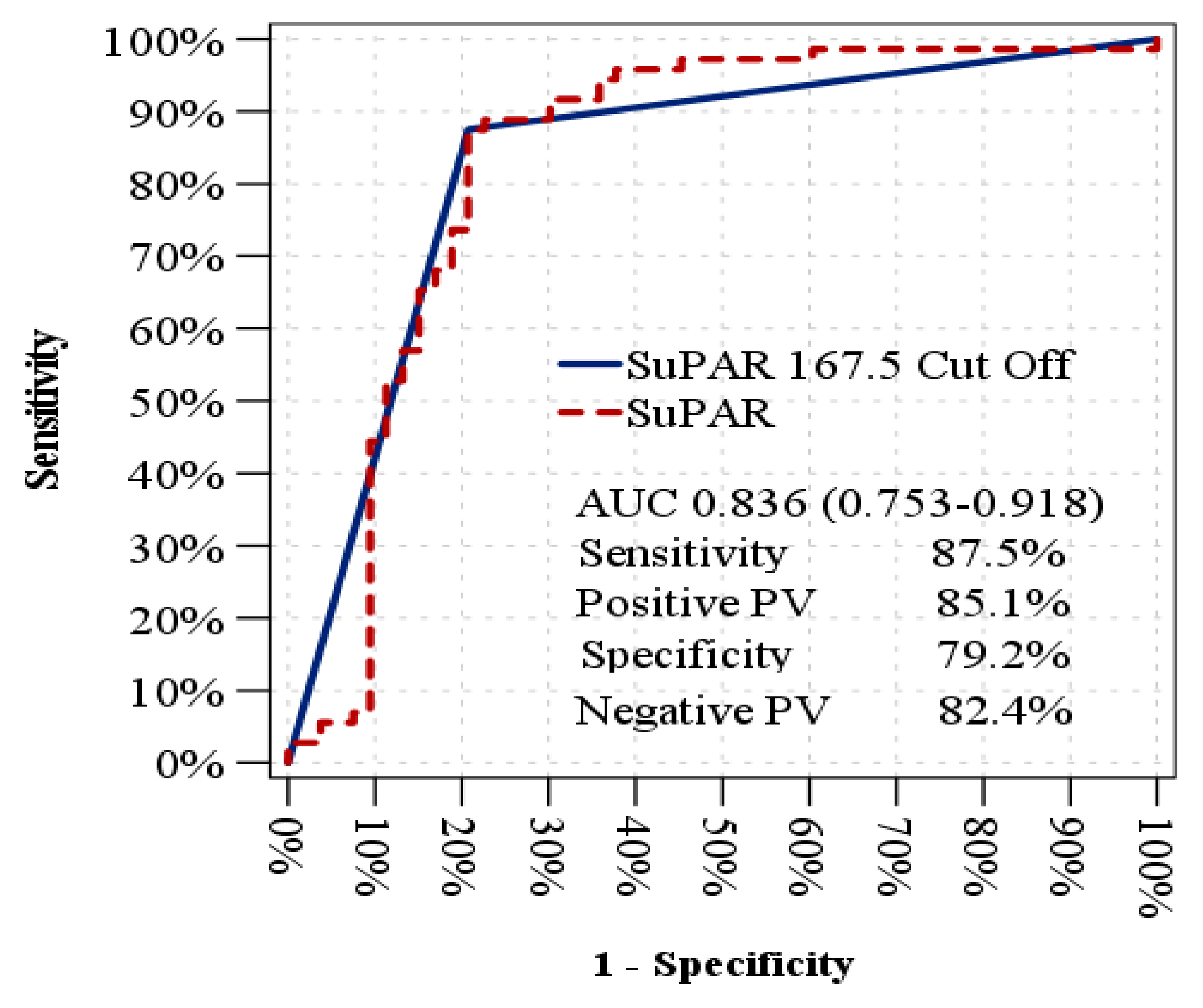
Figure 1. An analysis using receiver operating characteristic (ROC) curves to predict erectile dysfunction in diabetic patients.
Jcm 14 04029 g001
Figure 2. ROC analysis to distinguish DMED patients from healthy controls.
Jcm 14 04029 g002
Table 1. The demographic, anthropometric and clinical data of the patients.
Healthy Control
Group 1
(n = 46)
DM-NoED
Group 2
(n = 54)
DMED
Group 3
(n = 73)
p
Age, years 55 (51–61)57 (49–64)60 (51–64)0.055
Smoking status, n (%)
- Never 29 (63)38 (70)45 (63)0.583
- Ex-smoker 5 (11)4 (8)6 (8)
- Current smoker 12 (25)12 (22)22 (30)
Body mass index, kg/m223 (21.7–23.9)27.7 (25–30.5)28 (25.8–31)<0.001 *
Systolic BP, mmHg
Diastolic BP, mmHg
120 (118–130)124 (119–130)130 (120–130)0.221
78 (70–85)80 (72–85)80 (70–90)0.151
Diabetes duration, years -7 (3–14)14 (6–19.5)<0.001
ED duration, years --4.1
IIEF-5 score 23 (22–24)24 (23–25)11 (6–15)<0.001 **
ED severity, n (%)
- Mild ED
- Mild to moderate ED
- Moderate ED
- Severe ED
--15 (20.5)
--19 (26)
--16 (22)
--23 (31.5)
Comorbid diseases, n (%)
- Ischemic heart disease
- Hypertension
- Hyperlipidemia
-12 (22.6)18 (24.6)0.782
-23 (43)40 (54.7)0.394
-19 (35.8)33 (45.2)0.674
Medication, n (%)
- Metformin -43 (79.6)57 (78)0.959
- DPP4 inhibitors -27 (50)39 (53)0.695
- SGLT2 inhibitors -24 (44)39 (53)0.300
- Sulfonylureas -11 (20)13 (17)0.681
- Pioglitazone -5 (9.2)12 (16.4)0.180
- Insulin -12 (22)26 (35.6)0.072
- ACEI or ARB -12 (22.2)25 (34)0.103
- Statin and/or fibrate -14 (25.9)30 (41)0.053
DM-NoED: diabetes mellitus without erectile dysfunction; DMED: diabetes mellitus with erectile dysfunction; BP: blood pressure; ED: erectile dysfunction; IIEF-5: International Index of Erectile Function-5; DPP4: dipeptidyl peptidase 4; SGLT2: sodium–glucose cotransporter 2; ACEI: angiotensin converting enzyme inhibitor; ARB: angiotensin receptor blocker. Data are presented as median (interquartile range). Categorical parameters are expressed as n (percentage). * Post hoc analysis was performed with Dunn’s method. * p < 0.001 between Groups 1 and 2 and between groups 1 and 3. ** p < 0.001 between Groups 1 and 3 and between groups 2 and 3.
Table 2. Comparison of serum suPAR levels and relevant laboratory parameters between groups.
Healthy Control
Group 1
(n = 46)
DM-NoED
Group 2
(n = 54)
DMED
Group 3
(n = 73)
p
suPAR, pg/mL 107.9 (102–116)130.3 (112–165)218 (192–249)<0.001 *
FBG, mg/dL 92 (84–96)148 (104–245)175 (130–221)<0.001 **
HbA1c, % 5.7 (5.4–5.9)7.8 (6.8–9.6)8 (7.2–9.3)<0.001 **
HOMA-IR 2.2 (1.5–3)4 (2.2–10.3)4.9 (1.7–8.7)<0.001 **
LDLc, mg/dL 113 (91–137)98 (80–133)109 (80–133)0.368
HDLc, mg/dL 40 (36–45)39 (33–43)41 (34–48)0.204
TG, mg/dL 150 (107–200)145 (98–212)152 (104–234)0.582
T.testosterone, ng/mL 4.5 (2.9–5.5)3.4 (2.5–5)3.8 (2.8–5)0.131
Total PSA, µg/L 1.2 (0.8–2.7)1.3 (0.6–2.7)1.4 (0.5–2.5)0.413
Prolactin, µg/mL 11 (7.3–14.7)8.1 (6.8–12)9.9 (7.2–13.6)0.187
FSH, IU/L 8.8 (6.7–11.7)7.7 (5.7–9.4)8.1 (7–11.4)0.390
LH, IU/L 6.1 (5–7.9)6.2 (5.3–7.9)5.8 (4.7–7.1)0.508
TSH, mIU/L 1.5 (1–2.9)1.4 (1.1–2.5)2.1 (1.4–2.8)0.106
Leukocytes, 103/uL 7.8 (6.7–8.8)7.9 (6.4–9.8)8.3 (6.6–10)0.330
Hemoglobin, g/dL 14.9 (14.3–15.8)14.7 (13.6–15.7)14.8 (14–15.5)0.085
Platelets, 103/uL 242 (220–264)257 (212–293)249 (208–308)0.881
Creatinine, mg/dL 0.9 (0.8–1)0.9 (0.8–1)0.9 (0.6–1.1)0.664
eGFR, mL/min/1.73 m293 (84–101)92 (80–104)91 (75–100)0.091
ALT, U/L 20 (14–28)16 (12–27)18 (15–23)0.316
Total protein, g/L 7.5 (7.3–7.7)7.2 (7.1–7.5)7.3 (7.2–7.5)0.490
Albumin, g/L 4.4 (4.2–4.7)4.5 (4.3–4.7)4.2 (4–4.7)0.056
CRP, mg/L 2.1 (1.3–3.4)2.3 (1.1–4.4)1.8 (0.9–4.1)0.623
ESR, mm/h 8 (6–12)11 (6–17)9 (6–14)0.232
DM-NoED: diabetes mellitus without erectile dysfunction; DMED: diabetes mellitus with erectile dysfunction; suPAR: soluble urokinase plasminogen activator receptor; FBG: fasting blood glucose; HbA1c: hemoglobin A1c; HOMA-IR: homeostasis model assessment of insulin resistance; LDLc: low-density-lipoprotein cholesterol; HDLc: high-density-lipoprotein cholesterol; TG: triglyceride; T.testosterone: total testosterone; PSA: prostate-specific antigen; FSH: follicle-stimulating hormone; LH: luteinizing hormone; TSH: thyroid-stimulating hormone; eGFR: estimated glomerular filtration rate; ALT: alanine aminotransferase; CRP: C-reactive protein; ESR: erythrocyte sedimentation rate. * Post hoc analysis was performed with Dunn’s method, and p < 0.001 for all groups. ** p < 0.001 between Groups 1 and 2 and between groups 1 and 3.
Disclaimer/Publisher’s Note: The statements, opinions and data contained in all publications are solely those of the individual author(s) and contributor(s) and not of MDPI and/or the editor(s). MDPI and/or the editor(s) disclaim responsibility for any injury to people or property resulting from any ideas, methods, instructions or products referred to in the content.