
Clinical and Patient-Focused Outcomes After Percutaneous Screw Fixation of Pelvic Ring Fractures in Older Adults

 , and
, and
Abstract
1. Introduction
2. Methods
2.1. Study Design and Study Setting
2.2. Outcome Measures and Data Collection
2.3. Definition
2.4. Data Analysis
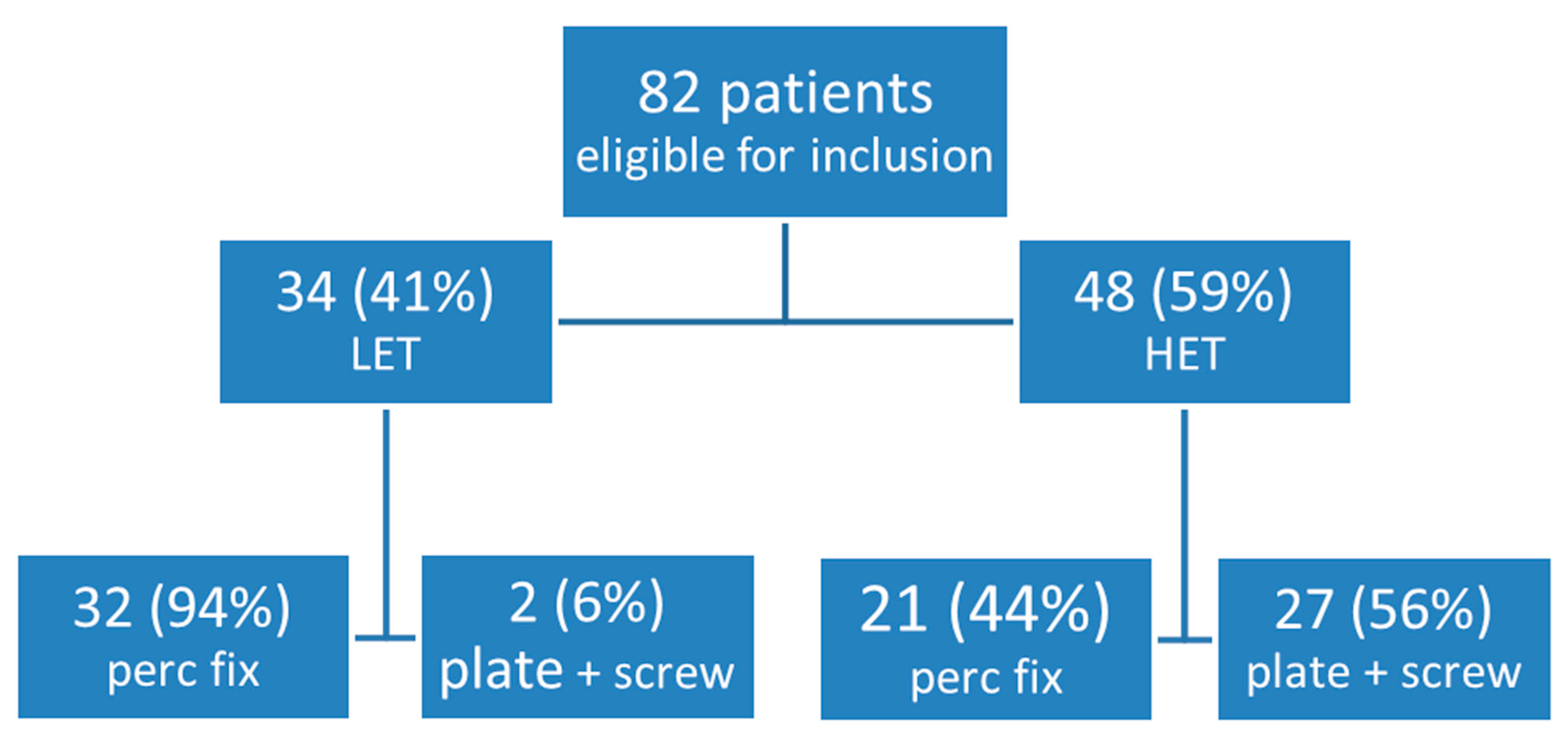
3. Results
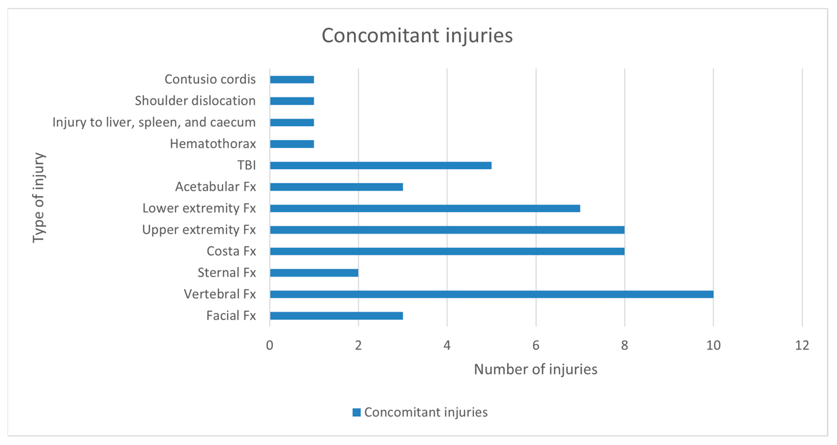
3.1. Operative Data, Direct Post-Operative Complications, and Follow-Up Imaging
3.2. Patient-Focused Outcomes
4. Discussion
Limitations and Strengths
5. Conclusions
Supplementary Materials
Author Contributions
Funding
Institutional Review Board Statement
Informed Consent Statement
Data Availability Statement
Conflicts of Interest
References
- Smits, C.H.; van den Beld, H.K.; Aartsen, M.J.; Schroots, J.J. Aging in the Netherlands: State of the art and science. Gerontologist 2014, 54, 335–343. [Google Scholar] [CrossRef] [PubMed]
- Neyens, J.; Halfens, R.; Spreeuwenberg, M.; Meijers, J.; Luiking, Y.; Verlaan, G.; Schols, J. Malnutrition is associated with an increased risk of falls and impaired activity in elderly patients in Dutch residential long-term care (LTC): A cross-sectional study. Arch. Gerontol. Geriatr. 2013, 56, 265–269. [Google Scholar] [CrossRef] [PubMed]
- Lundin, N.; Huttunen, T.T.; Berg, H.E.; Marcano, A.; Fellander-Tsai, L.; Enocson, A. Increasing incidence of pelvic and acetabular fractures. A nationwide study of 87,308 fractures over a 16-year period in Sweden. Injury 2021, 52, 1410–1417. [Google Scholar] [CrossRef]
- Kannus, P.; Parkkari, J.; Niemi, S.; Sievanen, H. Low-Trauma Pelvic Fractures in Elderly Finns in 1970–2013. Calcif. Tissue Int. 2015, 97, 577–580. [Google Scholar] [CrossRef]
- Elzohairy, M.M.; Salama, A.M. Open reduction internal fixation versus percutaneous iliosacral screw fixation for unstable posterior pelvic ring disruptions. Orthop. Traumatol. Surg. Res. 2017, 103, 223–227. [Google Scholar] [CrossRef]
- Giannoudis, P.V.; Tzioupis, C.C.; Pape, H.C.; Roberts, C.S. Percutaneous fixation of the pelvic ring: An update. J. Bone Jt. Surg. Br. 2007, 89, 145–154. [Google Scholar] [CrossRef]
- Benhenneda, R.; Letissier, H.; Dubrana, F.; Di Francia, R. Immediate full weight bearing after pelvic percutaneous fixation by screw for simple acetabular and pelvic ring fractures in patients older than sixty five years. Int. Orthop. 2022, 46, 2413–2421. [Google Scholar] [CrossRef] [PubMed]
- Rommens, P.M.; Hofmann, A. Comprehensive classification of fragility fractures of the pelvic ring: Recommendations for surgical treatment. Injury 2013, 44, 1733–1744. [Google Scholar] [CrossRef]
- Qoreishi, M.; Seyyed Hosseinzadeh, H.R.; Safdari, F. Clinical Results of Percutaneous Fixation of Pelvic and Acetabular Fractures: A Minimally Invasive Internal Fixation Technique. Arch. Bone Jt. Surg. 2019, 7, 284–290. [Google Scholar]
- Long, T.; Li, K.N.; Gao, J.H.; Liu, T.H.; Mu, J.S.; Wang, X.J.; Peng, C.; He, Z.Y. Comparative Study of Percutaneous Sacroiliac Screw with or without TiRobot Assistance for Treating Pelvic Posterior Ring Fractures. Orthop. Surg. 2019, 11, 386–396. [Google Scholar] [CrossRef]
- van Dijk, W.A.; Poeze, M.; van Helden, S.H.; Brink, P.R.; Verbruggen, J.P. Ten-year mortality among hospitalised patients with fractures of the pubic rami. Injury 2010, 41, 411–414. [Google Scholar] [CrossRef] [PubMed]
- Wilson, D.G.G.; Kelly, J.; Rickman, M. Operative management of fragility fractures of the pelvis—A systematic review. BMC Musculoskelet. Disord. 2021, 22, 717. [Google Scholar] [CrossRef] [PubMed]
- Burgess, A.R.; Eastridge, B.J.; Young, J.W.; Ellison, T.S.; Ellison, P.S., Jr.; Poka, A.; Bathon, G.H.; Brumback, R.J. Pelvic ring disruptions: Effective classification system and treatment protocols. J. Trauma 1990, 30, 848–856. [Google Scholar] [CrossRef]
- Ullrich, B.W.; Schnake, K.J.; Spiegl, U.J.A.; Schenk, P.; Mendel, T.; Behr, L.; Bula, P.; Flucht, L.B.; Franck, A.; Gercek, E.; et al. OF-Pelvis classification of osteoporotic sacral and pelvic ring fractures. BMC Musculoskelet. Disord. 2021, 22, 992. [Google Scholar] [CrossRef]
- Berian, J.R.; Mohanty, S.; Ko, C.Y.; Rosenthal, R.A.; Robinson, T.N. Association of Loss of Independence With Readmission and Death After Discharge in Older Patients After Surgical Procedures. JAMA Surg. 2016, 151, e161689. [Google Scholar] [CrossRef]
- Strout, K.; Crittenden, J.; O’Brien, L.M.; Howard, E.P. Assessing Older Adult Wellness: An Assessment of the Psychometric Properties of the Lifestyle Survey. J. Nurs. Meas. 2020, 28, 598–614. [Google Scholar] [CrossRef] [PubMed]
- Fried, T.R.; Bradley, E.H.; Towle, V.R.; Allore, H. Understanding the treatment preferences of seriously ill patients. N. Engl. J. Med. 2002, 346, 1061–1066. [Google Scholar] [CrossRef]
- Wiles, J.L.; Leibing, A.; Guberman, N.; Reeve, J.; Allen, R.E. The meaning of “aging in place” to older people. Gerontologist 2012, 52, 357–366. [Google Scholar] [CrossRef]
- Chappell, N.L.; Dlitt, B.H.; Hollander, M.J.; Miller, J.A.; McWilliam, C. Comparative costs of home care and residential care. Gerontologist 2004, 44, 389–400. [Google Scholar] [CrossRef]
- O’Brien, D.P.; Luchette, F.A.; Pereira, S.J.; Lim, E.; Seeskin, C.S.; James, L.; Miller, S.; Davis, K., Jr.; Hurst, J.M.; Johannigman, J.A.; et al. Pelvic fracture in the elderly is associated with increased mortality. Surgery 2002, 132, 710–714, discussion 714–715. [Google Scholar] [CrossRef]
- Morris, R.O.; Sonibare, A.; Green, D.J.; Masud, T. Closed pelvic fractures: Characteristics and outcomes in older patients admitted to medical and geriatric wards. Postgrad. Med. J. 2000, 76, 646–650. [Google Scholar] [CrossRef] [PubMed]
- Gruen, G.S.; Leit, M.E.; Gruen, R.J.; Garrison, H.G.; Auble, T.E.; Peitzman, A.B. Functional outcome of patients with unstable pelvic ring fractures stabilized with open reduction and internal fixation. J. Trauma 1995, 39, 838–844, discussion 844–835. [Google Scholar] [CrossRef]
- Van den Bosch, E.W.; Van der Kleyn, R.; Hogervorst, M.; Van Vugt, A.B. Functional outcome of internal fixation for pelvic ring fractures. J. Trauma 1999, 47, 365–371. [Google Scholar] [CrossRef] [PubMed]
- Routt, M.L., Jr.; Simonian, P.T.; Mills, W.J. Iliosacral screw fixation: Early complications of the percutaneous technique. J. Orthop. Trauma 1997, 11, 584–589. [Google Scholar] [CrossRef]
- Shuler, T.E.; Boone, D.C.; Gruen, G.S.; Peitzman, A.B. Percutaneous iliosacral screw fixation: Early treatment for unstable posterior pelvic ring disruptions. J. Trauma 1995, 38, 453–458. [Google Scholar] [CrossRef]
- Stevenson, A.J.; Swartman, B.; Bucknill, A.T. Percutaneous internal fixation of pelvic fractures. Unfallchirurg 2017, 120 (Suppl. 1), 10–18. [Google Scholar] [CrossRef]
- Gire, J.D.; Jiang, S.Y.; Gardner, M.J.; Bishop, J.A. Percutaneous Versus Open Treatment of Posterior Pelvic Ring Injuries: Changes in Practice Patterns Over Time. J. Orthop. Trauma 2018, 32, 457–460. [Google Scholar] [CrossRef]
- Routt, M.L., Jr.; Nork, S.E.; Mills, W.J. Percutaneous fixation of pelvic ring disruptions. Clin. Orthop. Relat. Res. 2000, 375, 15–29. [Google Scholar] [CrossRef] [PubMed]
- Scheyerer, M.J.; Osterhoff, G.; Wehrle, S.; Wanner, G.A.; Simmen, H.P.; Werner, C.M. Detection of posterior pelvic injuries in fractures of the pubic rami. Injury 2012, 43, 1326–1329. [Google Scholar] [CrossRef]
- Tile, M. Acute Pelvic Fractures: I. Causation and Classification. J. Am. Acad. Orthop. Surg. 1996, 4, 143–151. [Google Scholar] [CrossRef]
- Petryla, G.; Uvarovas, V.; Bobina, R.; Kurtinaitis, J.; Puronaite, R.; Kvederas, G.; Satkauskas, I. Comparison of One-Year Functional Outcomes and Quality of Life between Posterior Pelvic Ring Fixation and Combined Anterior-Posterior Pelvic Ring Fixation after Lateral Compression (B2 Type) Pelvic Fracture. Medicina 2021, 57, 204. [Google Scholar] [CrossRef] [PubMed]
- Zhang, B.F.; Shang, K.; Wang, P.F.; Ke, C.; Han, S.; Yang, K.; Fei, C.; Xu, X.; Zhang, K.; Zhuang, Y. Comparison of posterior ring fixation with combined anterior and posterior ring fixation for the treatment of lateral compression type 2 pelvic fractures. Int. Orthop. 2020, 44, 1187–1193. [Google Scholar] [CrossRef] [PubMed]
- Moussa, I.S.; Sallam, A.M.; Mahmoud, A.K.; Elzaher, E.H.; Nagy, A.M.; Eid, A.S. Combined anterior and posterior ring fixation versus posterior ring fixation alone in the management of unstable Tile B and C pelvic ring injuries: A randomized controlled trial. Chin. J. Traumatol. 2022, 26, 48–59. [Google Scholar] [CrossRef] [PubMed]
- Tucker, N.J.; Scott, B.L.; Heare, A.; Stacey, S.C.; Mauffrey, C.; Parry, J.A. Combined Anterior-Posterior Versus Posterior-Only Fixation of Stress-Positive Minimally Displaced Lateral Compression Type 1 (LC1) Pelvic Ring Injuries. J. Orthop. Trauma 2022, 37, 189–194. [Google Scholar] [CrossRef]
- Wang, P.; Ali, S.H.; Fei, C.; Zhang, B.; Wei, X.; Wang, H.; Cong, Y.; Deng, H.; Fu, Y.; Zhang, K.; et al. Management of LC Type I (LC-1) Pelvic Injuries with Complete Sacral Fracture: Comparison of Solitary Anterior Fixation with Combined Anterior-Posterior Fixation. Biomed. Res. Int. 2022, 2022, 3918794. [Google Scholar] [CrossRef]
- Herteleer, M.; Boudissa, M.; Hofmann, A.; Wagner, D.; Rommens, P.M. Plate fixation of the anterior pelvic ring in patients with fragility fractures of the pelvis. Eur. J. Trauma Emerg. Surg. 2022, 48, 3711–3719. [Google Scholar] [CrossRef]
- Basile, G.; Passeri, A.; Bove, F.; Accetta, R.; Gaudio, R.M.; Calori, G.M. Pelvic ring and acetabular fracture: Concepts of traumatological forensic interest. Injury 2022, 53, 475–480. [Google Scholar] [CrossRef]

| Questionnaire | |
|---|---|
| 1. Do you experience pain related to your pelvic fracture at rest? | NRS 0–10 |
| 2. Do you experience pain related to your pelvic fracture when walking? | NRS 0–10 |
| 3. What is your current level of independence during mobilization in your home? |
|
| 4. What is your current level of independence during mobilization outside? |
|
| 5. What is your current level of independence when going shopping/visiting a restaurant/or visiting family? |
|
| 6. Where do you live currently? |
|
| 7. Are you pleased with the results of the operation? |
|
| Pt | MOI | Descriptive | YB | Rommens | OF-Pelvic |
|---|---|---|---|---|---|
| 1 | HET | Sacrum bilateral + anterior ring unilateral | Other (LC1 + isolated sacral Fx) | NA | NA |
| 2 | LET | Sacrum bilateral + anterior ring bilateral | Other (LC1 bilateral) | 4b | 4 |
| 3 | LET | Sacrum bilateral + anterior ring unilateral | Other (LC1 + isolated sacral Fx) | 4b | 4 |
| 4 | HET | Sacrum bilateral | Other (Bilateral isolated sacral Fx) | NA | NA |
| 5 | LET | Ilium unilateral + anterior ring unilateral | LC2 | 3a | 5 |
| 6 | LET | Sacrum bilateral + anterior ring unilateral | Other (LC1 + isolated sacral Fx) | 4b | 4 |
| 7 | LET | Sacrum bilateral + anterior ring unilateral | Other (LC1 + sacral Fx) | 4b | 4 |
| 8 | HET | Sacrum unilateral + anterior ring unilateral | LC1 | NA | NA |
| 9 | LET | Sacrum unilateral + anterior ring bilateral | LC1 | 2c | 3 |
| 10 | LET | Sacrum bilateral + anterior ring bilateral | Other (LC1 bilateral) | 4b | 4 |
| 11 | HET | Sacrum unilateral + anterior ring unilateral | LC1 | NA | NA |
| 12 | LET | Ilium unilateral + anterior ring unilateral | LC2 | 3a | 5 |
| 13 | HET | Ilium unilateral + anterior ring unilateral | LC2 | NA | NA |
| 14 | LET | Ilium unilateral + anterior ring unilateral | LC2 | 3a | 5 |
| 15 | LET | Sacrum unilateral + anterior ring bilateral | LC1 | 2c | 3 |
| 16 | HET | Sacrum unilateral + anterior ring unilateral | LC1 | NA | NA |
| 17 | LET | Sacrum bilateral + anterior ring unilateral | Other (LC1 + isolated sacral Fx) | 4b | 4 |
| 18 | HET | Sacrum unilateral + anterior ring unilateral | LC1 | NA | NA |
| 19 | HET | Si-diastase unilateral | Other (Isolated unilateral SI-diastase) | NA | NA |
| 20 | HET | Sacrum bilateral + ilium unilateral + anterior ring bilateral | Other (LC2 + LC1) | NA | NA |
| 21 | HET | Ilium unilateral + anterior ring unilateral | LC2 | NA | NA |
| 22 | HET | Sacrum bilateral + anterior ring unilateral | Other (LC1 + isolated sacral Fx) | NA | NA |
| 23 | HET | Si-diastase bilateral + anterior ring bilateral | APC2 | NA | NA |
| 24 | LET | Sacrum bilateral + anterior ring unilateral | Other (LC1 + isolated sacral Fx) | 4b | 4 |
| 25 | LET | Sacrum bilateral + anterior ring unilateral | Other (LC1 + isolated sacral Fx) | 4b | 4 |
| 26 | LET | Sacrum unilateral + anterior ring unilateral | LC1 | 2c | 3 |
| 27 | HET | Sacrum unilateral + anterior ring unilateral | LC1 | NA | NA |
| 28 | LET | Sacrum unilateral + anterior ring unilateral | LC1 | NA | NA |
| 29 | HET | Sacrum unilateral + ilium unilateral + anterior ring unilateral | LC2 | NA | NA |
| 30 | HET | Ilium unilateral + anterior ring unilateral | LC2 | NA | NA |
| 31 | HET | Sacrum unilateral + anterior ring bilateral | LC1 | NA | NA |
| 32 | LET | Sacrum unilateral + anterior ring unilateral | LC1 | NA | NA |
| 33 | HET | Sacrum unilateral + anterior ring unilateral | LC1 | NA | NA |
| 34 | LET | Ilium unilateral + anterior ring unilateral | LC2 | NA | NA |
| 35 | LET | Sacrum unilateral + anterior ring unilateral | LC1 | NA | NA |
| 36 | LET | Sacrum unilateral + anterior ring unilateral | LC1 | NA | NA |
| 37 | HET | Sacrum bilateral + ilium unilateral + anterior ring bilateral | Other (LC2 + LC1) | NA | NA |
| 38 | HET | Sacrum unilateral + anterior ring unilateral | LC1 | NA | NA |
| 39 | LET | Sacrum bilateral + anterior ring unilateral | Other (LC1 + isolated sacral Fx) | 4b | 4 |
| 40 | LET | Sacrum bilateral + anterior ring bilateral | Other (LC1 bilateral) | 4b | 4 |
| 41 | HET | Sacrum unilateral + anterior ring bilateral | LC1 | NA | NA |
| 42 | LET | Sacrum bilateral + anterior ring bilateral | Other (LC1 bilateral) | NA | NA |
| 43 | LET | Sacrum bilateral + anterior ring unilateral | Other (LC1 bilateral) | NA | NA |
| 44 | LET | Sacrum unilateral + anterior ring unilateral | LC1 | NA | NA |
| 45 | HET | Sacrum unilateral + anterior ring unilateral | LC1 | NA | NA |
| 46 | LET | Sacrum unilateral + anterior ring unilateral | LC1 | NA | NA |
| 47 | LET | Ilium unilateral + anterior ring unilateral | LC2 | 3a | 5 |
| 48 | LET | Sacrum unilateral + anterior ring unilateral | LC2 | 2c | 5 |
| 49 | LET | Sacrum bilateral + anterior ring bilateral | Other (LC1 bilateral) | 4b | 4 |
| 50 | HET | Sacrum unilateral + ilium unilateral + anterior ring unilateral | LC2 | NA | NA |
| 51 | HET | Sacrum unilateral + anterior ring unilateral | LC1 | NA | NA |
| 52 | LET | Sacrum bilateral + anterior ring bilateral | Other (LC1 bilateral) | NA | NA |
| Pt | MOI | Fracture Pattern | Surgical Fixation | Weight-Bearing Regime |
|---|---|---|---|---|
| 1 | HET | Sacrum bilateral + anterior ring unilateral | Bilateral 1x SI-screw | WBAT |
| 2 | LET | Sacrum bilateral + anterior ring bilateral | Bilateral 1x TITS | WBAT |
| 3 | LET | Sacrum bilateral + anterior ring unilateral | Bilateral 1x SI-screw | WBAT |
| 4 | HET | Sacrum bilateral | Bilateral 1x SI-screw | WBAT |
| 5 | LET | Ilium unilateral + anterior ring unilateral | Unilateral 1x LC2-screw + unilateral pubic screw | 1 leg TTWB, 1 leg FWB 8w |
| 6 | LET | Sacrum bilateral + anterior ring unilateral | Unilateral pubic screw | WBAT |
| 7 | LET | Sacrum bilateral + anterior ring unilateral | Unilateral 1x TITS + unilateral pubic screw | WBAT |
| 8 | HET | Sacrum unilateral + anterior ring unilateral | Unilateral 1x TITS | WBAT |
| 9 | LET | Sacrum unilateral + anterior ring bilateral | Unilateral 2x TITS | WBAT |
| 10 | LET | Sacrum bilateral + anterior ring bilateral | Bilateral 1x TITS | WBAT |
| 11 | HET | Sacrum unilateral + anterior ring unilateral | Bilateral 1x SI-screw + unilateral pubic screw | WBAT |
| 12 | LET | Ilium unilateral + anterior ring unilateral | Unilateral 1x LC2-screw + unilateral pubic screw | WBAT |
| 13 | HET | Ilium unilateral + anterior ring unilateral | Unilateral 1x LC2-screw + unilateral pubic screw | 1 leg TTWB, 1 leg FWB 8w |
| 14 | LET | Ilium unilateral + anterior ring unilateral | Unilateral 2x iliac screw + unilateral pubic screw | WBAT |
| 15 | LET | Sacrum unilateral + anterior ring bilateral | Unilateral 2x SI-screw + bilateral pubic screw | WBAT |
| 16 | HET | Sacrum unilateral + anterior ring unilateral | Bilateral 1x TITS + bilateral pubic screw | 1 leg NWB, 1 leg FWB 8w |
| 17 | LET | Sacrum bilateral + anterior ring unilateral | Unilateral 1x TITS + unilateral pubic screw | WBAT |
| 18 | HET | Sacrum unilateral + anterior ring unilateral | Unilateral 2x SI-screw | WBAT |
| 19 | HET | Si-diastase unilateral | Unilateral 1x SI-screw | 1 leg NWB, 1 leg FWB 8w |
| 20 | HET | Sacrum bilateral + ilium unilateral + anterior ring bilateral | Bilateral 1x SI-screw + bilateral pubic screw | Both legs NWB 6w |
| 21 | HET | Ilium unilateral + anterior ring unilateral | Unilateral 1x LC2-screw + unilateral pubic screw | Both legs NWB 6w |
| 22 | HET | Sacrum bilateral + anterior ring unilateral | Bilateral 1x SI-screw + unilateral pubic screw | Both legs NWB due to calcaneal Fx |
| 23 | HET | Si-diastase bilateral + anterior ring bilateral | Unilateral 1x TITS + contralateral 1x SI-screw + bilateral pubic screw | Both legs NWB 8w |
| 24 | LET | Sacrum bilateral + anterior ring unilateral | Unilateral 1x SI-screw + 1x TITS + unilateral pubic screw | WBAT |
| 25 | LET | Sacrum bilateral + anterior ring unilateral | Bilateral 1x TITS + unilateral pubic screw | WBAT |
| 26 | LET | Sacrum unilateral + anterior ring unilateral | Unilateral 1x TITS + unilateral pubic screw | WBAT |
| 27 | HET | Sacrum unilateral + anterior ring unilateral | Unilateral 1x TITS + 1x SI-screw + unilateral pubic screw | WBAT |
| 28 | LET | Sacrum unilateral + anterior ring unilateral | Unilateral 1x TITS + unilateral pubic screw | WBAT |
| 29 | HET | Sacrum unilateral + ilium unilateral + anterior ring unilateral | Unilateral 2x TITS + unilateral pubic screw | WBAT |
| 30 | HET | Ilium unilateral + anterior ring unilateral | Unilateral 1x LC2-screw + unilateral pubic screw | 1 leg TTWB, 1 leg FWB 6w |
| 31 | HET | Sacrum unilateral + anterior ring bilateral | Unilateral 2x SI-screw + bilateral pubic screw | Both legs NWB 6w |
| 32 | LET | Sacrum unilateral + anterior ring unilateral | Unilateral 1x TITS | WBAT |
| 33 | HET | Sacrum unilateral + anterior ring unilateral | Unilateral 1x TITS + unilateral pubic screw | WBAT |
| 34 | LET | Ilium unilateral + anterior ring unilateral | Unilateral 1x TITS + 1x iliac screw + unilateral pubic screw | WBAT |
| 35 | LET | Sacrum unilateral + anterior ring unilateral | Unilateral 1x TITS + unilateral pubic screw | WBAT |
| 36 | LET | Sacrum unilateral + anterior ring unilateral | Unilateral 1x TITS + unilateral pubic screw | WBAT |
| 37 | HET | Sacrum bilateral + ilium unilateral + anterior ring bilateral | Bilateral 1x SI-screw + unilateral pubic screw | WBAT |
| 38 | HET | Sacrum unilateral + anterior ring unilateral | Unilateral 1x TITS + unilateral pubic screw | WBAT |
| 39 | LET | Sacrum bilateral + anterior ring unilateral | Unilateral 1x TITS + unilateral pubic screw | WBAT |
| 40 | LET | Sacrum bilateral + anterior ring bilateral | Unilateral 1x TITS + bilateral pubic screw | WBAT |
| 41 | HET | Sacrum unilateral + anterior ring bilateral | Unilateral 1x TITS + bilateral pubic screw | WBAT |
| 42 | LET | Sacrum bilateral + anterior ring bilateral | Unilateral 2x TITS + bilateral pubic screw | WBAT |
| 43 | LET | Sacrum bilateral + anterior ring unilateral | Bilateral 1x TITS + unilateral pubic screw | WBAT |
| 44 | LET | Sacrum unilateral + anterior ring unilateral | Unilateral 1x TITS | WBAT |
| 45 | HET | Sacrum unilateral + anterior ring unilateral | Unilateral 1x SI-screw + 1x TITS + unilateral pubic screw | WBAT |
| 46 | LET | Sacrum unilateral + anterior ring unilateral | Unilateral 1x TITS + unilateral pubic screw | WBAT |
| 47 | LET | Ilium unilateral + anterior ring unilateral | Unilateral 1x iliac screw + unilateral pubic screw | WBAT |
| 48 | LET | Sacrum unilateral + anterior ring unilateral | Unilateral 1x SI-screw + unilateral pubic screw | WBAT |
| 49 | LET | Sacrum bilateral + anterior ring bilateral | Unilateral 1x TITS + unilateral pubic screw | WBAT |
| 50 | HET | Sacrum unilateral + ilium unilateral + anterior ring unilateral | Unilateral 1x SI-screw + 1x TITS + unilateral pubic screw | WBAT |
| 51 | HET | Sacrum unilateral + anterior ring unilateral | Unilateral 2x TITS + unilateral pubic screw | WBAT |
| 52 | LET | Sacrum bilateral + anterior ring bilateral | Bilateral 1x TITS + bilateral pubic screw | WBAT |
Disclaimer/Publisher’s Note: The statements, opinions and data contained in all publications are solely those of the individual author(s) and contributor(s) and not of MDPI and/or the editor(s). MDPI and/or the editor(s) disclaim responsibility for any injury to people or property resulting from any ideas, methods, instructions or products referred to in the content. |
© 2025 by the authors. Licensee MDPI, Basel, Switzerland. This article is an open access article distributed under the terms and conditions of the Creative Commons Attribution (CC BY) license (https://creativecommons.org/licenses/by/4.0/).
Share and Cite
Mennen, A.H.M.; Boon, J.; Halm, J.A.; Peters, R.W.; Bloemers, F.W.; Van Embden, D. Clinical and Patient-Focused Outcomes After Percutaneous Screw Fixation of Pelvic Ring Fractures in Older Adults. J. Clin. Med. 2025, 14, 3919. https://doi.org/10.3390/jcm14113919
Mennen AHM, Boon J, Halm JA, Peters RW, Bloemers FW, Van Embden D. Clinical and Patient-Focused Outcomes After Percutaneous Screw Fixation of Pelvic Ring Fractures in Older Adults. Journal of Clinical Medicine. 2025; 14(11):3919. https://doi.org/10.3390/jcm14113919
Chicago/Turabian StyleMennen, Anna H. M., Jan Boon, Jens A. Halm, Rolf W. Peters, Frank W. Bloemers, and Daphne Van Embden. 2025. "Clinical and Patient-Focused Outcomes After Percutaneous Screw Fixation of Pelvic Ring Fractures in Older Adults" Journal of Clinical Medicine 14, no. 11: 3919. https://doi.org/10.3390/jcm14113919
APA StyleMennen, A. H. M., Boon, J., Halm, J. A., Peters, R. W., Bloemers, F. W., & Van Embden, D. (2025). Clinical and Patient-Focused Outcomes After Percutaneous Screw Fixation of Pelvic Ring Fractures in Older Adults. Journal of Clinical Medicine, 14(11), 3919. https://doi.org/10.3390/jcm14113919