Case Report

Bilateral Spontaneous Hemothorax: A Rare Case of Primary Pleural Angiosarcoma and Literature Review †

1
Department of Public Health and Infectious Diseases, Sapienza University of Rome, 00161 Rome, Italy
2
Department of Radiological, Oncological, and Pathological Sciences, Sapienza University of Rome, 00161 Rome, Italy
3
Department of Thoracic Surgery and Lung Transplantation, Sapienza University of Rome, 00161 Rome, Italy
4
Department of Clinical, Internal, Anaesthesiological and Cardiovascular Sciences, Sapienza University of Rome, 00161 Rome, Italy
5
Unit of Interventional Pulmonology, Department of Experimental and Clinical Medicine, Careggi University Hospital, University of Florence, 50134 Florence, Italy
*
Author to whom correspondence should be addressed.
This article is a revised and expanded version of a paper entitled “Bilateral spontaneous haemothorax: A rare case of primary pleural angiosarcoma and literature review”, which was presented at SIET (Società Italiana di Endoscopia Toracica) Conference, Florence, Italy, 17–19 October 2024.
J. Clin. Med. 2025, 14(10), 3377; https://doi.org/10.3390/jcm14103377
Submission received: 12 January 2025 / Revised: 31 January 2025 / Accepted: 4 February 2025 / Published: 12 May 2025
(This article belongs to the Section Respiratory Medicine)

Abstract

:
Introduction and case report: Angiosarcomas, rare soft tissue malignancies originating from endothelial cells, represent only 1–2% of all soft tissue sarcomas. Primary pleural angiosarcoma (PPA) is exceptionally rare, with only 43 reported cases since 1943. There are many diagnostic and therapeutic challenges due to the rarity of these tumors. We present the case of a 72-year-old man presenting with back pain, dyspnea and anemia. Conventional imaging revealed bilateral pleural effusion and a thickened parietal pleura, while contrast chest MR was able to identify pleural sites of contrast enhancement. Left chest tube placement evidenced a hemothorax, and the cytology result was negative. A thoracoscopic approach was chosen, allowing us to perform different parietal pleural biopsies. Radiological and pathological features led to the diagnosis of epithelioid PPA. Despite pleural drainage and blood transfusions, the patient died only 4 days after diagnosis. Objectives: To present a literature review, evaluating the disease epidemiology and the clinical, diagnostic and therapeutic features of PPA. Methods: We reviewed cases of PPA in the literature (1954–2024) by searching the PubMed database for the terms “pleural angiosarcoma” and “pleura + angiosarcoma”. Results: We found a total of 47 cases that were described between 1987 and 2024 with sufficient data to be included in our review. PPA was found to be a challenging diagnosis, found mostly in older Caucasian males. The cytology is mostly indeterminant, and an endoscopic approach is usually needed. Radical surgery is the most common treatment option, and chemotherapy and radiation therapy are also often used. However, the prognosis is poor. Conclusions: PPA is very rare, and complex cases such as this one showcase the importance of innovative approaches like MRI and emphasize the significance of multidisciplinary collaboration for optimal patient management. Bilateral spontaneous hemothorax, as seen in this case, is uncommon and poses additional challenges in disease management. Further research to advance the diagnostic capabilities and treatment efficacy is needed.

1. Introduction

Angiosarcomas are rare soft tissue malignancies derived from endothelial cells, which are part of blood or lymphatic vessels, accounting for only 1–2% of all soft tissue sarcomas. They carry a poor prognosis, also because they are characterized by a rapid growth and high metastatic potential. Also, they can arise anywhere in the body, but the most common site consists of the skin or soft tissues [1]. Rarely, they may involve the pleura [2,3]. Primary pleural angiosarcoma (PPA) is an extremely rare malignancy, with a total of 43 cases being reported in the literature since the first case was described in 1943 [2]. It is characterized by a wide variety of clinical presentations, often leading to delayed diagnosis and having poor prognosis. Also, the rarity of these tumors hinders the determination of an optimal treatment and prognosis. The description of new clinical cases of PPA can be useful in highlighting diagnostic novelties and new treatment strategies, especially in challenging cases such as our own. This case is one of the few described where PPA was complicated by bilateral spontaneous pleural bleeding, making it especially challenging in its diagnostic and therapeutic management. We also describe for the first time the use of chest MR in the diagnosis of PPA, which can be a promising tool [4,5].

2. Case Report

A 72-year-old Caucasian man presented to the emergency department with left-side back pain radiating anteriorly and worsening dyspnea in the past month. He had a medical history of hypertension; paraplegia secondary to spinal cord ischemia; endovascular repair of an abdominal aortic aneurysm with stent placement, complicated by type 2 endoleak; renal artery stent placement; and ureteral stent placement. Home therapy included double antiaggregant and anticoagulant therapy. The patient was a construction engineer with asbestos exposure and a former smoker of 40 pack-years. There was no history of tuberculosis or a family history of neoplastic diseases.
On admission to the emergency department, he was eupneic, with decreased respiratory sounds bilaterally at the bases on physical examination. The laboratory tests showed normocytic normochromic anemia (hemoglobin was 6.6 g/dL), requiring blood transfusion, and a chest radiography showed bilateral pleural effusion (Figure 1).
A chest CT was performed, and in accordance with the hypothesis of pulmonary or pleural malignancy, contrast was also performed, highlighting bilateral pleural effusion, hyperemia and thickening of the left parietal pleura (Figure 2).
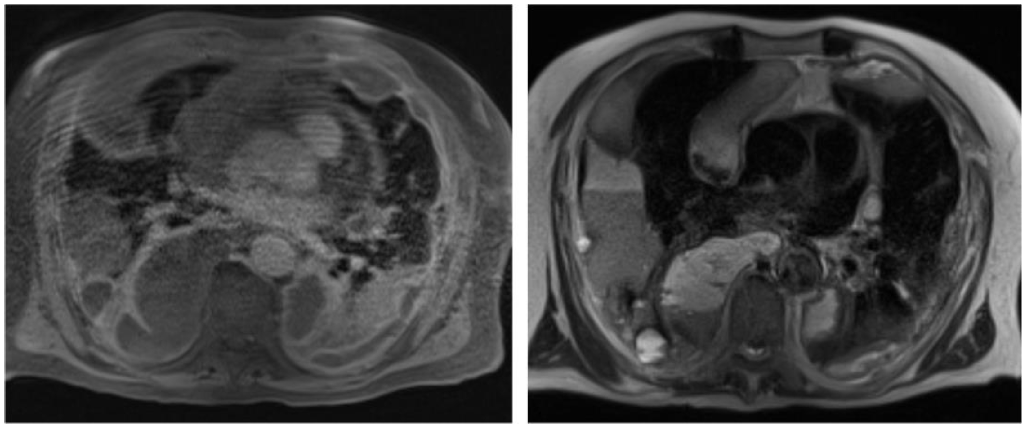
Also, to better characterize the pleural abnormalities and the pleural effusion, and with the aim to obtain a better planning for a surgical approach, a contrast chest MR was performed (Figure 3).
A 12 Fr chest tube was placed in the left hemithorax, and 6 L of hematic fluid (pleural fluid Hb 8.7 g/dL and hematocrit 27.4%) was evacuated, improving dyspnea and decreasing the oxygen requirement. The cytological examination excluded the presence of neoplastic cells. Microbiological tests performed on the pleural fluid were negative.
Due to the complexity of the disease, the case was subjected to a multidisciplinary hospital team evaluation, and it was also widely discussed on “Pleural Hub”, a Facebook platform that connects various international experts on chest diseases, also for discussing complex clinical cases. A thoracoscopic approach was widely advised in order to have a direct view of the parietal pleura and to choose the most adequate sites for biopsy. Also, thoracoscopy would have enabled a visualization of the active bleeding of the pleura, and talc poudrage could have been performed if advisable. Subsequently, a left video-assisted thoracoscopy (VATS) was performed. Unilateral VATS was chosen because of the patient’s critical condition, especially due to anemia, opting for the most simple surgical approach that could lead to a diagnosis. The parietal pleura appeared thickened, diffusely hyperemic and hemorrhagic. No nodules or masses were identified. Multiple biopsies of the parietal pleura were acquired, and a talc pleurodesis was performed. A 32 Fr drainage was positioned at the end of the procedure, and during the postoperative days, we assisted to reduce the amount of pleural effusion drainage, even if the patient continued to need blood transfusions.
The pathological examination enabled us to identify fibro-hematic material mixed with flaps of fibroadipose tissue with epithelioid neoplastic infiltration, partly necrotic, with the following immunophenotype: ERG+, CD31+, pancytokeratin+ (clone AE1-3), CD34−/+, D2-40−, CD15−, Ber-Ep4−, CEA−, TTF-1−, CK5/6−, calretinin−, WT1−, p16−, BAP1+ (maintained), p53+/− (wild-type), and a proliferation index (% Ki-67+) that was heterogeneously distributed and quantifiable in about 40% of neoplastic cells. The morphological and immunophenotypic findings described were consistent with a diagnosis of epithelioid angiosarcoma (Figure 4).
During hospitalization, the patient received several blood transfusions to maintain his hemoglobin level above 7 g/dL. Unfortunately, a specific oncologic treatment was never initiated due to the severe clinical condition of the patient. Exitus occurred only 4 days after the diagnosis.

3. Materials and Methods

We reviewed all described cases of primary pleural angiosarcoma in the recent literature (1954–2024) published in English language, using the PubMed database, by searching for the terms “pleural angiosarcoma” and “pleura + angiosarcoma”. We included in our database all cases that had enough information in order to describe the following: patients’ age, gender, main symptoms, presence of pleural effusion, outcome of pleural cytology investigation, presence of chest CT specific findings, performed diagnostic procedure and outcome, choice of treatment and time of survival. We found a total of 47 cases that included sufficient data to be analyzed and described in our review [6,7,8,9,10,11,12,13,14,15,16,17,18,19,20,21,22,23,24,25,26,27,28,29,30,31,32,33,34,35,36,37,38,39,40,41,42,43,44,45,46,47,48].

4. Results

All the results of our literature review are included in Table 1, which describes clinical, radiological and therapeutic aspects, summarized from a total of 47 cases that we found to be described in the literature between 1954 and 2024 [6,7,8,9,10,11,12,13,14,15,16,17,18,19,20,21,22,23,24,25,26,27,28,29,30,31,32,33,34,35,36,37,38,39,40,41,42,43,44,45,46,47,48].

5. Discussion

Angiosarcoma is a rare malignant tumor that accounts for only 1–2% of all soft tissue sarcomas. It arises from vascular endothelium and primarily affects the skin, breast, liver and heart [1]. PPA is an extremely rare entity [3], with only 46 cases being reported in the literature since its first description in 1943 [2]. It predominantly affects older Caucasian men. The diagnosis of PPA is challenging due to its nonspecific clinical presentation and imaging findings that may overlap with mesothelioma, lung carcinoma and metastatic adenocarcinoma. A cytological examination of pleural fluid has been negative in a significant proportion of PPA cases [3]. Thoracoscopy with biopsy is considered the most accurate diagnostic approach and should be performed promptly. A multidisciplinary evaluation is crucial for the accurate diagnosis and timely management of PPA. This involves collaboration between pulmonologists, thoracic surgeons, pathologists and oncologists. The prognosis of PPA is poor, with a median survival of approximately 4 months [2]. The presence of hemothorax is a negative prognostic factor. Radical surgery with complete resection is the primary treatment modality, while chemotherapy and radiation therapy may also be beneficial. Recently, the use of immunotherapy has been described, possibly representing a promising option for the treatment of pleural angiosarcoma [29]. However, despite these interventions, approximately 80% of patients succumb to PPA within 10 months from diagnosis [2].
The presented case highlights the diagnostic complexity of PPA, particularly in the setting of bilateral hemothorax and comorbid conditions that may preclude invasive procedures. This case highlights the limitations that are still present in diastolic pathways for PPA, especially when there is active bleeding and the patient presents with severe anemia, requiring a rapid diagnosis and prompt treatment. The patient’s imaging findings revealed bilateral pleural thickening without evident lesions, and the thoracoscopy demonstrated a thickened, hyperemic and hemorrhagic pleura without visible masses. Our case is the first ever described case that was also evaluated with MRI. Breast or cardiac angiosarcomas are frequently evaluated with MRI, as the exam is more sensitive in visualizing the lesion. Even if classic radiology techniques such as contrast chest Computed Tomography and PET/CT techniques are mostly used for the identification of pleural masses and for the planning of biopsy procedures, currently, MRI is considered the most accurate modality for characterization of a pleural lesion, local staging of the malignant tumor, and distinguishing between malignant and benign tumors [4,5]. MRI has always been underutilized in lung imaging due to two significant disadvantages: a low signal-to-noise ratio in lung tissues and susceptibility artifacts resulting from air, as well as cardiac and respiratory motion. On the other hand, this radiological technique can be useful in some cases such as the present one, allowing us to acquire a more precise localization of the tumor, when a clear lesion requiring biopsy was not seen on the CT. Bilateral involvement is a rare feature of PPA, as reported in a limited number of cases in the literature [2].

6. Conclusions

Primary pleural angiosarcoma is a rare and aggressive malignancy with a poor prognosis. This case is an example on how early diagnosis can be needed when the pathology is particularly aggressive, highlighting how multidisciplinary evaluation and prompt initiation of appropriate treatment are essential for optimizing patient outcomes. Our case emphasizes how further research is warranted to improve diagnostic accuracy, such as by developing new radiological approaches such as chest MR that can guide the clinician towards a rapid diagnosis, and to develop more effective treatment strategies and ultimately improve the survival of patients with this challenging disease.

Author Contributions

D.P.: Conceptualization, methodology, validation, formal analysis, investigation, data curation, writing—original draft preparation, writing—review and editing, supervision, and project administration; S.G.: formal analysis, investigation, data curation, writing—original draft preparation, writing—review and editing, and project administration; L.D.: investigation, data curation, and writing—review and editing; A.S.: investigation, data curation, and writing—review and editing; N.L.: investigation, data curation, and writing—review and editing; A.P.: investigation, data curation, and writing—review and editing; M.B. (Massimiliano Bassi): investigation, data curation, and writing—review and editing; C.C.: investigation, data curation, and writing—review and editing; D.D.: investigation, data curation, and writing—review and editing; F.V.: investigation, data curation, and writing—review and editing; P.G.: investigation, data curation, and writing—review and editing; P.P. (Pasquale Pignatelli): investigation, data curation, writing—review and editing, supervision, and project administration; L.C.: investigation, data curation, writing—review and editing, supervision, and project administration; M.B. (Matteo Bonini): investigation, data curation, writing—review and editing, supervision, and project administration; P.P. (Paolo Palange): investigation, data curation, writing—review and editing, supervision, and project administration. All authors have read and agreed to the published version of the manuscript.

Funding

This research received no external funding.

Institutional Review Board Statement

This study did not require ethical approval, being a retrospective description of a clinical case that was hospitalized in our ward and managed following up-to-date evidence, guidelines and clinical practice. No experimental intervention was part of the clinical management of the patient.

Informed Consent Statement

Informed consent was obtained from all subjects involved in the study.

Data Availability Statement

The original contributions presented in this study are included in the article. Further inquiries can be directed to the corresponding author.

Conflicts of Interest

The authors declare no conflicts of interest.

Abbreviations

The following abbreviations are used in this manuscript:
PPAprimary pleural angiosarcoma
VATSVideo-Assisted Thoracoscopy

References

  1. Lim, R.M.H.; Lee, J.Y.; Kannan, B.; Ko, T.K.; Chan, J.Y. Molecular and immune pathobiology of human angiosarcoma. Biochim. Biophys. Acta—Rev. Cancer 2024, 1879, 189159. [Google Scholar] [CrossRef] [PubMed]
  2. Wang, X.; Lu, Z.; Luo, Y.; Cai, J.; Wei, J.; Liu, A.; Zeng, Z. Characteristics and outcomes of primary pleural angiosarcoma: A retrospective study of 43 published cases. Medicine 2022, 101, e28785. [Google Scholar] [CrossRef] [PubMed] [PubMed Central]
  3. Young, R.J.; Brown, N.J.; Reed, M.W.; Hughes, D.; Woll, P.J. Angiosarcoma. Lancet Oncol. 2010, 11, 983–991. [Google Scholar] [CrossRef] [PubMed]
  4. Szczyrek, M.; Bitkowska, P.; Jutrzenka, M.; Szudy-Szczyrek, A.; Drelich-Zbroja, A.; Milanowski, J. Pleural Neoplasms-What Could MRI Change? Cancers 2023, 15, 3261. [Google Scholar] [CrossRef] [PubMed] [PubMed Central]
  5. Gil, B.M.; Chung, M.H.; Lee, K.N.; Jung, J.I.; Yoo, W.J.; Kwon, S.S.; Lee, H. Multidetector CT findings of primary pleural angiosarcoma: A systematic review, an additional cases report. Cancer Imaging 2022, 22, 6. [Google Scholar] [CrossRef] [PubMed] [PubMed Central]
  6. George, S.; Razak, A.A. Overview of the Initial Treatment of Metastatic Soft Tissue Sarcoma. Available online: https://www.uptodate.com/contents/overview-of-the-initial-treatment-of-metastatic-soft-tissue-sarcoma (accessed on 1 January 2025).
  7. Hassan, M.; Touman, A.A.; Grabczak, E.M.; Skaarup, S.H.; Faber, K.; Blyth, K.G.; Pochepnia, S. Imaging of pleural disease. Breathe 2024, 20, 230172. [Google Scholar] [CrossRef] [PubMed] [PubMed Central]
  8. Abu-Zaid, A.; Mohammed, S. Primary pleural angiosarcoma in a 63-year-old gentleman. Case Rep. Pulmonol. 2013, 2013, 974567. [Google Scholar] [CrossRef] [PubMed] [PubMed Central]
  9. Ai, L.; Li, J.; Wang, W.; Li, Y. Medical thoracoscopy-diagnosed pleural angiosarcoma after complete resection of brain meningioma in an adolescent male. Asian J. Surg. 2022, 45, 1075–1076. [Google Scholar] [CrossRef] [PubMed]
  10. Alexiou, C.; Clelland, C.A.; Robinson, D.; Morgan, W.E. Primary angiosarcomas of the chest wall and pleura. Eur. J. Cardio-Thorac. Surg. 1998, 14, 523–526. [Google Scholar] [CrossRef] [PubMed]
  11. Azzakhmam, M.; Elktaibi, A.; El Ochi, M.R.; Allaoui, M.; Albouzidi, A.; Oukabli, M. Primary epitheloid angiosarcoma of the pleura: An exceptional tumor location. Pan. Afr. Med. J. 2019, 33, 327. [Google Scholar] [CrossRef] [PubMed] [PubMed Central]
  12. Baisi, A.; Raveglia, F.; De Simone, M.; Cioffi, U. Primary multifocal angiosarcoma of the pleura. Interact. Cardiovasc. Thorac. Surg. 2011, 12, 1069–1070. [Google Scholar] [CrossRef] [PubMed]
  13. Cabibi, D.; Pipitone, G.; Porcasi, R.; Ingrao, S.; Benza, I.; Porrello, C.; Cajozzo, M.; Giannone, A.G. Pleural epithelioid angiosarcoma with lymphatic differentiation arisen after radiometabolic therapy for thyroid carcinoma: Immunohistochemical findings and review of the literature. Diagn. Pathol. 2017, 12, 60. [Google Scholar] [CrossRef] [PubMed] [PubMed Central]
  14. Chen, C.Y.; Wu, Y.C.; Chou, T.Y.; Yang, K.Y. Pleural angiosarcoma mimicking pleural haematoma. Interact. Cardiovasc. Thorac. Surg. 2013, 17, 886–888. [Google Scholar] [CrossRef] [PubMed] [PubMed Central]
  15. Chen, L.; Shih, H.J.; Seguerra, E., Jr.; Lin, J.H. Pathologic quiz case: A 39-year-old man with diffuse pleural thickening and massive hemothorax. Epithelioid angiosarcoma of pleura. Arch. Pathol. Lab. Med. 2004, 128, 1299–1300. [Google Scholar] [CrossRef] [PubMed]
  16. Dainese, E.; Pozzi, B.; Milani, M.; Rossi, G.; Pezzotta, M.G.; Vertemati, G.; Tricomi, P.; Sessa, F. Primary pleural epithelioid angiosarcoma. A case report and review of the literature. Pathol. Res. Pract. 2010, 206, 415–419. [Google Scholar] [CrossRef] [PubMed]
  17. Durani, U.; Gallo de Moraes, A.; Beachey, J.; Nelson, D.; Robinson, S.; Anavekar, N.S. Epithelioid angiosarcoma: A rare cause of pericarditis and pleural effusion. Respir. Med. Case Rep. 2018, 24, 77–80. [Google Scholar] [CrossRef] [PubMed] [PubMed Central]
  18. Falconieri, G.; Bussani, R.; Mirra, M.; Zanella, M. Pseudomesotheliomatous angiosarcoma: A pleuropulmonary lesion simulating malignant pleural mesothelioma. Histopathology 1997, 30, 419–424. [Google Scholar] [CrossRef] [PubMed]
  19. Filippiadis, D.K.; Kapetanakis, E.I.; Spiliopoulos, S.; Kostopanagiotou, K.; Tomos, P.; Kelekis, A. Bleeding Remission with Microwave Ablation in a Transfusion-Dependent Patient with Hemorrhaging Angiosarcoma of the Pleura. J. Vasc. Interv. Radiol. 2018, 29, 1298–1300. [Google Scholar] [CrossRef] [PubMed]
  20. Kao, Y.C.; Chow, J.M.; Wang, K.M.; Fang, C.L.; Chu, J.S.; Chen, C.L. Primary pleural angiosarcoma as a mimicker of mesothelioma: A case report. Diagn. Pathol. 2011, 6, 130. [Google Scholar] [CrossRef] [PubMed] [PubMed Central]
  21. Kimura, M.; Ito, H.; Furuta, T.; Tsumoto, T.; Hayashi, S. Pyothorax-associated angiosarcoma of the pleura with metastasis to the brain. Pathol. Int. 2003, 53, 547–551. [Google Scholar] [CrossRef] [PubMed]
  22. Kurtz, J.E.; Serra, S.; Duclos, B.; Brolly, F.; Dufour, P.; Bergerat, J.P. Diffuse primary angiosarcoma of the pleura: A case report and review of the literature. Sarcoma 2000, 8, 103–106. [Google Scholar] [CrossRef] [PubMed] [PubMed Central]
  23. Levi, G.; Orzes, N.; Uccelli, S.; Cettolo, F.; Arici, M.; Ciarfaglia, M.; Fisogni, S.; Marchetti, G.; Rocchetti, C. Spontaneous synchronous bilateral hemothorax as the only finding in primary pleural angiosarcoma: A case report and a literature review. Monaldi Arch. Chest Dis. 2021, 91, 1520. [Google Scholar] [CrossRef] [PubMed]
  24. Lorentziadis, M.; Sourlas, A. Primary de novo angiosarcoma of the pleura. Ann. Thorac. Surg. 2012, 93, 996–998. [Google Scholar] [CrossRef] [PubMed]
  25. Maglaras, G.C.; Katsenos, S.; Kakadelis, J.; Katsanos, C.; Metafratzi, Z.; Stefanou, D.G.; Vassiliou, M.P.; Constantopoulos, S.H. Primary angiosarcoma of the lung and pleura. Monaldi Arch. Chest Dis. 2004, 61, 234–236. [Google Scholar] [CrossRef] [PubMed]
  26. Matsuda, K.; Yamaryo, T.; Akazawa, Y.; Kawakami, K.; Nakashima, M. Primary pleural angiosarcoma associated with pneumoconiosis: An autopsy case. Pathol. Int. 2015, 65, 603–607. [Google Scholar] [CrossRef] [PubMed]
  27. Miller, R.; Mudambi, L.; Vial, M.R.; Kalhor, N.; Grosu, H.B. Radiation-induced Angiosarcoma as a Cause of Pleural Effusion. Am. J. Respir. Crit. Care Med. 2017, 196, e10–e11. [Google Scholar] [CrossRef] [PubMed]
  28. Miyazaki, H.; Goto, A.; Hino, R.; Ota, S.; Okudaira, R.; Murakawa, T.; Nakajima, J.; Fukayama, M. Pleural cavity angiosarcoma arising in chronic expanding hematoma after pneumonectomy. Hum. Pathol. 2011, 42, 1576–1579. [Google Scholar] [CrossRef] [PubMed]
  29. Nakamura, M.; Watanabe, K.; Nishimura, T.; Yoshida, K.; Fukumoto, K.; Hiyama, N.; Masuda, Y.; Morikawa, T.; Matsumoto, J.; Usui, K. Primary Pleural Angiosarcoma Treated with Nivolumab and Ipilimumab: A Case Report. Case Rep. Oncol. 2023, 16, 81–87. [Google Scholar] [CrossRef] [PubMed] [PubMed Central]
  30. Ogino, H.; Tobiume, M.; Kagawa, K.; Kawano, H.; Sakaguchi, S.; Saijo, A.; Matsumoto, D.; Takizawa, H.; Morikawa, Y.; Bando, Y.; et al. Radiation-associated Angiosarcoma Presenting as Massive Pleural Effusion. Intern. Med. 2022, 61, 1393–1397. [Google Scholar] [CrossRef] [PubMed] [PubMed Central]
  31. Onur, S.T.; Günlüoglu, Z.; Dalar, L.; Sökücü, S.; Altin, S.; Dinçer, I. Pleural angiosarcoma: A rare cause of spontaneous haemothorax. J. Pak. Med. Assoc. 2013, 63, 265–267. [Google Scholar] [PubMed]
  32. Otsubo, S.; Tanaka, S.; Kamiryo, Y.; Okumur, K.; Shimokama, T.; Kinjo, M. A case of primary pleural angiosarcoma metastasing to the bladder. Nihon Hinyokika Gakkai Zasshi 2011, 102, 686–690. (In Japanese) [Google Scholar] [CrossRef] [PubMed]
  33. Ott, R.A.; Eugene, J.; Kollin, J.; Kanas, R.J.; Conston, D.E.; Chi, J.C. Primary pulmonary angiosarcoma associated with multiple synchronous neoplasms. J. Surg. Oncol. 1987, 35, 269–276. [Google Scholar] [CrossRef] [PubMed]
  34. Patel, M.B.; Munzer, K.; Dougherty, M.; Williams, P.; Loiselle, A. Pleural Myiasis Associated with Pleural Angiosarcoma. Chest 2016, 149, e157–e160. [Google Scholar] [CrossRef] [PubMed]
  35. Quesada, A.; Quesada, J.; Khalil, K.; Ferguson, E.C.; Brown, R.E. Morphoproteomic study of primary pleural angiosarcoma of lymphangioendothelial lineage: A case report. Ann. Clin. Lab. Sci. 2013, 43, 317–322. [Google Scholar] [PubMed]
  36. Roh, M.S.; Seo, J.Y.; Hong, S.H. Epithelioid angiosarcoma of the pleura: A case report. J. Korean Med. Sci. 2001, 16, 792–795. [Google Scholar] [CrossRef] [PubMed] [PubMed Central]
  37. Saitou, M.; Niitsuma, K. A case of pyothorax-associated pleural angiosarcoma diagnosed by autopsy. Kekkaku 2009, 84, 531–534. (In Japanese) [Google Scholar] [PubMed]
  38. Sedhai, Y.R.; Basnyat, S.; Golamari, R.; Koirala, A.; Yuan, M. Primary pleural angiosarcoma: Case report and literature review. SAGE Open Med. Case Rep. 2020, 8, 2050313X20904595. [Google Scholar] [CrossRef] [PubMed] [PubMed Central]
  39. Sinkler, M.A.; Ullah, A.; Wehrle, C.J.; Ibrahim, M.A.; White, J. Primary Pleural Epithelioid Angiosarcoma With Extensive and Rapid Metastasis to Brain and Bilateral Adrenal Glands. Cureus 2020, 12, e9982. [Google Scholar] [CrossRef] [PubMed] [PubMed Central]
  40. Tsubouchi, K.; Yoshioka, H.; Ishida, T. Significant response to gemcitabine monotherapy in primary pleural epithelioid angiosarcoma. J. Thorac. Oncol. 2012, 7, 942–943. [Google Scholar] [CrossRef] [PubMed]
  41. Wang, X.; Wei, J.; Zeng, Z.; Cai, J.; Lu, Z.; Liu, A. Primary pleural epithelioid angiosarcoma treated successfully with anti-PD-1 therapy: A rare case report. Medicine 2021, 100, e27132. [Google Scholar] [CrossRef] [PubMed] [PubMed Central]
  42. Wen, J.; Gong, L.; Huang, X. A case description of primary pleural epithelioid angiosarcoma in an instance of sudden massive hemoptysis in a 52-year-old man. Quant. Imaging Med. Surg. 2021, 12, 2575–2578. [Google Scholar] [CrossRef] [PubMed] [PubMed Central]
  43. Woo, J.J.; Kim, Y.; An, J.K.; Lee, H. Primary pleural epithelioid angiosarcoma manifesting as a loculated hemothorax: A case report and literature review focusing on CT findings. Radiol. Case Rep. 2021, 16, 3072–3075. [Google Scholar] [CrossRef] [PubMed] [PubMed Central]
  44. Xu, J.; Liu, M.; Yuan, H.; Liu, Z.; Zhu, D. Case Report: Primary Pleural Angiosarcoma in a Patient with Klippel-Trenaunay Syndrome. Front. Genet. 2022, 13, 792466. [Google Scholar] [CrossRef] [PubMed] [PubMed Central]
  45. Yamaguchi, K.; Yoshino, K.; Imafuku, K.; Tsuboi, S.; Ohara, K. Case of primary pleural angiosarcoma with malignant seeding along the pleural tap tract. J. Dermatol. 2016, 44, e75–e76. [Google Scholar] [CrossRef] [PubMed]
  46. Yavuz, H.; Tekneci, A.K.; Akcam, T.I.; Turhan, K.; Akalın, T. Pleural angiosarcoma presenting with spontaneous hemothorax. Indian J. Thorac. Cardiovasc. Surg. 2023, 39, 543–546. [Google Scholar] [CrossRef] [PubMed] [PubMed Central]
  47. Zhang, S.; Zheng, Y.; Liu, W.; Yu, X. Primary epithelioid angiosarcoma of the pleura: A case report and review of literature. Int. J. Clin. Exp. Pathol. 2015, 8, 2153–2158. [Google Scholar] [PubMed] [PubMed Central]
  48. Zhu, C.; Yang, N.; Yao, J.; Du, X. Primary Pleural Epithelioid Angiosarcoma with Lung and Bone Metastases: A Case Report. Case Rep. Oncol. 2024, 17, 101–106. [Google Scholar] [CrossRef] [PubMed] [PubMed Central]
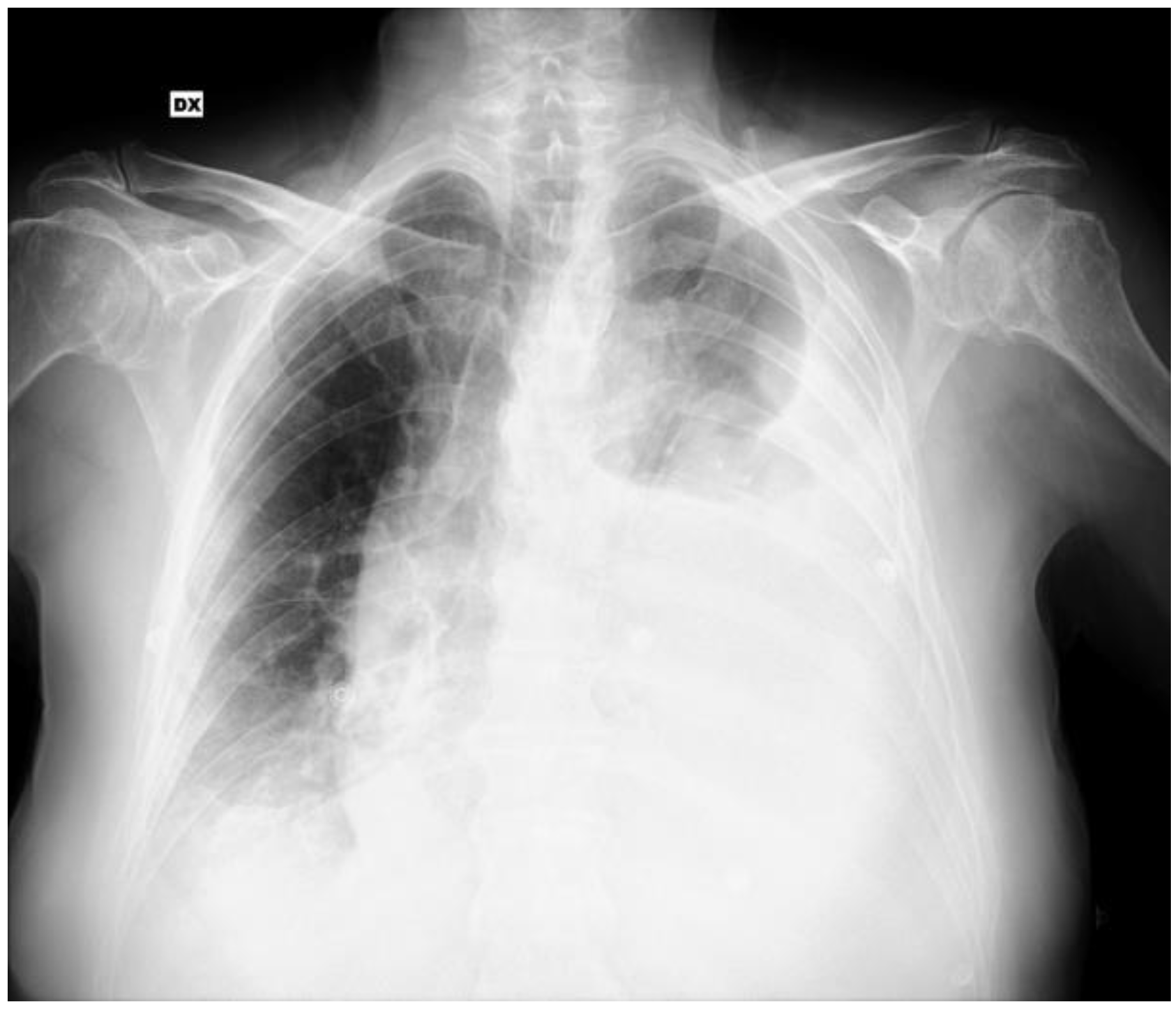
Figure 1. Chest radiography performed in the emergency department showing bilateral pleural effusion. DX: Right side.
Jcm 14 03377 g001
Figure 2. Contrast chest CT showing bilateral pleural effusion, consensual lung atelectasis, right-shifted cardiomediastinal axis, modest hyperemia, thickening of the left diaphragmatic pleural sheet and nodular thickenings of the left parietal pleura. These findings were suggestive of a malignancy involving the left parietal pleura and possibly neoplastic pleural effusion, prevalent on the left side.
Jcm 14 03377 g002
Figure 3. Chest contrast MR: bilateral pleural effusion with multiple saccular aspects as if due to partially bloody content. In the contrast phase, there were linear bilateral contrast enhancements of the pleura and laminar areas of restricted protonic diffusivity. These findings were consistent with pleural abnormalities, suggesting a neoplastic process, and with active bleeding in the pleural space.
Jcm 14 03377 g003
Figure 4. (a) Irregularly shaped anastomosing vascular channels, lined by atypical endothelial cells with a highly infiltrative architecture and poor demarcation (hematoxylin and eosin, magnification 10×); (b) solid growth pattern in poorly differentiated areas with epithelioid and pleomorphic cells (hematoxylin and eosin, magnification 20×); (c) in immunohistochemistry, neoplastic cells expressed ERG (endothelial marker). These findings, together with clinical and radiological data, were consistent with primary pleural epithelioid angiosarcoma.
Jcm 14 03377 g004
Table 1. Primary pleural angiosarcoma epidemiology and clinical, diagnostic and therapeutic features. The total number of cases is 47, unless otherwise specified.
Age (Mean ± SD)61.8 ± 15.3 years
SexMales: 39
Females: 10
SymptomsDyspnea: 22
Chest pain: 15
Hemoptysis: 6
Cough: 5
No respiratory symptoms: 2

(Total described cases: 39)
Pleural effusionNo:11 (23.9%)
Monolateral: 26 (56.5%)
Bilateral: 9 (19.6%)

Hematic effusion: 17 (48.6%)
Non-hematic effusion: 18 (51.4%)
Pleural effusion cytology, when performedDiagnostic: 3 (11.1%)
Not diagnostic: 24 (88.9%)

(Total described cases: 27)
CT specific findings,
when performed
(pleural masses or nodules)
Yes: 24 (57.1%)
No: 18 (42.9%)

(Total described cases: 42)
Diagnostic procedureCytology: 0 (0%)
Thoracoscopy and pleural biopsy: 33 (71.7%)
Autopsy: 7 (15.3%)
CT guided biopsy: 6 (13%)
TreatmentNo therapy: 11 (27.5%)
Chemotherapy: 18 (45%)
Radiotherapy: 3 (75%)
Surgery: 7 (17.5%)
Palliative care, including pleurodesis: 11 (27.5%)

(Total described cases: 40)
Survival from diagnosis
(mean ± SD)
20 ± 29.2 weeks
Disclaimer/Publisher’s Note: The statements, opinions and data contained in all publications are solely those of the individual author(s) and contributor(s) and not of MDPI and/or the editor(s). MDPI and/or the editor(s) disclaim responsibility for any injury to people or property resulting from any ideas, methods, instructions or products referred to in the content.