
Candida auris Infection, a Rapidly Emerging Threat in the Neonatal Intensive Care Units: A Systematic Review

 , , ,
, , ,  , , ,
, , , 
 and
and 
Abstract
1. Introduction
2. Materials and Methods
2.1. Inclusion Criteria
- Randomized controlled trials (RCTs), observational studies, case reports and case series;
- Population: neonates;
- Study design: studies focused on Candida auris infection, defined as positive blood, urine, CSF, or other sterile anatomical site culture in neonates.
2.2. Exclusion Criteria
- Surveys that included children and adults in the same group with neonates and did not offer information that solely involved the neonatal population;
- Review articles, systematic reviews and meta-analyses, and conference proceedings were excluded;
- Studies not published in the English language.
2.3. Research Objective
2.4. Main Outcome(s)
- Clinical presentation of Candida auris infection in neonates;
- Risk factors/characteristics of the affected population;
- Outcomes of the infection including mortality.
2.5. Search Strategy–Data Source
2.6. Resolving Conflicts
2.7. Data Synthesis and Presentation
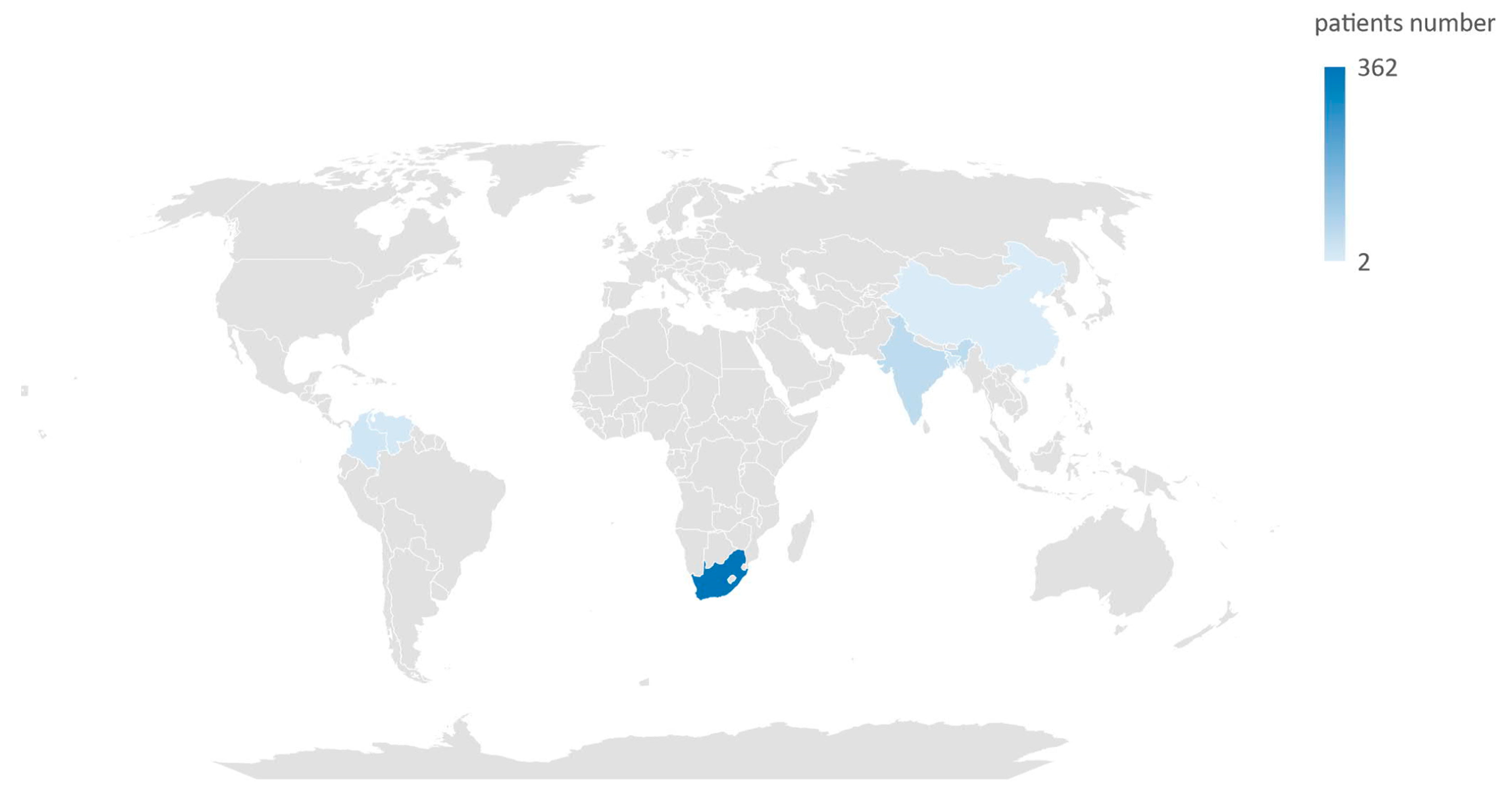
3. Results
4. Discussion
5. Conclusions
Supplementary Materials
Author Contributions
Funding
Institutional Review Board Statement
Informed Consent Statement
Data Availability Statement
Conflicts of Interest
References
- Spivak, E.S.; Hanson, K.E. Candida auris: An emerging fungal pathogen. J. Clin. Microbiol. 2018, 56, e01588-17. [Google Scholar] [CrossRef]
- Dahiya, S.; Chhillar, A.K.; Sharma, N.; Choudhary, P.; Punia, A.; Balhara, M.; Kaushik, K.; Parmar, V.S. Candida auris and Nosocomial Infection. Curr. Drug Targets 2020, 21, 365–373. [Google Scholar] [CrossRef]
- Sabino, R.; Veríssimo, C.; Pereira, Á.A.; Antunes, F. Candida auris, an agent of hospital-associated outbreaks: Which challenging issues do we need to have in mind? Microorganisms 2020, 8, 181. [Google Scholar] [CrossRef]
- Hu, S.; Zhu, F.; Jiang, W.; Wang, Y.; Quan, Y.; Zhang, G.; Gu, F.; Yang, Y. Retrospective Analysis of the Clinical Characteristics of Candida auris Infection Worldwide From 2009 to 2020. Front. Microbiol. 2021, 12, 658329. [Google Scholar] [CrossRef]
- Chen, J.; Tian, S.; Han, X.; Chu, Y.; Wang, Q.; Zhou, B.; Shang, H. Is the superbug fungus really so scary? A systematic review and meta-analysis of global epidemiology and mortality of Candida auris. BMC Infect. Dis. 2020, 20, 827. [Google Scholar] [CrossRef]
- Egger, N.B.; Kainz, K.; Schulze, A.; Bauer, M.A.; Madeo, F.; Carmona-Gutierrez, D. The rise of Candida auris: From unique traits to co-infection potential. Microb. Cell 2022, 9, 141–144. [Google Scholar] [CrossRef]
- Cristina, M.L.; Spagnolo, A.M.; Sartini, M.; Carbone, A.; Oliva, M.; Schinca, E.; Boni, S.; Pontali, E. An Overview on Candida auris in Healthcare Settings. J. Fungi 2023, 9, 913. [Google Scholar] [CrossRef]
- Satoh, K.; Makimura, K.; Hasumi, Y.; Nishiyama, Y.; Uchida, K.; Yamaguchi, H. Candida auris sp. nov., a novel ascomycetous yeast isolated from the external ear canal of an inpatient in a Japanese hospital. Microbiol. Immunol. 2009, 53, 41–44. [Google Scholar] [CrossRef] [PubMed]
- Kim, M.N.; Shin, J.H.; Sung, H.; Lee, K.; Kim, E.C.; Ryoo, N.; Lee, J.S.; Jung, S.I.; Park, K.H.; Kee, S.J.; et al. Candida haemulonii and closely related species at 5 university hospitals in Korea: Identification, antifungal susceptibility, and clinical features. Clin. Infect. Dis. 2009, 48, e57–e61. [Google Scholar] [CrossRef] [PubMed]
- Lee, W.G.; Shin, J.H.; Uh, Y.; Kang, M.G.; Kim, S.H.; Park, K.H.; Jang, H.C. First three reported cases of nosocomial fungemia caused by Candida auris. J. Clin. Microbiol. 2011, 49, 3139–3142. [Google Scholar] [CrossRef] [PubMed]
- Garcia-Bustos, V.; Cabanero-Navalon, M.D.; Ruiz-Saurí, A.; Ruiz-Gaitán, A.C.; Salavert, M.; Tormo, M.Á.; Pemán, J. What Do We Know about Candida auris? State of the Art, Knowledge Gaps, and Future Directions. Microorganisms 2021, 9, 2177. [Google Scholar] [CrossRef]
- CDC. Antibiotic Resistance Threats in the United States; U.S. Department of Health and Human Services, CDC: Atlanta, GA, USA, 2019. Available online: www.cdc.gov/DrugResistance/Biggest-Threats.html (accessed on 29 January 2024).
- Nelson, R. Emergence of resistant Candida auris. Lancet Microbe 2023, 4, e396. [Google Scholar] [CrossRef]
- Villanueva-Lozano, H.; Treviño-Rangel, R.D.J.; González, G.M.; Ramírez-Elizondo, M.T.; Lara-Medrano, R.; Aleman-Bocanegra, M.C.; Guajardo-Lara, C.E.; Gaona-Chávez, N.; Castilleja-Leal, F.; Torre-Amione, G.; et al. Outbreak of Candida auris infection in a COVID-19 hospital in Mexico. Clin. Microbiol. Infect. 2021, 27, 813–816. [Google Scholar] [CrossRef]
- Chowdhary, A.; Tarai, B.; Singh, A.; Sharma, A. Multidrug-Resistant Candida auris Infections in Critically Ill Coronavirus Disease Patients, India, April–July 2020. Emerg. Infect. Dis. 2020, 26, 2694–2696. [Google Scholar] [CrossRef]
- Moin, S.; Farooqi, J.; Rattani, S.; Nasir, N.; Zaka, S.; Jabeen, K. C. auris and non-C. auris candidemia in hospitalized adult and pediatric COVID-19 patients; single center data from Pakistan. Med. Mycol. 2021, 59, 1238–1242. [Google Scholar] [CrossRef]
- de Almeida, J.N.; Francisco, E.C.; Hagen, F.; Brandão, I.B.; Pereira, F.M.; Presta Dias, P.H.; de Miranda Costa, M.M.; de Souza Jordão, R.T.; de Groot, T.; Colombo, A.L. Emergence of Candida auris in Brazil in a COVID-19 Intensive Care Unit. J. Fungi 2021, 7, 220. [Google Scholar] [CrossRef]
- Prestel, C.; Anderson, E.; Forsberg, K.; Lyman, M.; Perio, M.; Kuhar, D.; Edwards, K.; Rivera, M.; Shugart, A.; Walters, M.; et al. Candida auris Outbreak in a COVID-19 Specialty Care Unit—Florida, July–August 2020. MMWR Morb. Mortal. Wkly. Rep. 2021, 70, 56–57. [Google Scholar] [CrossRef] [PubMed]
- Flores-Maldonado, O.; González, G.M.; Andrade, A.; Montoya, A.; Treviño-Rangel, R.; Silva-Sánchez, A.; Becerril-García, M.A. Dissemination of Candida auris to deep organs in neonatal murine invasive candidiasis. Microb. Pathog. 2021, 161, 105285. [Google Scholar] [CrossRef] [PubMed]
- Bagheri Lankarani, K.; Akbari, M.; Tabrizi, R.; Vali, M.; Sekhavati, E.; Heydari, S.T.; Khodadadi, H.; Ahmadizar, F. Candida auris: Outbreak fungal pathogen in COVID-19 pandemic: A systematic review and meta-analysis. Iran. J. Microbiol. 2022, 14, 276–284. [Google Scholar] [CrossRef] [PubMed]
- Warris, A.; Pana, Z.-D.; Oletto, A.; Lundin, R.; Castagnola, E.; Lehrnbecher, T.; Groll, A.H.; Roilides, E. Etiology and Outcome of Candidemia in Neonates and Children in Europe: An 11-year Multinational Retrospective Study. Pediatr. Infect. Dis. J. 2020, 39, 114–120. [Google Scholar] [CrossRef] [PubMed]
- Noni, M.; Stathi, A.; Vaki, I.; Velegraki, A.; Zachariadou, L.; Michos, A. Changing epidemiology of invasive candidiasis in children during a 10-year period. J. Fungi 2019, 5, 19. [Google Scholar] [CrossRef]
- Alvarado-Socarras, J.L.; Vargas-Soler, J.A.; Franco-Paredes, C.; Villegas-Lamus, K.C.; Rojas-Torres, J.P.; Rodriguez-Morales, A.J. A Cluster of Neonatal Infections Caused by Candida auris at a Large Referral Center in Colombia. J. Pediatr. Infect. Dis. Soc. 2021, 10, 549–555. [Google Scholar] [CrossRef]
- Armstrong, P.A.; Rivera, S.M.; Escandon, P.; Caceres, D.H.; Chow, N.; Stuckey, M.J.; Díaz, J.; Gomez, A.; Vélez, N.; Espinosa-Bode, A.; et al. Hospital-associated multicenter outbreak of emerging fungus Candida auris, colombia, 2016. Emerg. Infect. Dis. 2019, 25, 1339–1346. [Google Scholar] [CrossRef]
- Van Schalkwyk, E.; Mpembe, R.S.; Thomas, J.; Shuping, L.; Ismail, H.; Lowman, W.; Karstaedt, A.S.; Chibabhai, V.; Wadula, J.; Avenant, T.; et al. Epidemiologic shift in Candidemia driven by Candida auris, South Africa, 2016–2017. Emerg. Infect. Dis. 2019, 25, 1698–1707. [Google Scholar] [CrossRef]
- Kopanou Taliaka, P.; Tsantes, A.G.; Konstantinidi, A.; Houhoula, D.; Tsante, K.A.; Vaiopoulos, A.G.; Piovani, D.; Nikolopoulos, G.K.; Bonovas, S.; Iacovidou, N.; et al. Risk Factors, Diagnosis, and Treatment of Neonatal Fungal Liver Abscess: A Systematic Review of the Literature. Life 2023, 13, 167. [Google Scholar] [CrossRef]
- Hope, W.W.; Castagnola, E.; Groll, A.H.; Roilides, E.; Akova, M.; Arendrup, M.C.; Arikan-Akdagli, S.; Bassetti, M.; Bille, J.; Cornely, O.A.; et al. ESCMID* guideline for the diagnosis and management of Candida diseases 2012: Prevention and management of invasive infections in neonates and children caused by Candida spp. Clin. Microbiol. Infect. 2012, 18, 38–52. [Google Scholar] [CrossRef]
- Mantadakis, E.; Pana, Z.D.; Zaoutis, T. Candidemia in children: Epidemiology, prevention and management. Mycoses 2018, 61, 614–622. [Google Scholar] [CrossRef] [PubMed]
- Vasileiou, E.; Apsemidou, A.; Vyzantiadis, T.A.; Tragiannidis, A. Invasive candidiasis and candidemia in pediatric and neonatal patients: A review of current guidelines. Curr. Med. Mycol. 2018, 4, 28–33. [Google Scholar] [CrossRef] [PubMed]
- Hsu, J.F.; Lai, M.Y.; Lee, C.W.; Chu, S.M.; Wu, I.H.; Huang, H.R.; Lee, I.T.; Chiang, M.C.; Fu, R.H.; Tsai, M.H. Comparison of the incidence, clinical features and outcomes of invasive candidiasis in children and neonates. BMC Infect. Dis. 2018, 18, 194. [Google Scholar] [CrossRef] [PubMed]
- Bhattacharya, S.; Sae-Tia, S.; Fries, B.C. Candidiasis and mechanisms of antifungal resistance. Antibiotics 2020, 9, 312. [Google Scholar] [CrossRef] [PubMed]
- Chandramati, J.; Sadanandan, L.; Kumar, A.; Ponthenkandath, S. Neonatal Candida auris infection: Management and prevention strategies—A single centre experience. J. Paediatr. Child Health 2020, 56, 1565–1569. [Google Scholar] [CrossRef]
- Adilia, W. Candida auris, what do paediatricians need to know? Arch. Dis. Child. 2018, 103, 891. [Google Scholar] [CrossRef]
- Moher, D.; Liberati, A.; Tetzlaff, J.; Altman, D.G. Preferred reporting items for systematic reviews and meta-analyses: The PRISMA statement. J. Clin. Epidemiol. 2009, 62, 1006–1012. [Google Scholar] [CrossRef] [PubMed]
- Chowdhary, A.; Sharma, C.; Duggal, S.; Agarwal, K.; Prakash, A.; Singh, P.K.; Jain, S.; Kathuria, S.; Randhawa, H.S.; Hagen, F.; et al. New clonal strain of Candida auris, Delhi, India. Emerg. Infect. Dis. 2013, 19, 1670–1673. [Google Scholar] [CrossRef]
- Calvo, B.; Melo, A.S.; Perozo-Mena, A.; Hernandez, M.; Francisco, E.C.; Hagen, F.; Meis, J.F.; Colombo, A.L. First report of Candida auris in America: Clinical and microbiological aspects of 18 episodes of candidemia. J. Infect. 2016, 73, 369–374. [Google Scholar] [CrossRef]
- Lockhart, S.R.; Etienne, K.A.; Vallabhaneni, S.; Farooqi, J.; Chowdhary, A.; Govender, N.P.; Colombo, A.L.; Calvo, B.; Cuomo, C.A.; Desjardins, C.A.; et al. Simultaneous Emergence of Multidrug-Resistant Candida auris on 3 Continents Confirmed by Whole-Genome Sequencing and Epidemiological Analyses. Clin. Infect. Dis. 2017, 64, 134–140. [Google Scholar] [CrossRef]
- Rudramurthy, S.M.; Chakrabarti, A.; Paul, R.A.; Sood, P.; Kaur, H.; Capoor, M.R.; Kindo, A.J.; Marak, R.S.K.; Arora, A.; Sardana, R.; et al. Candida auris candidaemia in Indian ICUs: Analysis of risk factors. J. Antimicrob. Chemother. 2017, 72, 1794–1801. [Google Scholar] [CrossRef] [PubMed]
- Chen, Y.; Zhao, J.; Han, L.; Qi, L.; Fan, W.; Liu, J.; Wang, Z.; Xia, X.; Chen, J.; Zhang, L. Emergency of fungemia cases caused by fluconazole-resistant Candida auris in Beijing, China. J. Infect. 2018, 77, 561–571. [Google Scholar] [CrossRef]
- Dutta, S.; Rahman; Hossain, K.; Haq, J. Detection of Candida auris and its antifungal susceptibility: First report from Bangladesh. IMC J. Med. Sci. 2019, 13, 18–22. [Google Scholar] [CrossRef]
- Chakrabarti, A.; Sood, P.; Rudramurthy, S.M.; Chen, S.; Jillwin, J.; Iyer, R.; Sharma, A.; Harish, B.N.; Roy, I.; Kindo, A.J.; et al. Characteristics, outcome and risk factors for mortality of paediatric patients with ICU-acquired candidemia in India: A multicentre prospective study. Mycoses 2020, 63, 1149–1163. [Google Scholar] [CrossRef]
- Berrio, I.; Caceres, D.H.; Coronell, R.W.; Salcedo, S.; Mora, L.; Marin, A.; Varón, C.; Lockhart, S.R.; Escandón, P.; Berkow, E.L.; et al. Bloodstream Infections With Candida auris Among Children in Colombia: Clinical Characteristics and Outcomes of 34 Cases. J. Pediatr. Infect. Dis. Soc. 2021, 10, 151–154. [Google Scholar] [CrossRef] [PubMed]
- Withers, A.; Cronin, K.; Mabaso, M.; Brisighelli, G.; Gabler, T.; Harrison, D.; Patel, N.; Westgarth-Taylor, C.; Loveland, J. Neonatal surgical outcomes: A prospective observational study at a Tertiary Academic Hospital in Johannesburg, South Africa. Pediatr. Surg. Int. 2021, 37, 1061–1068. [Google Scholar] [CrossRef]
- Ramya, G.M.; Balakrishnan, U.; Chandrasekaran, A.; Abiramalatha, T.; Amboiram, P.; Sekar, U.; UshaDevi, R. Candida auris, an emerging pathogen—Challenge in the survival of microprimies. Indian J. Med. Microbiol. 2021, 39, 367–369. [Google Scholar] [CrossRef] [PubMed]
- Mishra, S.; Subramanian, A.; Kindo, A.J. Clinicomycological study of Candida isolates in a tertiary care hospital: A pilot study. J. Med. Soc. 2021, 35, 58. [Google Scholar]
- Shuping, L.; Mpembe, R.; Mhlanga, M.; Naicker, S.D.; Maphanga, T.G.; Tsotetsi, E.; Wadula, J.; Velaphi, S.; Nakwa, F.; Chibabhai, V.; et al. Epidemiology of Culture-confirmed Candidemia Among Hospitalized Children in South Africa, 2012–2017. Pediatr. Infect. Dis. J. 2021, 40, 730–737. [Google Scholar] [CrossRef]
- Chibabhai, V. Incidence of candidemia and prevalence of azole-resistant candidemia at a tertiary South African hospital—A retrospective laboratory analysis 2016–2020. S. Afr. J. Infect. Dis. 2022, 37, 326. [Google Scholar] [CrossRef]
- Mashau, R.C.; Meiring, S.T.; Dramowski, A.; Magobo, R.E.; Quan, V.C.; Perovic, O.; von Gottberg, A.; Cohen, C.; Velaphi, S.; van Schalkwyk, E.; et al. Culture-confirmed neonatal bloodstream infections and meningitis in South Africa, 2014-19: A cross-sectional study. Lancet Glob. Health 2022, 10, e1170–e1178. [Google Scholar] [CrossRef]
- Sathi, F.A.; Paul, S.K.; Ahmed, S.; Alam, M.M.; Nasreen, S.A.; Haque, N.; Islam, A.; Nila, S.S.; Afrin, S.Z.; Aung, M.S.; et al. Prevalence and Antifungal Susceptibility of Clinically Relevant Candida Species, Identification of Candida auris and Kodamaea ohmeri in Bangladesh. Trop. Med. Infect. Dis. 2022, 7, 211. [Google Scholar] [CrossRef]
- Cook, A.; Ferreras-Antolin, L.; Adhisivam, B.; Ballot, D.; Berkley, J.A.; Bernaschi, P.; Carvalheiro, C.G.; Chaikittisuk, N.; Chen, Y.; Chibabhai, V.; et al. Neonatal invasive candidiasis in low- and middle-income countries: Data from the NeoOBS study. Med. Mycol. 2023, 61, myad010. [Google Scholar] [CrossRef]
- Sathi, F.A.; Aung, M.S.; Paul, S.K.; Nasreen, S.A.; Haque, N.; Roy, S.; Ahmed, S.; Alam, M.M.; Khan, S.; Rabbany, M.A.; et al. Clonal Diversity of Candida auris, Candida blankii, and Kodamaea ohmeri Isolated from Septicemia and Otomycosis in Bangladesh as Determined by Multilocus Sequence Typing. J. Fungi 2023, 9, 658. [Google Scholar] [CrossRef]
- Singhal, T.; Shah, S.; Thakkar, P.; Ladi, S. Candida auris as the Predominant Species Causing Invasive Candidiasis in Neonates and Children. Indian J. Pediatr. 2023, 90, 946. [Google Scholar] [CrossRef]
- Shuping, L.; Maphanga, T.G.; Naicker, S.D.; Mpembe, R.; Ngoma, N.; Velaphi, S.; Nakwa, F.; Wadula, J.; Jaglal, P.; Govender, N.P. High Prevalence of Candida auris Colonization during Protracted Neonatal Unit Outbreak, South Africa. Emerg. Infect. Dis. 2023, 29, 1913–1916. [Google Scholar] [CrossRef]
- Ramdin, T.D.; Chibabhai, V.; Saggers, R.T.; Bandini, R.M.; Ballot, D.E. Epidemiology, risk factors and outcomes associated with candidaemia in very low birth weight infants at a tertiary South African Hospital over a 7-year period (2013–2019). Clin. Epidemiol. Glob. Health 2023, 20, 101247. [Google Scholar] [CrossRef]
- Stoll, B.J.; Hansen, N.; Fanaroff, A.A.; Wright, L.L.; Carlo, W.A.; Ehrenkranz, R.A.; Lemons, J.A.; Donovan, E.F.; Stark, A.R.; Tyson, J.E. Late-onset sepsis in very low birth weight neonates: The experience of the NICHD Neonatal Research Network. Pediatrics 2002, 110, 285–291. [Google Scholar] [CrossRef]
- Weimer, K.E.D.; Smith, P.B.; Puia-Dumitrescu, M.; Aleem, S. Invasive fungal infections in neonates: A review. Pediatr. Res. 2022, 91, 404–412. [Google Scholar] [CrossRef]
- Kelly, M.S.; Benjamin, D.K., Jr.; Smith, P.B. The epidemiology and diagnosis of invasive candidiasis among premature infants. Clin. Perinatol. 2015, 42, 105–117. [Google Scholar] [CrossRef] [PubMed]
- Ben-Ami, R.; Berman, J.; Novikov, A.; Bash, E.; Shachor-Meyouhas, Y.; Zakin, S.; Maor, Y.; Tarabia, J.; Schechner, V.; Adler, A. Multidrug-resistant Candida haemulonii and C. auris, tel aviv, Israel. Emerg. Infect. Dis. 2017, 23, 195. [Google Scholar] [CrossRef] [PubMed]
- Chowdhary, A.; Jain, K.; Chauhan, N. Candida auris Genetics and Emergence. Annu. Rev. Microbiol. 2023, 77, 583–602. [Google Scholar] [CrossRef]
- Ashkenazi-Hoffnung, L.; Rosenberg Danziger, C. Navigating the New Reality: A Review of the Epidemiological, Clinical, and Microbiological Characteristics of Candida auris, with a Focus on Children. J. Fungi 2023, 9, 176. [Google Scholar] [CrossRef]
- Munshi, A.; Almadani, F.; Ossenkopp, J.; Alharbi, M.; Althaqafi, A.; Alsaedi, A.; Al-Amri, A.; Almarhabi, H. Risk factors, antifungal susceptibility, complications, and outcome of Candida auris bloodstream infection in a tertiary care center in the western region of Saudi Arabia. J. Infect. Public Health 2024, 17, 182–188. [Google Scholar] [CrossRef] [PubMed]
- Vinayagamoorthy, K.; Pentapati, K.C.; Prakash, H. Prevalence, risk factors, treatment and outcome of multidrug resistance Candida auris infections in Coronavirus disease (COVID-19) patients: A systematic review. Mycoses 2022, 65, 613–624. [Google Scholar] [CrossRef]
- Horton, M.V.; Holt, A.M.; Nett, J.E. Mechanisms of pathogenicity for the emerging fungus Candida auris. PLoS Pathog. 2023, 19, e1011843. [Google Scholar] [CrossRef] [PubMed]
- Santana, D.J.; Anku, J.A.E.; Zhao, G.; Zarnowski, R.; Johnson, C.J.; Hautau, H.; Visser, N.D.; Ibrahim, A.S.; Andes, D.; Nett, J.E.; et al. A Candida auris-specific adhesin, Scf1, governs surface association, colonization, and virulence. Science 2023, 381, 1461–1467. [Google Scholar] [CrossRef] [PubMed]
- Fakhim, H.; Vaezi, A.; Dannaoui, E.; Chowdhary, A.; Nasiry, D.; Faeli, L.; Meis, J.F.; Badali, H. Comparative virulence of Candida auris with Candida haemulonii, Candida glabrata and Candida albicans in a murine model. Mycoses 2018, 61, 377–382. [Google Scholar] [CrossRef] [PubMed]
- Torres, S.R.; Pichowicz, A.; Torres-Velez, F.; Song, R.; Singh, N.; Lasek-Nesselquist, E.; De Jesus, M. Impact of Candida auris Infection in a Neutropenic Murine Model. Antimicrob. Agents Chemother. 2020, 64, e01625-19. [Google Scholar] [CrossRef]
- Staniszewska, M. Virulence Factors in Candida species. Curr. Protein Pept. Sci. 2020, 21, 313–323. [Google Scholar] [CrossRef]
- Pristov, K.E.; Ghannoum, M.A. Resistance of Candida to azoles and echinocandins worldwide. Clin. Microbiol. Infect. 2019, 25, 792–798. [Google Scholar] [CrossRef] [PubMed]
- Navarro-Arias, M.J.; Hernández-Chávez, M.J.; García-Carnero, L.C.; Amezcua-Hernández, D.G.; Lozoya-Pérez, N.E.; Estrada-Mata, E.; Martínez-Duncker, I.; Franco, B.; Mora-Montes, H.M. Differential recognition of Candida tropicalis, Candida guilliermondii, Candida krusei, and Candida auris by human innate immune cells. Infect. Drug Resist. 2019, 12, 783–794. [Google Scholar] [CrossRef]
- Netea, M.G.; Joosten, L.A.; van der Meer, J.W.; Kullberg, B.J.; van de Veerdonk, F.L. Immune defence against Candida fungal infections. Nat. Rev. Immunol. 2015, 15, 630–642. [Google Scholar] [CrossRef]
- Bruno, M.; Kersten, S.; Bain, J.M.; Jaeger, M.; Rosati, D.; Kruppa, M.D.; Lowman, D.W.; Rice, P.J.; Graves, B.; Ma, Z.; et al. Transcriptional and functional insights into the host immune response against the emerging fungal pathogen Candida auris. Nat. Microbiol. 2020, 5, 1516–1531. [Google Scholar] [CrossRef]
- Cortegiani, A.; Misseri, G.; Fasciana, T.; Giammanco, A.; Giarratano, A.; Chowdhary, A. Epidemiology, clinical characteristics, resistance, and treatment of infections by Candida auris. J. Intensive Care 2018, 6, 69. [Google Scholar] [CrossRef]
- Keighley, C.; Garnham, K.; Harch, S.A.J.; Robertson, M.; Chaw, K.; Teng, J.C.; Chen, S.C. Candida auris: Diagnostic Challenges and Emerging Opportunities for the Clinical Microbiology Laboratory. Curr. Fungal Infect. Rep. 2021, 15, 116–126. [Google Scholar] [CrossRef] [PubMed]
- Yamamoto, M.; Alshahni, M.M.; Tamura, T.; Satoh, K.; Iguchi, S.; Kikuchi, K.; Mimaki, M.; Makimura, K. Rapid detection of Candida auris based on loop-mediated isothermal amplification (LAMP). J. Clin. Microbiol. 2018, 56, e00591-18. [Google Scholar] [CrossRef]
- Sexton, D.J.; Bentz, M.L.; Welsh, R.M.; Litvintseva, A.P. Evaluation of a new T2 Magnetic Resonance assay for rapid detection of emergent fungal pathogen Candida auris on clinical skin swab samples. Mycoses 2018, 61, 786–790. [Google Scholar] [CrossRef] [PubMed]
- Briano, F.; Magnasco, L.; Sepulcri, C.; Dettori, S.; Dentone, C.; Mikulska, M.; Ball, L.; Vena, A.; Robba, C.; Patroniti, N. Candida auris candidemia in critically ill, colonized patients: Cumulative incidence and risk factors. Infect. Dis. Ther. 2022, 11, 1149–1160. [Google Scholar] [CrossRef] [PubMed]
- Bandara, N.; Samaranayake, L. Emerging and future strategies in the management of recalcitrant Candida auris. Med. Mycol. 2022, 60, myac008. [Google Scholar] [CrossRef] [PubMed]
- Ostrowsky, B.; Greenko, J.; Adams, E.; Quinn, M.; O’Brien, B.; Chaturvedi, V.; Berkow, E.; Vallabhaneni, S.; Forsberg, K.; Chaturvedi, S. Candida auris isolates resistant to three classes of antifungal medications—New York, 2019. Morb. Mortal. Wkly. Rep. 2020, 69, 6. [Google Scholar] [CrossRef]
- Kilburn, S.; Innes, G.; Quinn, M.; Southwick, K.; Ostrowsky, B.; Greenko, J.A.; Lutterloh, E.; Greeley, R.; Magleby, R.; Chaturvedi, V.; et al. Antifungal Resistance Trends of Candida auris Clinical Isolates in New York and New Jersey from 2016 to 2020. Antimicrob. Agents Chemother. 2022, 66, e0224221. [Google Scholar] [CrossRef]
- Zhang, D.; Xie, D.; He, N.; Wang, X.; Dong, W.; Lei, X. Prophylactic Use of Fluconazole in Very Premature Infants. Front. Pediatr. 2021, 9, 726769. [Google Scholar] [CrossRef]
- CDC. Treatment and Management of Infections and Colonization|Candida auris|Fungal Diseases|CDC. Available online: https://www.cdc.gov/fungal/candida-auris/c-auris-treatment.html (accessed on 30 December 2023).
- Tedersoo, L.; Bahram, M.; Põlme, S.; Kõljalg, U.; Yorou, N.S.; Wijesundera, R.; Ruiz, L.V.; Vasco-Palacios, A.M.; Thu, P.Q.; Suija, A.; et al. Global diversity and geography of soil fungi. Science 2014, 346, 1256688. [Google Scholar] [CrossRef]
- Ruiz-Gaitán, A.; Moret, A.M.; Tasias-Pitarch, M.; Aleixandre-López, A.I.; Martínez-Morel, H.; Calabuig, E.; Salavert-Lletí, M.; Ramírez, P.; López-Hontangas, J.L.; Hagen, F.; et al. An outbreak due to Candida auris with prolonged colonisation and 381 candidaemia in a tertiary care European hospital. Mycoses 2018, 61, 498–505. [Google Scholar] [CrossRef]
- O’Brien, B.; Liang, J.; Chaturvedi, S.; Jacobs, J.L.; Chaturvedi, V. Pan-resistant Candida auris: New York subcluster susceptible to antifungal combinations. Lancet Microbe 2020, 1, e193–e194. [Google Scholar] [CrossRef] [PubMed]
- Jaggavarapu, S.; Burd, E.M.; Weiss, D.S. Micafungin and amphotericin B synergy against Candida auris. Lancet Microbe 2020, 1, e314–e315. [Google Scholar] [CrossRef] [PubMed]
- O’Brien, B.; Chaturvedi, S.; Chaturvedi, V. In vitro evaluation of antifungal drug combinations against multidrug-resistant Candida auris isolates from New York outbreak. Antimicrob. Agents Chemother. 2020, 64, e02195-19. [Google Scholar] [CrossRef] [PubMed]
- Mesini, A.; Saffioti, C.; Mariani, M.; Florio, A.; Medici, C.; Moscatelli, A.; Castagnola, E. First case of Candida auris colonization in a preterm, extremely low-birth-weight newborn after vaginal delivery. J. Fungi 2021, 7, 649. [Google Scholar] [CrossRef] [PubMed]
- Maede, Y.; Ibara, S.; Nagasaki, H.; Inoue, T.; Tokuhisa, T.; Torikai, M.; Ishihara, C.; Matsui, T.; Kodaira, Y. Micafungin versus fluconazole for prophylaxis against fungal infections in premature infants. Pediatr. Int. 2013, 55, 727–730. [Google Scholar] [CrossRef] [PubMed]
- Bassetti, M.; Giacobbe, D.R.; Vena, A.; Esposito, S. An overview of micafungin as a treatment option for invasive candidiasis in pediatric patients younger than 4 months old. Expert Opin. Pharmacother. 2022, 23, 1987–1993. [Google Scholar] [CrossRef]
- Ascher, S.; Smith, P.B.; Benjamin, D.K., Jr. Safety of micafungin in infants: Insights into optimal dosing. Expert Opin. Drug Saf. 2011, 10, 281–286. [Google Scholar] [CrossRef]


| Authors | Country | Time Period | Type of Study | Number of Neonates | GA (Weeks) | Sex | BW (Grams) | Age at Diagnosis (Days) | Positive Blood Culture | Other Culture Site | Symptoms | Underlying Condition | Coinfection with | Treatment with | Survived | Died |
|---|---|---|---|---|---|---|---|---|---|---|---|---|---|---|---|---|
| Chowdhary [35], 2013 | India | 2009–2011 | Case series | 1 | NR | F | NR | 3 | 1 | NR | NR | TEF, ICH, bacterial sepsis | Bacterial sepsis (not specified) | CASP | 1 | |
| 1 | NR | F | NR | 10 | 1 | NR | NR | ELBW, bacterial sepsis | Bacterial sepsis (not specified) | AMB | 1 | |||||
| 1 | NR | F | NR | 28 | 1 | NR | NR | Pneumonia, late-onset sepsis | NR | AMB | 1 | |||||
| Calvo [36], 2016 | Venezuela | 03/2012–07/2013 | Case series | 1 | NR | F | NR | 30 | 1 | NR | Sepsis | NR | NR | AMB, VOR | 1 | |
| 1 | NR | F | NR | 13 | 1 | NR | Sepsis | colonic atresia | NR | AMB, VOR, CASP | 1 | |||||
| 1 | NR | M | NR | 17 | 1 | NR | Sepsis | NR | NR | FLU | 1 | |||||
| 1 | NR | F | NR | 23 | 1 | NR | Septic shock | NR | NR | CASP | 1 | |||||
| 1 | NR | M | NR | 18 | 1 | NR | Sepsis | NR | NR | FLU, VOR | 1 | |||||
| 1 | NR | M | NR | 2 | 1 | NR | NR | CHD and intestinal atresia | NR | CASP | 1 | |||||
| 1 | NR | M | NR | 12 | 1 | NR | Sepsis | HIE, NEC | NR | VOR, CASP | 1 | |||||
| 1 | NR | M | NR | 11 | 1 | NR | Sepsis | Surgical NEC | NR | AMB, CASP | 1 | |||||
| 1 | NR | F | NR | 18 | 1 | NR | Sepsis | Congenital abdominal wall defect | NR | VOR | 1 | |||||
| 1 | NR | F | NR | 10 | 1 | NR | Sepsis | HIE | NR | FLU | 1 | |||||
| 1 | NR | M | NR | 49 | 1 | NR | Sepsis | RDS | NR | VOR, CASP | 1 | |||||
| Lockhart [37], 2017 | Venezuela | 2012–2015 | Public health surveillance program | 3 | NR | NR | NR | NR | 3 | NR | NR | NR | NR | NR | NR | NR |
| Rudramurthy [38], 2017 | India | 2011–2012 | Retrospective observational study | 6 | NR | NR | NR | NR | 6 | NR | NR | NR | NR | NR | NR | NR |
| Chen [39], 2018 | China | 2018 | Case series | 1 | NR | M | 940 | 71 | 1 | NR | NR | NR | NR | FLU, then ITRA | 1 | |
| 1 | NR | F | 960 | 23 | 1 | NR | NR | NR | NR | FLU, then ITRA | 1 | |||||
| van Schalkwyk [25], 2019 | South Africa | 2006–2017 | Active national laboratory-based surveillance | 20 | NR | NR | NR | NR | 20 | NR | NR | NR | NR | NR | NR | NR |
| Dutta [40], 2019 | Bangladesh | NR | Observational study | 13 | NR | NR | NR | NR | 13 | NR | NR | NR | NR | NR | NR | NR |
| Armstrong [24], 2019 | Colombia | 01/2015–09/2016 | Outbreak investigation | 5 | 36 (29–39) | NR | NR | NR | 5 | NR | NR | NR | NR | NR | 4 | 1 |
| Chakrabarti [41], 2020 | India | 04/2011–09/2012 | Prospective observational study | 6 | NR | NR | NR | NR | 6 | NR | NR | NR | NR | NR | 4 | 2 |
| Berrio [42], 2020 | Colombia | 07/2014–10/2017 | Retrospective microbiological review | 7 | NR | 4M, 3F | NR | NR | 7 | NR | NR | NR | NR | 3 AMB, 3 azoles, 3 caspofungin | 3 | 4 |
| Chandramati [32], 2020 | India | 2016–2017 | Retrospective cohort study | 1 | 31 | M | 1250 | 19 (+/−10) | 1 | NR | Skin abscess, fever, tachycardia | RDS | NR | VOR | 1 | |
| 1 | 28 | F | 900 | 19 (+/−10) | 1 | NR | Respiratory distress, lethargy | RDS, pneumothorax | NR | VOR | 1 | |||||
| 1 | 28 | M | 850 | 19 (+/−10) | 1 | Urine | Respiratory distress, tachycardia | AREDF | NR | VOR, MIC | 1 | |||||
| 1 | 25 | M | 600 | 19 (+/−10) | 1 | Urine, lungs | Apnea, coagulopathy | RDS | NR | AMB, VOR | 1 | |||||
| 1 | 25 | M | 560 | 19 (+/−10) | 1 | Urine, CSF | Feeding intolerance | RDS, NEC | NR | VOR, MIC | 1 | |||||
| 1 | 36 | M | 2460 | 19 (+/−10) | 1 | NR | Lethargy, abdominal distension | CHD | NR | AMB, VOR | 1 | |||||
| 1 | 37 | M | 2650 | 19 (+/−10) | 1 | Urine | Respiratory distress, tachycardia | Anemia, hepatosplenomegaly | NR | FLU, VOR | 1 | |||||
| 1 | 31 | M | 1250 | 19 (+/−10) | 1 | NR | Apnea, lethargy, tachycardia | Inborn error of metabolism, renal failure | NR | FLU | 1 | |||||
| 1 | 25 | M | 500 | 19 (+/−10) | 1 | NR | Apnea, lethargy | RDS, coagulopathy | NR | FLU | 1 | |||||
| 1 | 40 | M | 2950 | 19 (+/−10) | 1 | Pus | Respiratory distress, tachycardia, skin abscess | MAS, BPD | NR | MIC, AMB | 1 | |||||
| 1 | 39 | F | 2750 | 19 (+/−10) | 1 | Urine, eye | Respiratory distress, tachycardia | CDH | NR | MIC, AMB | 1 | |||||
| 1 | 35 | M | 2520 | 19 (+/−10) | 1 | Urine | Respiratory distress, tachycardia | CDH | NR | MIC, AMB | 1 | |||||
| 1 | 31 | F | 1220 | 19 (+/−10) | 1 | Wound | Respiratory distress, tachycardia | Jejunal atresia | NR | MIC, AMB | 1 | |||||
| 1 | 35 | M | 2290 | 19 (+/−10) | 1 | NR | Respiratory distress | Jejunal atresia | NR | MIC, AMB | 1 | |||||
| 1 | 33 | F | 1430 | 19 (+/−10) | 1 | NR | Respiratory distress | TOF | NR | AMB, VOR | 1 | |||||
| 1 | 37 | M | 2670 | 19 (+/−10) | 1 | Urine | Fever, tachycardia | CDH | NR | VOR | 1 | |||||
| 1 | 36 | F | 2410 | 19 (+/−10) | 1 | NR | Fever, feeding intolerance | Jejunal atresia | NR | AMB, VOR | 1 | |||||
| Withers [43], 2021 | South Africa | 2019 | Prospective observational study | 14 | NR | NR | NR | NR | 14 | NR | NR | NR | NR | NR | NR | NR |
| Ramya [44], 2021 | India | 08/2019–09/2020 | Case series | 1 | 26 | F | 675 | 7 | 1 | NR | Long line induration | NR | NR | MIC | 1 | |
| 1 | 26 | M | 790 | 14 | 1 | Budding yeast in urine NOT specified | Apnea | NR | NR | MIC | 1 | |||||
| 1 | 25 | M | 620 | 9 | 1 | NR | Apnea | NR | NR | MIC, VOR | 1 | |||||
| 1 | 26 | M | 775 | 7 | 1 | Budding yeast in urine NOT specified | Abdominal distension | NR | NR | MIC, VOR | 1 | |||||
| 1 | 27 | F | 685 | 10 | 1 | NR | Abdominal distension | NR | NR | MIC, VOR | 1 | |||||
| Alvarado-Socarras [23], 2021 | Colombia | 2015–2017 | Case series | 1 | 35 | NR | 2300 | 12 | 1 | peritoneal fluid | Fever | NEC | NR | CASP | 1 | |
| 1 | 39 | NR | 3100 | 14 | 0 | Urine | Fever, thrombocytopenia | CHD | NR | FLU | 1 | |||||
| 1 | 38 | NR | 2800 | 15 | 0 | CHD, hydrocephalus, omphalocele | Kl.pneumoniae (wound) and A.Junii (blood) | 14 | CASP | 1 | ||||||
| 1 | 39 | NR | 3200 | 37 | 1 | Surgical sternal incision and pleural fluid | NR | CHD (hypoplastic left heart and anomalous venous return) | NR | FLU & CASP | 1 | |||||
| 1 | 38 | NR | 3400 | 14 | 1 | 0 | NR | CHD (Ebstein anomaly) | NR | CASP & FLU | 1 | |||||
| 1 | 39 | NR | 3100 | 9 | 1 | Urine (positive for C.tropicalis and R. ornitinolytica) | Unspecified respiratory symptoms | Dilated cardiomyopathy | NR | CASP | 1 | |||||
| 1 | 38 | NR | 3300 | 16 | 0 | urine | Thrombocytopenia | CHD (d-TGA) | NR | 1 | ||||||
| 1 | 39 | NR | 3400 | 13 | 0 | Central venous catheter tip | Thrombocytopenia | CHD and right hydronephrosis, NEC, Meckel’s diverticulum | NR | CASP | 1 | |||||
| Mishra [45], 2021 | India | 08/2020–09/2020 | Cross-sectional study | 1 | 27 | NR | NR | NR | 1 | NR | Sepsis, refractory shock | NEC | NR | MIC, VOR | 1 | |
| Shuping [46], 2021 | South Africa | 2012–2017 | Laboratory-based surveillance | 7 | NR | NR | NR | NR | 7 | NR | NR | NR | NR | NR | NR | NR |
| Chibabhai [47], 2022 | South Africa | 01/2016–12/2020 | Retrospective laboratory analysis | 14 | NR | NR | NR | NR | 14 | NR | NR | NR | NR | NR | NR | NR |
| Mashau [48], 2022 | South Africa | 2014–2019 | Cross-sectional study | 60 | NR | NR | NR | NR | 59 | 1 (CSF) | NR | NR | NR | NR | NR | NR |
| Sathi [49], 2022 | Bangladesh | 2021 | Cross-sectional study | 1 | NR | M | NR | 15 | 1 | NR | NR | NR | NR | VOR | 1 | |
| 1 | NR | M | NR | 2 | 1 | NR | NR | NR | NR | VOR | 1 | |||||
| 1 | NR | M | NR | 12 | 1 | NR | NR | NR | NR | VOR | 1 | |||||
| Cook [50], 2023 | Various low- and middle-income countries | 2018–2020 | Prospective observational cohort study | 18 | NR | NR | NR | NR | 18 | NR | NR | NR | NR | NR | 13 | 5 |
| Sathi [51], 2023 | Bangladesh | 2021 | Retrospective study | 1 | NR | F | LBW | 6 | 1 | NR | Septicaemia | NR | NR | NR | 1 | |
| 1 | NR | M | LBW | 15 | 1 | NR | Septicaemia | NR | NR | NR | 1 | |||||
| 1 | NR | M | LBW | 30 | 1 | NR | Septicaemia | NR | NR | NR | 1 | |||||
| 1 | NR | M | NR | 2 | 1 | NR | Septicaemia | NR | NR | NR | 1 | |||||
| 1 | NR | M | LBW | 1 | 1 | NR | Septicaemia | NR | NR | NR | 1 | |||||
| 1 | NR | M | NR | 22 | 1 | NR | Septicaemia | NR | NR | NR | 1 | |||||
| 1 | NR | M | LBW | 2 | 1 | NR | Septicaemia | NR | NR | NR | 1 | |||||
| Singhal [52], 2023 | India | 2015–2022 | Retrospective observational study | 6 | >34 | NR | NR | NR | 6 | NR | NR | NR | NR | NR | NR | NR |
| Shuping [53], 2023 | South Africa | 06/2019–06/2022 | Point prevalence surveys | 208 | NR | 102 F, 97 M | NR | 21 days (IQR, 14–36) | NR | NR | NR | NR | NR | NR | NR | NR |
| Ramdin [54], 2023 | South Africa | 01/2013–12/2019 | Retrospective cohort study | 32 | NR | NR | NR | NR | 32 | NR | NR | NR | NR | NR | NR | NR |
| Reported Data | Missing Data | ||
|---|---|---|---|
| Gestational age (weeks) | Preterm | 20/31 (64.5) | 445/476 (93.5) |
| Term | 11/31 (35.5) | ||
| Gender | Male | 132/254 (52.0) | 222/476 (46.6) |
| Female | 122/254 (48.0) | ||
| Predisposing factors | Prematurity | 55/71 (77.5) | 405/476 (85.1) |
| Total parenteral nutrition administration | 25/94 (26.6) | 382/476 (80.3) | |
| Central line catheter placement | 41/94 (43.6) | 382/476 (80.3) | |
| Mechanical ventilation | 17/94 (18.1) | 382/476 (80.3) | |
| Broad-spectrum antibiotic administration | 18/94 (19.1) | 382/476 (80.3) |
| Isolation Site | Reported Data | Missing Data |
|---|---|---|
| Blood specimen | 263/268 (98.1) | 208/476 (43.7) |
| Other isolation sites | ||
| Urine | 14/85 (16.5) | 391/476 (82.1) |
| Peritoneal fluid specimen | 2/85 (2.4) | |
| Cerebrospinal fluid specimen | 2/85 (2.4) | |
| Central venous catheter tip | 1/85 (1.2) | |
| Surgical sternal incision and pleural fluid | 1/85 (1.2) | |
| Pus | 1/85 (1.2) | |
| Wound | 1/85 (1.2) | |
| Antifungal Therapy | Patients (n Reported Data/n all Cases, %) |
|---|---|
| Amphotericin B monotherapy | 15/57 (26.3) |
| Amphotericin B and voriconazole | 7/57 (12.3) |
| Amphotericin B and caspofungin | 1/57 (1.8) |
| Fluconazole | 5/57 (8.8) |
| Fluconazole and voriconazole | 2/57 (3.5) |
| Fluconazole and caspofungin | 2/57 (3.5) |
| Fluconazole and itraconazole | 2/57 (3.5) |
| Micafungin | 2/57 (3.5) |
| Micafungin and voriconazole | 8/57 (14.0) |
| Micafungin and amphotericin B | 2/57 (3.5) |
| Voriconazole | 4/57 (7.0) |
| Voriconazole, amphotericin B, and caspofungin | 1/57 (1.8) |
| Survived | 54/93 (58.1) |
| Died | 39/93 (41.9) |
Disclaimer/Publisher’s Note: The statements, opinions and data contained in all publications are solely those of the individual author(s) and contributor(s) and not of MDPI and/or the editor(s). MDPI and/or the editor(s) disclaim responsibility for any injury to people or property resulting from any ideas, methods, instructions or products referred to in the content. |
© 2024 by the authors. Licensee MDPI, Basel, Switzerland. This article is an open access article distributed under the terms and conditions of the Creative Commons Attribution (CC BY) license (https://creativecommons.org/licenses/by/4.0/).
Share and Cite
Sokou, R.; Palioura, A.E.; Kopanou Taliaka, P.; Konstantinidi, A.; Tsantes, A.G.; Piovani, D.; Tsante, K.A.; Gounari, E.A.; Iliodromiti, Z.; Boutsikou, T.; et al. Candida auris Infection, a Rapidly Emerging Threat in the Neonatal Intensive Care Units: A Systematic Review. J. Clin. Med. 2024, 13, 1586. https://doi.org/10.3390/jcm13061586
Sokou R, Palioura AE, Kopanou Taliaka P, Konstantinidi A, Tsantes AG, Piovani D, Tsante KA, Gounari EA, Iliodromiti Z, Boutsikou T, et al. Candida auris Infection, a Rapidly Emerging Threat in the Neonatal Intensive Care Units: A Systematic Review. Journal of Clinical Medicine. 2024; 13(6):1586. https://doi.org/10.3390/jcm13061586
Chicago/Turabian StyleSokou, Rozeta, Alexia Eleftheria Palioura, Paschalia Kopanou Taliaka, Aikaterini Konstantinidi, Andreas G. Tsantes, Daniele Piovani, Konstantina A. Tsante, Eleni A. Gounari, Zoi Iliodromiti, Theodora Boutsikou, and et al. 2024. "Candida auris Infection, a Rapidly Emerging Threat in the Neonatal Intensive Care Units: A Systematic Review" Journal of Clinical Medicine 13, no. 6: 1586. https://doi.org/10.3390/jcm13061586
APA StyleSokou, R., Palioura, A. E., Kopanou Taliaka, P., Konstantinidi, A., Tsantes, A. G., Piovani, D., Tsante, K. A., Gounari, E. A., Iliodromiti, Z., Boutsikou, T., Tsantes, A. E., Bonovas, S., & Iacovidou, N. (2024). Candida auris Infection, a Rapidly Emerging Threat in the Neonatal Intensive Care Units: A Systematic Review. Journal of Clinical Medicine, 13(6), 1586. https://doi.org/10.3390/jcm13061586