Article

Comparative Uptake Patterns of Radioactive Iodine and [18F]-Fluorodeoxyglucose (FDG) in Metastatic Differentiated Thyroid Cancers

by
Devan Diwanji
1,2,
Emmanuel Carrodeguas
1,
Youngho Seo
1,3,4,5,
Hyunseok Kang
6,
Myat Han Soe
7,
Janet M. Chiang
7,8,9,
Li Zhang
6,10,
Chienying Liu
7,
Spencer C. Behr
1 and
Robert R. Flavell
1,11,12,*
1
Department of Radiology and Biomedical Imaging, University of California, San Francisco, CA 94143, USA
2
Medical Scientist Training Program, University of California, San Francisco, CA 94143, USA
3
Molecular Biophysics and Integrated Bioimaging Division, Lawrence Berkeley National Laboratory, Berkeley, CA 94720, USA
4
Joint Graduate Group in Bioengineering, University of California, San Francisco, CA 94720, USA
5
Department of Nuclear Engineering, University of California, Berkeley, CA 94720, USA
6
Division of Hematology/Oncology, Department of Medicine, University of California, San Francisco, CA 94143, USA
7
Division of Endocrinology, Department of Medicine, University of California, San Francisco, CA 94143, USA
8
Division of Endocrinology, Department of Medicine, San Francisco VA Healthcare System, San Francisco, CA 94121, USA
9
Division of Endocrinology, Department of Medicine, Zuckerberg San Francisco General Hospital and Trauma Center, San Francisco, CA 94110, USA
10
Department of Epidemiology and Biostatistics, University of California, San Francisco, CA 94143, USA
11
Molecular Imaging and Therapeutics Clinical Section, Department of Radiology and Biomedical Imaging, University of California, San Francisco, CA 94143, USA
12
Department of Pharmaceutical Chemistry, University of California, San Francisco, CA 94143, USA
*
Author to whom correspondence should be addressed.
J. Clin. Med. 2024, 13(13), 3963; https://doi.org/10.3390/jcm13133963
Submission received: 10 June 2024 / Revised: 26 June 2024 / Accepted: 1 July 2024 / Published: 6 July 2024
(This article belongs to the Section Oncology)

Abstract

:
Background: Metastatic differentiated thyroid cancer (DTC) represents a molecularly heterogeneous group of cancers with varying radioactive iodine (RAI) and [18F]-fluorodeoxyglucose (FDG) uptake patterns potentially correlated with the degree of de-differentiation through the so-called “flip-flop” phenomenon. However, it is unknown if RAI and FDG uptake patterns correlate with molecular status or metastatic site. Materials and Methods: A retrospective analysis of metastatic DTC patients (n = 46) with radioactive 131-iodine whole body scan (WBS) and FDG-PET imaging between 2008 and 2022 was performed. The inclusion criteria included accessible FDG-PET and WBS studies within 1 year of each other. Studies were interpreted by two blinded radiologists for iodine or FDG uptake in extrathyroidal sites including lungs, lymph nodes, and bone. Cases were stratified by BRAF V600E mutation status, histology, and a combination of tumor genotype and histology. The data were analyzed by McNemar’s Chi-square test. Results: Lung metastasis FDG uptake was significantly more common than iodine uptake (WBS: 52%, FDG: 84%, p = 0.04), but no significant differences were found for lymph or bone metastases. Lung metastasis FDG uptake was significantly more prevalent in the papillary pattern sub-cohort (WBS: 37%, FDG: 89%, p = 0.02) than the follicular pattern sub-cohort (WBS: 75%, FDG: 75%, p = 1.00). Similarly, BRAF V600E+ tumors with lung metastases also demonstrated a preponderance of FDG uptake (WBS: 29%, FDG: 93%, p = 0.02) than BRAF V600E− tumors (WBS: 83%, FDG: 83%, p = 1.00) with lung metastases. Papillary histology featured higher FDG uptake in lung metastasis (WBS: 39%, FDG: 89%, p = 0.03) compared with follicular histology (WBS: 69%, FDG: 77%, p = 1.00). Patients with papillary pattern disease, BRAF V600E+ mutation, or papillary histology had reduced agreement between both modalities in uptake at all metastatic sites compared with those with follicular pattern disease, BRAF V600E− mutation, or follicular histology. Low agreement in lymph node uptake was observed in all patients irrespective of molecular status or histology. Conclusions: The pattern of FDG-PET and radioiodine uptake is dependent on molecular status and metastatic site, with those with papillary histology or BRAF V600E+ mutation featuring increased FDG uptake in distant metastasis. Further study with an expanded cohort may identify which patients may benefit from specific imaging modalities to recognize and surveil metastases.

1. Introduction

Differentiated thyroid cancers (DTCs) consists of histologically and, more recently, molecularly defined variants [1,2,3,4]. Nearly 10–30% of DTCs metastasize or recur, and up to 50% of metastatic cases are refractory to the standard radioactive iodine (RAI) therapy [5,6,7]. Metastatic DTCs carry a 60% 5-year survival compared with 99% in patients without metastases [8]. Identifying which patients may benefit from RAI or alternative therapies may improve patient outcomes.
Imaging with 18F-Fluorodeoxyglucose (FDG) positron emission technology (PET) and RAI whole body scan (WBS) play an important role in the identification, staging, and assessment of treatment response in metastatic DTCs. Previous studies have shown that FDG positivity correlates with a worse prognosis irrespective of RAI uptake and that FDG uptake is often inversely correlated with RAI uptake through the “flip-flop phenomenon” [9,10,11,12,13,14]. The flip-flop phenomenon observed in thyroid and certain neuroendocrine malignancies describes an imaging finding in which the uptake of a tissue-specific tracer (RAI for thyroid) is inversely related to the uptake of a non-tissue-specific tracer (FDG, for example) [15,16]. Although the molecular basis of the flip-flop phenomenon remains under investigation, FDG uptake in DTCs may serve as a marker for tumor de-differentiation, increased aggressiveness, and decreased responsiveness to empiric RAI therapy [12,17,18,19,20].
Recent efforts in uncovering the molecular pathogenesis of DTCs have led to the identification oncogenic driver mutations and enabled precise therapeutic targeting. Histologic classification schemes have been supplemented by the presence or absence of mutations in oncogenes such as BRAF, RAS, and RET [4,21]. For example, papillary thyroid cancers (PTCs) constituting nearly 80% of DTC cases are now divided into BRAF V600E-like (BVL) or RAS-like (RL) tumors with notable differences between the two groups [4,21,22,23]. BVL tumors which signal through mitogen-activated protein kinase (MAPK) pathways are more de-differentiated and associated with RAI refractoriness compared with RL tumors [24,25,26,27,28,29]. Studies have shown that redifferentiation by kinase specific inhibitors restores RAI sensitivity [30,31,32,33,34,35]. Furthermore, TERT promoter mutations are associated with more clinically aggressive disease independent of BRAF status [36].
Understanding the flip-flop phenomenon may help guide the imaging modality used to surveil metastases or recurrence as well as identify DTC patients who are refractory to RAI treatment and who could benefit from alternative therapies. Although molecular subtype closely relates to tumor dedifferentiation, no study to date has elucidated the relationship between molecular subtype and the flip-flop phenomenon at specific metastatic sites which may facilitate the appropriate selection of imaging modality. In this study, we investigated the flip-flop phenomenon in metastatic DTCs based on molecular subtype (presence or absence of BRAF V600E), histologic classification (papillary or follicular), a combination of molecular and histologic data (papillary pattern and follicular pattern), and metastatic site (lymph node, lung, and bone).

2. Materials and Methods

2.1. Cohort Selection

A retrospective, IRB-approved (Protocols #10-02005 and 11-05345) study was performed at the University of California, San Francisco (UCSF) with patients diagnosed with biopsy-confirmed metastatic DTC who had both radioactive iodine WBS and FDG-PET within 12 months of each other. A 12-month imaging timeframe was chosen to limit the effects of potential tumor evolution between imaging studies without sacrificing statistical power. WBS imaging was conducted with I-131 post-therapy scans after either rhTSH (recombinant human thyroid stimulating hormone) or thyroid hormone withdrawal. Imaging studies were conducted from November 2008 to March 2022. Patients who received interval surgical, RAI, or medical management for thyroid cancer were excluded from the study. A total of 46 patients meeting the inclusion criteria were analyzed in the study (Table 1). Per patient data including histologic classification, classification, tumor genetics, and consensus RAI or FDG uptake are presented in Supplemental Table S1.

2.2. Histology and Molecular Data Classification

Histology and molecular data of the primary tumor, if available, were obtained through chart review. A total of 24 patients (52%) had papillary histology (13 classical, 4 Tall, and 1 sclerosing subtypes with 6 unknown subtypes), and 22 patients (48%) had follicular histology (including 10 with follicular thyroid cancer, 9 papillary cancer with follicular-like architecture (PTC-FV), and 3 with mixed papillary and follicular features) (Table 1). Patients with Hurthle cell histology were excluded from this analysis given the insufficient cohort size (n = 3) and molecular signatures distinct from standard follicular or papillary-like metastatic DTC [37].
A total of 29 (63%) patients had explicit BRAF testing, while 17 (37%) did not. Of those with BRAF testing, 20 patients (69%) were BRAF+, while 9 patients (31%) were BRAF−. For patients in whom BRAF testing was not available, it was presumed that either the test was performed but the results were not available upon chart review, or testing was not performed with a histopathologic diagnosis. A subset of patients (11 patients, 24% of entire cohort) also received tissue genomic panel analysis (UCSF500) [2,24,26] (Table 1). RAS and TERT promoter mutation status were obtained through tissue genomic panel testing and/or chart review.
Tumors were classified as papillary pattern (PP) or follicular pattern (FP) to integrate molecular and histologic tumor features. Papillary pattern tumors met at least one of the following criteria: (1) BRAF V600E+ mutation or BRAF fusion, (2) presence of RET fusion, (3) confirmed RAS negative mutation, or (4) papillary histology (excluding PTC-FV or mixed subtypes). Tumors in the papillary pattern sub-cohort most closely resemble BRAF V600E-like (BVL) tumors given that 20/27 (74%) featured BRAF V600E mutation or RET fusion [4]. Follicular pattern tumors met at least one of the following criteria: (1) presence of RAS mutation, (2) presence of BRAF mutation other than V600E or other mutation consistent with RAS-like axis dependence, or (3) PTC-FV or follicular histology. Tumors in the follicular pattern sub-cohort most closely resemble RAS-like (RL) tumors. If molecular testing and DTC subtype were unavailable, the primary histology was used to classify the tumor into papillary or follicular pattern. For example, DTCs from Patient ID 10 and 15 (see Supplementary Table S1) were classified as papillary pattern tumors given that only papillary histology was present in the pathology report and molecular testing was not available. A total of 27 patients (59% of entire cohort) were classified with papillary pattern tumors and 19 with follicular pattern tumors (41% of entire cohort).

3. Image Review

Each imaging study was independently read by two radiologists (E.C., a fellow with four years of radiology experience and one year of dedicated NM training, and S.C.B., a nuclear radiologist with 1 year of dedicated NM fellowship training and 10 years of independent practice), blinded to molecular status and histology. Each study was assessed for the presence of radiotracer uptake (qualitatively apparent uptake above background blood pool SUVmax) on a regional basis in the lung, lymphatic tissue, and bone. Uptake in other organs was not assessed. Discordant reads were decided by a third blinded radiologist (R.R.F., a nuclear radiologist with 1 year of fellowship training and 6 years of dedicated NM practice). Consensus uptake at each site for both modalities is provided in Supplementary Table S1.

3.1. Statistical Analyses

McNemar’s Chi-square analysis in R (v4.0.5) was used to compare WBS and FDG uptake between molecular or histologic sub-cohorts. Statistical significance was defined as a two-sided alpha <0.05.

3.2. Agreement Analyses

Agreement between imaging modalities was assessed on an individual patient level by calculating the proportion of discordant uptake patterns in patients that had positive uptake by at least one modality at a particular metastatic site. Patients who did not have uptake at a particular site by either WBS or FDG were excluded from the analysis. Higher discordant rates indicate reduced agreement between imaging modalities.

4. Results

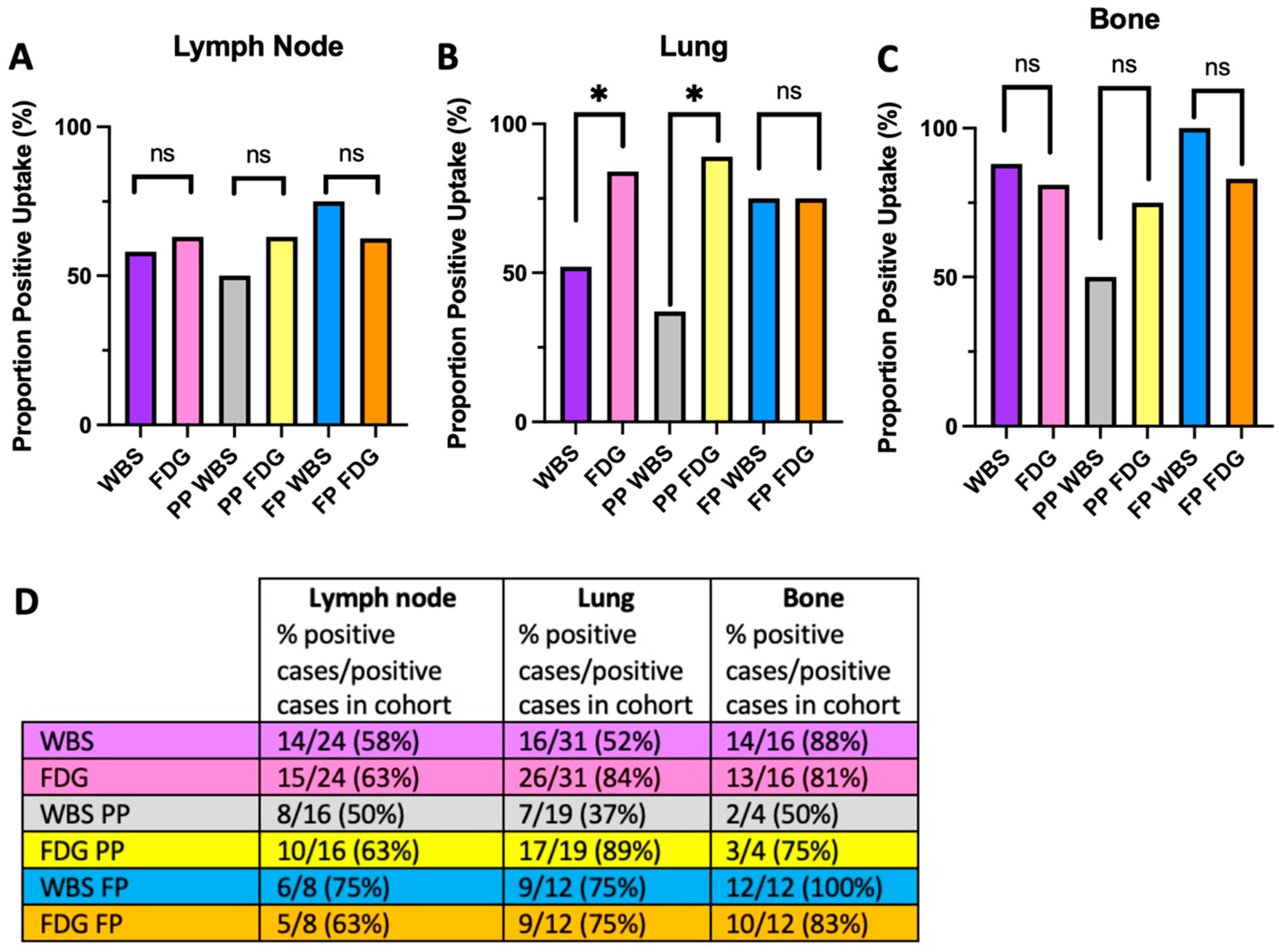
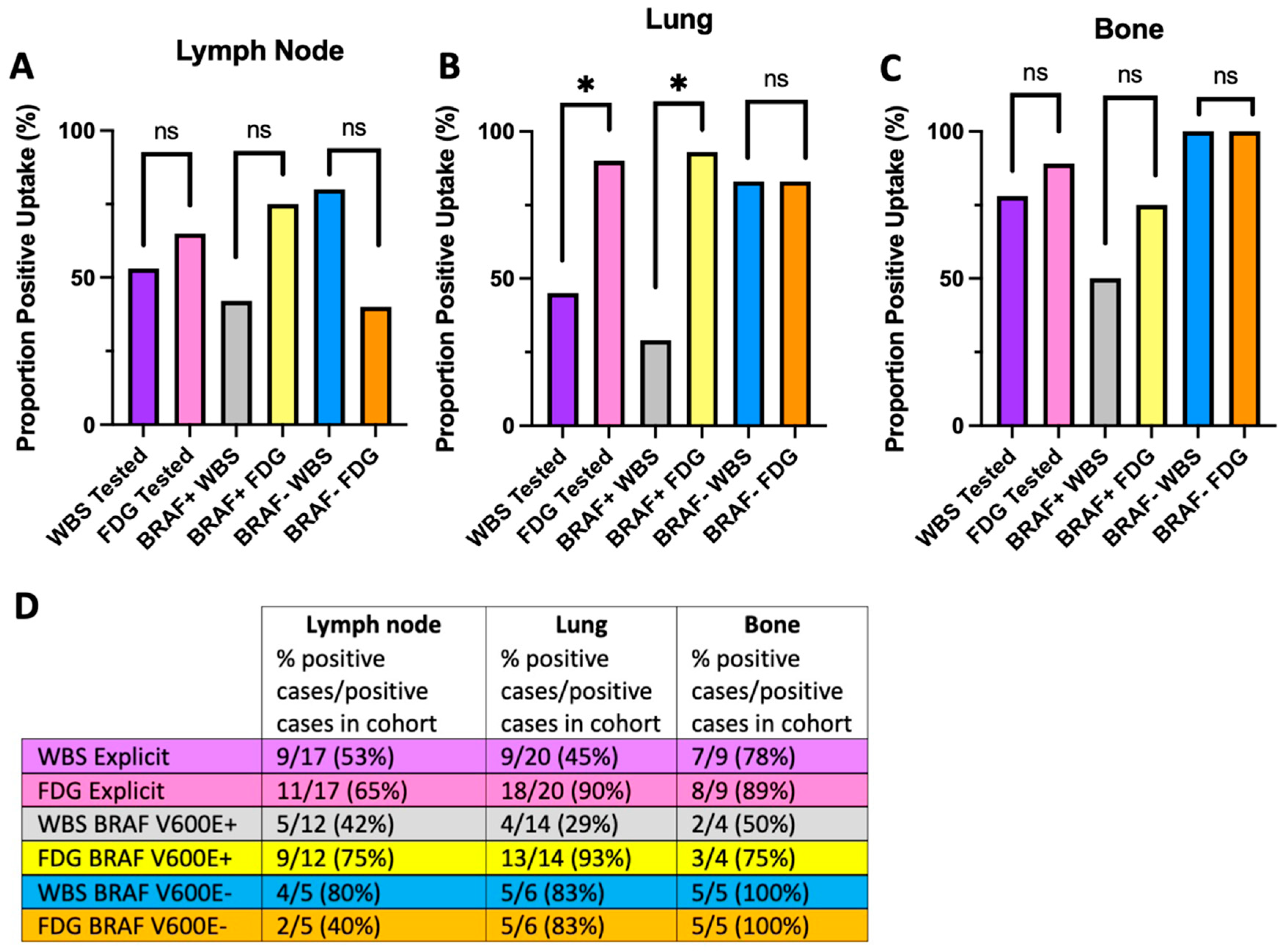
4.1. The Flip-Flop Phenomenon Varies by Metastatic Site and Molecular Status

We assessed for radiotracer uptake at three extra-thyroidal metastatic sites: lymphatic tissue, lung, and bone. In this cohort, 24 patients (52%) had lymph node uptake by either RAI or FDG, 31 (67%) had lung uptake, and 16 (35%) had bone uptake (Table 1).
When considering lymph node uptake, no significant differences were observed in the entire cohort (WBS: n = 14/24, 58%; FDG: n = 15/24, 63%; p = 1.00), nor in the PP (WBS: n = 8/16, 50%; FDG: n = 10/16, 63%; p0.79) or FP (WBS: n = 6/8, 75%; FDG: n = 5/8, 63%; p = 1.00) sub-cohorts (Figure 1A,D). With respect to lung, significant differences in uptake were observed when considering the entire cohort (WBS: n = 16/31, 52%; FDG: n = 26/31, 84%; p = 0.04) supporting the flip-flop phenomenon at distant metastatic sites (Figure 1B,D). However, differences in uptake were maintained only in the PP sub-cohort (WBS: n = 7/19, 37%; FDG: n = 17/19, 89%; p = 0.02) and not in the FP sub-cohort (WBS: n = 9/12, 75%; FDG: n = 9/12, 75%; p = 1.00) (Figure 1B,D).
Like lymphatic tissue, no significant differences in bone uptake were observed in the entire cohort (WBS: n = 14/16, 88%; FDG: n = 13/16, 81%; p = 1.00), nor PP (WBS: n = 2/4, 50%; FDG: n = 3/4, 75%; p = 1.00) or FP sub-cohorts (WBS: n = 12/12, 100%; FDG: n = 10/12, 83%; p = 0.48) (Figure 1C,D). The flip-flop phenomenon is therefore not as evident in lymph or bone metastatic sites as it is in lung.
We repeated the analysis excluding patients without BRAF testing (Figure 2). A similar pattern emerges in which BRAF V600E+ tumors demonstrate robust FDG uptake in lung metastases (WBS: n = 4/14, 29%; FDG: n = 13/14, 93%; p = 0.02) compared with that of BRAF V600E− tumors (WBS: n = 5/6, 83%; FDG: n = 5/6, 83%; p = 1.00) (Figure 2B,D). Example WBS and FDG images from patients with lung metastasis in BRAF V600E+ and BRAF V600E− disease are shown in Figure 3.
TERT promoter molecular testing was also available for a subset of patients (n = 14, 30% of entire cohort). A total of 10 patients (22% of entire cohort) tested positive for mutations in the TERT promotor (TERT+), while 4 tested did not have TERT promoter mutations (TERT−). No differences between TERT sub-cohorts were noted in lymph nodes (TERT+ WBS: n = 4/6, 67%; FDG: n = 4/6, 67%; p = 1.00) (TERT− WBS: n = 1/1, 100%; FDG: n = 0/1, 0%; p = 1.00) or bone (TERT+ WBS: n = 3/5, 60%; FDG: n = 5/5, 100%; p = 0.48) (TERT− WBS: n = 1/1, 100%; FDG: n = 1/1, 100%; p = 1.00), although our results are limited by the small sample size (Supplementary Figure S1A,C,D). Like BRAF V600E+ and papillary pattern tumors, TERT+ tumors exhibited FDG avidity in the lung (WBS: n = 4/8, 50%; FDG: n = 8/8, 100%; p = 0.08) compared with TERT− tumors (WBS: n = 3/3, 100%; FDG: n = 2/3, 67%; p = 1.00) (Supplementary Figure S2B,D).

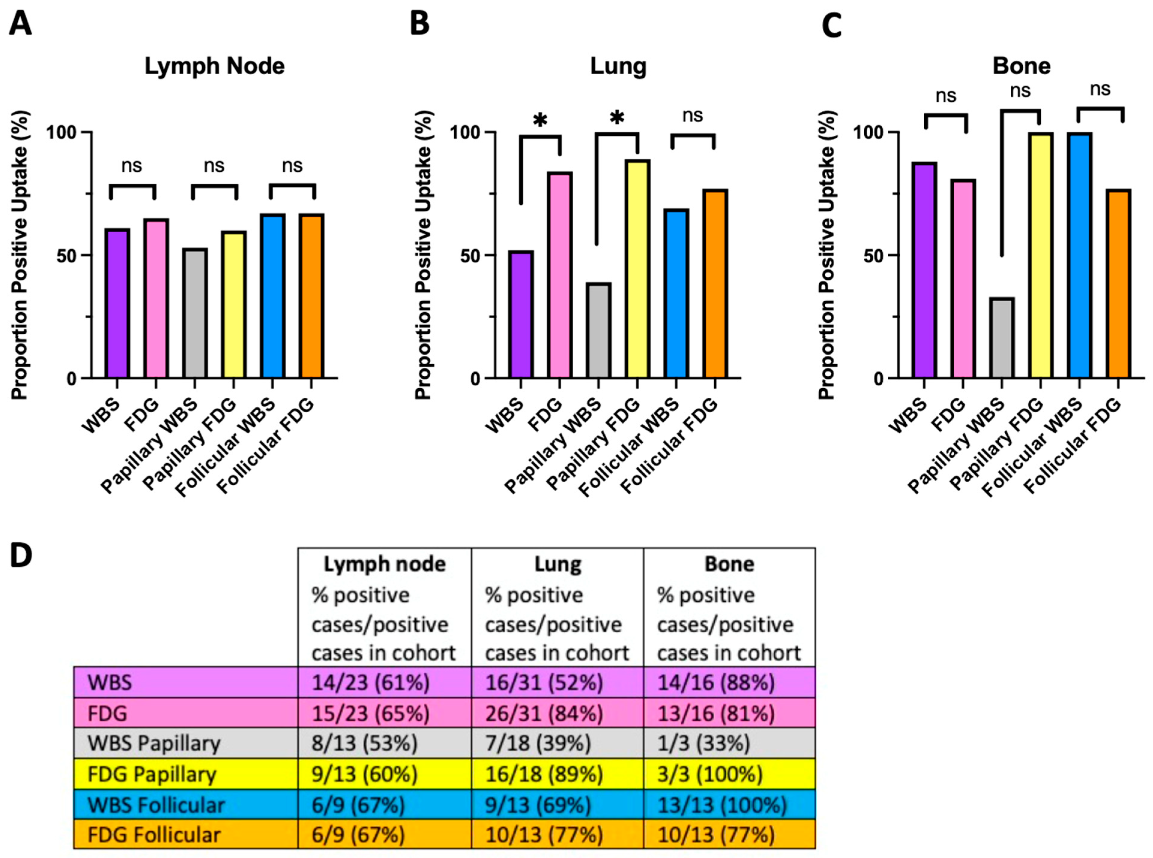
4.2. The Flip-Flop Phenomenon and Histologic Classification

We next subdivided the cohort by tumor histologic classification instead of molecular status. Radiotracer uptake in the lymphatic tissues remained similar in both imaging modalities across follicular (WBS: n = 6/9, 67%; FDG: n = 6/9, 67%; p = 1.00) and papillary (WBS: n = 8/15, 53%; FDG: n = 9/15, 60%; p = 1.00) tumor histology (Figure 4A,D). In the lung, primary tumors with follicular histology did not show differences in radiotracer uptake (WBS: n = 9/13, 69%; FDG: n = 10/13, 77%; p = 1.00); however, those with papillary histology showed a higher propensity for FDG uptake than iodine uptake (WBS: n = 7/18, 39%; FDG: n = 16/18, 89%; p = 0.03) (Figure 4B,D). No significant differences were observed in bone uptake in follicular (WBS: n = 13/13, 100%; FDG: n = 10/13, 77%; p = 0.25) or papillary (WBS: n = 1/3, 33%; FDG: n = 3/3, 100%; p = 0.48) tumors (Figure 4C,D). Overall, uptake by histologic classification largely mirrored the uptake patterns of molecular classification.

4.3. Agreement between Imaging Modalities

We determined the agreement between imaging modalities on an individual patient level (see Section 2).
In this analysis, discordant findings are consistent with the previously described flip-flop phenomenon, while concordant positive findings are not. In considering the entire cohort, lymphatic tissues displayed the highest degree of discordance (79%), followed by lung (64%), and bone (31%) (Figure 5A). In the papillary pattern sub-cohort, discordance at lymph, lung, and bone metastatic sites increased to 88%, 74%, and 75%, respectively (Figure 5B). The opposite pattern was identified in the follicular pattern sub-cohort with decreased discordance compared with the entire patient population: lymph (63%), lung (42%), and bone (17%) (Figure 5C).
In considering agreement between modalities stratified by BRAF V600E mutation status, BRAF V600E+ tumors resemble the papillary pattern sub-cohort with high lung (79%) and bone (75%) discordance (Supplemental Figure S2B). BRAF V600E−tumors resemble the follicular pattern sub-cohort with lower discordance in in lymph node (80%), lung (33%), and bone (0%) (Supplemental Figure S2C). Similarly, higher discordance is seen among tumors with papillary histology (lymph node 87%, lung 72%, bone 67%) across all sites compared with follicular histology tumors (lymph node 67%, lung 54%, bone 23%) (Supplemental Figure S3B,C). Notably, discordance at lymph node was the highest amongst all three sites irrespective of integrated pattern, BRAF V600E status, and histology.

5. Discussion

In this retrospective cohort study, we interrogate the effect of molecular subtype, histologic classification, and metastatic site on the flip-flop phenomenon in patients with metastatic DTCs. Our analysis reveals that the flip-flop phenomenon is not universally applicable across DTC molecular subtypes, histology, or metastatic sites.
Consistently with previous studies of BVL subtypes exhibiting greater dedifferentiation, RAI refractoriness, and tumor aggressiveness than RL subtypes [24,25,26,27,28], we find that tumors with papillary pattern, BRAF V600E mutation, and papillary histology have a higher propensity for FDG uptake compared with follicular pattern, BRAF V600E−, and follicular histology tumors, particularly in lung metastases. Previous studies support the link between molecular status, histology, and radiotracer avidity. For example, Ha et al. identified that the majority of RAI-negative and recurrent DTC were BRAF V600E+ and featured metastatic FDG avidity [38]. Soe et al. found that PTC with papillary architecture on histology were enriched in a cohort of non-iodine avid disease, further supporting the idea that BVL tumors are much more likely to be FDG avid [25]. Given that the majority of the papillary histology cohort consisted of BRAF V600E+ tumors (17/24, 71%) compared with that of the follicular cohort (3/22, 14%), it is not surprising that tumors with papillary histology largely behave like the BVL population and those with follicular histology are more congruent with the RL population by radiotracer imaging [28]. Although not reaching the level of statistical significance and limited by the cohort size in our study, TERT+ tumors behaved similarly as papillary and BRAF V600E+ tumors. Several studies have shown that BRAF and TERT promoter mutations promote the loss of RAI uptake, function cooperatively, and may adopt a more dedifferentiated phenotype than RL tumors [4,24,25,26,27]. Our results are therefore consistent with previous studies tying molecular status and histologic classification to dedifferentiation [24,26,27] and allow us to form two broader groups with internally consistent uptake patterns: (1) BRAF V600E+/TERT+/papillary histology and (2) BRAF V600E−/TERT−/follicular histology.
Our study adds to the existing literature by analyzing radiotracer uptake across common DTC metastatic sites. By combining information from histology or molecular subtype with metastatic site, our results may facilitate the selection of an appropriate radiotracer for the surveillance of tumor recurrence or progression. A summary of potential imaging recommendations is presented in Figure 5. For all molecular and histologic subtypes, the low agreement between both modalities at lymph node metastases renders both WBS and FDG-PET important for surveillance since neither modality may be sufficiently sensitive to reliably detect lymph node metastasis. In lung metastasis, papillary pattern, BRAF V600E+, and papillary histology tumors featured low agreement with strong FDG avidity such that FDG-PET may be considered the primary imaging modality. In contrast, for follicular pattern, BRAF V600E−, and follicular histology tumors, higher agreement and roughly equivalent RAI and FDG uptake renders either imaging modality appropriate. A previous study involving 83 patients with DTC lung metastases found that 30% (25/83) of patients had lung FDG uptake, while 55% (46/83) had lung radioiodine avidity, although this study did not differentiate follicular histology and PTC-FV from classical PTC, nor provide molecular correlates [39]. A population of tumors enriched in PTC-FV, follicular, or RL tumors may bias the cohort towards more differentiated phenotypes and thus higher rates of RAI avidity. In our study, FDG and RAI uptake was equivalent in bone for follicular pattern, BRAF V600E−, or follicular histology with high agreement for either imaging modality, an observation previously noted and attributed to the presence of mixed differentiated and dedifferentiated tumor tissues [40]. We observed lower agreement in bone metastasis for papillary pattern, BRAF V600E+, or papillary histology tumors which could represent a propensity for these tumors to be dedifferentiated, but our sample size is limited.
Beyond guiding the imaging modality for surveillance, our study may help stratify metastatic DTC patients who could benefit from alternative therapies rather than empiric RAI ablation (Figure 6). We show that increased FDG avidity is associated with the BRAF V600E+/TERT+ molecular subtypes and papillary histology which, in turn, is indicative of dedifferentiated tumor tissue, increased aggressiveness, RAI insensitivity, and a worse prognosis [9,10,11,13,21,36]. For example, a patient with a BVL primary tumor and lung metastasis is much less likely to have iodine avid disease and tumor tissue capable of concentrating iodine. Such a patient may first benefit from iodine “resensitization” therapy with kinase inhibitors [31,32,33,34,35,41] prior to RAI ablation therapy. An interval follow-up with FDG-PET would be recommended given that FDG avidity is an independently poor prognostic factor in metastatic DTCs. In contrast, a patient with bone metastasis, regardless of subtype, may benefit from a combination of RAI and alternative therapies given that our radiotracer imaging findings are consistent with a mixture of differentiated states in bone metastasis.
The limitations of our study largely derive from cohort size, particularly with patients who have been explicitly tested for TERT promoter, the asynchronous imaging timeline (albeit limited to less than a 1-year inclusion criteria), and the potential for metastases to drift from the molecular signatures of the primary tumor.
Our study highlights the heterogeneous nature of the flip-flop phenomenon and identifies molecular subtype, histologic classification, and metastatic site as key variables for discrepancies in radiotracer uptake in metastatic DTCs. This knowledge of uptake patterns may be harnessed to guide the choice of appropriate modality for tumor surveillance, medical treatment, and the assessment of treatment response.

Supplementary Materials

The following supporting information can be downloaded at: https://www.mdpi.com/article/10.3390/jcm13133963/s1, Figure S1: WBS and FDG uptake stratified by TERT promoter mutation status. Figure S2: Agreement between imaging modalities by BRAF status. Figure S3: Agreement between imaging modalities by histology. Table S1: Per-patient histology, classification, tumor genetics, and consensus uptake.

Author Contributions

Conceptualization, D.D. and R.R.F.; methodology, D.D., C.L. and R.R.F.; Analysis, D.D., E.C., L.Z., S.C.B. and R.R.F.; original draft preparation, D.D.; writing—review and editing, D.D., E.C., Y.S., H.K., M.H.S., J.M.C., L.Z., C.L., S.C.B. and R.R.F. All authors have read and agreed to the published version of the manuscript.

Funding

This research was funded by National Institute of Health and National Cancer Institute, F30CA247147, for D.D.

Institutional Review Board Statement

The study was conducted in accordance with the Declaration of Helsinki, and approved by the Institutional Review Board of University of California San Francisco (protocol code 10-02005, approval date 20 August 2010 and 11-05345, approval date 2 March 2011).

Informed Consent Statement

Patient consent was not required for participation in the study, as it this is a secondary analysis of data obtained as part of standard of care.

Data Availability Statement

Data are available upon request and correspondence with Robert R. Flavell, MD, PhD at robert.flavell@ucsf.edu.

Conflicts of Interest

The authors declare no conflicts of interest.

Abbreviations

BVLBRAF V600E-Like
DTCDifferentiated thyroid cancer
FDG18Fluorodeoxyglucose
FPFollicular pattern
MAPKMitogen-activated protein kinases
PETPositron emission technology
PPPapillary pattern
RAIRadioactive iodine
RLRAS-like
TERTTelomerase reverse transcriptase
WBSWhole body scan

References

  1. Fagin, J.A.; Wells, S.A., Jr. Biologic and Clinical Perspectives on Thyroid Cancer. N. Engl. J. Med. 2016, 375, 1054–1067. [Google Scholar] [CrossRef] [PubMed]
  2. Nikiforov, Y.E.; Nikiforova, M.N. Molecular genetics and diagnosis of thyroid cancer. Nat. Rev. Endocrinol. 2011, 7, 569–580. [Google Scholar] [CrossRef] [PubMed]
  3. Baloch, Z.W.; Asa, S.L.; Barletta, J.A.; Ghossein, R.A.; Juhlin, C.C.; Jung, C.K.; LiVolsi, V.A.; Papotti, M.G.; Sobrinho-Simoes, M.; Tallini, G.; et al. Overview of the 2022 WHO Classification of Thyroid Neoplasms. Endocr Pathol 2022, 33, 27–63. [Google Scholar] [CrossRef] [PubMed]
  4. Cancer Genome Atlas Research, N. Integrated genomic characterization of papillary thyroid carcinoma. Cell 2014, 159, 676–690. [Google Scholar] [CrossRef]
  5. Suh, S.; Goh, T.S.; Kim, Y.H.; Oh, S.O.; Pak, K.; Seok, J.W.; Kim, I.J. Development and Validation of a Risk Scoring System Derived from Meta-Analyses of Papillary Thyroid Cancer. Endocrinol. Metab. 2020, 35, 435–442. [Google Scholar] [CrossRef] [PubMed]
  6. Cho, S.W.; Choi, H.S.; Yeom, G.J.; Lim, J.A.; Moon, J.H.; Park, D.J.; Chung, J.K.; Cho, B.Y.; Yi, K.H.; Park, Y.J. Long-term prognosis of differentiated thyroid cancer with lung metastasis in Korea and its prognostic factors. Thyroid 2014, 24, 277–286. [Google Scholar] [CrossRef]
  7. Wang, H.; Dai, H.; Li, Q.; Shen, G.; Shi, L.; Tian, R. Investigating (18)F-FDG PET/CT Parameters as Prognostic Markers for Differentiated Thyroid Cancer: A Systematic Review. Front. Oncol. 2021, 11, 648658. [Google Scholar] [CrossRef]
  8. Goffredo, P.; Sosa, J.A.; Roman, S.A. Differentiated thyroid cancer presenting with distant metastases: A population analysis over two decades. World J. Surg. 2013, 37, 1599–1605. [Google Scholar] [CrossRef]
  9. Robbins, R.J.; Wan, Q.; Grewal, R.K.; Reibke, R.; Gonen, M.; Strauss, H.W.; Tuttle, R.M.; Drucker, W.; Larson, S.M. Real-time prognosis for metastatic thyroid carcinoma based on 2-[18F]fluoro-2-deoxy-D-glucose-positron emission tomography scanning. J. Clin. Endocrinol. Metab. 2006, 91, 498–505. [Google Scholar] [CrossRef]
  10. Deandreis, D.; Al Ghuzlan, A.; Leboulleux, S.; Lacroix, L.; Garsi, J.P.; Talbot, M.; Lumbroso, J.; Baudin, E.; Caillou, B.; Bidart, J.M.; et al. Do histological, immunohistochemical, and metabolic (radioiodine and fluorodeoxyglucose uptakes) patterns of metastatic thyroid cancer correlate with patient outcome? Endocr. Relat. Cancer 2011, 18, 159–169. [Google Scholar] [CrossRef]
  11. Gaertner, F.C.; Okamoto, S.; Shiga, T.; Ito, Y.; Uchiyama, Y.; Manabe, O.; Hattori, N.; Tamaki, N. FDG PET Performed at Thyroid Remnant Ablation Has a Higher Predictive Value for a Long-Term Survival of High-Risk Patients with Well-Differentiated Thyroid Cancer Than Radioiodine Uptake. Clin. Nucl. Med. 2015, 40, 378–383. [Google Scholar] [CrossRef] [PubMed]
  12. Salvatore, B.; Klain, M.; Nicolai, E.; D’Amico, D.; De Matteis, G.; Raddi, M.; Fonti, R.; Pellegrino, T.; Storto, G.; Cuocolo, A.; et al. Prognostic role of FDG PET/CT in patients with differentiated thyroid cancer treated with 131-iodine empiric therapy. Medicine 2017, 96, e8344. [Google Scholar] [CrossRef] [PubMed]
  13. Feine, U.; Lietzenmayer, R.; Hanke, J.-P.; Held, J.; Wohrle, H.; Muller-Schauenburg, W. Fluorine-18-FDG and Iodine-131-Iodide Uptake in Thyroid Cancer. J. Nucl. Med. 1996, 37, 1468–1472. [Google Scholar]
  14. Kang, S.Y.; Bang, J.I.; Kang, K.W.; Lee, H.Y.; Chung, J.K. FDG PET/CT for the early prediction of RAI therapy response in patients with metastatic differentiated thyroid carcinoma. PLoS ONE 2019, 14, e0218416. [Google Scholar] [CrossRef] [PubMed]
  15. Flavell, R.; Naeger, D.; Aparici, C.; Hawkins, R.; Pampaloni, M.; Behr, S. Malignancies with Low Fluoro-deoxyglucose Uptake at PET/CT: Pitfalls and Prognostic Importance. Radiographics 2016, 36, 293–294. [Google Scholar] [CrossRef]
  16. Bahri, H.; Laurence, L.; Edeline, J.; Leghzali, H.; Devillers, A.; Raoul, J.L.; Cuggia, M.; Mesbah, H.; Clement, B.; Boucher, E.; et al. High prognostic value of 18F-FDG PET for metastatic gastroenteropancreatic neuroendocrine tumors: A long-term evaluation. J. Nucl. Med. 2014, 55, 1786–1790. [Google Scholar] [CrossRef] [PubMed]
  17. Haugen, B.R.; Alexander, E.K.; Bible, K.C.; Doherty, G.M.; Mandel, S.J.; Nikiforov, Y.E.; Pacini, F.; Randolph, G.W.; Sawka, A.M.; Schlumberger, M.; et al. 2015 American Thyroid Association Management Guidelines for Adult Patients with Thyroid Nodules and Differentiated Thyroid Cancer: The American Thyroid Association Guidelines Task Force on Thyroid Nodules and Differentiated Thyroid Cancer. Thyroid 2016, 26, 1–133. [Google Scholar] [CrossRef] [PubMed]
  18. Wang, W.; Larson, S.M.; Tuttle, R.M.; Kalaigian, H.; Kolbert, K.; Sonenberg, M.; Robbins, R.J. Resistance of [18F]-Fluorodeoxyglucose-Avid Metastatic Thyroid Cancer Lesions to Treatment with High-Dose Radioactive Iodine. Thyroid 2001, 11, 1169–1175. [Google Scholar] [CrossRef]
  19. Rosario, P.W.; Mourao, G.F.; dos Santos, J.B.; Calsolari, M.R. Is empirical radioactive iodine therapy still a valid approach to patients with thyroid cancer and elevated thyroglobulin? Thyroid 2014, 24, 533–536. [Google Scholar] [CrossRef]
  20. Blaser, D.; Maschauer, S.; Kuwert, T.; Prante, O. In Vitro Studies on the Signal Transduction of Thyroidal Uptake of 18F-FDG and 131I-Iodide. J. Nucl. Med. 2006, 47, 1382–1388. [Google Scholar]
  21. Giordano, T.J. Follicular cell thyroid neoplasia: Insights from genomics and The Cancer Genome Atlas research network. Curr. Opin. Oncol. 2016, 28, 1–4. [Google Scholar] [CrossRef]
  22. Al-Qurayshi, Z.; Sullivan, C.B.; Pagedar, N.; Lee, G.S.; Tufano, R.; Kandil, E. Prevalence and Risk of Metastatic Thyroid Cancers and Management Outcomes: A National Perspective. Laryngoscope 2021, 131, 237–244. [Google Scholar] [CrossRef] [PubMed]
  23. Bongiovanni, M.; Paone, G.; Ceriani, L.; Pusztaszeri, M. Cellular and molecular basis for thyroid cancer imaging in nuclear medicine. Clin. Transl. Imaging 2013, 1, 149–161. [Google Scholar] [CrossRef]
  24. Liu, J.; Liu, R.; Shen, X.; Zhu, G.; Li, B.; Mingzhao, Z. The Genetic Duet of BRAF V600E and TERT Promoter Mutations Robustly Predicts Loss of Radioiodine Avidity in Recurrent Papillary Thyroid Cancer. J. Nucl. Med. 2020, 61, 177–182. [Google Scholar] [CrossRef]
  25. Soe, M.H.; Chiang, J.M.; Flavell, R.R.; Khanafshar, E.; Mendoza, L.; Kang, H.; Liu, C. Non-Iodine-Avid Disease Is Highly Prevalent in Distant Metastatic Differentiated Thyroid Cancer With Papillary Histology. J. Clin. Endocrinol. Metab. 2022, 107, e3206–e3216. [Google Scholar] [CrossRef] [PubMed]
  26. Yang, X.; Li, J.; Li, X.; Liang, Z.; Gao, W.; Liang, J.; Cheng, S.; Lin, Y. TERT Promoter Mutation Predicts Radioiodine-Refractory Character in Distant Metastatic Differentiated Thyroid Cancer. J. Nucl. Med. 2017, 58, 258–265. [Google Scholar] [CrossRef] [PubMed]
  27. Durante, C.; Puxeddu, E.; Ferretti, E.; Morisi, R.; Moretti, S.; Bruno, R.; Barbi, F.; Avenia, N.; Scipioni, A.; Verrienti, A.; et al. BRAF mutations in papillary thyroid carcinomas inhibit genes involved in iodine metabolism. J. Clin. Endocrinol. Metab. 2007, 92, 2840–2843. [Google Scholar] [CrossRef]
  28. Kebebew, E.; Weng, J.; Bauer, J.; Ranvier, G.; Clark, O.H.; Duh, Q.Y.; Shibru, D.; Bastian, B.; Griffin, A. The prevalence and prognostic value of BRAF mutation in thyroid cancer. Ann. Surg. 2007, 246, 466–470. [Google Scholar] [CrossRef]
  29. Boucai, L.; Saqcena, M.; Kuo, F.; Grewal, R.K.; Socci, N.; Knauf, J.A.; Krishnamoorthy, G.P.; Ryder, M.; Ho, A.L.; Ghossein, R.A.; et al. Genomic and Transcriptomic Characteristics of Metastatic Thyroid Cancers with Exceptional Responses to Radioactive Iodine Therapy. Clin. Cancer Res. 2023, 29, 1620–1630. [Google Scholar] [CrossRef]
  30. Chakravarty, D.; Santos, E.; Ryder, M.; Knauf, J.A.; Liao, X.H.; West, B.L.; Bollag, G.; Kolesnick, R.; Thin, T.H.; Rosen, N.; et al. Small-molecule MAPK inhibitors restore radioiodine incorporation in mouse thyroid cancers with conditional BRAF activation. J. Clin. Investig. 2011, 121, 4700–4711. [Google Scholar] [CrossRef] [PubMed]
  31. Ho, A.L.; Grewal, R.K.; Leboeuf, R.; Sherman, E.J.; Pfister, D.G.; Deandreis, D.; Pentlow, K.S.; Zanzonico, P.B.; Haque, S.; Gavane, S.; et al. Selumetinib-enhanced radioiodine uptake in advanced thyroid cancer. N. Engl. J. Med. 2013, 368, 623–632. [Google Scholar] [CrossRef]
  32. Rothenberg, S.M.; McFadden, D.G.; Palmer, E.L.; Daniels, G.H.; Wirth, L.J. Redifferentiation of iodine-refractory BRAF V600E-mutant metastatic papillary thyroid cancer with dabrafenib. Clin. Cancer Res. 2015, 21, 1028–1035. [Google Scholar] [CrossRef]
  33. Jaber, T.; Waguespack, S.G.; Cabanillas, M.E.; Elbanan, M.; Vu, T.; Dadu, R.; Sherman, S.I.; Amit, M.; Santos, E.B.; Zafereo, M.; et al. Targeted Therapy in Advanced Thyroid Cancer to Resensitize Tumors to Radioactive Iodine. J. Clin. Endocrinol. Metab. 2018, 103, 3698–3705. [Google Scholar] [CrossRef]
  34. Iravani, A.; Solomon, B.; Pattison, D.A.; Jackson, P.; Ravi Kumar, A.; Kong, G.; Hofman, M.S.; Akhurst, T.; Hicks, R.J. Mitogen-Activated Protein Kinase Pathway Inhibition for Redifferentiation of Radioiodine Refractory Differentiated Thyroid Cancer: An Evolving Protocol. Thyroid 2019, 29, 1634–1645. [Google Scholar] [CrossRef] [PubMed]
  35. Dunn, L.A.; Sherman, E.J.; Baxi, S.S.; Tchekmedyian, V.; Grewal, R.K.; Larson, S.M.; Pentlow, K.S.; Haque, S.; Tuttle, R.M.; Sabra, M.M.; et al. Vemurafenib Redifferentiation of BRAF Mutant, RAI-Refractory Thyroid Cancers. J. Clin. Endocrinol. Metab. 2019, 104, 1417–1428. [Google Scholar] [CrossRef]
  36. Melo, M.; da Rocha, A.G.; Vinagre, J.; Batista, R.; Peixoto, J.; Tavares, C.; Celestino, R.; Almeida, A.; Salgado, C.; Eloy, C.; et al. TERT promoter mutations are a major indicator of poor outcome in differentiated thyroid carcinomas. J. Clin. Endocrinol. Metab. 2014, 99, E754–E765. [Google Scholar] [CrossRef]
  37. Ganly, I.; Ricarte Filho, J.; Eng, S.; Ghossein, R.; Morris, L.G.; Liang, Y.; Socci, N.; Kannan, K.; Mo, Q.; Fagin, J.A.; et al. Genomic dissection of Hurthle cell carcinoma reveals a unique class of thyroid malignancy. J. Clin. Endocrinol. Metab. 2013, 98, E962–E972. [Google Scholar] [CrossRef]
  38. Ha, L.N.; Iravani, A.; Nhung, N.T.; Hanh, N.T.M.; Hutomo, F.; Son, M.H. Relationship between clinicopathologic factors and FDG avidity in radioiodine-negative recurrent or metastatic differentiated thyroid carcinoma. Cancer Imaging 2021, 21, 8. [Google Scholar] [CrossRef]
  39. Zhu, X.; Wu, S.; Yuan, X.; Wang, H.; Ma, C. Progression free survival related to 18F-FDG PET/CT uptake and 131I uptake in lung metastases of differentiated thyroid cancer. Hell. J. Nucl. Med. 2019, 22, 123–130. [Google Scholar]
  40. Wang, D.; Bai, Y.; Huo, Y.; Ma, C. FDG PET Predicts the Effects of (131)I and Prognosis for Patients with Bone Metastases from Differentiated Thyroid Carcinoma. Cancer Manag. Res. 2020, 12, 13223–13232. [Google Scholar] [CrossRef]
  41. Leboulleux, S.; Dupuy, C.; Lacroix, L.; Attard, M.; Grimaldi, S.; Corre, R.; Ricard, M.; Nasr, S.; Berdelou, A.; Hadoux, J.; et al. Redifferentiation of a BRAF(K601E)-Mutated Poorly Differentiated Thyroid Cancer Patient with Dabrafenib and Trametinib Treatment. Thyroid 2019, 29, 735–742. [Google Scholar] [CrossRef]
Figure 1. WBS and FDG uptake stratified by molecular subtype. WBS: radioactive iodine whole body scan; FDG: 18Fluorodeoxyglucose-PET; PP: papillary pattern; FP: follicular pattern. (A) No significant differences were observed in lymphatic tissue metastatic radiotracer uptake in any cohort. (B) Significantly increased FDG over iodine uptake was observed in the entire cohort in lung tissue and papillary pattern (PP) sub-cohort, but not in the follicular pattern (FP) sub-cohort. (C) No significant differences in radiotracer uptake were observed in bone in any cohort. (D) Table summary with proportions of patients which have metastases at any given site and proportions of patients that had FDG or WBS positivity. Asterisk (*) indicates p < 0.05; ns = not significant.
Jcm 13 03963 g001
Figure 2. WBS and FDG uptake stratified by BRAF V600E status. WBS: radioactive iodine whole body scan; FDG: 18Fluorodeoxyglucose-PET. Only patients with BRAF testing are included in the cohort analysis (purple and pink for “WBS Tested” and “FDG Tested”, respectively). (A) No significant differences were observed in lymphatic tissue metastatic radiotracer uptake in any cohort. (B) Significantly increased FDG over iodine uptake was observed in the entire BRAF tested cohort in lung tissue and BRAF+ sub-cohort, but not in the BRAF− sub-cohort. (C) No significant differences in radiotracer uptake were observed in bone in any cohort. (D) Table summary with proportions of patients which have metastases at any given site and proportions of patients that had FDG or WBS positivity. Asterisk (*) indicates p < 0.05; ns = not significant.
Jcm 13 03963 g002
Figure 3. Example WBS and FDG PET scans of DTC lung metastasis. RAI WBS: radioactive iodine whole body scan; FDG: 18Fluorodeoxyglucose-PET. (A,B) Top row images are exemplary of DTC lung metastasis with FDG > WBS uptake in a patient with BRAF+ disease, illustrative of the “flip-flop” phenomenon in a less differentiated tumor. Despite clear focal FDG avidity, iodine radiotracer is at background levels in WBS. (C,D) Middle row images demonstrate a concordant BRAF+ case with iodine and FDG radiotracer avidity in the lung. (E,F) Bottom row images demonstrate a BRAF− case showing both low level FDG and iodine avidity in lung metastasis, both clearly above background levels, although more conspicuous by WBS.
Jcm 13 03963 g003
Figure 4. WBS and FDG uptake stratified by histology. (A) No significant differences were observed in lymphatic tissue metastatic radiotracer uptake in any cohort. (B) Significantly increased FDG over iodine uptake was observed in the entire cohort in lung tissue and papillary sub-cohort, but not in the follicular sub-cohort. (C) No significant differences in radiotracer uptake were observed in bone in any cohort. (D) Table summary with proportions of patients which have metastases at any given site and proportions of patients that had FDG or WBS positivity. Asterisk (*) indicates p < 0.05; ns = not significant.
Jcm 13 03963 g004
Figure 5. Agreement between imaging modalities by papillary or follicular pattern. Agreement is measured by percent discordance between WBS and FDG in patients who had positive uptake by at least one imaging modality. Discordance is inversely proportional to agreement. Below each bar chart, pie charts express the classification of each case into WBS+/FDG+, WBS+ only, and FDG+ only at each metastatic site. Discordance is the calculated sum of WBS+ only and FDG+ only. (A) In the entire cohort, percent discordant rates for lymph nodes (79%), lung (64%), and bone (31%). (B) In patients with papillary patterned thyroid cancer, lymph node (88%), lung (74%), and bone (75%). (C) In patients with follicular patterned thyroid cancer, lymph node (63%), lung (42%), and bone (17%).
Jcm 13 03963 g005
Figure 6. Schematic of recommended initial imaging modalities by molecular classification and histology.
Jcm 13 03963 g006
Table 1. Patient characteristics, tumor histology, and molecular type.
CharacteristicsValue
Number of patients (N)46
Female24 (52%)
Male22 (48%)
Age at imaging [median (interquartile range)]63 (55–73%)
Prior RAI (% of cohort)12 (26%)
Primary Histology (% of cohort)
Papillary (Tall, Classical, Sclerosing, Unknown Subtypes)24 (52%)
Follicular (Follicular, Papillary-Follicular, Mixed Subtypes)22 (48%)
Histology by Subtype (% of cohort)
Papillary-Follicular (PTC-FV)9 (20%)
Papillary-Tall4 (9%)
Papillary Classical13 (28%)
Papillary-Mixed3 (7%)
Papillary-Sclerosing1 (2%)
Papillary-Unknown6 (13%)
Follicular (FTC)10 (22%)
Integrated Classification (% of cohort)
Papillary Pattern27 (59%)
Follicular Pattern19 (41%)
Tumor Molecular Characteristics
BRAF V600E+20 (44%)
BRAF V600E−9 (20%)
BRAF V600E Untested17 (37%)
RAS Mutation+5 (11%)
RAS Mutation–9 (20%)
RAS Untested32 (70%)
TERT Mutation+10 (22%)
TERT Mutation–4 (9%)
TERT Untested32 (70%)
Metastatic Uptake (on either WBS or FDG)
Lymph node (% of cohort)24 (52%)
Lung (% of cohort)31 (67%)
Bone (% of cohort)16 (35%)
RAI: radioactive iodine ablative therapy; PTC-FV: papillary thyroid cancer follicular variant; FTC: follicular thyroid cancer; WBS: radioactive iodine whole body scan; FDG: 18Fluorodeoxyglucose-PET; TERT: telomerase reverse transcriptase promoter.
Disclaimer/Publisher’s Note: The statements, opinions and data contained in all publications are solely those of the individual author(s) and contributor(s) and not of MDPI and/or the editor(s). MDPI and/or the editor(s) disclaim responsibility for any injury to people or property resulting from any ideas, methods, instructions or products referred to in the content.

