


Developing the Benchmark: Establishing a Gold Standard for the Evaluation of AI Caries Diagnostics
 ,
,  ,
,
Abstract
1. Introduction
2. Materials and Methods
2.1. Ethical Aspects
2.2. Trial Profile
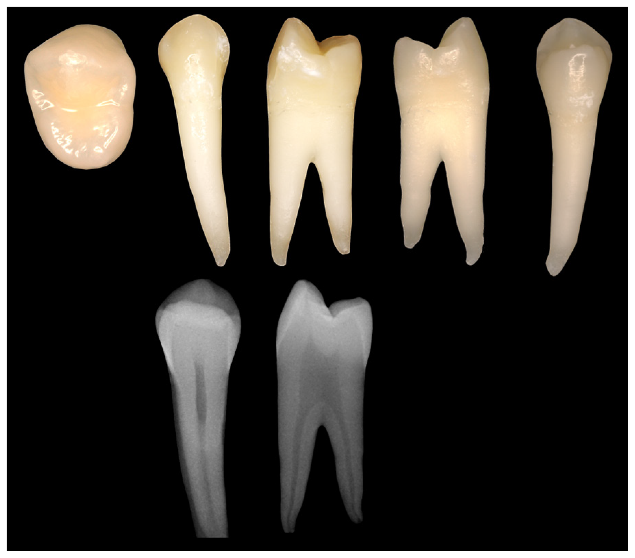
2.3. Tooth Selection
2.4. Preparation of Artificial Defects
2.5. Bitewing Design
2.6. Preparation of Histological Samples
2.7. Lesion Classification of the Histological Samples
2.8. Radiographic Caries Diagnostic by Dental Examiners
2.9. Statistical Analysis and Performance Metrics
2.10. Sample Sice Planning
3. Results
3.1. Examiner Characteristics
3.2. Reliability of Histological Lesion Classification
3.3. Examiners Performance Metrics
3.4. AUC
3.5. MCC by Lesion Class
3.6. Gender Specific MCC
3.7. MCC by Occupation
3.8. MCC by Experience
3.9. Influence of Eccentricity on MCC
3.10. Differentiation between Carious Lesions and Artifically Induced Lesions
3.11. Tooth Classification
4. Discussion
5. Conclusions
Author Contributions
Funding
Institutional Review Board Statement
Informed Consent Statement
Data Availability Statement
Conflicts of Interest
Abbreviations
| AI | Artificial intelligence |
| AUC | Area under the curve |
| EtOH | Ethanol |
| ICC | Intraclass correlation coefficient |
| NPV | Negative predictive value |
| MCC | Matthews correlation coefficient |
| PPV | Positive predictive value |
| ROC | Receiver operating characteristics |
| STARD | Standards for Reporting of Diagnostic Accuracy Studies |
| UV | Ultraviolet |
References
- Schwendicke, F.; Samek, W.; Krois, J. Artificial Intelligence in Dentistry: Chances and Challenges. J. Dent. Res. 2020, 99, 769–774. [Google Scholar] [CrossRef] [PubMed]
- Ahmed, N.; Abbasi, M.S.; Zuberi, F.; Qamar, W.; Halim, M.S.B.; Maqsood, A.; Alam, M.K. Artificial Intelligence Techniques: Analysis, Application, and Outcome in Dentistry-A Systematic Review. Biomed Res. Int. 2021, 2021, 9751564. [Google Scholar] [CrossRef]
- Jiang, F.; Jiang, Y.; Zhi, H.; Dong, Y.; Li, H.; Ma, S.; Wang, Y.; Dong, Q.; Shen, H.; Wang, Y. Artificial intelligence in healthcare: Past, present and future. Stroke Vasc. Neurol. 2017, 2, 230–243. [Google Scholar] [CrossRef]
- Khanagar, S.B.; Al-Ehaideb, A.; Maganur, P.C.; Vishwanathaiah, S.; Patil, S.; Baeshen, H.A.; Sarode, S.C.; Bhandi, S. Developments, application, and performance of artificial intelligence in dentistry—A systematic review. J. Dent. Sci. 2021, 16, 508–522. [Google Scholar] [CrossRef]
- Schwendicke, F.; Rossi, J.G.; Göstemeyer, G.; Elhennawy, K.; Cantu, A.G.; Gaudin, R.; Chaurasia, A.; Gehrung, S.; Krois, J. Cost-effectiveness of Artificial Intelligence for Proximal Caries Detection. J. Dent. Res. 2021, 100, 369–376. [Google Scholar] [CrossRef]
- Schwendicke, F.; Paris, S.; Stolpe, M. Detection and treatment of proximal caries lesions: Milieu-specific cost-effectiveness analysis. J. Dent. 2015, 43, 647–655. [Google Scholar] [CrossRef]
- Alam, M.K.; Alftaikhah, S.A.A.; Issrani, R.; Ronsivalle, V.; Lo Giudice, A.; Cicciù, M.; Minervini, G. Applications of artificial intelligence in the utilisation of imaging modalities in dentistry: A systematic review and meta-analysis of in-vitro studies. Heliyon 2024, 10, e24221. [Google Scholar] [CrossRef]
- Obuchowicz, R.; Strzelecki, M.; Piórkowski, A. Clinical Applications of Artificial Intelligence in Medical Imaging and Image Processing—A Review. Cancers 2024, 16, 1870. [Google Scholar] [CrossRef]
- Bayraktar, Y.; Ayan, E. Diagnosis of interproximal caries lesions with deep convolutional neural network in digital bitewing radiographs. Clin. Oral Investig. 2022, 26, 623–632. [Google Scholar] [CrossRef] [PubMed]
- Zhu, H.; Cao, Z.; Lian, L.; Ye, G.; Gao, H.; Wu, J. CariesNet: A deep learning approach for segmentation of multi-stage caries lesion from oral panoramic X-ray image. Neural Comput. Appl. 2022, 35, 1–9. [Google Scholar] [CrossRef]
- Park, E.Y.; Cho, H.; Kang, S.; Jeong, S.; Kim, E.-K. Caries detection with tooth surface segmentation on intraoral photographic images using deep learning. BMC Oral Health 2022, 22, 573. [Google Scholar] [CrossRef] [PubMed]
- Chen, X.; Guo, J.; Ye, J.; Zhang, M.; Liang, Y. Detection of Proximal Caries Lesions on Bitewing Radiographs Using Deep Learning Method. Caries Res. 2022, 56, 455–463. [Google Scholar] [CrossRef] [PubMed]
- Obuchowicz, R.; Nurzynska, K.; Obuchowicz, B.; Urbanik, A.; Piórkowski, A. Caries detection enhancement using texture feature maps of intraoral radiographs. Oral Radiol. 2020, 36, 275–287. [Google Scholar] [CrossRef] [PubMed]
- Anil, S.; Porwal, P.; Porwal, A. Transforming Dental Caries Diagnosis Through Artificial Intelligence-Based Techniques. Cureus 2023, 15, e41694. [Google Scholar] [CrossRef] [PubMed]
- Roosanty, A.; Widyaningrum, R.; Diba, S.F. Artificial intelligence based on Convolutional Neural Network for detecting dental caries on bitewing and periapical radiographs. J. Radiol. Dentomaksilofasial Indones. (JRDI) 2022, 6, 89–94. [Google Scholar] [CrossRef]
- Kunz, F.; Stellzig-Eisenhauer, A.; Zeman, F.; Boldt, J. Artificial intelligence in orthodontics: Evaluation of a fully automated cephalometric analysis using a customized convolutional neural network. J. Orofac. Orthop. 2020, 81, 52–68. [Google Scholar] [CrossRef] [PubMed]
- Kunz, F.; Stellzig-Eisenhauer, A.; Boldt, J. Applications of Artificial Intelligence in Orthodontics—An Overview and Perspective Based on the Current State of the Art. Appl. Sci. 2023, 13, 3850. [Google Scholar] [CrossRef]
- Mohammad-Rahimi, H.; Motamedian, S.R.; Rohban, M.H.; Krois, J.; Uribe, S.E.; Mahmoudinia, E.; Rokhshad, R.; Nadimi, M.; Schwendicke, F. Deep learning for caries detection: A systematic review. J. Dent. 2022, 122, 104115. [Google Scholar] [CrossRef] [PubMed]
- Ammar, N.; Kühnisch, J. Diagnostic performance of artificial intelligence-aided caries detection on bitewing radiographs: A systematic review and meta-analysis. Jpn. Dent. Sci. Rev. 2024, 60, 128–136. [Google Scholar] [CrossRef]
- Gomez, J. Detection and diagnosis of the early caries lesion. BMC Oral Health 2015, 15 (Suppl. 1), S3. [Google Scholar] [CrossRef]
- Grieco, P.; Jivraj, A.; Da Silva, J.; Kuwajima, Y.; Ishida, Y.; Ogawa, K.; Ohyama, H.; Ishikawa-Nagai, S. Importance of bitewing radiographs for the early detection of interproximal carious lesions and the impact on healthcare expenditure in Japan. Ann. Transl. Med. 2021, 10, 2. [Google Scholar] [CrossRef] [PubMed]
- Prados-Privado, M.; García Villalón, J.; Martínez-Martínez, C.H.; Ivorra, C.; Prados-Frutos, J.C. Dental Caries Diagnosis and Detection Using Neural Networks: A Systematic Review. J. Clin. Med. 2020, 9, 3579. [Google Scholar] [CrossRef]
- Khanagar, S.B.; Alfouzan, K.; Awawdeh, M.; Alkadi, L.; Albalawi, F.; Alfadley, A. Application and Performance of Artificial Intelligence Technology in Detection, Diagnosis and Prediction of Dental Caries (DC)-A Systematic Review. Diagnostics 2022, 12, 1083. [Google Scholar] [CrossRef] [PubMed]
- Albano, D.; Galiano, V.; Basile, M.; Di Luca, F.; Gitto, S.; Messina, C.; Cagetti, M.G.; Del Fabbro, M.; Tartaglia, G.M.; Sconfienza, L.M. Artificial intelligence for radiographic imaging detection of caries lesions: A systematic review. BMC Oral Health 2024, 24, 274. [Google Scholar] [CrossRef] [PubMed]
- Alzubaidi, L.; Zhang, J.; Humaidi, A.J.; Al-Dujaili, A.; Duan, Y.; Al-Shamma, O.; Santamaría, J.; Fadhel, M.A.; Al-Amidie, M.; Farhan, L. Review of deep learning: Concepts, CNN architectures, challenges, applications, future directions. J. Big Data 2021, 8, 53. [Google Scholar] [CrossRef]
- Bossuyt, P.M.; Reitsma, J.B.; Bruns, D.E.; Gatsonis, C.A.; Glasziou, P.P.; Irwig, L.; Lijmer, J.G.; Moher, D.; Rennie, D.; Vet, H.C.W.d.; et al. STARD 2015: An Updated List of Essential Items for Reporting Diagnostic Accuracy Studies. Radiology 2015, 277, 826–832. [Google Scholar] [CrossRef] [PubMed]
- Schwendicke, F.; Göstemeyer, G. Conventional Bitewing Radiographs. In Detection and Assessment of Dental Caries: A Clinical Guide, 1st ed.; Ferreira Zandona, A., Longbottom, C., Eds.; Springer International Publishing: Cham, Switzerland, 2019; pp. 109–117. [Google Scholar] [CrossRef]
- Devlin, H.; Williams, T.; Graham, J.; Ashley, M. The ADEPT study: A comparative study of dentists’ ability to detect enamel-only proximal caries in bitewing radiographs with and without the use of AssistDent artificial intelligence software. Br. Dent. J. 2021, 231, 481–485. [Google Scholar] [CrossRef]
- Vaarkamp, J.; Ten Bosch, J.J.; Verdonschot, E.H.; Bronkhorst, E.M. The Real Performance of Bitewing Radiography and Fiber-Optic Transillumination in Approximal Caries Diagnosis. J. Dent. Res. 2000, 79, 1747–1751. [Google Scholar] [CrossRef] [PubMed]
- Bader, J.D.; Shugars, D.A.; Bonito, A.J. Systematic reviews of selected dental caries diagnostic and management methods. J. Dent. Educ. 2001, 65, 960–968. [Google Scholar] [CrossRef]
- Hegde, S.; Gao, J.; Vasa, R.; Cox, S. Factors affecting interpretation of dental radiographs. Dentomaxillofacial Radiol. 2022, 52, 20220279. [Google Scholar] [CrossRef]
- Stroud, J.L.; English, J.; Buschang, P.H. Enamel thickness of the posterior dentition: Its implications for nonextraction treatment. Angle Orthod. 1998, 68, 141–146. [Google Scholar] [PubMed]
- Kamburoglu, K.; Kolsuz, E.; Murat, S.; Yüksel, S.; Ozen, T. Proximal caries detection accuracy using intraoral bitewing radiography, extraoral bitewing radiography and panoramic radiography. Dentomaxillofacl Radiol. 2012, 41, 450–459. [Google Scholar] [CrossRef]
- Wenzel, A. Bitewing and digital bitewing radiography for detection of caries lesions. J. Dent. Res. 2004, 83, 72–75. [Google Scholar] [CrossRef]
- Donath, K. Die Trenn-Dünnschliff-Technik zur Herstellung Histologischer Präparate von nicht schneidbaren Geweben und Materialien: Apparate- und Methodenbeschreibung; EXAKT-Kulzer-Druckschriften: Norderstedt, Germany, 1988. [Google Scholar]
- Cantu, A.G.; Gehrung, S.; Krois, J.; Chaurasia, A.; Rossi, J.G.; Gaudin, R.; Elhennawy, K.; Schwendicke, F. Detecting caries lesions of different radiographic extension on bitewings using deep learning. J. Dent. 2020, 100, 103425. [Google Scholar] [CrossRef]
- Bayrakdar, I.S.; Orhan, K.; Akarsu, S.; Çelik, Ö.; Atasoy, S.; Pekince, A.; Yasa, Y.; Bilgir, E.; Sağlam, H.; Aslan, A.F.; et al. Deep-learning approach for caries detection and segmentation on dental bitewing radiographs. Oral Radiol. 2022, 38, 468–479. [Google Scholar] [CrossRef]
- Moran, M.; Faria, M.; Giraldi, G.; Bastos, L.; Oliveira, L.; Conci, A. Classification of Approximal Caries in Bitewing Radiographs Using Convolutional Neural Networks. Sensors 2021, 21, 5192. [Google Scholar] [CrossRef]
- Lee, S.; Oh, S.I.; Jo, J.; Kang, S.; Shin, Y.; Park, J.W. Deep learning for early dental caries detection in bitewing radiographs. Sci. Rep. 2021, 11, 16807. [Google Scholar] [CrossRef]
- Mao, Y.-C.; Chen, T.-Y.; Chou, H.-S.; Lin, S.-Y.; Liu, S.-Y.; Chen, Y.-A.; Liu, Y.-L.; Chen, C.-A.; Huang, Y.-C.; Chen, S.-L.; et al. Caries and Restoration Detection Using Bitewing Film Based on Transfer Learning with CNNs. Sensors 2021, 21, 4613. [Google Scholar] [CrossRef] [PubMed]
- Walsh, T. Fuzzy gold standards: Approaches to handling an imperfect reference standard. J. Dent. 2018, 74, 47–49. [Google Scholar] [CrossRef]
- Jablonski-Momeni, A.; Stachniss, V. Serial sectioning of teeth and microscopy in cariology research. In Microscopy: Science, Technology, Applications and Education, 4th ed.; Méndez-Vilas, A., Díaz, J., Eds.; FORMATEX: Badajoz, Spain, 2010; Volume 3, pp. 785–791. [Google Scholar]
- Pitts, N.B. Clinical diagnosis of dental caries: A European perspective. J. Dent. Educ. 2001, 65, 972–978. [Google Scholar] [CrossRef]
- Li, G.; Qu, X.-m.; Chen, Y.; Zhang, J.; Zhang, Z.-y.; Ma, X.-c. Diagnostic accuracy of proximal caries by digital radiographs: An in vivo and in vitro comparative study. Oral Surg. Oral Med. Oral Pathol. Oral Radiol. Endodontology 2010, 109, 463–467. [Google Scholar] [CrossRef]
- Hintze, H.; Wenze, A. Clinical and laboratory radiographic caries diagnosis. A study of the same teeth. Dentomaxillofacl Radiol. 1996, 25, 115–118. [Google Scholar] [CrossRef] [PubMed]
- Suzuki, T.; Finger, W.J. Dentin adhesives: Site of dentin vs. bonding of composite resins. Dent. Mater. 1988, 4, 379–383. [Google Scholar] [CrossRef]
- O’Brien, J.A., 3rd; Retief, D.H.; Bradley, E.L.; Denys, F.R. Shear bond strength of a new dentin bonding restorative system. Dent. Mater. 1988, 4, 179–183. [Google Scholar] [CrossRef]
- Haller, B.; Hofmann, N.; Klaiber, B.; Bloching, U. Effect of storage media on microleakage of five dentin bonding agents. Dent. Mater. 1993, 9, 191–197. [Google Scholar] [CrossRef]
- Söderholm, K.J.M. Correlation of in vivo and in vitro performance of adhesive restorative materials: A report of the ASC MD156 task group on test methods for the adhesion of restorative materials. Dent. Mater. 1991, 7, 74–83. [Google Scholar] [CrossRef]
- Wenzel, A.; Hintze, H. Comparison of microscopy and radiography as gold standards in radiographic caries diagnosis. Dentomaxillofacl Radiol. 1999, 28, 182–185. [Google Scholar] [CrossRef] [PubMed]
- Rodrigues, J.A.; Neuhaus, K.W.; Diniz, M.B.; Hug, I.; Stich, H.; Karlsson, L.; Lussi, A. Comparison among gold standard techniques used for the validation of methods for occlusal caries detection. Microsc. Res. Tech. 2012, 75, 605–608. [Google Scholar] [CrossRef] [PubMed]
- Schulze, R.K.; Nackat, D.; D’Hoedt, B. In vitro carious lesion detection on D-, E-, and F-speed radiographic films. Oral Surg. Oral Med. Oral Pathol. Oral Radiol. Endodontology 2004, 97, 529–534. [Google Scholar] [CrossRef]
- Kay, E.J.; Knill-Jones, R. Variation in restorative treatment decisions: Application of Receiver Operating Characteristic curve (ROC) analysis. Community Dent. Oral Endodontology 1992, 20, 113–117. [Google Scholar] [CrossRef]
- Mileman, P.A.; van der Weele, L.T. Accuracy in radiographic diagnosis: Dutch practitioners and dental caries. J. Dent. 1990, 18, 130–136. [Google Scholar] [CrossRef] [PubMed]
- Peers, A.; Hill, F.J.; Mitropoulos, C.M.; Holloway, P.J. Validity and reproducibility of clinical examination, fibre-optic transillumination, and bite-wing radiology for the diagnosis of small approximal carious lesions: An in vitro study. Caries Res. 1993, 27, 307–311. [Google Scholar] [CrossRef]
- Deprá Lde, C.; Vessoni Iwaki, L.C.; Chicarelli, M.; Takeshita, W.M. Influence of Image Filters and Variation in Horizontal Angle of Incidence of X-ray Beam in Digital Interproximal Radiographs for Diagnosis of Secondary Caries in Esthetic Restorations. J. Contemp. Dent. Pract. 2015, 16, 805–812. [Google Scholar] [CrossRef] [PubMed]
- Chadwick, B.L.; Dummer, P.M.; van der Stelt, P.F. The effect of alterations in horizontal X-ray beam angulation and bucco-lingual cavity width on the radiographic depth of approximal cavities. J. Oral Rehabil. 1999, 26, 292–301. [Google Scholar] [CrossRef] [PubMed]
- Geibel, M.-A.; Carstens, S.; Braisch, U.; Rahman, A.; Herz, M.; Jablonski-Momeni, A. Radiographic diagnosis of proximal caries—Influence of experience and gender of the dental staff. Clin. Oral Investig. 2017, 21, 2761–2770. [Google Scholar] [CrossRef]
- Jordan, A.; Micheelis, W.; Cholmakow-Bodechtel, C.; Füßl-Grünig, E.; Geyer, S.; Hertrampf, K.; Hoffmann, T.; Holtfreter, B.; Kocher, T.; Nitschke, I.; et al. Fünfte Deutsche Mundgesundheitsstudie (DMS V); Deutscher Zahnärzteverlag: Köln, Germany, 2016. [Google Scholar]
- Dinga, R.; Penninx, B.W.J.H.; Veltman, D.J.; Schmaal, L.; Marquand, A.F. Beyond accuracy: Measures for assessing machine learning models, pitfalls and guidelines. bioRxiv 2019. [Google Scholar] [CrossRef]
- Chicco, D.; Jurman, G. The advantages of the Matthews correlation coefficient (MCC) over F1 score and accuracy in binary classification evaluation. BMC Genom. 2020, 21, 6. [Google Scholar] [CrossRef]


| Time | Chemical Solution | Volume Ratio | Storage |
|---|---|---|---|
| Day 1 | EtOH 70%/purif. H2O | 100 | 50 mL plastic flacons |
| Day 2 | EtOH 80%/purif. H2O | 100 | 50 mL plastic flacons |
| Day 3 | EtOH 90%/purif. H2O | 100 | 50 mL plastic flacons |
| Day 4 | EtOH 96%/purif. H2O | 100 | 50 mL plastic flacons |
| Day 5 and 6 | EtOH 99% | 100 | 50 mL plastic flacons |
| Day 7 | EtOH 99%/Technovit 7200 VLC | 50/50 | 50 mL plastic flacons (in darkness) |
| Day 8 | Technovit 7200 VLC | 100 | Snap-on lid tablet glass (in darkness) |
| Day 9 | Technovit 7200 VLC | 100 | Snap-on lid tablet glass (in darkness) |
| Day 10 | Technovit 7200 VLC | 100 | Snap-on lid tablet glass (in darkness) |
| Day 11 | Technovit 7200 VLC | 100 | Snap-on lid tablet glass (in darkness) |
| Day 12 | Technovit 7200 VLC | 100 | Snap-on lid tablet glass (in darkness) |
| Classification of Caries | Carious Lesion Extension |
|---|---|
| E1 | Caries limited to the outer half of the enamel |
| E2 | Caries extending to the inner half of the enamel |
| D1 | Caries in the outer third of dentin |
| D2 | Caries in the middle third of dentin |
| D3 | Caries in the dentinal third close to the pulp or up to the pulp |
| Caries Classification | Proximal Surfaces | Percentage |
|---|---|---|
| E1 | 15 | 22.1% |
| E2 | 8 | 11.8% |
| D1 | 8 | 11.8% |
| D2 | 18 | 26.4% |
| D3 | 19 | 27.9% |
| 68 | 100% |
| Occupation | Experience | Gender | |||||
|---|---|---|---|---|---|---|---|
| Private Practitioners | Clinicians | Students | <5 Years | ≥5 Years | Male | Female | |
| Occupation | |||||||
| Private practitioners | 10 | - | - | 6 | 4 | 6 | 4 |
| Clinicians | - | 10 | - | 5 | 5 | 5 | 5 |
| Students | - | - | 10 | - | - | 2 | 8 |
| Parameter | |
|---|---|
| Accuracy | 0.799 |
| Sensitivity | 0.565 |
| Specificity | 0.956 |
| PPV | 0.896 |
| NPV | 0.765 |
| F1 score | 0.693 |
| MCC | 0.578 |
| AUC | 76.1 |
| Lesion Classification | |||||
|---|---|---|---|---|---|
| E1 | E2 | D1 | D2 | D3 | |
| Lesion classification | |||||
| E1 | - | 1 | <0.001 * | <0.001 * | <0.001 * |
| E2 | 1 | - | <0.001 * | <0.001 * | <0.001 * |
| D1 | <0.001 * | <0.001 * | - | 0.008 * | <0.001 * |
| D2 | <0.001 * | <0.001 * | 0.008 * | - | <0.001 * |
| D3 | <0.001 * | <0.001 * | <0.001 * | <0.001 * | - |
Disclaimer/Publisher’s Note: The statements, opinions and data contained in all publications are solely those of the individual author(s) and contributor(s) and not of MDPI and/or the editor(s). MDPI and/or the editor(s) disclaim responsibility for any injury to people or property resulting from any ideas, methods, instructions or products referred to in the content. |
© 2024 by the authors. Licensee MDPI, Basel, Switzerland. This article is an open access article distributed under the terms and conditions of the Creative Commons Attribution (CC BY) license (https://creativecommons.org/licenses/by/4.0/).
Share and Cite
Boldt, J.; Schuster, M.; Krastl, G.; Schmitter, M.; Pfundt, J.; Stellzig-Eisenhauer, A.; Kunz, F. Developing the Benchmark: Establishing a Gold Standard for the Evaluation of AI Caries Diagnostics. J. Clin. Med. 2024, 13, 3846. https://doi.org/10.3390/jcm13133846
Boldt J, Schuster M, Krastl G, Schmitter M, Pfundt J, Stellzig-Eisenhauer A, Kunz F. Developing the Benchmark: Establishing a Gold Standard for the Evaluation of AI Caries Diagnostics. Journal of Clinical Medicine. 2024; 13(13):3846. https://doi.org/10.3390/jcm13133846
Chicago/Turabian StyleBoldt, Julian, Matthias Schuster, Gabriel Krastl, Marc Schmitter, Jonas Pfundt, Angelika Stellzig-Eisenhauer, and Felix Kunz. 2024. "Developing the Benchmark: Establishing a Gold Standard for the Evaluation of AI Caries Diagnostics" Journal of Clinical Medicine 13, no. 13: 3846. https://doi.org/10.3390/jcm13133846
APA StyleBoldt, J., Schuster, M., Krastl, G., Schmitter, M., Pfundt, J., Stellzig-Eisenhauer, A., & Kunz, F. (2024). Developing the Benchmark: Establishing a Gold Standard for the Evaluation of AI Caries Diagnostics. Journal of Clinical Medicine, 13(13), 3846. https://doi.org/10.3390/jcm13133846