


Abstract
The definitive diagnosis of Cushing’s disease (CD) in the presence of pituitary microadenoma remains a continuous challenge. Novel available pituitary imaging techniques are emerging. This study aimed to provide a structured analysis of the diagnostic accuracy as well as the clinical use of molecular imaging in patients with ACTH-dependent Cushing’s syndrome (CS). We also discuss the role of multidisciplinary counseling in decision making. Additionally, we propose a complementary diagnostic algorithm for both de novo and recurrent or persistent CD. A structured literature search was conducted and two illustrative CD cases discussed at our Pituitary Center are presented. A total of 14 CD (n = 201) and 30 ectopic CS (n = 301) articles were included. MRI was negative or inconclusive in a quarter of CD patients. 11C-Met showed higher pituitary adenoma detection than 18F-FDG PET–CT (87% versus 49%). Up to 100% detection rates were found for 18F-FET, 68Ga-DOTA-TATE, and 68Ga-DOTA-CRH, but were based on single studies. The use of molecular imaging modalities in the detection of pituitary microadenoma in ACTH-dependent CS is of added and complementary value, serving as one of the available tools in the diagnostic work-up. In selected CD cases, it seems justified to even refrain from IPSS.
1. Introduction
Cushing’s syndrome (CS) is a rare condition characterized by prolonged increased exposure to cortisol, leading to a classic clinical appearance that is accompanied by multiple comorbidities. The most prevalent cause of Cushing’s syndrome is excess exogenous cortisol as a consequence of glucocorticoid use. When exogenous Cushing’s syndrome is excluded, screening for endogenous cortisol is warranted. For this purpose, several screening tests are available. In the case of the biochemical confirmation of hypercortisolism, the measurement of ACTH concentrations is pivotal to determine whether the Cushing’s syndrome is ACTH-dependent or not. Low ACTH values are indicative of an adrenal source of hypercortisolism, while normal or high ACTH (“ACTH-dependent CS”) points to a pituitary (“Cushing’s disease”, CD) or ectopic source (“ectopic Cushing’s syndrome”, ECS). Since pituitary adenoma (CD) forms the majority (approx. 70%) of the cases with ACTH-dependent CS, a pituitary MRI is recommended [1]. However, previous studies have shown a negative or inconclusive scan in a considerable number of these patients [1,2,3,4].
Until recently, it was advocated to perform subsequent bilateral inferior petrosal sinus sampling (IPSS) when MRI results were negative, inconclusive, or showed a microadenoma (<10 mm), and IPSS is considered the golden standard to distinguish CD from ECS. In 2003, a consensus statement on the diagnosis of Cushing’s syndrome described, for the first time, the possibility of refraining from a further evaluation using IPSS [5] in case of an adenoma with a diameter of more than 6 mm in combination with the clinical suspicion and dynamic biochemical confirmation of a pituitary source [5]. The cut-off diameter of 6 mm was based on studies from the 1990s, reporting a considerable number of pituitary lesions of unknown significance (“incidentalomas”) and artifacts in the general population using 1.5-Tesla MRI. Incidentalomas usually did not exceed 6 mm in diameter [6,7,8]. The previously proposed cut-off diameter to distinguish ACTH-producing pituitary adenomas (corticotropinoma) from pituitary incidentalomas in ECS was confirmed in a more recent study by Yogi-Morren et al., showing 96% specificity for a cut-off diameter of 6 mm [9]. According to a recent update of the international diagnostic guideline, CD can be presumed when biochemical tests are in line with a pituitary source of ACTH secretion in the presence of a pituitary macroadenoma (≥10 mm). In these cases, there is a consensus that no further IPSS is justified [10]. In the case of a microadenoma smaller than 6 mm, there still is a strong recommendation to perform IPSS. Expert opinions differ on recommendations for the indication of IPSS in the case of a microadenoma between 6 and 9 mm. The majority (up to 90%) of CD adenomas are microadenomas; therefore, invasive diagnostic modalities are currently recommended in most patients [11,12].
Moreover, IPSS is currently recommended for CD patients with persistent disease after surgery (approximately a quarter of the patients) and without histological confirmation of a corticotroph adenoma [10,13]. Since long-term recurrence rates of CD vary between 15–44%, lifelong follow-up is strongly advised, and IPSS can also be reconsidered as a diagnostic tool in selected cases of CD recurrence without previous histological confirmation [10,14,15,16].
Centers vary in their institutionally based experience with diagnostic (both biochemical and IPSS) modalities for ACTH-dependent hypercortisolism as well as in the used cut-off values. Moreover, the decreasing availability of corticotropin-releasing hormone (CRH) worldwide also hampers the possibility of performing a CRH stimulation test, or CRH stimulation during IPSS. In addition, the availability of an experienced multidisciplinary team consisting of endocrinologists, neurosurgeons, (nuclear and intervention) radiologists, and pathologists can shift the diagnostic approach in CS from a static protocol-based approach to a more personalized approach.
Newly available imaging techniques with increased accuracy (both structural and molecular) may substitute IPSS in some cases. Bashari et al. recently proposed a stepwise approach to optimize the MRI protocol with additional sequences and magnetic field strengths to improve adenoma detection [17]. When improved structural imaging modalities still fail to detect a pituitary adenoma, the next diagnostic consideration could be molecular imaging, enabling a combination of anatomical (computed tomography, CT/magnetic resonance imaging, and MRI) with functional (metabolic) tissue information (positron emission tomography and PET). The results, though still based on small patient cohorts, are promising [17,18].
However, (interpretation of) these new diagnostic (imaging) techniques are not yet available in a substantial part of referral centers, and differences in available diagnostic capabilities may lead to different strategies between (referral) centers.
This study aimed to provide a structured analysis of the diagnostic accuracy as well as the clinical use of molecular imaging in patients with ACTH-dependent CS. We also discuss the role of multidisciplinary counseling within diagnostic decision making. Additionally, we propose a complementary diagnostic algorithm, both for de novo and recurrent or persistent CD.
2. Materials and Methods
2.1. Search Strategy and Selection Criteria
A structured PubMed search was conducted in January 2023 to collect relevant studies describing molecular imaging in ACTH-dependent CS using the following terms: “positron emission tomography” AND “Cushing”. This resulted in 199 articles. After the screening of titles and abstracts, 143 articles were excluded. Exclusion criteria were abstracts, poster presentations, case reports, reviews, meta-analyses, and articles written in languages other than English. The remaining 56 articles were screened for eligibility criteria—we only included peer-reviewed, original prospective, and retrospective cohort studies. Since some of the found studies were from the same research group, we checked if duplication of cohorts was described. If not, the studies were included. In total 33 articles could be included after this process. Hereafter, we searched within these 33 included articles for additional relevant references, and were able to include 8 additional articles for review, leading to a total of 41 articles.
2.2. Data Extraction
Data extraction included the following variables: study (year, research group, and design), population (number, age, sex, pituitary adenoma size or tumor with ectopic ACTH secretion), imaging modalities (including used sequences and magnetic field strength), tracer (including dosage), data and results (true positive, true negative, false negative, and false positive; sensitivity and specificity), and the conclusions of authors.
2.3. Data Analysis and Synthesis
Descriptive data per included study were presented in tables. Data were synthesized in diagnostic accuracy tables, giving the total amount and percentage of true positives, false positives, true negatives, and false negatives for different PET tracers.
2.4. Illustrative Patient Cases
Two illustrative CD patient cases, which were discussed at the Amsterdam Pituitary Center multidisciplinary team (MDT) meeting and thereafter underwent molecular imaging, are presented to emphasize the value of proposed complementary diagnostic algorithms.
3. Results
3.1. Molecular Imaging in Cushing’s Disease (See Also Table 1)
The use of molecular imaging in Cushing’s disease was described in a total of 201 patients in 14 articles between 2006 and 2022, including 6 prospective (of which 1 as a pilot) and 8 retrospective cohorts, as shown in Table 1. Tracers included 18F-fluorodeoxyglucose (18F-FDG, eight articles including one with and without CRH stimulation), 11C-methionine (11C-Met, five articles), 68Gallium-DOTA-TATE (68Ga-DOTA-TATE, one article), 68Gallium-pentixafor (68Ga-pentifaxor, one article), 68Gallium-DOTA-CRH (68Ga-DOTA-CRH, one article), 18F-fluorethyltyrosine (18F-FET, one article), and 13N-ammonia (one article). Functional imaging modalities combined with MRI were PET in four articles, PET–CT in eight articles, and PET–MR in two articles. We did not include studies describing octreotide/single-photon emission computerized tomography (SPECT) because of their inferior spatial resolution compared to PET. Concerning the magnetic field strength of MRI in these 14 studies; 6 used 1.5-Tesla, 4 used 3.0-Tesla, 1 did not use MRI, and, for 4 studies, the MRI field strength was unknown. A total of 156 (78%) de novo and 45 (22%) recurrent cases of CD were reviewed. Age ranged between 11 and 77 years, and a female sex preponderance was found (136 females, 57 males, and unknown for 8). Of the pituitary adenomas, MRI was negative or inconclusive in approximately a quarter of patients (55/194 = 28%, with a similar percentage when including only an MRI sequence with presumed higher sensitivity [spoiled gradient recalled echo sequence, SPGR]: 23/94 = 24%). Adenoma size was unknown for 51/201 (25%) patients. Of the remaining 150 adenomas described in the studies, 39 (26%) were macroadenoma and 111 (74%) were microadenomas. Within the microadenoma group, adenoma size was not further specified in 56/111 (50%), 35/111 (32%) were sized ≤6 mm, and 20/111 (18%) were sized 7 to 9 mm.
Table 1.
Original studies on molecular imaging in Cushing’s disease.
Diagnostic accuracy: Table 2 shows the diagnostic accuracies for pituitary adenoma detection in CD for the different tracers. The only tracers that were studied multiple times were 18F-FDG and 11C-Met. While the overall sensitivity of adenoma detection for 11C-Met was higher (87% versus 49%), slightly more false positives were also reported (3% versus 0%). CRH stimulation before 18F-FDG led to somewhat higher diagnostic accuracy compared to 18F-FDG without CRH stimulation. Based on single studies, a diagnostic accuracy of 100% was found for 18F-FET, 68Ga-DOTA-TATE, and 68Ga-DOTA-CRH, and was also high in 68Ga-pentixafor (86%) and 13N-ammonia (90%)—both with one false negative. The exclusion of studies in which histological confirmation was not described (three studies), led to minor changes in results. Diagnostic accuracy was higher for de novo than recurrent pituitary corticotropinomas in mainly 18F-FDG (56% versus 35%) and 68Ga-pentixafor (100% versus 68%). When MRI was negative or inconclusive, molecular imaging detected a pituitary adenoma in 100% (68Ga-DOTA-CRH), 67% (11C-Met and 68Ga-pentixafor), 22% (18F-FDG–PET after CRH stimulation), 17% (18F-FDG–PET) and 0 (13N-ammonia) patients. For both 18F-FDG (± CRH stimulation) and 11C-Met, macroadenoma detection was not better than in adenomas sized 7–9 mm (62% versus 82% and 100% versus 100%), while sensitivity for detection of adenomas ≤6 mm was lower (45% and 83%). This was not true for 68Ga-DOTA-CRH and 13N-ammonia, and the latter even showed lower sensitivity in macroadenoma.
Table 2.
Diagnostic accuracy of molecular imaging in Cushing’s disease.
3.2. Illustrative Cases
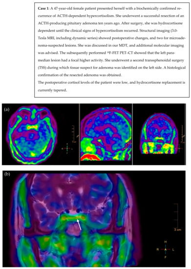
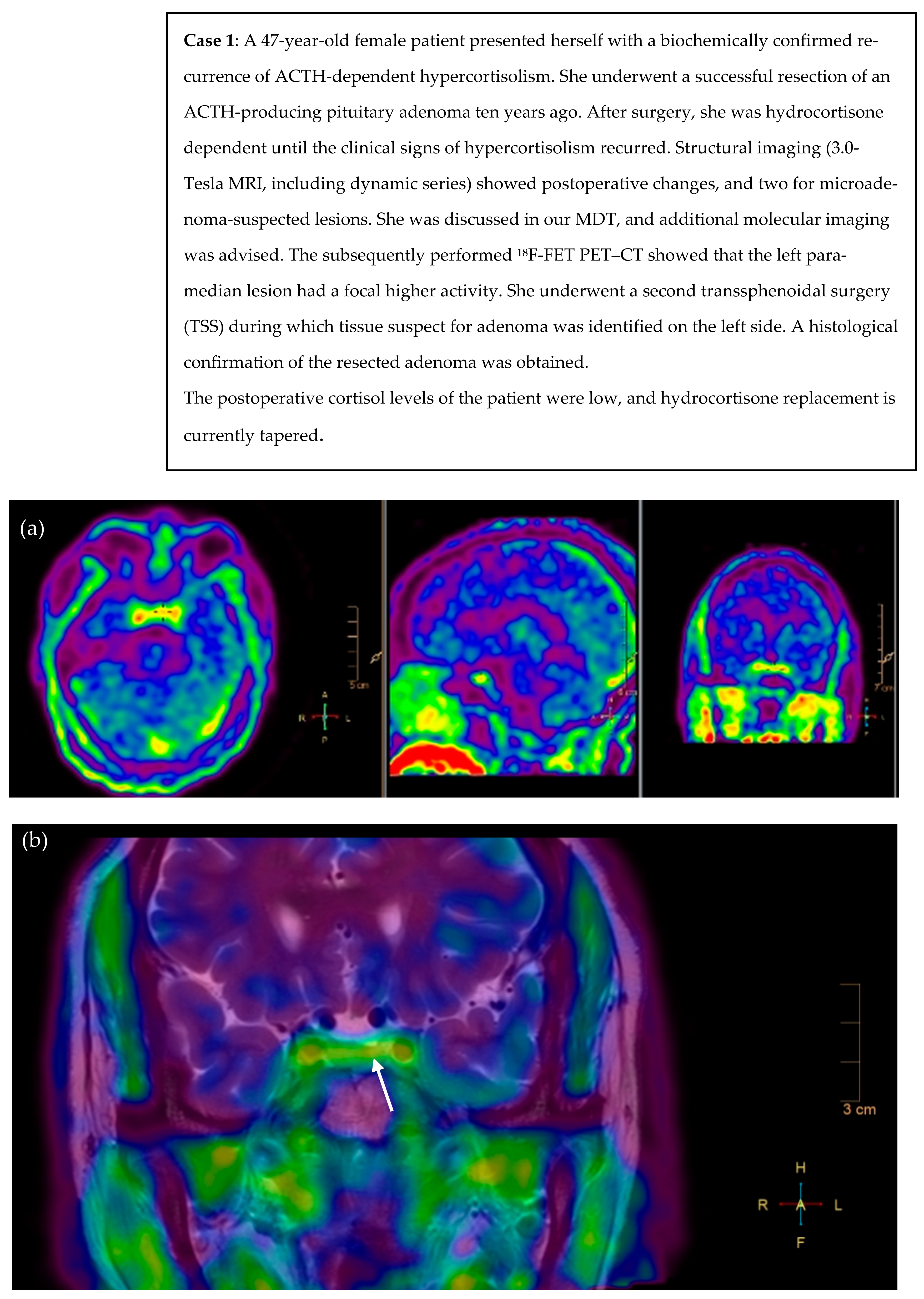
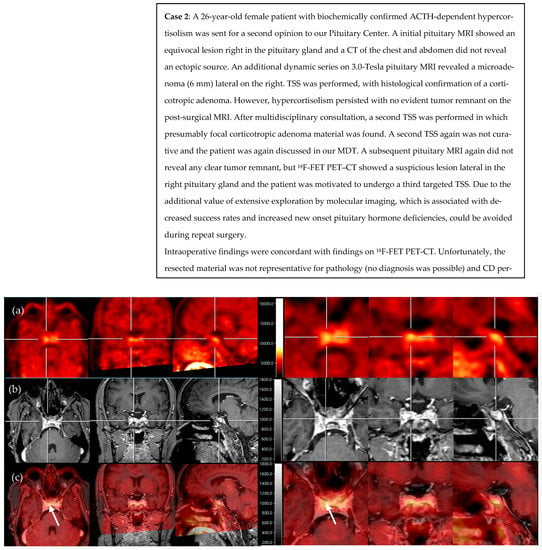
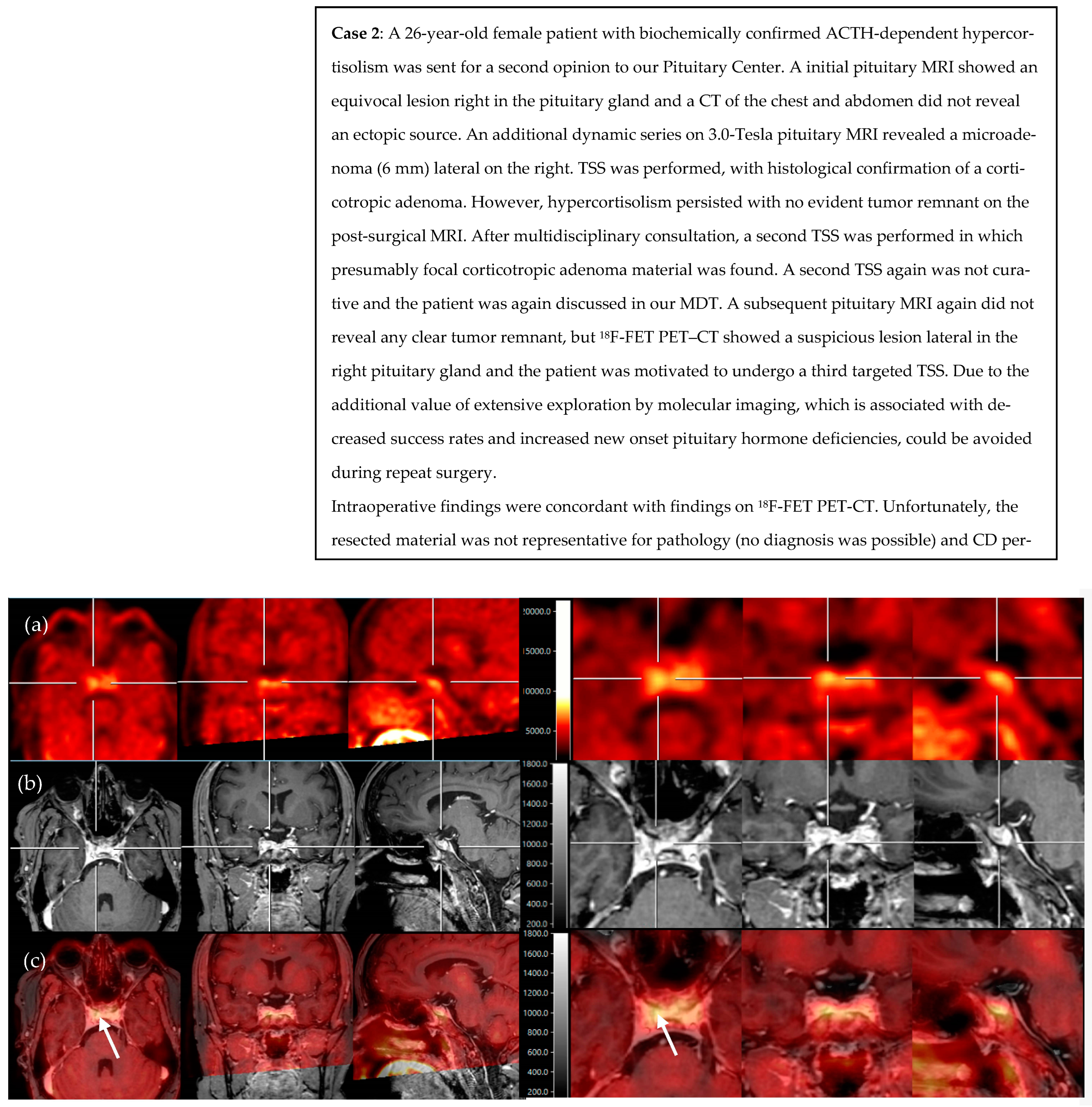
We present two illustrative CD cases discussed during multidisciplinary counseling in our Pituitary Center, that underwent molecular imaging. Due to pragmatic reasons (reliable production of radioligand and former permission of the Health and Youth Care Inspectorate) and the promising previous results of Berkmann et al., we chose to use 18F-FET PET–CT [30]. Case 1 included a patient with CD recurrence and several suspicious lesions on a postoperative pituitary MRI (Figure 1). Case 2 included a patient with persistent CD, with no clear tumor remnant on a postoperative pituitary MRI (Figure 2).
Figure 1.
18F-FET PET scan and fusion of PET with T2 MR images reveal asymmetrical focal high uptake (in orange) in a pituitary adenoma left paramedian, as shown with the crosshair (a) and white arrow (b). (a) axial, sagittal, and coronal view of PET scan after infusion of 219 MBq 18F-FET, (b) fusion of PET/MRI (T2-weighted coronal view, pre-gadolinium). L = left, R = right, H = head, F = feet.
Figure 2.
18F-FET PET and fusion of PET with post-contrast T1 MR images reveal asymmetrical focal high uptake (in yellow) lateral in the right pituitary, as centered with the crosshair (a–c) and shown with the white arrows (c). (a) axial, coronal, and sagittal view of PET scan after infusion of 192 MBq 18F-FET, (b) axial, coronal, and sagittal view of 3D T1-weighted, post-gadolinium MRI, and (c) axial, coronal, and sagittal view of PET/MRI fusion. Scans on the right side present the same lesion but in more detail.
3.3. Molecular Imaging in Ectopic Cushing’s Syndrome (See Also Table A1 and Table A2)
A total of 301 patients in 30 articles were described between 2004 and 2022, four of which were prospective (see also Table A1). Tracers included 68Ga-SSTR (18 articles, including 8 DOTA-TATE, 6 DOTA-TOC, and 6 DOTA-NOC), 18F-FDG (18 articles), 18F-DOPA (3 articles), 68Ga-DOTA-CRH (1 article), 68Ga-pentixafor (1 article), 11C-5-HTP (1 article), and 11C-Met (1 article). Functional imaging modality was PET (9 articles) and PET–CT (21 articles). We did not include studies describing octreotide/single-photon emission computerized tomography (SPECT) because of their inferior spatial resolution compared to PET. The age ranged from 1 to 80 years, but the vast majority of studies included adult patients with mean ages between 38 and 58 years. Similar to the CD studies, more female than male patients were included, but this difference was less pronounced (1.5:1—146 versus 108; unknown in 47). As expected, most frequently encountered tumors with ectopic ACTH secretion were of pulmonary origin (bronchus carcinoid, pulmonary/small cell lung carcinoma), others included thymic (thymoma/thymic carcinoma), gastro-intestinal (insulinoma, gastrinoma, somatostatinoma, gastric NEC, small bowel carcinoma NET, and carcinoma), pancreatic NET, medullary thyroid carcinoid, olfactory neuroblastoma, and metastatic foci of unknown origin. Rarely encountered underlying tumors were carcinoids of the right atrium, cervix, urinary bladder NET, breast tumor, and paraganglioma/pheochromocytoma.
Diagnostic accuracy: Table A2 shows the diagnostic accuracy for tumor detection in ECS for the different tracers. Tracers that were studied multiple times were 18F-FDG, 68Ga-SSTR (68Ga-DOTA-TATE/-TOC and/-NOC), and 18F-DOPA. Among these, 68Ga-SSTR showed superior overall sensitivity (59%) compared to 18F-FDG (46%) and 18F-DOPA (32%). The rate of false positives was high in 18F-FDG (23%), while low in 68Ga-SSTR (6%) and 18F-DOPA (0). The remaining tracers (68Ga-pentixafor, 68Ga-CRH, 11C-Met, and 11C-5-HTP) were only studied in very small sample sizes (n ≤ 3) but showed very high accuracy. The exclusion of studies that used only PET (and not PET–CT) did not lead to higher accuracy. For both 18F-FDG and 68Ga-SSTR, sensitivity was higher in recurrent/persistent than de novo CS.
3.4. Proposal of Diagnostic Algorithms in ACTH-Dependent Cushing’s Syndrome (See Also Figure 3a,b)
Based on the current literature, we suggest that molecular imaging can be included in the diagnostic algorithm for Cushing’s disease and, therefore, made amendments to the recently proposed algorithms of the consensus statement by Fleseriu et al. [10]. Multidisciplinary counseling remains key, which forms the basis to enable individualization of the diagnostic and treatment approach, and which is also guided by the availability of, and experience with, different diagnostic modalities. In selected cases, we propose the potential to refrain from IPSS and first perform molecular imaging (PET–CT with 11C-methionine, 68Ga-SSTR, or 18F-FET, and if not available, 18F-FDG) for de novo CD:
- If (optimized) structural imaging remains negative or equivocal or shows a microadenoma (<10 mm), and clinical presentation including biochemical testing is suggestive of Cushing’s disease (high “pretest probability”; young women with gradual onset and mildly elevated ACTH levels);
- If CRH and desmopressin test and whole-body CT (or whole-body 68Ga-SSTR PET–CT) in the search for ECS is inconclusive;
- Presence of contraindications to IPSS (renal failure, blood clotting disorders, or allergy to dye contrast).
Figure 3.
(a) Diagnostic algorithm for de novo diagnosis of Cushing’s syndrome.1 optimized as proposed by Bashari et al. [17]: Step 1a = conventional MRI: spin echo (SE) pre- and post-gadolinium, if no clear (micro)adenoma >, Step 1b = subsequent MRI sequences: gadolinium-enhanced 3D-spoiled gradient (recalled) echo (3D-SGE/3D-GRE) or gadolinium-enhanced dynamic (dMRI), if still not clear >, Step 2 = subsequent MRI sequences: fluid-attenuated inversion recovery (FLAIR) with gadolinium contrast, constructive interference in steady state (CISS) or isotropic 3D-fast turbo SE and/or use of the ultra-high field (7.0-Tesla) MRI. 2 optimally performed with CRH stimulation. 3 Alternative: whole-body 68Gallium-somatostatin receptor PET–CT. (b) Algorithm for persistent or recurrent Cushing’s disease. * In selected cases of Cushing’s disease with the equivocal biochemical response after surgery.
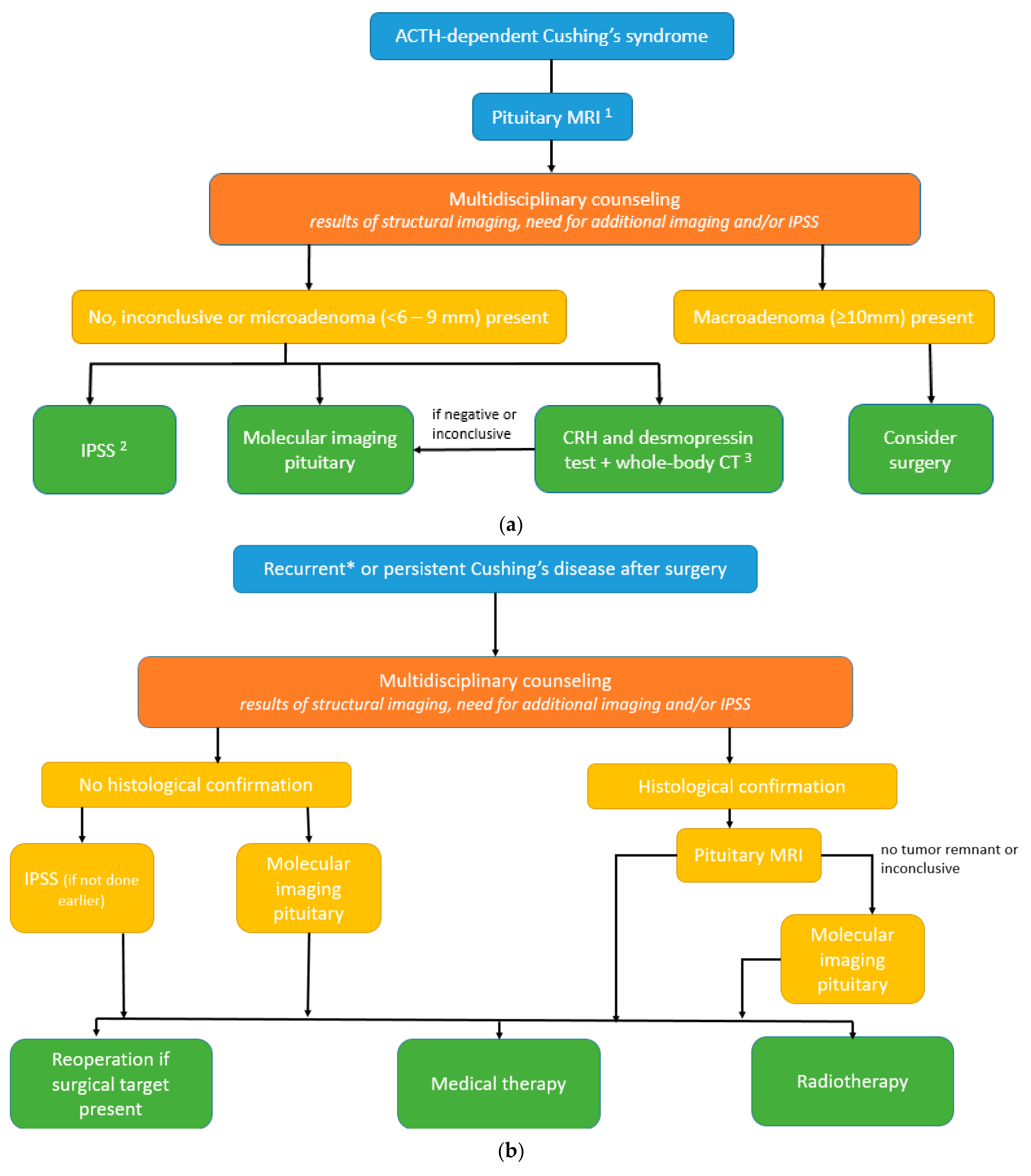
Next to the results of structural imaging and the need for additional imaging or IPSS, other relevant aspects such as age, child wish, and remaining pituitary function (hypopituitarism) are also discussed during multidisciplinary counseling in persistent or recurrent CD. We propose the potential to perform functional imaging (PET–CT with 11C-methionine, 68Ga-SSTR, or 18F-FET, and if not available, 18F-FDG) in the following cases:
- Persistent or selected cases of recurrent Cushing’s disease (equivocal biochemical response) after transsphenoidal surgery (TSS) and without histological confirmation.
- Persistent or selected cases of recurrent Cushing’s disease (equivocal biochemical response) after TSS and with histological confirmation, but no or inconclusive adenoma remnant localization on the pituitary MRI (illustrative cases 1 and 2).
4. Discussion
The definitive diagnosis of Cushing’s disease in the presence of pituitary microadenoma remains a continuous challenge in individual cases since most corticotroph adenomas are microadenomas. While the diagnostic accuracy of structural imaging in the detection of these microadenomas has improved over the last decades, the diagnostic accuracy to detect all microadenomas is still limited. On the other hand, improved sensitivity of structural imaging may lead to the detection of incidentalomas (false positives) [9,25]. This structured review clearly supports the added value of molecular imaging—co-registered or combined with structural MRI—in the diagnostic work-up of ACTH-dependent CS in selected cases. In de novo patients, we propose potentially refraining from IPSS and performing molecular imaging first (PET–CT with 11C-methionine, 68Ga-SSTR, or 18F-FET, and if not available, 18F-FDG) in the following cases: (1) if (optimized) structural imaging remains negative or equivocal or shows a microadenoma (<10 mm) and clinical presentation including biochemical testing is suggestive of Cushing’s disease (high “pretest probability”), (2) if CRH and desmopressin whole-body CT, in search for ECS, is inconclusive, or (3) presence of contraindications to IPSS (renal failure, blood clotting disorder, or allergy to dye contrast). In persistent or recurrent CD, we propose the possibility of performing molecular imaging in the following cases: (1) persistent or selected cases of recurrent Cushing’s disease (equivocal biochemical response) after TSS and without histological confirmation, before the use of IPSS (if not performed previously) or (2) in persistent or selected cases of recurrent Cushing’s disease (equivocal biochemical response) after TSS and with histological confirmation, but with equivocal tumor remnant localization on structural imaging (illustrative cases).
At present, the international consensus guideline allows for different diagnostic modalities after the biochemical confirmation of ACTH-dependent CS [10]. This allows for increased tailor-made diagnostic strategies, depending on, among others, the institutional availability and experience with both noninvasive and invasive diagnostic tests and modalities. Several alternative noninvasive diagnostic strategies after the optimization of pituitary MRI protocols, but before the use of IPSS, have been described. Isidori et al. found that a combination of dynamic testing, using both the CRH test and dexamethasone suppression test, led to a sensitivity of 97% and specificity of 94% in the correct distinguishing of CD from an ectopic source if both tests are positive [33]. In the published literature up to date, this was not confirmed for the high dose dexamethasone suppression test (which exists in multiple variants), as recently reviewed by Ferriere and Tabarin [34], while other studies indeed show high diagnostic accuracy of CRH testing in the differentiation between CD and ECS, so the usefulness of the CRH test and the high dose dexamethasone suppression test remains controversial. Another alternative noninvasive strategy was proposed by Frete et al., using CRH and desmopressin tests in combination with pituitary MRI and thin-slice whole-body CT. They found very high diagnostic accuracy when both tests and imaging were conclusive (e.g., CD: negative pituitary MRI and CT in combination with a positive CRH and desmopressin test, ECS: negative pituitary MRI and negative CT and a negative CRH and desmopressin test) and calculated that (recommendation of) IPSS could be omitted in about half of the patients [35]. The latter alternative strategy has now been incorporated in the update of the international clinical guidelines and states that if both tests are positive and no focus on the whole-body CT scan is found, CD can be assumed, while the opposite accounts for ECS, especially in the setting of a high pretest probability [10]. It should be mentioned that expert opinions differed on this last proposal and it warrants further investigation. Above that, these alternative diagnostic approaches also have their shortcomings. As mentioned before, the availability of CRH is decreasing (and the alternative desmopressin requires further research) and tests may also be discordant, again leading to the need for invasive IPSS.
It should be noted that although the diagnostic accuracy of IPSS to distinguish CD from ECS is very high, IPSS also gives false negatives (approximately 10–15%) and false positive results, leading to an estimated specificity between 90 and 95% [36,37]. Given that IPSS also is an invasive procedure with associated risks, only reliable in highly experienced hands, and lacks correct pituitary adenoma lateralization, there is a persistent unmet need to improve the stepwise (noninvasive) diagnostic approach in ACTH-dependent CS [37,38].
Part of this need is fulfilled by optimization of the dedicated pituitary MRI (thin slice, small field of view, dynamic contrast acquisition) and using higher magnetic field strengths (3.0 and even 7.0 Tesla), leading to increased detection of microadenomas. However, in this structured review, we found that MRI, even in those studies that included the more sensitive additional SPGR sequence, still failed to clearly detect a pituitary adenoma in 24 to 28% of CD patients. Molecular imaging, combined with CT or MRI, or co-registered with MRI, combines both anatomical and functional tissue information and appears to provide added value also for the imaging of pituitary adenomas. Large cohorts as well as individual small case series, report on the amount of (incidental) pituitary uptake of radioligands and their clinical significance, in both the general population and in different pituitary conditions such as (functioning) adenomas or carcinomas [22,39,40,41,42,43,44]. More specifically, the number of original studies on molecular imaging modalities in the detection of CD and ECS is increasing. While molecular imaging is already studied and used in ECS detection for a longer period, most studies on the clinical use of CD are from the last years. One of the advantages of molecular imaging is that detection of adenomas seems less reliant on tumor size, which is particularly relevant for corticotropinomas with their predominantly small sizes [23,24]. In this structured review, we also found that detection rates did not decrease notably in adenomas sized ≤ 6 mm in comparison to larger adenomas. Approximately half and 87% of pituitary adenomas were detected using 18F-FDG and 11C-Met PET(–CT), respectively. PET–CT using 18F-FET and three 68Ga-labeled radioligands even showed higher sensitivity (up to 100%), but were only studied in smaller samples: n = 9 for 18F-FET, n = 7, 7, and 24 for 68Ga-DOTA-TATE, -pentixafor and –DOTA-CRH, respectively. This should be taken into consideration when choosing an appropriate radioligand for molecular imaging in the detection and localization of pituitary microadenoma. Not many false positives were reported. With regard to the 18F-FDG studies included in this review, most authors concluded that its diagnostic use in CD is mainly complementary since some extra cases were detected on PET–CT that were not (clearly) seen on conventional MRI [20,22,23]. Stimulation with CRH can lead to increased 18F-FDG uptake, possibly leading to higher detection rates of pituitary adenoma in CD [24]. In studies that reported on both 18F-FDG and 11C-Met, diagnostic accuracy was higher in 11C-Met [21,26]. Four studies reported superior adenoma detection and localization using 11C-Met compared to other (structural) imaging techniques, while in another study, this appeared not true when using the (additional) SPGR MRI sequence [19,21,25,26,30]. Given the high predictive value for adenoma detection and localization, multiple studies proposed a useful role of 11C-Met PET–CT in treatment planning and/or in recurrent/residual cases to distinguish postoperative changes from adenoma tissue; a distinction less easily made by MRI [19,25,30]. This also accounts for 68Ga-DOTA-TATE and 68Ga-DOTA-CRH [29,31]. The specifically developed 68Ga-DOTA-CRH and 68Ga-pentixafor for the detection and localization of corticotroph adenoma showed very promising results, and sensitivity was higher than optimized MRI (SPGR/dynamic) with 100% and 86% sensitivity, respectively [29,32]. Overall, several authors suggested that molecular imaging with PET–CT could be a complementary diagnostic tool to MRI and/or IPSS in Cushing’s adenoma, especially when not available or inconclusive, in difficult cases, or when IPSS can even be omitted [20,21,25,26].
Besides these promising results, molecular imaging also has some limitations and disadvantages compared to IPSS. For instance, the sensitivity of molecular imaging in the diagnosis of CD ranged between 49% (18F-FDG) and 100% (68Ga-DOTA-TATE, 68Ga-DOTA-CRH, and 18F-FET, see also Table 2), which is lower than the sensitivity range reported for IPSS (80–100%) [36,37]. Concerning specificity, false positive cases are very rare in IPSS and to date have only been reported in two patients using molecular imaging (11C-Met). Theoretically, other sellar lesions, such as (non-)functioning pituitary adenoma, can lead to false positives when using molecular imaging for the diagnosis of CD in ACTH-dependent hypercortisolism, since the uptake of 18F-FDG and 11C-Met has also been described in these lesions [19,39]. In addition, the interpretation of molecular imaging requires specific expertise that, as is the case for any new technique, is subject to standardization, optimization, and a learning curve. Consequently, outcomes are still reviewer dependent. This can lead to interrater variability, as shown by the study of Boyle et al., in which 4/27 (18F-FDG hrPET without CRH stimulation) and 1/27 (18F-FDG hrPET with CRH stimulation) corticotropinoma were reviewed as such by only one of two neuroradiologists [24]. As mentioned before, accessibility to molecular imaging is currently limited to some specialized referral centers, and some isotopes, such as 11C-Met, require the availability of an expensive cyclotron, and, as stated above, experienced nuclear radiologists and validation for correct interpretation are required per (expert) center. Therefore, we conclude that molecular imaging is still complementary, serving as part of the whole in the diagnostic work-up of ACTH-dependent CS.
For the performance of additional (molecular) imaging, such as in the case of IPSS, patients need to be referred to expert centers with demonstrable expertise in this specific technique. In ACTH-dependent CS, but also for rare diseases in general, it is, therefore, of the utmost importance to share knowledge about and availability of diagnostic opportunities, which is (made) possible in a network context. With the establishment of such networks, complex cases can be discussed within a broad team of (multidisciplinary) experts and physicians who can clearly inform their patients about possibilities for further diagnostic or treatment options after extensive and optimal counseling, including shared decision making.
The diagnostic use of molecular imaging has been studied more extensively and for a longer period in the case of neuroendocrine tumors with ECS. The tumors are also rare and represent a heterogeneous group of patients in which tumors can occur throughout the whole body, but are mostly found in the chest or abdomen. Former studies already advocated a more prominent role of molecular imaging in diagnostic algorithms [45,46,47]. In the present structured review, we found sensitivity rates of 46%, 59%, and 32% for 18F-FDG, 68Ga-SSTR, and 18F-DOPA PET(-CT), respectively. Most authors of included ECS studies on 18F-FDG PET(–CT), concluded that it should be used as a complementary diagnostic tool since detection rates were not better than (less costly) conventional imaging modalities (CT or MRI) [45,48,49,50,51]. However, in the case of negative conventional imaging, or to distinguish true from false positive lesions in inconclusive scans, it can be very helpful [49,52,53,54,55]. Two studies highlighted the dependency of 18F-FDG on metabolic activity, such as tumor proliferation, with aggressive and invasive tumors being better visualized than tumors with low metabolic activity [47,50]. 18F-FDG was also found superior to 68Ga-SSTR imaging in suspected metastasis or in the differentiation between pulmonary infections and ACTH-secreting bronchial tumors in found lung nodules [56,57], while 68Ga-SSTR appeared superior to 18F-FDG for de novo ECS tumor detection [51,57]. Conclusions of authors of studies on 68Ga-SSTR imaging in the tumor detection of ECS were not concordant: while some studies reported high diagnostic accuracy [51,58,59,60] and found its use helpful in tumor staging and treatment decision making [59,60,61] or even limiting the need for invasive diagnostic procedures [58], others stated that its use should be complementary when conventional imaging is negative or to enhance the positive predictive value of previously found lesions [31,55,62,63,64]. Dutta et al. reported that 68Ga-DOTA-TOC PET–CT was not useful in the detection of thymic carcinoid, and Varlamov et al. suggested that previously reported results on 68Ga-SSTR imaging for tumor detection in ECS are probably subject to publication bias [65,66]. Overall, we conclude that molecular imaging, especially 68Ga-SSTR PET–CT, can be of additional value in the diagnostic work-up for ECS.
Recently, three reviews highlighted the potential use of new emerging imaging techniques in Cushing’s syndrome [17,18,67]. The current paper, however, is the first that structurally reviews all of the available literature, providing an up-to-date synergistic overview, addressing multiple aspects of the diagnostic work-up of ACTH-dependent CS, and proposing amendments to the diagnostic algorithms of the current consensus statement. Since most studies were retrospective and included a limited number of patients, we propose future research to confirm results in a prospective study including a larger cohort, ideally also comparing diagnostic accuracy to the results of IPSS. This is in line with one of the recommendations of the current consensus statement on the diagnosis of CD, which suggests that combining structural with molecular imaging will likely improve diagnostic work-up, but more data on the clinical use of molecular imaging are needed [10].
5. Conclusions
Altogether, with the upcoming availability of highly sensitive and discriminative imaging modalities in the detection of pituitary microadenoma in ACTH-dependent CS, the potential to refrain from IPSS is rising. This seems justified for selected cases, including even microadenomas smaller than 6 mm, in which the MDT agrees on the high pretest probability of Cushing’s disease and provided that the center has validated and demonstrated expertise with specific techniques. Centers differ in their diagnostic capabilities and work-up strategies, which makes good communication and sharing knowledge within a Cushing’s network key for this challenging disease.
Author Contributions
Conceptualization: A.M.P., M.L.D. and T.N.A.S.; methodology: A.M.P., M.L.D. and T.N.A.S.; systemic literature search and data synthesis: T.N.A.S.; writing—original draft preparation: T.N.A.S.; writing—review and editing: T.N.A.S., D.J.S., E.v.d.G., S.D.R., M.M.L.d.W., J.C.J.B., E.A., R.P., J.H., M.L.D. and A.M.P.; visualization: E.v.d.G.; supervision: A.M.P. and M.L.D. All authors have read and agreed to the published version of the manuscript.
Funding
This research received no external funding.
Institutional Review Board Statement
Not applicable.
Informed Consent Statement
Not applicable.
Data Availability Statement
Not applicable.
Conflicts of Interest
The authors declare no conflict of interest.
Appendix A
Table A1.
Original studies on functional imaging in ectopic Cushing’s syndrome.
Appendix B
Table A2.
Diagnostic accuracy of molecular imaging in ectopic Cushing’s syndrome.
References
- Boscaro, M.; Barzon, L.; Fallo, F.; Sonino, N. Cushing’s syndrome. Lancet 2001, 357, 783–791. [Google Scholar] [CrossRef] [Scilit] [PubMed]
- Grober, Y.; Grober, H.; Wintermark, M.; Jane, J.A.; Oldfield, E.H. Comparison of MRI techniques for detecting microadenomas in Cushing’s disease. J. Neurosurg. JNS 2018, 128, 1051–1057. [Google Scholar] [CrossRef] [Scilit] [PubMed]
- Invitti, C.; Giraldi, F.P.; De Martin, M.; Cavagnini, F. Diagnosis and Management of Cushing’s Syndrome: Results of an Italian Multicentre Study1. J. Clin. Endocrinol. Metab. 1999, 84, 440–448. [Google Scholar] [CrossRef] [Scilit] [PubMed]
- Buchfelder, M.; Nistor, R.; Fahlbusch, R.; Huk, W.J. The accuracy of CT and MR evaluation of the sella turcica for detection of adrenocorticotropic hormone-secreting adenomas in Cushing disease. Am. J. Neuroradiol. 1993, 14, 1183–1190. [Google Scholar] [PubMed]
- Arnaldi, G.; Angeli, A.; Atkinson, A.B.; Bertagna, X.; Cavagnini, F.; Chrousos, G.P.; Fava, G.A.; Findling, J.W.; Gaillard, R.C.; Grossman, A.B.; et al. Diagnosis and Complications of Cushing’s Syndrome: A Consensus Statement. J. Clin. Endocrinol. Metab. 2003, 88, 5593–5602. [Google Scholar] [CrossRef] [Scilit]
- Hall, W.A.; Luciano, M.G.; Doppman, J.L.; Patronas, N.J.; Oldfield, E.H. Pituitary Magnetic Resonance Imaging in Normal Human Volunteers: Occult Adenomas in the General Population. Ann. Intern. Med. 1994, 120, 817–820. [Google Scholar] [CrossRef] [Scilit]
- Elster, A.D. Sellar susceptibility artifacts: Theory and implications. Am. J. Neuroradiol. 1993, 14, 129–136. [Google Scholar]
- Chong, B.W.; Kucharczyk, W.; Singer, W.; George, S. Pituitary gland MR: A comparative study of healthy volunteers and patients with microadenomas. Am. J. Neuroradiol. 1994, 15, 675–679. [Google Scholar]
- Yogi-Morren, D.; Habra, M.A.; Faiman, C.; Bena, J.; Hatipoglu, B.; Kennedy, L.; Weil, R.J.; Hamrahian, A.H. Pituitary mri findings in patients with pituitary and ectopic acth-dependent cushing syndrome: Does a 6-mm pituitary tumor size cut-off value exclude ectopic acth syndrome? Endocr. Pract. 2015, 21, 1098–1103. [Google Scholar] [CrossRef] [Scilit]
- Fleseriu, M.; Auchus, R.; Bancos, I.; Ben-Shlomo, A.; Bertherat, J.; Biermasz, N.R.; Boguszewski, C.L.; Bronstein, M.D.; Buchfelder, M.; Carmichael, J.D. Consensus on diagnosis and management of Cushing’s disease: A guideline update. Lancet Diabetes Endocrinol. 2021, 9, 847–875. [Google Scholar] [CrossRef] [Scilit]
- Woo, Y.S.; Isidori, A.M.; Wat, W.Z.; Kaltsas, G.A.; Afshar, F.; Sabin, I.; Jenkins, P.J.; Monson, J.P.; Besser, G.M.; Grossman, A.B. Clinical and Biochemical Characteristics of Adrenocorticotropin-Secreting Macroadenomas. J. Clin. Endocrinol. Metab. 2005, 90, 4963–4969. [Google Scholar] [CrossRef] [Scilit]
- Katznelson, L.; Bogan, J.S.; Trob, J.R.; Schoenfeld, D.A.; Hedley-Whyte, E.T.; Hsu, D.W.; Zervas, N.T.; Swearingen, B.; Sleeper, M.; Klibanski, A. Biochemical Assessment of Cushing’s Disease in Patients with Corticotroph Macroadenomas1. J. Clin. Endocrinol. Metab. 1998, 83, 1619–1623. [Google Scholar] [CrossRef] [Scilit]
- Dabrh, A.; Ospina, N.S.; Nofal, A.; Farah, W.; Barrionuevo, P.; Sarigianni, M.; Mohabbat, A.; Benkhadra, K.; Leon, B.C.; Gionfriddo, M. Predictors of biochemical remission and recurrence after surgical and radiation treatments of Cushing disease: A systematic review and meta-analysis. Endocr. Pract. 2016, 22, 466–475. [Google Scholar] [CrossRef] [Scilit]
- Atkinson, A.B.; Kennedy, A.; Wiggam, M.I.; McCance, D.R.; Sheridan, B. Long-term remission rates after pituitary surgery for Cushing’s disease: The need for long-term surveillance. Clin. Endocrinol. 2005, 63, 549–559. [Google Scholar] [CrossRef] [Scilit]
- Hofmann, B.M.; Hlavac, M.; Martinez, R.; Buchfelder, M.; Müller, O.A.; Fahlbusch, R. Long-term results after microsurgery for Cushing disease: Experience with 426 primary operations over 35 years. J. Neurosurg. 2008, 108, 9–18. [Google Scholar] [CrossRef] [Scilit]
- Alexandraki, K.I.; Kaltsas, G.A.; Isidori, A.M.; Storr, H.L.; Afshar, F.; Sabin, I.; Akker, S.A.; Chew, S.L.; Drake, W.M.; Monson, J.P. Long-term remission and recurrence rates in Cushing’s disease: Predictive factors in a single-centre study. Eur. J. Endocrinol. 2013, 168, 639–648. [Google Scholar] [CrossRef] [Scilit]
- Bashari, W.A.; Gillett, D.; MacFarlane, J.; Powlson, A.S.; Kolias, A.G.; Mannion, R.; Scoffings, D.J.; Mendichovszky, I.A.; Jones, J.; Cheow, H.K.; et al. Modern imaging in Cushing’s disease. Pituitary 2022, 25, 709–712. [Google Scholar] [CrossRef] [Scilit]
- Senanayake, R.; Gillett, D.; MacFarlane, J.; Van de Meulen, M.; Powlson, A.; Koulouri, O.; Casey, R.; Bashari, W.; Gurnell, M. New types of localization methods for adrenocorticotropic hormone-dependent Cushing’s syndrome. Best Pract. Res. Clin. Endocrinol. Metab. 2021, 35, 101513. [Google Scholar] [CrossRef] [Scilit]
- Tang, B.N.T.; Levivier, M.; Heureux, M.; Wikler, D.; Massager, N.; Devriendt, D.; David, P.; Dumarey, N.; Corvilain, B.; Goldman, S. 11C-methionine PET for the diagnosis and management of recurrent pituitary adenomas. Eur. J. Nucl. Med. Mol. Imaging 2006, 33, 169–178. [Google Scholar] [CrossRef] [Scilit]
- Alzahrani, A.S.; Farhat, R.; Al-Arifi, A.; Al-Kahtani, N.; Kanaan, I.; Abouzied, M. The diagnostic value of fused positron emission tomography/computed tomography in the localization of adrenocorticotropin-secreting pituitary adenoma in Cushing’s disease. Pituitary 2009, 12, 309. [Google Scholar] [CrossRef] [Scilit]
- Ikeda, H.; Abe, T.; Watanabe, K. Usefulness of composite methionine–positron emission tomography/3.0-tesla magnetic resonance imaging to detect the localization and extent of early-stage Cushing adenoma: Clinical article. J. Neurosurg. JNS 2010, 112, 750–755. [Google Scholar] [CrossRef] [Scilit] [PubMed]
- Seok, H.; Lee, E.Y.; Choe, E.Y.; Yang, W.I.; Kim, J.Y.; Shin, D.Y.; Cho, H.J.; Kim, T.S.; Yun, M.J.; Lee, J.D. Analysis of 18F-fluorodeoxyglucose positron emission tomography findings in patients with pituitary lesions. Korean J. Intern. Med. 2013, 28, 81. [Google Scholar] [CrossRef] [Scilit] [PubMed]
- Chittiboina, P.; Montgomery, B.K.; Millo, C.; Herscovitch, P.; Lonser, R.R. High-resolution18F-fluorodeoxyglucose positron emission tomography and magnetic resonance imaging for pituitary adenoma detection in Cushing disease. J. Neurosurg. JNS 2015, 122, 791–797. [Google Scholar] [CrossRef] [Scilit] [PubMed]
- Boyle, J.; Patronas, N.J.; Smirniotopoulos, J.; Herscovitch, P.; Dieckman, W.; Millo, C.; Maric, D.; Chatain, G.P.; Hayes, C.P.; Benzo, S.; et al. CRH stimulation improves 18F-FDG-PET detection of pituitary adenomas in Cushing’s disease. Endocrine 2019, 65, 155–165. [Google Scholar] [CrossRef] [Scilit] [PubMed]
- Koulouri, O.; Steuwe, A.; Gillett, D.; Hoole, A.C.; Powlson, A.S.; Donnelly, N.A.; Burnet, N.G.; Antoun, N.M.; Cheow, H.; Mannion, R.J.; et al. A role for 11C-methionine PET imaging in ACTH-dependent Cushing’s syndrome. Eur. J. Endocrinol. 2015, 173, M107–M120. [Google Scholar] [CrossRef] [Scilit]
- Feng, Z.; He, D.; Mao, Z.; Wang, Z.; Zhu, Y.; Zhang, X.; Wang, H. Utility of 11C-Methionine and 18F-FDG PET/CT in Patients With Functioning Pituitary Adenomas. Clin. Nucl. Med. 2016, 41, e130–e134. [Google Scholar] [CrossRef] [Scilit]
- Wang, Z.; Mao, Z.; Zhang, X.; He, D.; Wang, X.; Du, Q.; Xiao, Z.; Zhu, D.; Zhu, Y.; Wang, H. Utility of 13N-Ammonia PET/CT to Detect Pituitary Tissue in Patients with Pituitary Adenomas. Acad. Radiol. 2019, 26, 1222–1228. [Google Scholar] [CrossRef] [Scilit]
- Zhou, J.; Ju, H.; Zhu, L.; Pan, Y.; Lv, J.; Zhang, Y. Value of fluorine-18-fluorodeoxyglucose PET/CT in localizing the primary lesion in adrenocorticotropic hormone-dependent Cushing syndrome. Nucl. Med. Commun. 2019, 40, 539–544. [Google Scholar] [CrossRef] [Scilit]
- Walia, R.; Gupta, R.; Bhansali, A.; Pivonello, R.; Kumar, R.; Singh, H.; Ahuja, C.; Chhabra, R.; Singh, A.; Dhandapani, S.; et al. Molecular Imaging Targeting Corticotropin-releasing Hormone Receptor for Corticotropinoma: A Changing Paradigm. J. Clin. Endocrinol. Metab. 2020, 106, 1816–1826. [Google Scholar] [CrossRef] [Scilit]
- Berkmann, S.; Roethlisberger, M.; Mueller, B.; Christ-Crain, M.; Mariani, L.; Nitzsche, E.; Juengling, F. Selective resection of cushing microadenoma guided by preoperative hybrid 18-fluoroethyl-L-tyrosine and 11-C-methionine PET/MRI. Pituitary 2021, 24, 878–886. [Google Scholar] [CrossRef] [Scilit]
- Novruzov, F.; Aliyev, A.; Wan, M.Y.S.; Syed, R.; Mehdi, E.; Aliyeva, I.; Giammarile, F.; Bomanji, J.B.; Kayani, I. The value of [68Ga]Ga-DOTA-TATE PET/CT in diagnosis and management of suspected pituitary tumors. Eur. J. Hybrid Imaging 2021, 5, 10. [Google Scholar] [CrossRef] [Scilit]
- Ding, J.; Tong, A.; Hacker, M.; Feng, M.; Huo, L.; Li, X. Usefulness of 68Ga-Pentixafor PET/CT on Diagnosis and Management of Cushing Syndrome. Clin. Nucl. Med. 2022, 47, 669–676. [Google Scholar] [CrossRef] [Scilit]
- Isidori, A.M.; Kaltsas, G.A.; Mohammed, S.; Morris, D.G.; Jenkins, P.; Chew, S.L.; Monson, J.P.; Besser, G.M.; Grossman, A.B. Discriminatory Value of the Low-Dose Dexamethasone Suppression Test in Establishing the Diagnosis and Differential Diagnosis of Cushing’s Syndrome. J. Clin. Endocrinol. Metab. 2003, 88, 5299–5306. [Google Scholar] [CrossRef] [Scilit]
- Ferriere, A.; Tabarin, A. Biochemical testing to differentiate Cushing’s disease from ectopic ACTH syndrome. Pituitary 2022, 25, 705–708. [Google Scholar] [CrossRef] [Scilit]
- Frete, C.; Corcuff, J.-B.; Kuhn, E.; Salenave, S.; Gaye, D.; Young, J.; Chanson, P.; Tabarin, A. Non-invasive diagnostic strategy in ACTH-dependent Cushing’s syndrome. J. Clin. Endocrinol. Metab. 2020, 105, 3273–3284. [Google Scholar] [CrossRef] [Scilit]
- Giraldi, F.P.; Cavallo, L.M.; Tortora, F.; Pivonello, R.; Colao, A.; Cappabianca, P.; Mantero, F. The role of inferior petrosal sinus sampling in ACTH-dependent Cushing’s syndrome: Review and joint opinion statement by members of the Italian Society for Endocrinology, Italian Society for Neurosurgery, and Italian Society for Neuroradiology. Neurosurg. Focus FOC 2015, 38, E5. [Google Scholar] [CrossRef] [Scilit]
- Zampetti, B.; Grossrubatscher, E.; Dalino Ciaramella, P.; Boccardi, E.; Loli, P. Bilateral inferior petrosal sinus sampling. Endocr. Connect. 2016, 5, R12–R25. [Google Scholar] [CrossRef] [Scilit]
- Perlman, J.E.; Johnston, P.C.; Hui, F.; Mulligan, G.; Weil, R.J.; Recinos, P.F.; Yogi-Morren, D.; Salvatori, R.; Mukherjee, D.; Gallia, G.; et al. Pitfalls in Performing and Interpreting Inferior Petrosal Sinus Sampling: Personal Experience and Literature Review. J. Clin. Endocrinol. Metab. 2021, 106, e1953–e1967. [Google Scholar] [CrossRef] [Scilit]
- Jeong, S.Y.; Lee, S.-W.; Lee, H.J.; Kang, S.; Seo, J.-H.; Chun, K.A.; Cho, I.H.; Won, K.S.; Zeon, S.K.; Ahn, B.-C.; et al. Incidental pituitary uptake on whole-body 18F-FDG PET/CT: A multicentre study. Eur. J. Nucl. Med. Mol. Imaging 2010, 37, 2334–2343. [Google Scholar] [CrossRef] [Scilit]
- Hyun, S.H.; Choi, J.Y.; Lee, K.-H.; Choe, Y.S.; Kim, B.-T. Incidental Focal 18F-FDG Uptake in the Pituitary Gland: Clinical Significance and Differential Diagnostic Criteria. J. Nucl. Med. 2011, 52, 547–550. [Google Scholar] [CrossRef] [Scilit]
- Tomura, N.; Saginoya, T.; Mizuno, Y.; Goto, H. Accumulation of (11)C-methionine in the normal pituitary gland on (11)C-methionine PET. Acta Radiol 2017, 58, 362–366. [Google Scholar] [CrossRef] [Scilit] [PubMed]
- Bergström, M.; Muhr, C.; Lundberg, P.; Långström, B. PET as a tool in the clinical evaluation of pituitary adenomas. J. Nucl. Med. Off. Publ. Soc. Nucl. Med. 1991, 32, 610–615. [Google Scholar]
- Garmes, H.M.; Carvalheira, J.B.C.; Reis, F.; Queiroz, L.S.; Fabbro, M.D.; Souza, V.F.P.; Santos, A.O. Pituitary carcinoma: A case report and discussion of potential value of combined use of Ga-68 DOTATATE and F-18 FDG PET/CT scan to better choose therapy. Surg. Neurol. Int. 2017, 8, 162. [Google Scholar] [CrossRef] [Scilit] [PubMed]
- Xiangsong, Z.; Dianchao, Y.; Anwu, T. Dynamic 13N-ammonia PET: A new imaging method to diagnose hypopituitarism. J. Nucl. Med. 2005, 46, 44–47. [Google Scholar] [PubMed]
- Ejaz, S.; Vassilopoulou-Sellin, R.; Busaidy, N.L.; Hu, M.I.; Waguespack, S.G.; Jimenez, C.; Ying, A.K.; Cabanillas, M.; Abbara, M.; Habra, M.A. Cushing syndrome secondary to ectopic adrenocorticotropic hormone secretion. Cancer 2011, 117, 4381–4389. [Google Scholar] [CrossRef] [Scilit]
- Deldycke, A.; Haenebalcke, C.; Taes, Y. Paraneoplastic Cushing syndrome, case-series and review of the literature. Acta Clin. Belg. 2018, 73, 298–304. [Google Scholar] [CrossRef] [Scilit]
- Boddaert, G.; Grand, B.; Le Pimpec-Barthes, F.; Cazes, A.; Bertagna, X.; Riquet, M. Bronchial Carcinoid Tumors Causing Cushing’s Syndrome: More Aggressive Behavior and the Need for Early Diagnosis. Ann. Thorac. Surg. 2012, 94, 1823–1829. [Google Scholar] [CrossRef] [Scilit]
- Pacak, K.; Ilias, I.; Chen, C.C.; Carrasquillo, J.A.; Whatley, M.; Nieman, L.K. The Role of [18F]Fluorodeoxyglucose Positron Emission Tomography and [111In]-Diethylenetriaminepentaacetate-d-Phe-Pentetreotide Scintigraphy in the Localization of Ectopic Adrenocorticotropin-Secreting Tumors Causing Cushing’s Syndrome. J. Clin. Endocrinol. Metab. 2004, 89, 2214–2221. [Google Scholar] [CrossRef] [Scilit]
- Xu, H.; Zhang, M.; Zhai, G.; Zhang, M.; Ning, G.; Li, B. The role of integrated 18F-FDG PET/CT in identification of ectopic ACTH secretion tumors. Endocrine 2009, 36, 385–391. [Google Scholar] [CrossRef] [Scilit]
- Doi, M.; Sugiyama, T.; Izumiyama, H.; Yoshimoto, T.; Hirata, Y. Clinical features and management of ectopic ACTH syndrome at a single institute in Japan. Endocr. J. 2010, 57, 1061–1069. [Google Scholar] [CrossRef] [Scilit]
- Zisser, L.; Kulterer, O.C.; Itariu, B.; Fueger, B.; Weber, M.; Mazal, P.; Vraka, C.; Pichler, V.; Kautzky-Willer, A.; Hacker, M.; et al. Diagnostic Role of PET/CT Tracers in the Detection and Localization of Tumours Responsible for Ectopic Cushing’s Syndrome. Anticancer Res. 2021, 41, 2477–2484. [Google Scholar] [CrossRef] [Scilit]
- Gabriel, S.; Garrigue, P.; Dahan, L.; Castinetti, F.; Sebag, F.; Baumstark, K.; Archange, C.; Jha, A.; Pacak, K.; Guillet, B.; et al. Prospective evaluation of 68Ga-DOTATATE PET/CT in limited disease neuroendocrine tumours and/or elevated serum neuroendocrine biomarkers. Clin. Endocrinol. 2018, 89, 155–163. [Google Scholar] [CrossRef] [Scilit]
- Kumar, J.; Spring, M.; Carroll, P.V.; Barrington, S.F.; Powrie, J.K. 18Flurodeoxyglucose positron emission tomography in the localization of ectopic ACTH-secreting neuroendocrine tumours. Clin. Endocrinol. 2006, 64, 371–374. [Google Scholar] [CrossRef] [Scilit]
- Moraes, A.B.; Taboada, G.F.; Carneiro, M.P.; Neto, L.V.; Wildemberg, L.E.A.; Madi, K.; Domingues, R.C.; Gadelha, M.R. Utility of [18F] fluoro-2-deoxy-d-glucose positron emission tomography in the localization of ectopic ACTH-secreting tumors. Pituitary 2008, 12, 380. [Google Scholar] [CrossRef] [Scilit]
- Kakade, H.R.; Kasaliwal, R.; Jagtap, V.S.; Bukan, A.; Budyal, S.R.; Khare, S.; Lila, A.R.; Bandgar, T.; Menon, P.S.; Shah, N.S. Ectopic ACTH-secreting syndrome: A single-center experience. Endocr. Pract. 2013, 19, 1007–1014. [Google Scholar] [CrossRef] [Scilit]
- Hou, G.; Jiang, Y.; Li, F.; Cheng, X. Use of 18F-FDG PET/CT to Differentiate Ectopic Adrenocorticotropic Hormone-Secreting Lung Tumors From Tumor-Like Pulmonary Infections in Patients With Ectopic Cushing Syndrome. Front. Oncol. 2021, 11, 762327. [Google Scholar] [CrossRef] [Scilit]
- Zhang, B.; He, Q.; Long, Y.; Zhang, Y.; Wang, X.; Chen, Z.; Liu, J.; Zhang, X. Comparison of diagnostic efficacy of (18)F-FDG PET/CT and (68)Ga-DOTANOC PET/CT in ectopic adrenocorticotropic hormone syndrome. Front Endocrinol. 2022, 13, 962800. [Google Scholar] [CrossRef] [Scilit]
- Gilardi, L.; Colandrea, M.; Fracassi, S.L.; Sansovini, M.; Paganelli, G. 68Ga- DOTA0-Tyr3octreotide (DOTATOC) positron emission tomography (PET)/CT in five cases of ectopic adrenocorticotropin-secreting tumours. Clin. Endocrinol. 2014, 81, 152–153. [Google Scholar] [CrossRef] [Scilit]
- Venkitaraman, B.; Karunanithi, S.; Kumar, A.; Bal, C.; Ammini, A.C.; Kumar, R. 68Ga-DOTATOC PET-CT in the localization of source of ectopic ACTH in patients with ectopic ACTH-dependent Cushing’s syndrome. Clin. Imaging 2014, 38, 208–211. [Google Scholar] [CrossRef] [Scilit]
- Wannachalee, T.; Turcu, A.F.; Bancos, I.; Habra, M.A.; Avram, A.M.; Chuang, H.H.; Waguespack, S.G.; Auchus, R.J. The Clinical Impact of [68Ga]-DOTATATE PET/CT for the Diagnosis and Management of Ectopic Adrenocorticotropic Hormone-Secreting Tumours. Clin. Endocrinol. 2019, 91, 288–294. [Google Scholar] [CrossRef] [Scilit]
- Bélissant Benesty, O.; Nataf, V.; Ohnona, J.; Michaud, L.; Zhang-Yin, J.; Bertherat, J.; Chanson, P.; Reznik, Y.; Talbot, J.-N.; Montravers, F. 68Ga-DOTATOC PET/CT in detecting neuroendocrine tumours responsible for initial or recurrent paraneoplastic Cushing’s syndrome. Endocrine 2020, 67, 708–717. [Google Scholar] [CrossRef] [Scilit] [PubMed]
- Özkan, Z.G.; Kuyumcu, S.; Balköse, D.; Özkan, B.; Aksakal, N.; Yılmaz, E.; Şanlı, Y.; Türkmen, C.; Aral, F.; Adalet, I. The value of somatostatin receptor imaging with In-111 octreotide and/or Ga-68 DOTATATE in localizing ectopic ACTH producing tumors. Mol. Imaging Radionucl. Ther. 2013, 22, 49. [Google Scholar] [CrossRef] [Scilit] [PubMed]
- Goroshi, M.R.; Jadhav, S.S.; Lila, A.R.; Kasaliwal, R.; Khare, S.; Yerawar, C.G.; Hira, P.; Phadke, U.; Shah, H.; Lele, V.R.; et al. Comparison of 68Ga-DOTANOC PET/CT and contrast-enhanced CT in localisation of tumours in ectopic ACTH syndrome. Endocr. Connect. 2016, 5, 83–91. [Google Scholar] [CrossRef] [Scilit] [PubMed]
- Ceccato, F.; Cecchin, D.; Gregianin, M.; Ricci, G.; Campi, C.; Crimì, F.; Bergamo, M.; Versari, A.; Lacognata, C.; Rea, F.; et al. The role of 68Ga-DOTA derivatives PET-CT in patients with ectopic ACTH syndrome. Endocr. Connect. 2020, 9, 337–345. [Google Scholar] [CrossRef] [Scilit]
- Varlamov, E.; Hinojosa-Amaya, J.M.; Stack, M.; Fleseriu, M. Diagnostic utility of Gallium-68-somatostatin receptor PET/CT in ectopic ACTH-secreting tumors: A systematic literature review and single-center clinical experience. Pituitary 2019, 22, 445–455. [Google Scholar] [CrossRef] [Scilit]
- Dutta, R.; Kumar, A.; Julka, P.K.; Mathur, S.R.; Kaushal, S.; Kumar, R.; Jindal, T.; Suri, V. Thymic neuroendocrine tumour (carcinoid): Clinicopathological features of four patients with different presentation. Interact. CardioVascular Thorac. Surg. 2010, 11, 732–736. [Google Scholar] [CrossRef] [Scilit]
- Bashari, W.A.; Senanayake, R.; MacFarlane, J.; Gillett, D.; Powlson, A.S.; Kolias, A.; Mannion, R.J.; Koulouri, O.; Gurnell, M. Using Molecular Imaging to Enhance Decision Making in the Management of Pituitary Adenomas. J. Nucl. Med. 2021, 62, 57S–62S. [Google Scholar] [CrossRef] [Scilit]
- Zemskova, M.S.; Gundabolu, B.; Sinaii, N.; Chen, C.C.; Carrasquillo, J.A.; Whatley, M.; Chowdhury, I.; Gharib, A.M.; Nieman, L.K. Utility of Various Functional and Anatomic Imaging Modalities for Detection of Ectopic Adrenocorticotropin-Secreting Tumors. J. Clin. Endocrinol. Metab. 2010, 95, 1207–1219. [Google Scholar] [CrossRef] [Scilit]
- Wahlberg, J.; Ekman, B. Atypical or typical adrenocorticotropic hormone-producing pulmonary carcinoids and the usefulness of 11C-5-hydroxytryptophan positron emission tomography: Two case reports. J. Med. Case Rep. 2013, 7, 80. [Google Scholar] [CrossRef] [Scilit]
- Karageorgiadis, A.S.; Papadakis, G.Z.; Biro, J.; Keil, M.F.; Lyssikatos, C.; Quezado, M.M.; Merino, M.; Schrump, D.S.; Kebebew, E.; Patronas, N.J.; et al. Ectopic Adrenocorticotropic Hormone and Corticotropin-Releasing Hormone Co-Secreting Tumors in Children and Adolescents Causing Cushing Syndrome: A Diagnostic Dilemma and How to Solve It. J. Clin. Endocrinol. Metab. 2015, 100, 141–148. [Google Scholar] [CrossRef] [Scilit]
- Sathyakumar, S.; Paul, T.V.; Asha, H.S.; Gnanamuthu, B.R.; Paul, M.; Abraham, D.T.; Rajaratnam, S.; Thomas, N. Ectopic Cushing syndrome: A 10-year experience from a tertiary care center in Southern India. Endocr. Pract. 2017, 23, 907–914. [Google Scholar] [CrossRef] [Scilit]

Disclaimer/Publisher’s Note: The statements, opinions and data contained in all publications are solely those of the individual author(s) and contributor(s) and not of MDPI and/or the editor(s). MDPI and/or the editor(s) disclaim responsibility for any injury to people or property resulting from any ideas, methods, instructions or products referred to in the content. |
© 2023 by the authors. Licensee MDPI, Basel, Switzerland. This article is an open access article distributed under the terms and conditions of the Creative Commons Attribution (CC BY) license (https://creativecommons.org/licenses/by/4.0/).