Visual Prognostic Factors in Eyes with Subretinal Fluid Associated with Branch Retinal Vein Occlusion
Abstract
1. Introduction
2. Materials and Methods
2.1. Ethics
2.2. Patients
- Presence of hemicentral retinal vein occlusion.
- Presence of significant media opacity that prevents evaluation of OCT findings.
- Presence of other retinal diseases, such as diabetic retinopathy or epiretinal membrane.
- Presence of severe glaucoma.
- Patients after vitrectomy (i.e., non-vitreous eyes).
- History of intra-ocular surgery (such as cataract and glaucoma surgery) 6 months prior to the study.
- Patients who underwent treatment, including intravitreal injection of steroids or other anti-vascular endothelial growth factor agents, or who were treated with a sub-Tenon injection of triamcinolone acetonide.
- Patients who could not perform adequately on the one + PRN regimen of aflibercept despite the presence of ME secondary to BRVO.
- Patients who were not followed for 12 months.
2.3. Examinations
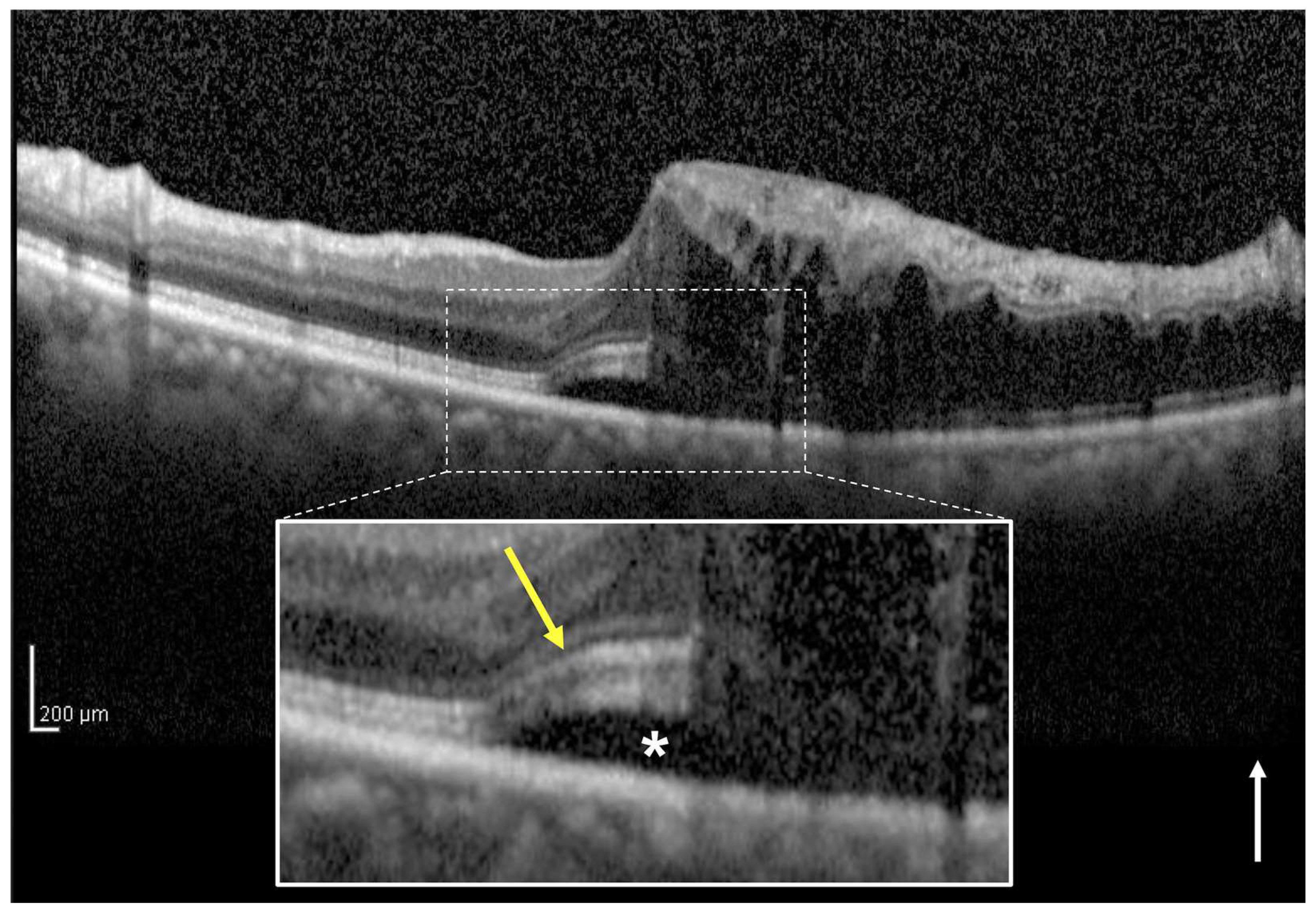
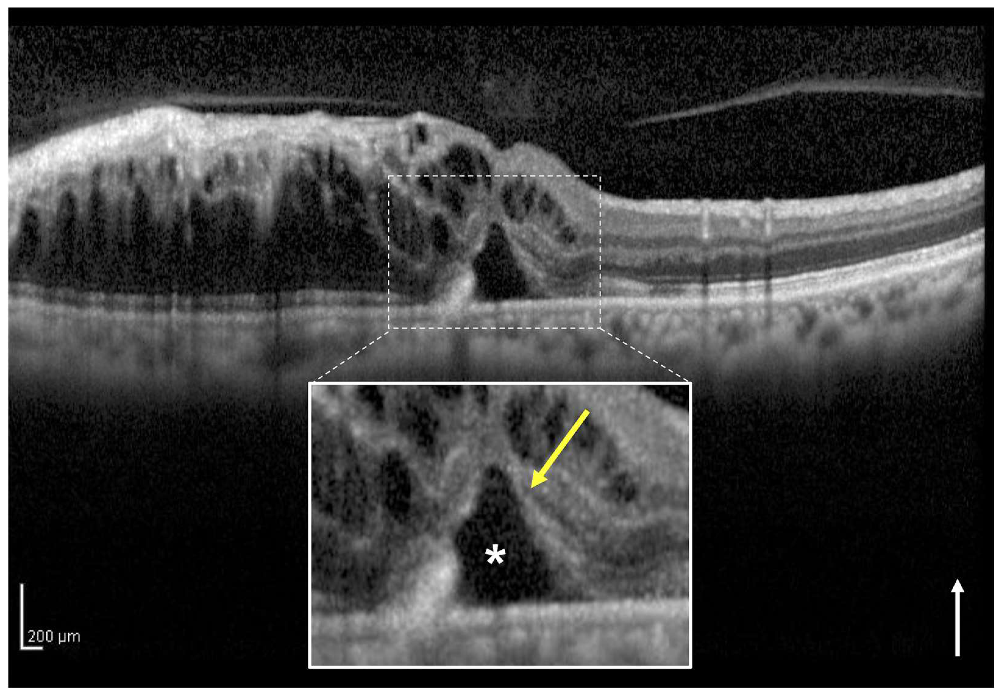
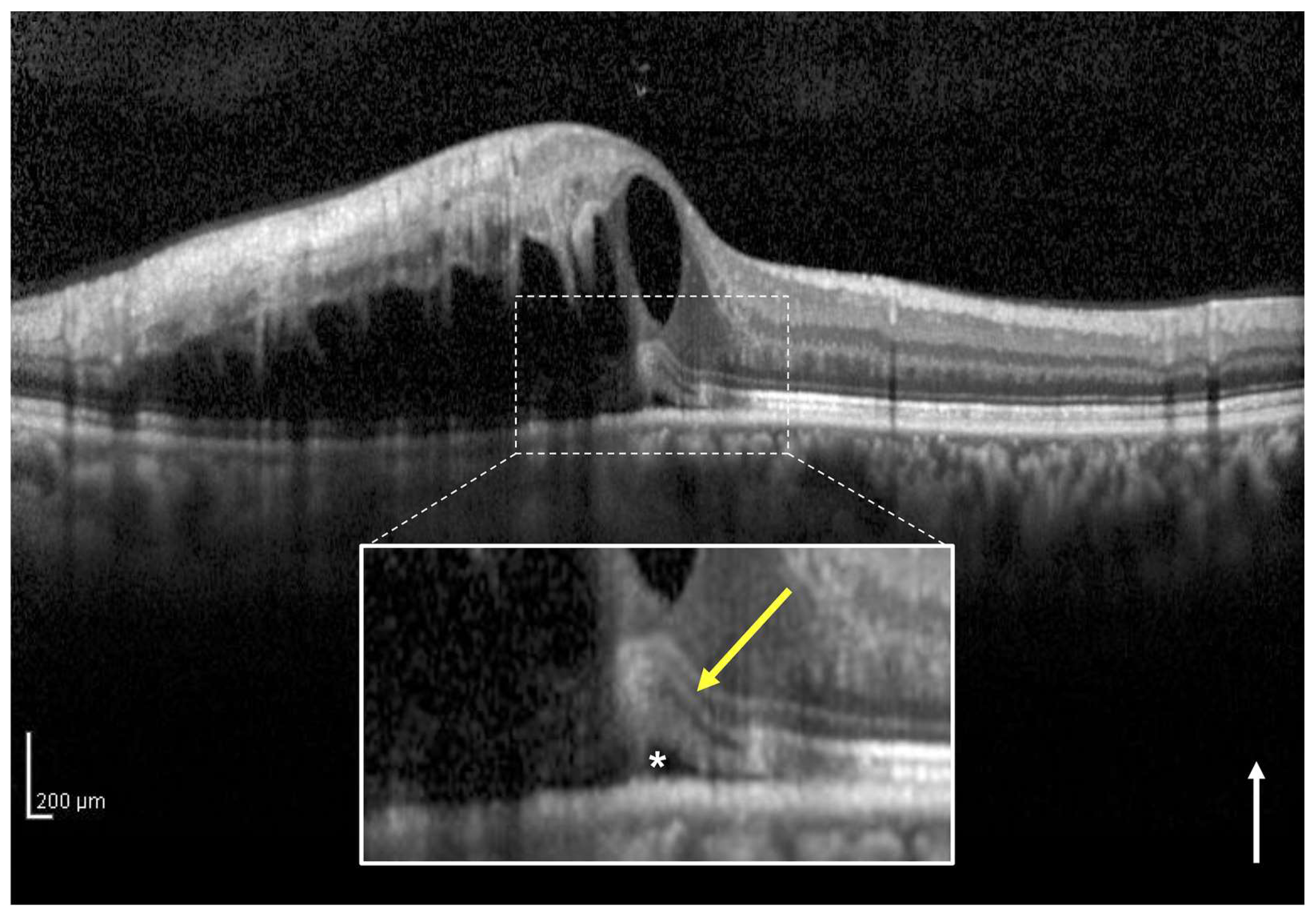
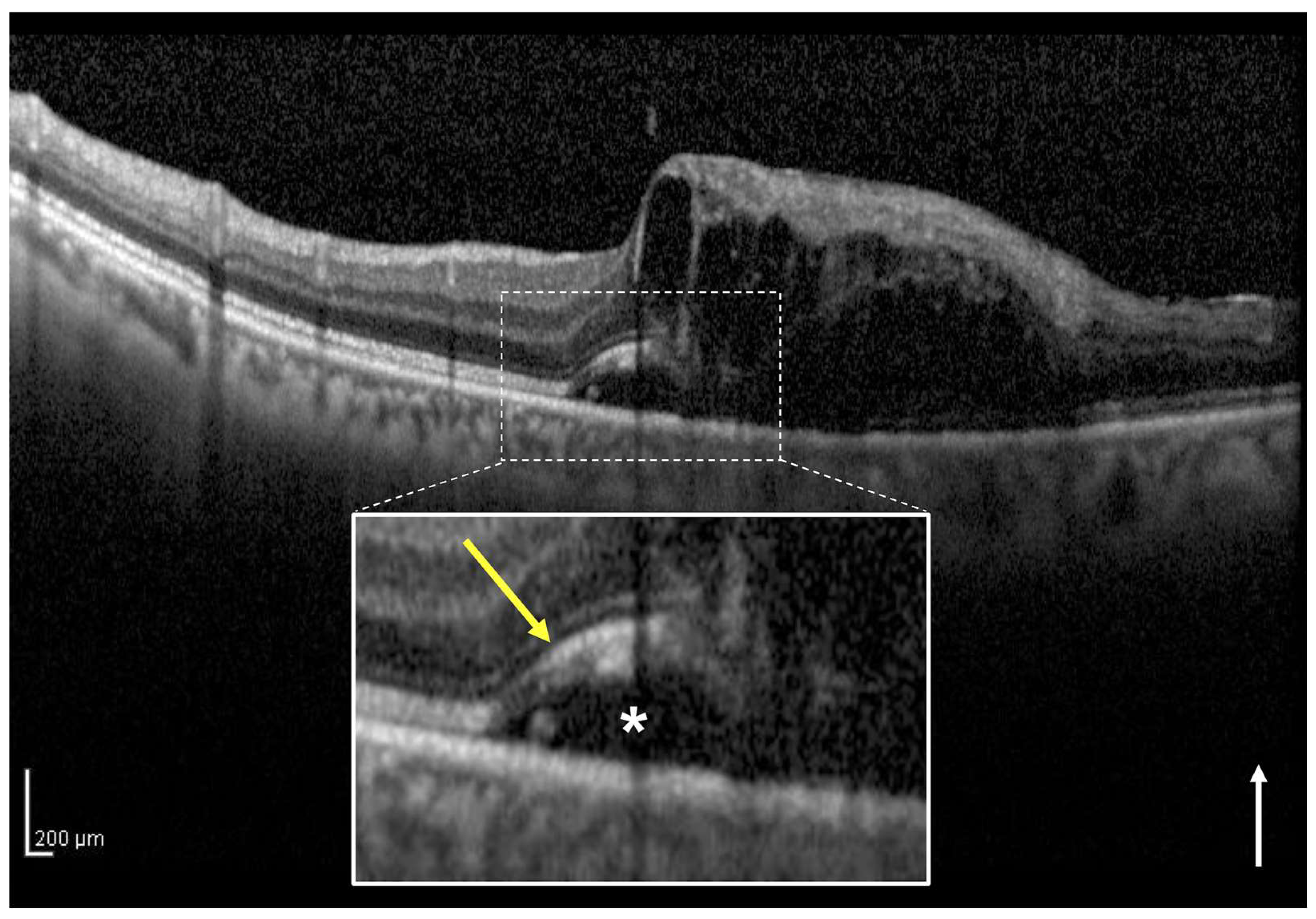
2.4. Definition and Classification of the Two Groups Using OCT
2.5. How to Divide Intact Ellipsoid Zone Band into Two Groups Using OCT
2.6. Assessment of the Foveal Ellipsoid Zone Band at Month 12
2.7. Statistical Analysis
3. Results
3.1. Patient Characteristics
3.2. Comparison of the Two Groups According to Foveal Ellipsoid Zone Band Status at the Initial Visit
3.3. Comparison of the Two Groups According to Whether the Intact Ellipsoid Zone Band Is Clear or Blurred at the Initial Visit
3.4. Baseline Characteristics Which Were Associated with Visual Acuity at Month 12
4. Discussion
5. Conclusions
Author Contributions
Funding
Institutional Review Board Statement
Informed Consent Statement
Data Availability Statement
Conflicts of Interest
References
- Kondo, M.; Kondo, N.; Ito, Y.; Kachi, S.; Kikuchi, M.; Yasuma, T.R.; Ota, I.; Kensaku, M.; Terasaki, H. Intravitreal injection of bevacizumab for macular edema secondary to branch retinal vein occlusion: Results after 12 months and multiple regression analysis. Retina 2009, 29, 1242–1248. [Google Scholar] [CrossRef]
- Jaissle, G.B.; Szurman, P.; Feltgen, N.; Spitzer, B.; Pielen, A.; Rehak, M.; Spital, G.; Heimann, H.; Meyer, C.H.; Retinal Vein Occlusion Study Group. Predictive factors for functional improvement after intravitreal bevacizumab therapy for macular edema due to branch retinal vein occlusion. Graefe’s Arch. Clin. Exp. Ophthalmol. 2011, 249, 183–192. [Google Scholar] [CrossRef]
- Iida-Miwa, Y.; Muraoka, Y.; Iida, Y.; Ooto, S.; Murakami, T.; Suzuma, K.; Tsujikawa, A. Branch retinal vein occlusion: Treatment outcomes according to the retinal nonperfusion area, clinical subtype, and crossing pattern. Sci. Rep. 2019, 9, 6569. [Google Scholar] [CrossRef]
- Scott, I.U.; VanVeldhuisen, P.C.; Oden, N.L.; Ip, M.S.; Blodi, B.A.; Hartnett, M.E.; Cohen, G.; Standard Care versus COrticosteroid for REtinal Vein Occlusion Study Investigator Group. Baseline predictors of visual acuity and retinal thickness outcomes in patients with retinal vein occlusion: Standard care versus corticosteroid for retinal vein occlusion study Report 10. Ophthalmology 2011, 118, 345–352. [Google Scholar] [CrossRef]
- Sasajima, H.; Zako, M.; Maeda, R.; Murotani, K.; Ishida, H.; Ueta, Y. Foveal intraretinal fluid localization affects the visual prognosis of branch retinal vein occlusion. J. Clin. Med. 2022, 11, 3540. [Google Scholar] [CrossRef]
- Kang, H.M.; Chung, E.J.; Kim, Y.M.; Koh, H.J. Spectral-domain optical coherence tomography (SD-OCT) patterns and response to intravitreal bevacizumab therapy in macular edema associated with branch retinal vein occlusion. Graefe’s Arch. Clin. Exp. Ophthalmol. 2013, 251, 501–508. [Google Scholar] [CrossRef]
- Shiono, A.; Kogo, J.; Sasaki, H.; Yomoda, R.; Jujo, T.; Tokuda, N.; Kitaoka, Y.; Takagi, H. Optical coherence tomography findings as a predictor of clinical course in patients with branch retinal vein occlusion treated with ranibizumab. PLoS ONE 2018, 13, e0199552. [Google Scholar] [CrossRef]
- Spaide, R.F.; Lee, J.K.; Klancnik, J.K., Jr.; Gross, N.E. Optical coherence tomography of branch retinal vein occlusion. Retina 2003, 23, 343–347. [Google Scholar] [CrossRef]
- Shroff, D.; Mehta, D.K.; Arora, R.; Narula, R.; Chauhan, D. Natural history of macular status in recent-onset branch retinal vein occlusion: An optical coherence tomography study. Int. Ophthalmol. 2008, 28, 261–268. [Google Scholar] [CrossRef]
- Tsujikawa, A.; Sakamoto, A.; Ota, M.; Kotera, Y.; Oh, H.; Miyamoto, K.; Kita, M.; Yoshimura, N. Serous retinal detachment associated with retinal vein occlusion. Am. J. Ophthalmol. 2010, 149, 291–301.e5. [Google Scholar] [CrossRef] [PubMed]
- Ohashi, H.; Oh, H.; Nishiwaki, H.; Nonaka, A.; Takagi, H. Delayed absorption of macular edema accompanying serous retinal detachment after grid laser treatment in patients with branch retinal vein occlusion. Ophthalmology 2004, 111, 2050–2056. [Google Scholar] [CrossRef]
- Gallego-Pinazo, R.; Dolz-Marco, R.; Pardo-López, D.; Martínez-Castillo, S.; Lleó-Pérez, A.; Arévalo, J.F.; Díaz-Llopis, M. Ranibizumab for serous macular detachment in branch retinal vein occlusions. Graefe’s Arch. Clin. Exp. Ophthalmol. 2013, 251, 9–14. [Google Scholar] [CrossRef] [PubMed]
- Schmidt-Erfurth, U.; Garcia-Arumi, J.; Gerendas, B.S.; Midena, E.; Sivaprasad, S.; Tadayoni, R.; Wolf, S.; Loewenstein, A. Guidelines for the management of retinal vein occlusion by the European Society of Retina Specialists (EURETINA). Ophthalmologica 2019, 242, 123–162. [Google Scholar] [CrossRef]
- The Branch Vein Occlusion Study Group. Argon laser photocoagulation for macular edema in branch vein occlusion. The Branch Vein Occlusion Study Group. Am. J. Ophthalmol. 1984, 98, 271–282. [Google Scholar] [CrossRef]
- Hayreh, S.S.; Zimmerman, M.B. Fundus changes in branch retinal vein occlusion. Retina 2015, 35, 1016–1027. [Google Scholar] [CrossRef]
- Miwa, Y.; Muraoka, Y.; Osaka, R.; Ooto, S.; Murakami, T.; Suzuma, K.; Takahashi, A.; Iida, Y.; Yoshimura, N.; Tsujikawa, A. Ranibizumab for macular edema after branch retinal vein occlusion: One initial injection versus three monthly injections. Retina 2017, 37, 702–709. [Google Scholar] [CrossRef]
- Murata, T.; Kondo, M.; Inoue, M.; Nakao, S.; Osaka, R.; Shiragami, C.; Sogawa, K.; Mochizuki, A.; Shiraga, R.; Ohashi, Y.; et al. The randomized ZIPANGU trial of ranibizumab and adjunct laser for macular edema following branch retinal vein occlusion in treatment-naïve patients. Sci. Rep. 2021, 11, 551. [Google Scholar] [CrossRef] [PubMed]
- Ota, M.; Tsujikawa, A.; Murakami, T.; Kita, M.; Miyamoto, K.; Sakamoto, A.; Yamaike, N.; Yoshimura, N. Association between integrity of foveal photoreceptor layer and visual acuity in branch retinal vein occlusion. Br. J. Ophthalmol. 2007, 91, 1644–1649. [Google Scholar] [CrossRef]
- Murakami, T.; Tsujikawa, A.; Ohta, M.; Miyamoto, K.; Kita, M.; Watanabe, D.; Takagi, H.; Yoshimura, N. Photoreceptor status after resolved macular edema in branch retinal vein occlusion treated with tissue plasminogen activator. Am. J. Ophthalmol. 2007, 143, 171–173. [Google Scholar] [CrossRef] [PubMed]
- Tso, M.O. Pathology of cystoid macular edema. Ophthalmology 1982, 89, 902–915. [Google Scholar] [CrossRef]
- Lee, Y.H.; Kim, Y.C. Clinical characteristics of branch retinal vein occlusion with increased retinal haemorrhage during treatment for macular edema. Sci. Rep. 2020, 10, 10272. [Google Scholar] [CrossRef] [PubMed]
- Iijima, H. Mechanisms of vision loss in eyes with macular edema associated with retinal vein occlusion. Jpn. J. Ophthalmol. 2018, 62, 265–273. [Google Scholar] [CrossRef]
- Nagasato, D.; Muraoka, Y.; Tanabe, M.; Nishigori, N.; Osaka, R.; Mitamura, Y.; Tabuchi, H.; Kadomoto, S.; Murakami, T.; Ooto, S.; et al. Foveal thickness fluctuation in anti-VEGF treatment for branch retinal vein occlusion: A long-term study. Ophthalmol. Retina 2022, 6, 567–574. [Google Scholar] [CrossRef] [PubMed]
- Kriechbaum, K.; Michels, S.; Prager, F.; Georgopoulos, M.; Funk, M.; Geitzenauer, W.; Schmidt-Erfurth, U. Intravitreal avastin for macular oedema secondary to retinal vein occlusion: A prospective study. Br. J. Ophthalmol. 2008, 92, 518–522. [Google Scholar] [CrossRef] [PubMed]
- Ota, M.; Tsujikawa, A.; Murakami, T.; Yamaike, N.; Sakamoto, A.; Kotera, Y.; Miyamoto, K.; Kita, M.; Yoshimura, N. Foveal photoreceptor layer in eyes with persistent cystoid macular edema associated with branch retinal vein occlusion. Am. J. Ophthalmol. 2008, 145, 273–280. [Google Scholar] [CrossRef] [PubMed]
- Hoeh, A.E.; Ruppenstein, M.; Ach, T.; Dithmar, S. OCT patterns of macular edema and response to bevacizumab therapy in retinal vein occlusion. Graefe’s Arch. Clin. Exp. Ophthalmol. 2010, 248, 1567–1572. [Google Scholar] [CrossRef]


| Parameters | Value | p-Value |
|---|---|---|
| Total no. of eyes | 38 | |
| Baseline | ||
| Age (years) | 66.4 (12.7) | |
| Gender (male/female) | 12/26 | |
| Affected eye (right/left) | 22/16 | |
| Lens status (phakic/pseudophakic) | 29/9 | |
| Hypertension (%) | 21 (55.3) | |
| Diabetes mellitus (%) | 9 (23.7) | |
| Dyslipidemia (%) | 8 (21.1) | |
| Subtype (major/macular) | 30/8 | |
| Perfusion status (ischemic/perfused) | 25/13 | |
| Duration before initial treatment (weeks) | 5 (5.2) | |
| LogMAR BCVA | 0.38 (0.32) | |
| Central subfield thickness (μm) | 583.5 (133.3) | |
| Subretinal fluid thickness (μm) | 155.9 (121.5) | |
| Intact EZ band/disruptive EZ band | 26/12 | |
| Subretinal hemorrhage (%) | 18 (47.4) | |
| 12 months following the initial treatment | ||
| Total no. of injections | 4.2 (1.7) | |
| Scatter laser photocoagulations during the follow-up periods (%) | 20 (52.6) | |
| LogMAR BCVA | 0.089 (0.29) | <0.001 * |
| Central subfield thickness (μm) | 273.8 (29.2) | <0.001 * |
| Foveal EZ band disruption (%) | 8 (21.1) | |
| Parameters | Intact EZ | Disruptive EZ | p-Value |
|---|---|---|---|
| Total no. of eyes | 26 | 12 | |
| Baseline | |||
| Age (years) | 65.3 (12.4) | 68.9 (13.6) | 0.42 |
| Gender (female) | 18 | 8 | 1 |
| Affected eye (right) | 14 | 8 | 0.5 |
| Lens status (pseudophakic) | 7 | 2 | 0.69 |
| Hypertension (%) | 15 (57.7) | 6 (50) | 0.73 |
| Diabetes mellitus (%) | 8 (30.8) | 1 (8.3) | 0.22 |
| Dyslipidemia (%) | 6 (23.1) | 2 (16.7) | 1 |
| Subtype (major) | 19 | 11 | 0.39 |
| Perfusion status (ischemic/perfused) | 15/11 | 10/2 | 0.16 |
| Duration before initial treatment (weeks) | 4.5 (5.7) | 6.1 (3.9) | 0.39 |
| LogMAR BCVA | 0.23 (0.2) | 0.7 (0.3) | <0.001 |
| Central subfield thickness (μm) | 556.2 (124.3) | 642.8 (138.1) | 0.062 |
| Subretinal fluid thickness (μm) | 125 (116.6) | 223 (107.8) | 0.019 |
| Subretinal hemorrhage (%) | 10 (38.5) | 8 (66.7) | 0.16 |
| 12 months following the initial treatment | |||
| Total no. of injections | 4.0 (1.7) | 4.4 (1.8) | 0.54 |
| Scatter laser photocoagulations during the follow-up periods (%) | 12 (46.2) | 8 (66.7) | 0.31 |
| LogMAR BCVA | −0.045 (0.1) | 0.38 (0.3) | <0.001 |
| Change in logMAR BCVA | −0.27 (0.2) | −0.32 (0.4) | 0.64 |
| Central subfield thickness (μm) | 274.9 (20.5) | 271.4 (43.6) | 0.74 |
| Foveal EZ band disruption (%) | 1 (3.8) | 7 (58.3) | <0.001 |
| Parameters | Recovered EZ | Non-Recovered EZ | p-Value |
|---|---|---|---|
| Total no. of eyes | 5 | 7 | |
| Age (years) | 66 (12.0) | 71 (15.1) | 0.55 |
| Duration before initial treatment (weeks) | 6.2 (4.4) | 6 (3.8) | 0.93 |
| Baseline logMAR BCVA | 0.58 (0.3) | 0.78 (0.4) | 0.33 |
| Baseline central subfield thickness (μm) | 625.4 (44.0) | 655.1 (182.4) | 0.73 |
| Baseline subretinal fluid thickness (μm) | 201.4 (53.5) | 238.4 (136.9) | 0.58 |
| Baseline subretinal hemorrhage (%) | 3 (60) | 5 (71.4) | 1 |
| Total no. of injections | 4 (1.4) | 4.7 (2.1) | 0.53 |
| Parameters | Clear EZ | Blurred EZ | p-Value |
|---|---|---|---|
| Total no. of eyes | 15 | 11 | |
| Baseline | |||
| Age (years) | 66.3 (11.8) | 63.9 (13.7) | 0.63 |
| Gender (female) | 10 | 8 | 1 |
| Affected eye (right) | 9 | 5 | 0.69 |
| Lens status (pseudophakic) | 3 | 4 | 0.41 |
| Hypertension (%) | 9 (60) | 6 (54.5) | 1 |
| Diabetes mellitus (%) | 5 (33.3) | 3 (27.3) | 1 |
| Dyslipidemia (%) | 4 (26.7) | 2 (18.2) | 1 |
| Subtype (major) | 12 | 7 | 0.41 |
| Perfusion status (ischemic/perfused) | 11/4 | 4/7 | 0.11 |
| Duration before initial treatment (weeks) | 3.3 (4.5) | 6.1 (6.9) | 0.23 |
| LogMAR BCVA | 0.23 (0.2) | 0.22 (0.2) | 0.95 |
| Central subfield thickness (μm) | 511.9 (100.1) | 616.5 (132.8) | 0.031 |
| Subretinal fluid thickness (μm) | 45.5 (28.7) | 233.4 (102.1) | <0.001 |
| Subretinal hemorrhage (%) | 0 (0) | 10 (90.9) | <0.001 |
| 12 months following the initial treatment | |||
| Total no. of injections | 4.1 (1.7) | 4.0 (1.7) | 0.92 |
| Scatter laser photocoagulations during the follow-up periods (%) | 7 (46.7) | 5 (45.5) | 1 |
| LogMAR BCVA | −0.03 (0.1) | −0.066 (0.1) | 0.39 |
| Change in logMAR BCVA | −0.26 (0.2) | −0.29 (0.2) | 0.65 |
| Central subfield thickness (μm) | 274.4 (21.7) | 275.6 (19.7) | 0.88 |
| Foveal EZ band disruption (%) | 1 (6.7) | 0 (0) | 1 |
| Explanatory Variables | Coefficient (95% CI) | p-Value | Standardized Coefficient |
|---|---|---|---|
| Intact EZ (compared to disruptive EZ) | −0.31 (−0.5 to −0.11) | 0.0028 | −0.51 |
| Age (years) | 0.004 (−0.002 to 0.001) | 0.15 | 0.18 |
| Baseline logMAR BCVA | 0.14 (−0.16 to 0.44) | 0.35 | 0.16 |
| Baseline central subfield thickness (μm) | 0.0004 (−0.00007 to 0.0009) | 0.09 | 0.2 |
Disclaimer/Publisher’s Note: The statements, opinions and data contained in all publications are solely those of the individual author(s) and contributor(s) and not of MDPI and/or the editor(s). MDPI and/or the editor(s) disclaim responsibility for any injury to people or property resulting from any ideas, methods, instructions or products referred to in the content. |
© 2023 by the authors. Licensee MDPI, Basel, Switzerland. This article is an open access article distributed under the terms and conditions of the Creative Commons Attribution (CC BY) license (https://creativecommons.org/licenses/by/4.0/).
Share and Cite
Sasajima, H.; Zako, M.; Murotani, K.; Ishida, H.; Ueta, Y.; Tachi, N.; Suzuki, T.; Watanabe, Y.; Hashimoto, Y. Visual Prognostic Factors in Eyes with Subretinal Fluid Associated with Branch Retinal Vein Occlusion. J. Clin. Med. 2023, 12, 2909. https://doi.org/10.3390/jcm12082909
Sasajima H, Zako M, Murotani K, Ishida H, Ueta Y, Tachi N, Suzuki T, Watanabe Y, Hashimoto Y. Visual Prognostic Factors in Eyes with Subretinal Fluid Associated with Branch Retinal Vein Occlusion. Journal of Clinical Medicine. 2023; 12(8):2909. https://doi.org/10.3390/jcm12082909
Chicago/Turabian StyleSasajima, Hirofumi, Masahiro Zako, Kenta Murotani, Hidetoshi Ishida, Yoshiki Ueta, Naoko Tachi, Takafumi Suzuki, Yuji Watanabe, and Yoshihiro Hashimoto. 2023. "Visual Prognostic Factors in Eyes with Subretinal Fluid Associated with Branch Retinal Vein Occlusion" Journal of Clinical Medicine 12, no. 8: 2909. https://doi.org/10.3390/jcm12082909
APA StyleSasajima, H., Zako, M., Murotani, K., Ishida, H., Ueta, Y., Tachi, N., Suzuki, T., Watanabe, Y., & Hashimoto, Y. (2023). Visual Prognostic Factors in Eyes with Subretinal Fluid Associated with Branch Retinal Vein Occlusion. Journal of Clinical Medicine, 12(8), 2909. https://doi.org/10.3390/jcm12082909


