Article

Exploring the Bio-Functional Effect of Single Nucleotide Polymorphisms in the Promoter Region of the TNFSF4, CD28, and PDCD1 Genes

1
Department of Laboratory Medicine, Linkou Chang Gung Memorial Hospital at Linkou, Taoyuan City 333, Taiwan
2
Department of Medical Biotechnology and Laboratory Science, College of Medicine, Chang Gung University at Linkou, Taoyuan City 333, Taiwan
3
Graduate Institute of Clinical Medical Sciences, College of Medicine, Chang Gung University at Linkou, Taoyuan City 333, Taiwan
4
School of Medicine, National Tsing Hua University, Hsinchu 30013, Taiwan
5
School of Medicine, Chang Gung University at Linkou, Taoyuan City 333, Taiwan
*
Author to whom correspondence should be addressed.
J. Clin. Med. 2023, 12(6), 2157; https://doi.org/10.3390/jcm12062157
Submission received: 18 January 2023 / Revised: 1 March 2023 / Accepted: 6 March 2023 / Published: 10 March 2023
(This article belongs to the Section Clinical Laboratory Medicine)

Abstract

:
In a prior study, we discovered that hematopoietic stem cell transplantation (HSCT) and/or autoimmune diseases, such as systemic lupus erythematosus, were associated with the rs1234314 C/G and rs45454293 C/T polymorphisms of TNFSF4, the rs5839828 C > del and rs36084323 C > T polymorphisms of PDCD1, and the rs28541784C/T, rs200353921A/T, rs3181096C/T, and rs3181098 G/A polymorphisms of CD28. However, the association does not imply causation. These single nucleotide polymorphisms (SNPs) are all located in the promoter region of these genes, so we used the dual-luminescence reporter assay to explore the effect of single nucleotide polymorphisms (SNPs) on transcriptional activity. For each promoter–reporter with a single SNP mutation, more than 10 independent experiments were carried out, and the difference in transcription activity was compared using one-way ANOVA and Tukey’s honestly significant difference test. The results showed that the G-allele of rs1234314 had 0.32 ± 0.09 times the average amount of relative light units (RLU) compared to the C-allele (p = 0.003), the T-allele of rs45454293 had 4.63 ± 0.92 times the average amount of RLU compared to the C-allele (p < 0.001), the del-allele of rs5839828 had 1.37 ± 0.24 times the average amount of RLU compared to the G-allele (p < 0.001), and the T-allele of rs36084323 had 0.68 ± 0.07 times the average amount of RLU compared to the C-allele (p < 0.001). The CD28 SNPs studied here did not affect transcriptional activity. In conclusion, the findings of this study could only confirm that the SNP had a bio-functional effect on gene expression levels. According to the findings, several SNPs in the same gene have bio-functions that affect transcriptional activity. However, some increase transcriptional activity while others decrease it. Consequently, we inferred that the final protein level should be the integration result of the co-regulation of all the SNPs with the effect on transcriptional activity.

1. Introduction

Even though hematopoietic stem cell transplantation (HSCT) has become the gold standard therapy for hematological diseases and cancer, post-HSCT complications limit its therapeutic potential. Although standardization of human leukocyte antigen (HLA) matching between donor and patient before transplantation has greatly improved HSCT outcomes, the mortality rate caused by many complications after transplantation, such as graft-versus-host disease (GVHD), infection, and disease relapse, remains significantly higher than that of the general population [1,2]. Accumulating evidence supports that non-HLA gene variations are an important cause of post-HSCT outcomes [3,4,5]. The immune system’s role is largely responsible for the immune system’s reaction to alloantigens and infection complications after HSCT. Co-stimulatory molecules are one of the most important components in immune regulation, and they are likely to be affected by gene polymorphisms [5,6]. It is known that T-cell activation requires two necessary signals. The first signal is produced when the T-cell receptor interacts with the antigen peptide presented by the major histocompatibility complex molecule on the antigen-presenting cells (APCs), and the second is a positive regulation signal of T-cell activation produced by the interaction between the receptor expressed on the surface of T cells and its ligand expressed on the APCs [7,8,9,10]. The second signal will reduce T-cell proliferation and cytokine production, promote T-cell dysfunction or apoptosis, and activate regulatory T cells [11].
Imbalance of the co-stimulatory system is one of the immune escape mechanisms in blood cancers. Many studies [12,13,14,15] have shown a link between HSCT and co-stimulatory molecules, such as tumor necrosis factor superfamily 4 (TNFSF4), programmed cell death protein 1 (PDCD1; PD-1; CD279), and CD28. These genes were also associated with the development of autoimmune diseases and cancers [16]. According to some studies, CD28 and PDCD1 play an important role in the immune system and transplantation [8,17,18,19]. Furthermore, genetic variations, such as SNPs in HLA and non-HLA genes, were linked to HSCT success or failure in various ethnic groups [20].
Systemic lupus erythematosus (SLE) is an autoimmune disease (AD) characterized by the production of autoantibodies. Its pathogenesis is still unclear. Several studies have shown that the overactivation of autoreactive T cells is the primary cause of Alzheimer’s disease [21,22]. Although the current study found that the HLA gene usually had the strongest correlation with AD [23], other genes located out of the HLA region may also be risk factors for AD. SNPs in genes encoding T-cell and B-cell function-related proteins were identified as SLE susceptibility loci. Among them, the CD28 gene, which continuously expresses on T cells and provides the second stimulation signal to promote T-cell activation and make it aggressive after binding with CD80/CD86 on APCs, is the most important [24,25]. PDCD1 is a type of immune checkpoint. When PD-1 protein binds to its ligand on APCs, it activates an immunoreceptor tyrosine-based inhibitory motif on the cytoplasmic tail of PD-1, thus inhibiting T-cell activation [26]. In addition, TNFSF4 (OX40L) interacts with its receptor (OX40) and can also provide signals to promote T-cell activation, with previous research showing that OX40L is capable of stimulating T-cell response as well as promoting the pathogenesis of SLE [27].
We previously discussed the association between SNPs of the CD28, TNFSF4, and PDCD1 genes and the outcomes of post-cord blood transplantation (CBT) and HSCT and the development of SLE [28,29,30], finding that many SNPs located in the promoter region had statistically significant differences. However, the association does not imply causation. As a result, fluorescence analysis for SNPs in the promoter regions of the TNFSF4, PDCD1, and CD28 genes was performed to investigate the impact of these SNP changes on transcription activity.

2. Materials and Methods

2.1. SNP Selection

In a previous study, the promoter regions of CD28, PDCD1, and TNFSF4 were amplified to investigate the relationship between SNPs in these regions and HSCT, CBT, and SLE. Of interest were the TNFSF4 polymorphisms rs1234314 C/G and rs45454293 C/T, PDCD1 polymorphisms rs5839828 C/del and rs36084323 C/T, and CD28 polymorphisms rs28541784 C/T, rs200353921 A/T, rs3181096 C/T, and rs3181098 G/A. Thus, these promoter SNPs were selected to analyze the effect on transcription activity. For a detailed PCR program and the primers used for amplifying the promoter regions of CD28, PDCD1, and TNFSF4, please refer to ref. [28,29,30].

2.2. Promoter–Reporter Assay

2.2.1. Constructing the Template of Promoter–Reporter and Plasmid

To begin, a normal human DNA template was used, and primers with specific restriction enzyme cleavage sites (as shown in Table 1) for the promoter region of the TNFSF4, CD28, and PDCD1 genes were designed. After the DNA sequence was confirmed to be correct by sequencing, the promoter fragment was transferred into plasmids using the TOPO TA cloning kit (Invitrogen). Plasmid DNA was then extracted and sequenced in an E. coli competent cell (TOP10 or DH5α) to determine the promoter sequence.

2.2.2. Construction of Luciferase Expression Vectors and Transformation

The TNFSF4/CD28/PDCD1 promoter plasmid with restriction enzyme cleavage sites (SacI and EcoRV for TNFSF4 and PDCD1; SacI and HindIII for CD28) and the fluorescent expression vector pNL1.1 [Nluc] (Promega, Madison, WI, USA) were reacted with a restriction enzyme for one hour at 37 °C. Electrophoresis was carried out with 1% agarose gel for 20 min. The pNL1.1 vector with a specific promoter fragment was cut off in the gel, using a PCR/gel purification kit, and the two fragments were then joined with a T4 ligase-connecting enzyme to create a promoter vector with NanoLuc® luciferase expression (Promega). The vector was transformed to competent cells (TOP10 or DH5α). After screening and culture of the successfully transformed cells (LB agar plate), the NanoLuc® luciferase expression vector with the correct promoter fragment was extracted. The promoter report assay used the vector with the correct sequence as the control group, and the plasmid DNA served as the template for site-directed mutagenesis PCR (QuikChange II site-directed Mutagenesis Kit, Stratagene). The report vectors of rs1234314 C > G, rs45454293C > T, rs5839828 C > del, rs36084323 C > T, rs28541784C/T, rs200353921A/T, rs3181096C/T, and rs3181098G/A (Table 1) were constructed in the same way as above.

2.2.3. Dual-Luciferase Reporter Assay

A total of 0.5 μg of the construct promoter–reporter vector, 0.5 μg of the PGL 4.5 [Luc2/TK] vector (Promega, Madison, WI, USA with firefly luciferase used as an internal control to correct transformation efficiency, and Lipofectamine2000 (Invitrogen, Carlsbad, CA, USA) were mixed evenly and allowed to stand for 20 min. They were then co-cultured with 5 × 10⁵ K562 cells in a 24-well plate of RPMI 1640 medium containing 10% fetal bovine serum, 50 U/mL of penicillin, and 50 μg/mL of streptomycin in a CO2 incubator for 48 h. The cultured cells were evenly mixed and 80 uL of them were extracted. They were placed in a white flat-bottomed 96-well plate, treated with Promega Passive Lysis Buffer, and allowed to stand for 15 min. The Nano-Glo Dual-Luciferase Reporter with the Dual Luminescent Enzyme Detection Kit Assay System (Promega) was then used for the reaction, followed by luminescence detection with a luminescence enzyme detector (GloMax Discover System, Promega, CA, USA).

2.3. Statistical Analysis

The promoter–reporter assay of each SNP mutation was conducted 11–16 times in parallel. We calculated the wild type of each gene by dividing the value of pNL1.1 (NanoLuc) by the value of PGL 4.5 (firefly). The relative light units (RLU) of wild-type constructs were corrected to 1. There was one SNP variation between wild-type constructs and the SNP–reporter construct in order to verify the transcription activity level of the specific SNP. Data beyond plus or minus 2 standard deviations were excluded. The average amount of RLU corresponding to the promoter construct of each SNP was compared using one-way ANOVA in the SPSS 17.0 software package (SPSS Inc., Chicago, IL, USA), and Tukey’s honestly significant difference test was used for post hoc testing. The significance level was set as 0.05.

3. Results

3.1. TNFSF4 Promoter–Reporter Assay

Both the rs1234314 C > G and rs45454293 C > T reporter assays were tested 16 times in a row. The C-allele of rs1234314 and C-allele of rs45454293 were defined as wild type. It was discovered that rs1234314 C > G had 0.32 ± 0.09 times the RLU of rs1234314 with the C-allele and rs45454293 C > T had 4.630.92 times the RLU of rs45454293 with the C-allele. In ANOVA analysis, the main effect of constructs (F(2,43) = 83.255, p < 0.001) was revealed. Following a post hoc analysis, it was discovered that rs1234314 C > G (p = 0.003) and rs45454293C > T (p < 0.001) have statistical significance with the wild type (Supplementary Table S1 and Figure 1).

3.2. PDCD-1 Promoter–Reporter Assay

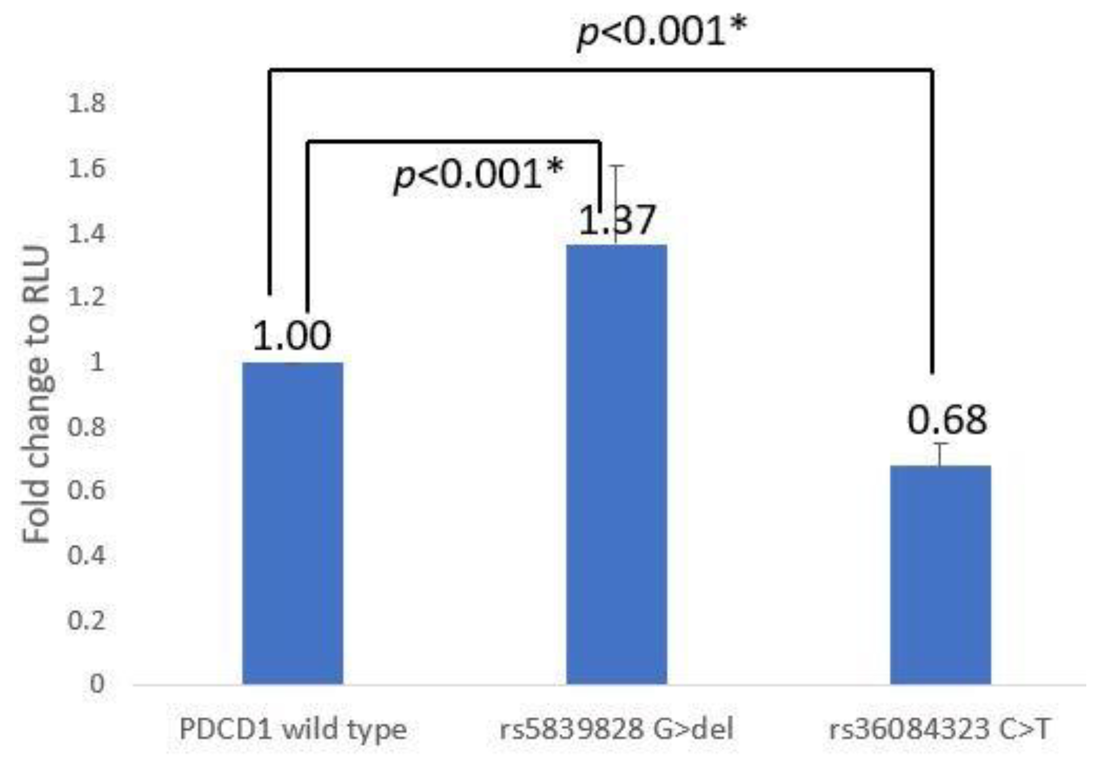
Both the rs5839828 G > del and rs36084323 C > T reporter assays were conducted 11 times as independent tests. The del-allele of rs5839828 had 1.37 ± 0.24 times the RLU of the G-allele, and the T-allele of rs36084323 had 0.68 ± 0.07 times the RLU of the C-allele. In ANOVA analysis, it was also shown that there was a main effect between constructs (F(2,34) = 70.093, p < 0.001). Following a post hoc analysis, it was discovered that rs5839828 G > del (p < 0.001) and rs36084323 C > T (p < 0.001) have statistical significance with the wild type (Supplementary Table S2 and Figure 2).

3.3. CD28 Promoter–Reporter Assay

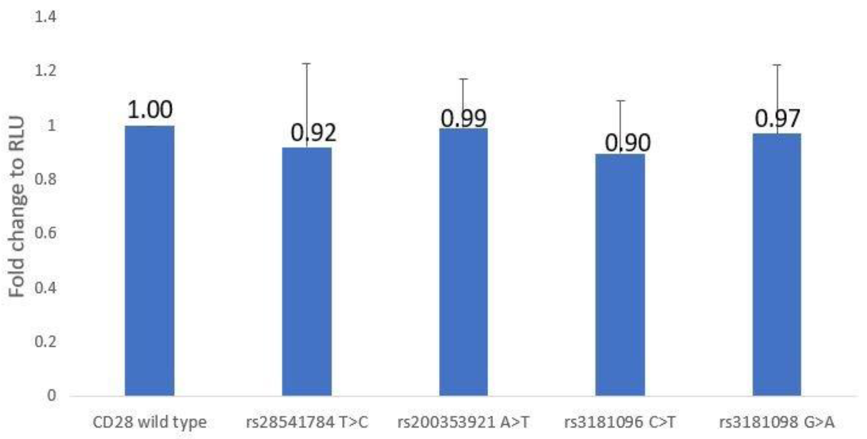
In the CD28 promoter region, four SNPs were examined (rs28541784T > C, rs200353921A > T, rs3181096C > T, and rs3181098G > A), with each promoter–reporter receiving 15 independent tests. It was shown that these SNPs had little effect on transcriptional activity. The RLU value for rs28541784 with the C-allele was 0.92 ± 0.31 times that of rs28541784 with the T-allele, the RLU value of rs200353921 with the T-allele was 0.99 ± 0.18 times that of rs200353921 with the A-allele, and the RLU of rs3181096 with the T-allele was 0.90 ± 0.20 times that of rs3181096 with the C-allele and 0.97 ± 0.25 times that of rs3181098 with the G-allele. In ANOVA analysis, there was no effect between these CD28 promoter–reporter constructs (F(4,66) = 0.644, p = 0.633) (Supplementary Table S3 and Figure 3).

4. Discussion

Co-stimulatory molecules are important in immune regulation and have been linked to the pathogenesis of autoimmune diseases, cancer, and transplant rejection [31]. On antigen-presenting cells, CD28 interacts with CD80/CD86 to provide a second stimulation signal required for T-cell activation [32]. PDCD1 plays a negative regulatory role in the activation process of T cells. It functions as an immune checkpoint and promotes immune tolerance, which can prevent the development of autoimmune diseases; cancer cells can also evade the immune system’s pursuit [33]. In addition, the OX40 ligand encoded by TNFSF4 is the key to coordinating innate and adaptive immune cells and plays an important role in differentiation, activation, inhibition, and apoptosis in the life cycle of immune cells [34].

4.1. About TNFSF4 Analysis

Regarding TNFSF4, it was discovered that rs1234314 and rs45454293 of TNSFS4 were associated with post-HSCT GVHD III-IV in acute myeloid leukemia (AML) patients in our previously published HSCT (including bone marrow transplantation and peripheral blood stem cell transplantation) research analysis [29]. Grafts with the rs1234314 C-allele had a 7.39-fold increased risk of GVHD III-IV compared with the G-allele (p = 0.011), and the rs45454293 T-allele had a 4.86-fold increased risk of GVHD III-IV compared with the C-allele (p = 0.010). In a CBT research study [28], rs1234314 of the TNFSF4 gene was associated with mortality after CBT (p < 0.001), and having the CC genotype increased the risk of mortality by 15.4 times. In SLE research analysis [30], the genotype frequency (CC vs. CG vs. GG) of rs1234314 was significantly different between cases and controls (p = 0.005). When compared to the CC genotype, the GG genotype was associated with a 4.4-fold increased risk of developing SLE (p = 0.004). Analysis of the promoter–reporter assay showed that rs1234314 C > G significantly reduced transcription activity by 0.32 times, while rs45454293 C > T significantly increased activity by 4.63 times.
TNFSF4 expression is known to drive T-cell proliferation, differentiation, and cytokine production [35]. Thus, the higher promoter activity of rs1234314 with the C-allele and rs45454293 with the T-allele was consistent with our previously published findings that suggest that they increase the risk of GVHD III-IV after HSCT in AML patients, as well as the risk of mortality after CBT [28,29]. Furthermore, Tripathi et al. discovered in 2019 that the OX40L(TNFSF4)-OX40 interaction on T cells was linked to the induction and development of acute GVHD (aGVHD) in HSCT, and that treatment with anti-human OX40L mAb could effectively prevent and reduce the severity of aGVHD [36]. Our findings revealed that transplanting the graft with the rs1234314 C-allele and rs45454293 T-allele increased the risk of GVHD III-IV and death because these two alleles had higher transcription activity.
The rs1234314 C to G variation reduced promoter activity by 68% (0.32), which seems to contradict the result for rs1234314 GG where the risk of SLE increased by 4.4 times [30]. However, a Chinese study published in 2017 discovered that the CC genotype of rs1234314 protected against allergic rhinitis in the Chinese Han population [37]. We also found that the risk of SLE in the GG genotype was 4.4 times higher than that in the CC genotype in results of patient analyses. In other words, CC protected against SLE. It has been established that GVHD is caused by the activation of allogeneic T cells in response to recognition of the allogeneic antigen presented by mismatched MHC molecules, resulting in the graft attacking the host [38]. Autoimmune diseases are caused by a loss of immune tolerance that is caused by the overactivation of autologous reactive T cells [39]. Based on this knowledge, we inferred that rs1234314 may play different roles in the prognosis of CBT, HSCT, and autoimmune diseases, which needs to be further verified in the future.

4.2. About PDCD1 Analysis

Previous research on the relationship between SNPs and HSCT outcomes [29] found that the C-allele of rs36084323 in the promoter region of the PDCD1 gene in donors was associated with a higher risk of CMV (p = 0.0265) and relapse (p = 0.0356) in ALL patients, while the G-allele of rs5839828 (p = 0.0265) increased the risk of relapse in ALL patients. According to analysis of the effectiveness of CBT [28], when the graft carried at least one T-allele in rs36084323, the CBT case had a 4.2 times greater risk of relapse (95% CI = 1.331–13.320, p = 0.012). For SLE [30], it was shown that the T-allele of rs36084323 provided a protective effect.
The PDCD1 promoter–reporter assay revealed that rs5839828 with the del-allele had 1.37 times the transcriptional activity of rs5839828 with the G-allele, whereas rs36084323 C > T decreased transcriptional activity. It is known that the role of PD1 is similar to that of CTLA4, which plays a negative regulatory role in T-cell activation to produce immune tolerance [40]. This regulatory mechanism can help to prevent autoimmune diseases, but it can also keep the immune system from killing cancer cells, resulting in disease relapse [26,41]. Our result showed that rs36084323 T-allele had significantly lower PDCD1 transcriptional activity. As a result, we concluded that transplanting a graft with rs36084323 T-allele may reduce PDCD1 expression, thus reducing T-cell activation and increasing the risk of relapse in ALL patients. In addition, the risk of CMV infection may also increase due to the decrease in T-cell activation. However, SLE patients had a higher frequency of at least one T-allele (CT + TT) in rs36084323 than controls. The possible reason may be the same as the conclusion in the above paragraph: the role of rs36084323 in autoimmune disease and transplantation was not consistent.
Aside from HSCT and CBT outcomes and SLE, rs36084323 has been linked to several cancers and immune abnormalities, including breast cancer, ovarian cancer, esophageal cancer, rheumatoid arthritis, and abortion [42]. A 2014 study showed that non-small-cell lung cancer patients with CC genotype rs36084323 had significantly poorer prognosis [43]. This could be due to rs36084323 SNP variation affecting PDCD1 transcriptional activity. In the reporter assay of rs36084323, we found that rs36084323 C-allele had higher transcription activity than T-allele. In other words, when rs36084323 is associated with the CC genotype, PDCD1 expression is increased. Additionally, PDCD1 is an inhibitory regulator of T-cell activation, which may hurt the elimination of cancer cells.
The effect of rs36084323 G > A (C > T) on transcriptional activity assessed by Ishizaki et al. [44] using a dual-luciferase reporter assay was consistent with ours. Both of our results showed that rs36084323 G > A (C > T) could reduce the transcriptional activity of PDCD1. Furthermore, a 2014 study by Jiao et al. found that rs36084323 with the GG genotype had higher mRNA expression in the PBMCs of SLE patients compared to the AA genotype [45]. Therefore, these reproducible results could prove that our experimental results were correct.

4.3. About CD28 Analysis

Previously, we found that rs200353921, rs28541784, rs3181096, and rs3181098 located in the promoter region of CD28 were associated with HSCT effectiveness. AML patients with T-alleles at rs200353921 had a higher risk of relapse (OR = 2.1, 95% CI = 1.06–4.18, p = 0.0343). Grafts with the T-allele at rs28541784 increase the risk of chronic GVHD (p = 0.0303) in ALL patients (OR = 2.78, 95% CI = 1.11–6.98). The T-allele of rs3181096 and the A-allele of rs3181098 decreased the risk of GVHD in AML patients and ALL patients, respectively. However, in the CD28 gene promoter–reporter assay, these SNP variations were found to not affect transcriptional activity.
GVHD is the most important adverse complication after HSCT. It is an immune system disease that affects many organ systems, including the gastrointestinal tract, liver, skin, and lungs, and has an impact on HSCT outcomes. Research shows that the pathogenesis and severity of GVHD are related to the activation of T cells [46]. CD28-mediated co-stimulatory signals are essential for the initiation and maintenance of T-cell activation. Animal experiments have also found that CD28 is related to the onset and severity of GVHD, and it can even reduce the severity of GVHD by blocking the function of CD28 on T cells [47].
Although there were no statistically significant changes in CD28 SNP and transcriptional activity in this study, we previously discovered that the CD28 SNP rs3181097 G > A could reduce CD28 transcriptional activity and was associated with a significantly lower risk of transfusion reactions [48]. It was proposed that, in addition to these four SNPs, there may be other SNPs in the CD28 gene that regulate gene expression.

4.4. The Significant and Further Study

It is well known that correlation does not imply causation. After association analysis, we verified the effect of the SLE- or HSCT-associated SNPs located in the promoter region of genes on transcription activity through reporter assays. Based on the findings, it was determined that several SNPs in one gene had biological functions affecting transcriptional activity (some increasing and others decreasing) and that the final protein level should be the integration result of the co-regulation of these SNPs with biological function. Therefore, it is necessary to test the effect of the haplotype. Next, we will investigate whether the SNPs/haplotypes with a functional effect on transcription activity influence T-cell activation or T-cell differentiation through a cell study model based on the T cells extracted from patients and explore the mechanism of diseases caused by the SNPs/haplotypes through animal models. However, gene expression in vitro does not imply in vivo expression. Thus, the proteins expressed in the serum or on the surface of T cells need to be tested. Additionally, how these SNPs affect transcription activity remains to be clarified, such as exploration through silico studies of the extent to which they alter transcription factor binding sites.

5. Conclusions

In summary, the results of this study verify that some promoter SNPs have the biological function of regulating transcriptional activity. As a result, in the future, it will be necessary to thoroughly investigate data related to the genes and proteins involved in autoimmune diseases or outcomes of HSCT to clarify the mechanism of SNPs on diseases. Furthermore, the findings regarding SNP–disease association can only be used to develop a genetic testing kit to aid clinical diagnosis and cannot explain the direct causal relationship between SNPs and diseases.

Supplementary Materials

The following supporting information can be downloaded at: https://www.mdpi.com/article/10.3390/jcm12062157/s1. Table S1: The RLU of TNFSF4 promoter reporter assay (16 independent tests); Table S2: The RLU of PDCD1 promoter reporter assay (11 independent tests); Table S3: The RLU of CD28 promoter reporter assay (15 independent tests).

Author Contributions

Conceptualization and writing of the manuscript, D.-P.C.; expression and data curation, W.-T.W.; analyzed and interpreted data, Y.-H.W. and W.-T.L. All authors have read and agreed to the published version of the manuscript.

Funding

This research was funded by Chang Gung Memorial Hospital, grant number CMRPG3M0571.

Institutional Review Board Statement

The study was conducted in accordance with the Declaration of Helsinki and approved by the Institutional Review Board of Chang Gung Memorial Hospital with the approval ID of 202101505B0.

Informed Consent Statement

Informed consent was obtained from all subjects involved in the study.

Data Availability Statement

The data presented in this study are openly available in reference number [28,29,30].

Conflicts of Interest

The authors declare no conflict of interest.

References

  1. Bhatia, S.; Francisco, L.; Carter, A.; Sun, C.-L.; Baker, K.S.; Gurney, J.G.; McGlave, P.B.; Nademanee, A.; O’Donnell, M.; Ramsay, N.K.C.; et al. Late mortality after allogeneic hematopoietic cell transplantation and functional status of long-term survivors: Report from the Bone Marrow Transplant Survivor Study. Blood 2007, 110, 3784–3792. [Google Scholar] [CrossRef] [PubMed] [Green Version]
  2. Bieri, S.; Roosnek, E.; Ozsahin, H.; Huguet, S.; Ansari, M.; Trombetti, A.; Helg, C.; Chapuis, B.; Miralbell, R.; Passweg, J.; et al. Outcome and risk factors for late onset complications 24 months beyond allogeneic hematopoietic stem cell transplantation. Eur. J. Haematol. 2011, 87, 138–147. [Google Scholar] [CrossRef] [PubMed]
  3. Hansen, J.A.; Chien, J.W.; Warren, E.H.; Zhao, L.P.; Martin, P.J. Defining genetic risk for graft versus-host disease and mortality following allogeneic hematopoietic stem cell transplantation. Curr. Opin. Hematol. 2010, 17, 483–492. [Google Scholar] [CrossRef] [PubMed]
  4. Dickinson, A.M.; Holler, E. Polymorphisms of cytokine and innate immunity genes and GVHD. Best Pract. Res. Clin. Haematol. 2008, 21, 149–164. [Google Scholar] [CrossRef] [PubMed]
  5. Conway, S.E.; Abdi, R. Immunoregulatory gene polymorphisms and graft versus-host disease. Exp. Rev. Clin. Immunol. 2009, 5, 523–534. [Google Scholar] [CrossRef]
  6. Ting, C.; Alterovitz, G.; Merlob, A.; Abdi, R. Genomic studies of GVHD lessons learned thus far. Bone Marrow Transpl. 2013, 48, 4–9. [Google Scholar] [CrossRef] [Green Version]
  7. Bonnefoy-Berard, N.; Besnard, V.; Morel, P.; Hmama, Z.; Verrier, B.; Mandrand, B.; Vincent, C.; Revillard, J.P. Second signal for T lymphocyte activation: Multiple targets for pharmacological modulation. Dev. Biol. Stand. 1992, 77, 41–48. [Google Scholar]
  8. Linsley, P.S.; Ledbetter, J.A. The role of the CD28 receptor during T cell responses to antigen. Annu. Rev. Immunol. 1993, 11, 191–212. [Google Scholar] [CrossRef]
  9. Bluestone, J.A. New perspectives of CD28-B7-mediated T cell costimulation. Immunity 1995, 2, 555–559. [Google Scholar] [CrossRef] [Green Version]
  10. Rothstein, D.M.; Sayegh, M.H. T-cell costimulatory pathways in allograft rejection and tolerance. Immunol. Rev. 2003, 196, 85–108. [Google Scholar] [CrossRef]
  11. Boenisch, O.; Sayegh, M.H.; Najafian, N. Negative T-cell costimulatory pathways: Their role in regulating alloimmune responses. Curr. Opin. Organ Transpl. 2008, 13, 373–378. [Google Scholar] [CrossRef] [PubMed]
  12. Jindra, P.T.; Conway, S.E.; Ricklefs, S.M.; Porcella, S.F.; Anzick, S.L.; Haagenson, M.; Wang, T.; Spellman, S.; Milford, E.; Kraft, P.; et al. Analysis of a genetic polymorphism in the costimulatory molecule TNFSF4 with hematopoietic stem cell transplant outcomes. Biol. Blood Marrow Transpl. 2016, 22, 27–36. [Google Scholar] [CrossRef] [PubMed] [Green Version]
  13. Yu, X.Z.; Martin, P.J.; Anasetti, C. Role of CD28 in acute graft-versus-host disease. Blood 1998, 92, 2963–2970. [Google Scholar] [CrossRef] [PubMed]
  14. Wallace, P.M.; Johnson, J.S.; MacMaster, J.F.; Kennedy, K.A.; Gladstone, P.; Linsley, P.S. CTLA4Ig treatment ameliorates the lethality of murine graft-versus-host disease across major histocompatibility complex barriers. Transplantation 1994, 58, 602–610. [Google Scholar] [CrossRef] [PubMed]
  15. Giannopoulos, K. Targeting immune signaling checkpoints in acute myeloid leukemia. J. Clin. Med. 2019, 8, 236. [Google Scholar] [CrossRef] [PubMed] [Green Version]
  16. O’Neill, R.E.; Cao, X. Co-stimulatory and co-inhibitory pathways in cancer immunotherapy. Adv. Cancer Res. 2019, 143, 145–194. [Google Scholar]
  17. Riella, L.V.; Paterson, A.M.; Sharpe, A.H.; Chandraker, A. Role of the PD-1 pathway in the immune response. Am. J. Transpl. 2012, 12, 2575–2587. [Google Scholar] [CrossRef] [Green Version]
  18. Simonetta, F.; Pradier, A.; Bosshard, C.; Masouridi-Levrat, S.; Dantin, C.; Koutsi, A.; Tirefort, Y.; Roosnek, E.; Chalandon, Y. Dynamics of expression of programmed cell death protein-1 (PD-1) on T cells after allogeneic hematopoietic stem cell transplantation. Front. Immunol. 2019, 10, 1034. [Google Scholar] [CrossRef] [Green Version]
  19. Baroni, M.L.; Sanchez Martinez, D.; Gutierrez Aguera, F.; Roca Ho, H.; Castella, M.; Zanetti, S.R.; Hernandez, T.V.; de la Guardia, R.D.; Castaño, J.; Anguita, E.; et al. 41BB-based and CD28-based CD123-redirected T-cells ablate human normal hematopoiesis in vivo. J. Immunother. Cancer 2020, 8, e000845. [Google Scholar] [CrossRef]
  20. Harkensee, C.; Oka, A.; Onizuka, M.; Middleton, P.G.; Inoko, H.; Hirayasu, K.; Kashiwase, K.; Yabe, T.; Nakaoka, H.; Gennery, A.R.; et al. Single nucleotide polymorphisms and outcome risk in unrelated mismatched hematopoietic stem cell transplantation: An exploration study. Blood 2012, 119, 6365–6372. [Google Scholar] [CrossRef] [Green Version]
  21. Bluestone, J.A.; Bour-Jordan, H.; Cheng, M.; Anderson, M. T cells in the control of organ-specific autoimmunity. J. Clin. Investig. 2015, 125, 2250–2260. [Google Scholar] [CrossRef] [Green Version]
  22. Dornmair, K.; Goebels, N.; Weltzien, H.U.; Wekerle, H.; Hohlfeld, R. T-cell-mediated autoimmunity: Novel techniques to characterize autoreactive T-cell receptors. Am. J. Pathol. 2003, 163, 1215–1226. [Google Scholar] [CrossRef]
  23. Gough, S.C.; Simmonds, M.J. The HLA Region and Autoimmune Disease: Associations and Mechanisms of Action. Curr. Genom. 2007, 8, 453–465. [Google Scholar]
  24. Kobata, T.; Azuma, M.; Yagita, H.; Okumura, K. Role of costimulatory molecules in autoimmunity. Rev. Immunogenet. 2000, 2, 74–80. [Google Scholar]
  25. Lenschow, D.J.; Walunas, T.L.; Bluestone, J.A. CD28/B7 system of T cell costimulation. Annu. Rev. Immunol. 1996, 14, 233–258. [Google Scholar] [CrossRef] [PubMed]
  26. Francisco, L.M.; Sage, P.T.; Sharpe, A.H. The PD-1 pathway in tolerance and autoimmunity. Immunol. Rev. 2010, 236, 219–242. [Google Scholar] [CrossRef] [PubMed]
  27. Jacquemin, C.; Schmitt, N.; Contin-Bordes, C.; Liu, Y.; Narayanan, P.; Seneschal, J.; Maurouard, T.; Dougall, D.; Davizon, E.S.; Dumortier, H.; et al. OX40 ligand contributes to human lupus pathogenesis by promoting T follicular helper response. Immunity 2015, 42, 1159–1170. [Google Scholar] [CrossRef] [PubMed] [Green Version]
  28. Chen, D.P.; Jaing, T.H.; Hour, A.L.; Lin, W.T.; Hsu, F.P. Single-nucleotide polymorphisms within non-HLA regions are associated with engraftment effectiveness for patients with unrelated cord blood transplantation. Front. Immunol. 2022, 13, 888204. [Google Scholar] [CrossRef] [PubMed]
  29. Chen, D.P.; Chang, S.W.; Wang, P.N.; Lin, W.T.; Hsu, F.P.; Wang, W.T.; Tseng, C.P. The association between single-nucleotide polymorphisms of co-stimulatory genes within non-HLA region and the prognosis of leukemia patients with hematopoietic stem cell transplantation. Front. Immunol. 2021, 12, 730507. [Google Scholar] [CrossRef] [PubMed]
  30. Chen, D.P.; Lin, W.T.; Yu, K.H. Investigation of the association between the genetic polymorphisms of the co-stimulatory system and systemic lupus erythematosus. Front. Immunol. 2022, 13, 946456. [Google Scholar] [CrossRef]
  31. Chen, L.; Flies, D.B. Molecular mechanisms of T cell co-stimulation and co-inhibition. Nature reviews. Immunology 2013, 13, 227–242. [Google Scholar] [PubMed]
  32. Syn, N.L.; Teng, M.W.L.; Mok, T.S.K.; Soo, R.A. De-novo and acquired resistance to immune checkpoint targeting. Lancet Oncol. 2017, 18, e731–e741. [Google Scholar] [CrossRef] [PubMed]
  33. Tanhapour, M.; Vaisi-Raygani, A.; Khazaei, M.; Rahimi, Z.; Pourmotabbed, T. Cytotoxic T-lymphocyte associated antigen-4 (CTLA-4) polymorphism, cancer, and autoimmune diseases. AIMS Med. Sci. 2017, 4, 395–412. [Google Scholar] [CrossRef]
  34. Fu, Y.; Lin, Q.; Zhang, Z.; Zhang, L. Therapeutic strategies for the costimulatory molecule OX40 in T-cell-mediated immunity. Acta Pharm. Sin. B 2020, 10, 414–433. [Google Scholar] [CrossRef] [PubMed]
  35. Redmond, W.L.; Ruby, C.E.; Weinberg, A.D. The role of OX40-mediated co-stimulation in T-cell activation and survival. Crit. Rev. Immunol. 2009, 29, 187–201. [Google Scholar] [CrossRef] [Green Version]
  36. Tripathi, T.; Yin, W.; Xue, Y.; Zurawski, S.; Fujita, H.; Hanabuchi, S.; Liu, Y.-J.; Oh, S.; Joo, H. Central roles of OX40L-OX40 interaction in the induction and progression of human T cell-driven acute graft-versus-host disease. Immuno Horiz. 2019, 3, 110–120. [Google Scholar] [CrossRef] [Green Version]
  37. Shen, Y.; Liu, Y.; Wang, X.Q.; Ke, X.; Kang, H.Y.; Hong, S.L. Association between TNFSF4 and BLK gene polymorphisms and susceptibility to allergic rhinitis. Mol. Med. Rep. 2017, 16, 3224–3232. [Google Scholar] [CrossRef] [Green Version]
  38. Karl, F.; Hudecek, M.; Berberich-Siebelt, F.; Mackensen, A.; Mougiakakos, D. T-cell metabolism in graft versus host disease. Front. Immunol. 2021, 12, 760008. [Google Scholar] [CrossRef]
  39. Krovi, S.H.; Kuchroo, V.K. Activation pathways that drive CD4+ T cells to break tolerance in autoimmune diseases. Immunol. Rev. 2022, 307, 161–190. [Google Scholar] [CrossRef]
  40. Riley, J.L. PD-1 signaling in primary T cells. Immunol. Rev. 2009, 229, 114–125. [Google Scholar] [CrossRef]
  41. Fife, B.T.; Pauken, K.E. The role of the PD-1 pathway in autoimmunity and peripheral tolerance. Ann. N. Y. Acad. Sci. 2011, 1217, 45–59. [Google Scholar] [CrossRef] [PubMed]
  42. Boguszewska-Byczkiewicz, K.; Kołacińska-Wow, A. Liquid biopsy in targeting gene polymorphism related to the response within immunocheckpoint inhibitors therapeutic regimen. Med. Res. J. 2021, 6, 245–248. [Google Scholar] [CrossRef]
  43. Sasaki, H.; Tatemaysu, T.; Okuda, K.; Moriyama, S.; Yano, M.; Fujii, Y. PD-1 gene promoter polymorphisms correlate with a poor prognosis in non-small cell lung cancer. Mol. Clin. Oncol. 2014, 2, 1035–1042. [Google Scholar] [CrossRef] [PubMed] [Green Version]
  44. Ishizaki, Y.; Yukaya, N.; Kusuhara, K.; Kira, R.; Torisu, H.; Ihara, K.; Sakai, Y.; Sanefuji, M.; Pipo-Deveza, J.R.; Silao, C.L.T.; et al. PD1 as a common candidate susceptibility gene of subacute sclerosing panencephalitis. Hum. Genet. 2010, 127, 411–419. [Google Scholar] [CrossRef] [PubMed]
  45. Wei, J.F.; Wang, Y.Q.; Luo, H.R. Drug-related genomics in cancer and immunological diseases. Int. J. Genom. 2014, 2014, 306980. [Google Scholar] [CrossRef] [PubMed]
  46. Ferrara, J.L.; Levine, J.E.; Reddy, P.; Holler, E. Graft-versus-host disease. Lancet 2009, 373, 1550–1561. [Google Scholar] [CrossRef] [PubMed]
  47. Li, J.; Semple, K.; Suh, W.-K.; Liu, C.; Chen, F.; Blazar, B.R.; Yu, X.-Z. Roles of CD28, CTLA4, and inducible costimulator in acute graft-versus-host disease in mice. Biology of blood and marrow transplantation. Biol. Blood Marrow Transpl. 2011, 17, 962–969. [Google Scholar] [CrossRef] [Green Version]
  48. Wen, Y.H.; Lin, W.T.; Wang, W.T.; Hen, D.P. CD28 Gene Polymorphisms in the Promoter Region Are Associated with Transfusion Reactions: A Functional Study. J. Clin. Med. 2021, 10, 871. [Google Scholar] [CrossRef]
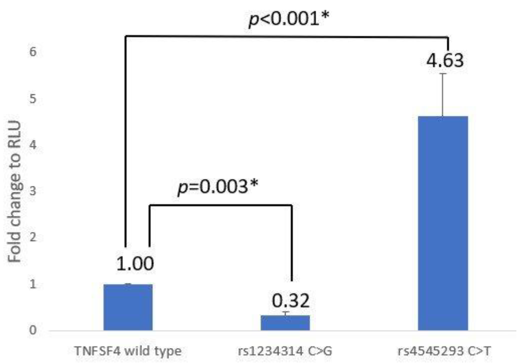
Figure 1. Luciferase reporter assay of the effect of SNPs rs1234314 (C/G) and rs45454293 (C/T) on TNFSF4 promoter activity. “*” means p < 0.05.
Jcm 12 02157 g001
Figure 2. Luciferase reporter assay of the effect of SNPs rs5839828 (G/del) and rs36084323 (C/T) on PDCD1 promoter activity. “*” means p < 0.05.
Jcm 12 02157 g002
Figure 3. Luciferase reporter assay of the effect of SNPs rs28541784 (T/C), rs200353921 (A/T), rs3181096 (C/T), and rs3181098 (G/A) on CD28 promoter activity.
Jcm 12 02157 g003
Table 1. The primer pairs used for site-directed mutagenesis PCR.
PrimerSequenceNCBI Position
SacI-TNFSF4F5′-GGCG GAGCTC CT CAA CAC CAG TAT GTT CTC C-3′173208582
EcoRV-TNFSF4R5-GGCG GATATC AA TAG GCA AAG GTC CCA GGG C-3′173207292
Rs1234314CF5′-TAC ATC ACA TGA GCC TGG CAC TGT ACT GGA-3′173208253
Rs1234314CR5′-TCC AGT ACA GTG CCA G GC TCA TGT GAT GTA
Rs4545293TF5′-CTT TCT TTG AGG TTG TGG CTG GCC TCA GAA-3′173208097
Rs4545293TR5′-TTC TGA GGC CAG CCA CAA CCT CAA AGA AAG-3′
SacI-PD1F5′-ACTG GAGCTC CA ACC AAC AGT TCT CCA GCC C-3′241858839
EcoRV-PD1R5-TTATC GATATC G CCT GGA GCA GCC CCA CCA G-3′241860275
Rs36084323TF5′-AAG GGG GAT GGG CCA GGA AGG CAG AGG CCA-3′241859444
Rs36084323TR5′-TGG CCT CTG CCT TCC TGG CCC ATC CCC CTT-3′
Rs5839828delF5′-ACCGCCCCAGCCCCCCGTCAGGCTGTTGCAGGCAT-3′241859601
Rs5839828delR5′-ATGCCTGCAACAGCCTGACGGGGGGCTGGGGCGGT-3′
SacI-CD28F 5′-TAT GAGCTC AGC AGT TGG CCG TGC TGG TGG AAT-3′203705243
HindIII-CD28P5′-TTA T AAGCTT GG GTT CCA GCC CCT CCT CCC CGA-3′203706675
Rs28541784CF5′-CCTTCCCTCCCTCCCTCTCTCTTTCTTTCCATCTT-3′203705806
Rs28541784CR5′-AAGATGGAAAGAAAGAGAGAGGGAGGGAGGGAAGG-3′
Rs200353921TF5′-CCCTCCCTCTTTCTTTCTTTCCTTCTTTCTTTCTTTC-3′203705818
Rs200353921TR5′-GAAAGAAAGAAAGAAGGAAAGAAAGAAAGAGGGAGGG-3′
Rs3181096TF5′-CTCCTTTTGTGCCCTATTATTTAACCTTGAGGG-3′203705369
Rs3181096TR5′-CCCTCAAGGTTAAATAATAGGGCACAAAAGGAG-3′
Rs3181098AF5′-GTAACTCCTTTAAACATTTATGCAGATGTTTCCC-3′203705655
Rs3181098AR5′-GGGAAACATCTGCATAAATGTTTAAAGGAGTTAC-3′
NCBI position according to GRCh38.p13. The underlined mutagenesis primer sequence refers to the position recognized by the specific restriction enzyme. The bold sequence refers to the position of site-directed mutagenesis.
Disclaimer/Publisher’s Note: The statements, opinions and data contained in all publications are solely those of the individual author(s) and contributor(s) and not of MDPI and/or the editor(s). MDPI and/or the editor(s) disclaim responsibility for any injury to people or property resulting from any ideas, methods, instructions or products referred to in the content.