Review

Angiographic Outcomes for Arterial and Venous Conduits Used in CABG

1
Department of Cardiothoracic Surgery, Weill Cornell Medicine, New York, NY 10065, USA
2
Department of Cardiothoracic Surgery, New York Presbyterian Queens Hospital, Queens, New York, NY 11355, USA
*
Author to whom correspondence should be addressed.
These authors contributed equally to this work.
J. Clin. Med. 2023, 12(5), 2022; https://doi.org/10.3390/jcm12052022
Submission received: 17 January 2023 / Revised: 25 February 2023 / Accepted: 2 March 2023 / Published: 3 March 2023
(This article belongs to the Special Issue Advances in Coronary Surgery Using Arterial Conduits)

Abstract

:
Coronary artery bypass grafting is the most commonly performed cardiac surgical procedure. Conduit selection is crucial to achieving early optimal outcomes, with graft patency being likely the main driver to long-term survival. We present a review of current evidence on the patency of arterial and venous bypass conduits and of differences in angiographic outcomes.

1. Introduction

Coronary artery bypass grafting (CABG) is the most commonly performed cardiac surgery and the treatment of choice in patients with complex and multivessel coronary artery disease (CAD) [1,2]. However, there is still discussion regarding optimal strategies for CABG, with the choice of conduit being a particularly highly debated topic.
The long-term survival benefit provided by CABG likely relies on the long-term patency of the grafts utilized during the procedure. Arterial grafts have been suggested to provide better long-term patency by being less susceptible to atherosclerotic disease and, therefore, occlusion compared to venous grafts [3]. However, current national registries in the United States, United Kingdom, and globally report low use of arterial grafts in patients undergoing CABG [4,5,6].
The aim of this review is to provide a state-of-the-art summary of the current evidence on the patency of grafts used in CABG.

2. Saphenous Vein

The saphenous vein graft (SVG) was first used by Favaloro in 1968 [7] and is currently the most commonly used conduit for revascularization. This is due to its ease of availability, its accessibility, and its length. While the left internal thoracic artery (LITA) remains the first choice of conduit in CABG, the high usage rate of the SVG may also be related to the lack of consensus on either the radial artery (RA) or the right internal thoracic artery (RITA) as the second conduit of choice in CABG.
Graft failure represents the key problem with SVG use: 10–25% of SVGs fail within the first year after CABG, another 5–10% fail between one and five years after CABG, and up to 25% fail in the following years, leading to an overall failure rate of almost 50% at 10 years [8,9,10,11]. Early graft failure is generally related to technical issues and graft thrombosis [8]. The late failure phase is instead ascribed to the development of disease intrinsic to the graft, such as intimal hyperplasia and progressing atherosclerosis [8]. Several additional factors can impact the short- and long-term patency of SVGs.

2.1. The Impact of the Harvesting Technique

The observation that injury and stress of the endothelium, which is incurred intraoperatively during graft harvesting, could be behind the suboptimal patency of SVGs prompted the development of new harvesting approaches. In the 1990s, the no-touch (NT) technique was first described: the SVG is atraumatically harvested with the surrounding pedicle [12], which prevents graft spasm and retains the biological properties of the vein, such as the production of nitric oxide [13]. In contrast to the conventional technique, manual dilatation of the SVG is not needed with NT harvesting, sparing the SVG of mechanical endothelial injuries [12,14]. Finally, the surrounding adipose-connective tissue can act as a supporting scaffold that prevents the graft from kinking.
A single-centered randomized clinical trial investigated the patency rate of SVGs in 156 patients randomized to conventional SVG harvesting with conduit distension, conventional SVG harvesting without conduit distension, or NT harvesting. The study consistently demonstrated at 1.5, 8.5, and 16 years that NT-SVG had the lowest rate of graft failures, with patency (83%) comparable to that of the internal thoracic artery (ITA) [15,16,17].
Another small, randomized trial, the Impact of Perivascular Tissue on Endothelial Function in Coronary Artery Bypass Grafting (IMPROVE-CABG), randomized 100 patients undergoing CABG to either conventional or NT-SVG harvesting. The early results showed that the occlusion rates were not different (conventional 1.2% vs. NT 0.8%), and the prevalence of harvest site infection was comparable in the two groups (4%) [18]. The five-year angiographic results of this trial are anticipated.
The SUrgical and Pharmacological novel intERventions to Improve Overall Results of Saphenous Vein Graft Patency (SUPERIOR SVG) trial was the first multicenter angiographic randomized clinical trial comparing the conventional versus the NT technique in 250 patients undergoing CABG in 12 centers [19]. The incidence of the primary composite outcome of SVG occlusion and all-cause mortality one year after surgery was not different between the two techniques (conventional 10.6% vs. NT 5.5%; odds ratio [OR] 0.49, 95% confidence interval [CI] 0.19–1.28). Similarly, there was no difference in the secondary composite outcome of significant stenosis or complete occlusion (conventional 15% vs. NT 7.8%; OR 0.48, 95% CI 0.20–1.19). Importantly, the incidence of leg (harvest site) adverse events, defined as infection, necrosis, dehiscence, drainage, and fluid collection, were higher in the NT group (NT 23.3% vs. conventional 9.5%; p < 0.01). However, the cumulative incidence of wound infection became comparable at one year [19]. When the results of these three trials were pooled meta-analytically, the estimate for the composite of graft occlusion and significant stenosis favored the NT-SVG harvesting technique (OR 0.47, 95% CI 0.26–0.84) [19].
The Graft Patency Between no-touch Vein Harvesting Technique and Conventional Approach in Coronary Artery Bypass Graft Surgery (PATENCY) trial randomized 2655 patients undergoing CABG in seven centers in China to either NT or conventional SVG harvesting [20]. The odds of vein graft occlusion at 12 months were found to be decreased with the NT harvesting technique (6.5% vs. 3.7%; OR 0.56, 95% CI 0.41–0.77). Moreover, recurrence of angina was significantly lower in the NT group, while the rates of major adverse cardiac and cerebrovascular events were not different between the two groups (conventional 4.3% vs. NT 3.8%; OR 0.89, 95% CI 0.61–1.29). Before discharge, patients in the NT group were more likely to develop exudation, numbness, or edema at the harvest site, but the rate of wound re-suture was not different compared to the conventional harvesting approach. At one year follow-up, there was no difference in terms of wound pain and wound surgical intervention between the groups. An important caveat of the study is that vein harvesting was performed by senior residents who had performed at least 100 SVG harvests and had undergone standardized training for NT technique prior to trial enrollment [20].
Further evidence will be available after the publication of the results of the ongoing SWEDEGRAFT trial, which is investigating the two-year patency results of the NT and conventional SVG harvesting techniques, as well as the two-year rates of leg wound complications [21].
Endoscopic vein harvesting (EVH) was introduced to decrease the incidence of leg wound complications. In a meta-analysis of 22 studies, including 27,911 patients, harvest-site complications were less common in the EVH group compared to the open harvesting group (0.75% vs. 2.92%; OR 0.19, 95% CI 0.12–0.30; p < 0.001) [22]. There was also no difference in the incidence of all-cause mortality and major adverse cardiac events between open and EVH, despite the finding that the EVH was associated with a lower SVG patency rate at one to five years (OR 0.80; 95% CI 0.70–0.91) and at 5–7 years (OR 0.15, 95% CI 0.04–0.61).

2.2. The Impact of Pharmacotherapy

The use of antiplatelet agents (aspirin and P2Y12-inhibitors) and statins are aimed at preventing thrombosis within the graft secondary to the activation of the endothelium and at preventing the development of intimal hyperplasia and atherosclerosis within the graft, respectively. The use of these medications is the cornerstone of medical management after CABG in order to maintain graft patency.
In a network meta-analysis including 16 studies and 3133 patients, the use of aspirin compared to placebo was associated with a significant improvement in SVG patency (OR 1.90, 95% credible interval: 1.3–2.8). A similar favoring trend, albeit not a significant difference, was shown when aspirin alone was compared to aspirin plus clopidogrel or ticagrelor [23]. However, the meta-analysis did not formally investigate clinical outcomes, such as bleeding and cerebrovascular accidents. It is reasonable to hypothesize that there is an enhanced antiplatelet effect with the concomitant use of aspirin and ticagrelor or clopidogrel (DAPT, dual antiplatelet therapy). However, studies comparing aspirin alone and DAPT reported conflicting results [24,25,26].
The Different Antiplatelet Therapy Strategy after CABG Surgery (DACAB) was a multicenter, randomized clinical trial that enrolled 500 patients assigned to either aspirin alone, ticagrelor alone, or ticagrelor DAPT within 24 h from CABG. At one year, there was a significant difference in SVG patency between aspirin alone (76.5%) and ticagrelor DAPT (88.7%; difference 12.2%, 95% CI 5.3–19.2%; p = 0.01), while there was no difference between aspiring alone and ticagrelor alone (82.8%; difference 6.3%, 95% CI −1.1–13.7; P0.10). Notably, there was no difference in the rate of major bleeding between the three groups [25]. Different results were demonstrated in the POPular CABG trial (The Effect of Ticagrelor on Saphenous Vein Graft Patency in Patients Undergoing Coronary Artery Bypass Grafting Surgery), which enrolled 499 patients randomized to receive either ticagrelor or placebo in addition to standard aspirin therapy. The incidence of SVG occlusion was 10.5% in the ticagrelor DAPT group and 9.1% in the placebo group, leading to a non-significant treatment effect (OR 1.29, 95% CI 0.73–2.30). The rate of major bleeding was not different between the two groups (2.0% vs. 2.0%; p > 0.99) [26].
In the recently published Ticagrelor Antiplatelet therapy to Reduce Graft Events and Thrombosis (TARGET) trial, 250 patients were randomly assigned to receive either aspirin or ticagrelor after CABG. At two years after surgery, the angiographic follow-up demonstrated that neither SVG occlusion (aspirin 15.7% vs. ticagrelor 13.2%; p = 0.71) nor the presence of any SVG disease (aspirin 19.4% vs. ticagrelor 19.8%; p = 1.00) were different between the two groups. Similar to previous studies, the incidence of bleeding was not different between aspirin alone (2.3%) and ticagrelor alone (5.8%) [27].
All these trials were limited by the small sample size and by being underpowered for the detection of relevant clinical outcomes, such as bleeding. The use of an individual patient-data meta-analysis merging those studies was eventually able to provide more solid results. A total of 1316 patients and 1668 SVGs were included and compared the SVG failure rate between aspirin alone, ticagrelor alone and ticagrelor DAPT [28]. The authors found that ticagrelor DAPT was associated with a risk of SVG failure that was nearly half that of aspirin, a significant difference (OR 0.51, 95% CI 0.35–0.74; p  <  0.001), but that ticagrelor DAPT was also associated with an increased risk of bleeding when compared with aspirin (OR 2.98; 95% CI 1.99–4.47; p  <  0.001). Ticagrelor alone was not associated with SVG failure or a higher incidence of bleeding. Therefore, the authors concluded that the risk of failure of the SVG should be balanced carefully within each patient, with the risk of bleeding arising from DAPT [28].
Current North American guidelines recommend initiating aspirin within six hours postoperatively and continuing indefinitely as a strategy to reduce SVG occlusion and averse cardiovascular death (Class I, level of evidence [LOE] A) [1]. In selected patients, such as those undergoing off-pump CABG, DAPT should be considered (Class 2b, LOE B-R) to improve vein graft patency as compared to aspirin alone [1].
Statins have been proven to reduce the incidence of SVG occlusion through their demonstrated cholesterol-lowering effect [29,30,31].
A small, single-center angiographic study showed that patients who received statins preoperatively had a significantly reduced number of new lesions and a lower incidence of SVG occlusion (p = 0.02) one year after CABG, which translated into a significant decrease in the incidence of myocardial infarction (MI) [31].
The widespread habit of initiating long-term statin therapy after bypass surgery was validated in a retrospective study including 7503 patients who underwent CABG between 1995 and 2004. Compared to no statin use, the commencement of statin therapy within one month postoperatively was associated with lower all-cause mortality (HR 0.82, 95% CI 0.72–0.94) and a lower risk of major adverse cardiovascular events (HR 0.89, 95% CI 0.81–0.98) [32]. These findings were confirmed in a retrospective analysis including 35,193 patients who underwent first-time isolated CABG in Sweden from 2006 to 2017. The benefit of initiating a statin therapy also translated into a markedly reduced risk of undergoing new angiography (HR 0.81, 95% CI 0.74–0.88) and new revascularization (HR 0.79, 95% CI 0.70–0.88) [33].
Lastly, while no doubt remains regarding the efficacy of statins in reducing SVG occlusion, debate still surrounds the dose to obtain this benefit. The Aggressive Cholesterol Therapy to Inhibit Vein Graft Events (ACTIVE) trial showed that an 80 mg dose did not confer a significant advantage in terms of vein graft occlusion one year after CABG compared to a 10 mg dose (11.4% for 80 mg versus 12.9% for 10 mg) [34].
Current North American guidelines recommend all patients undergoing CABG should receive statin therapy prior to and early after surgery, to be indefinitely continued unless contraindicated [1]. Dose intensity may be adjusted based on the patient’s age, LDL target, statin intolerance status, and the presence of drug interactions [1].

2.3. The Impact of External Stenting

SVG failure after one year following CABG is mainly due to intimal hyperplasia, which predisposes the graft to accelerated atherosclerosis [8]. Attempts to attenuate this pathophysiologic mechanism have led to the emergence of a novel surgical strategy based on the use of cobalt-chromium mesh external stents that have been shown to be promising in preclinical testing by reducing wall tension, improving lumen uniformity, and creating a protective “neo adventitia” layer rich with microvasculature [5,35,36].
First experiences with such devices required gluing, suturing or both during the implantation procedure and yielded high rates of graft occlusion at up to one-year follow-up [37,38,39,40]. This initial failure prompted the industry to improve the design of external stents and led to the conduction of the Venous External Support Trials (VEST). In the VEST I trial, 30 patients were enrolled and received one stented SVG (30 grafts), and one or more non-stented SVG (39 grafts) [41]. The intimal hyperplasia area, assessed by intravascular ultrasonography, was lower in the stented group (mean 4.37 mm2) compared to nonstented grafts (mean 5.12 mm2; p = 0.04). The one-year angiographic follow-up, completed in 97% of patients, showed similar failure rates between stented and non-stented grafts (30% vs. 28.2%; p = 0.55). The five-year follow-up (VEST IV) confirmed similar patency rates between the two groups (30% vs. 23%; p = 0.42), while the intimal hyperplasia area was still significantly smaller in stented SVGs (p < 0.001) [42].
The VEST II trial investigated factors that could impact the patency of stented SVG supplying the right coronary territory finding that the use of only suture ligation and avoidance of fixation of stents to the proximal or distal anastomosis led to similar patency rates for stented and non-stented grafts (86.2% vs. 88.8%) at 3–6 months [43].
The VEST III trial enrolled a larger cohort of patients (n = 184) and showed similar patency rates between stented and non-stented SVGs (78.3% vs. 82.2%; p = 0.43) at two years [44]. Therefore, based on the available evidence, external stenting certainly plays a relevant role in preventing the development and progression of graft atherosclerosis. However, further long-term data and device refinement are warranted to improve upon this promising technique, as angiographic outcomes seem to be in favor of non-stented SVGs.

2.4. The Impact of the Anastomotic Technique

SVGs can be grafted as an individual anastomosis or as a sequential anastomosis, which allows for a greater extent of myocardial revascularization. However, there is limited data on the impact of sequential grafting on SVG patency.
In a study of 2515 patients, the clinical and angiographic outcomes in 946 sequential SVGs and 1366 individual SVGs were compared [45]. The graft failure rates were 10.3% and 17.7% at five years and 20.9% and 33.6% at 10 years in the sequential and individual groups, respectively. After propensity score adjustment, the risk of graft failure was lower in the sequential group at a median follow-up of 88 months (HR 0.69, 95% CI 0.50–0.95; p = 0.02). The risk of the composite outcome of death, nonfatal MI, and repeat revascularization was numerically lower in the sequential group (36.8% vs. 41.4%; HR 0.91, 95% CI 0.75–1.09; p = 0.30). Conversely, in a sub-study of the Project of Ex-Vivo Vein Graft Engineering via Transfection (PREVENT) IV trial, which enrolled 3014 patients to receive either edifoligide-treated SVGs or placebo, investigators found a higher risk of graft occlusion with sequential anastomosis at one year (OR 1.24, 95% CI 1.03–1.48; p = 0.025) [46].
A recent meta-analysis including 15 cohort studies, totaling 10,681 patients and 12,957 grafts found that patency was higher for sequential SVGs compared to individual SVGs (relative risk [RR] 1.11, 95% CI 1.03–1.21; p = 0.01) [47].
Due to conflicting results, more solid evidence, potentially from RCTs, is welcomed to explore the impact of individual vs. sequential anastomosis on SVGs patency.

3. Internal Thoracic Artery

The use of the LITA has been the gold standard for surgical myocardial revascularization since Loop et al.’s study demonstrating a long-term benefit in terms of mortality, MI, and cardiac reintervention in patients receiving a single ITA to the left anterior descending coronary artery (LAD) compared with patients receiving only venous grafts [48]. Further studies have later corroborated this evidence, in particular supporting the hypothesis of the underlying mechanism for better long-term results: better graft patency [49,50]. The use of LITA for revascularization of the LAD is now recommended by international guidelines on myocardial revascularization due to its exceptional durability [1,2]. Long-term patency rates for the LITA are outstanding: 93% at 10 years and 88% at 15 years in the latest reports [51,52].
The right internal thoracic artery (RITA) has not achieved a similar widespread success compared to its contralateral counterpart, and its use is still mainly concentrated in the hands of surgeons who routinely perform bilateral internal thoracic artery (BITA) grafting. The long-term patency rates for the RITA are reported to be between 80–90% at 10 years [53,54].
There may be several explanations for the failure of BITA to achieve widespread adoption, but the neutrality of the Arterial Revascularization Trial (ART) played a pivotal role. The ART was a multicenter, double-arm study investigating the outcomes of 3102 patients undergoing CABG and randomized to either single internal thoracic artery (SITA) or BITA grafting. At the ten-year follow-up, the intention-to-treat analysis showed no difference in all-cause mortality and event-free survival between the two groups. After the publication of this trial, important issues within the trial were highlighted. First, the crossover rate in the two groups was notably high, with 14% of patients in the BITA group actually receiving SITA, diluting the treatment effect of the BITA. Second, there was no a priori criteria to guide surgeons in the choice of concomitant arterial conduits, leading to 21% of the patients in the SITA group receiving an RA as well. In the as-treated analyses, there was a significantly lower risk of mortality in the BITA group. However, these findings can only be considered hypothesis-generating.
The results from ART contrasted substantially with the findings from observational studies, which had identified a benefit from the use of the two ITAs [55]. Among other factors impacting the overall adoption of the BITA are the higher complexity and longer times required for harvest [56] and the perceived higher risk of sternal wound complications. Prior reports have also highlighted the dependency of BITA outcomes on operator experience [57], possibly limiting adoption among surgeons who fear they lack the requisite expertise. The combination of these issues has led to limited use of BITA: around 5% in the U.S. [4] and 15% in Europe [58].
Current North American guidelines indicate that BITA should be used by experienced surgeons in appropriately selected patients (Class 2a, LOE B-NR) [1], which is aligned with current European recommendations [2].

3.1. The Impact of the Harvesting Technique

The ITA can be harvested either as a pedicled or as a skeletonized graft. Traditionally, this conduit has been harvested as a pedicled graft, meaning that the accompanying veins, the endothoracic fascia, and parts of the parietal pleura and of the transversus thoracic muscle are taken together with the artery. Conversely, skeletonized ITA entails the dissection of the artery as an isolated conduit free from the aforementioned surrounding tissues. Skeletonization is more challenging and technically demanding, with a steep learning curve and longer harvesting time (15–20 min compared to pedicled harvesting) [59,60]. In addition, the need to dissect more closely to the ITA wall can increase the risk of injuries induced by electrocautery or manipulation of the vessel (i.e., hematoma, vessel dissection, thrombosis), which can impact graft patency.
These two techniques were recently at the center of the debate in the cardiac-surgical community following the publication of two post-hoc analyses utilizing data from randomized clinical trials.
The Cardiovascular Outcomes for People using Anticoagulation Strategies (COMPASS) clinical trial originally randomized 27,395 patients with stable CAD or peripheral artery disease to receive rivaroxaban and aspirin or rivaroxaban and placebo or aspirin and placebo for secondary cardiovascular prevention [61]. Of those, 1002 patients undergoing CABG were included in this analysis [62]. The ITA was most commonly harvested as a pedicle graft (72%). After a mean follow-up of 23 months, patients receiving a pedicled ITA had a higher risk of major adverse cardiovascular events (cardiovascular mortality, MI, stroke, or revascularization; HR 3.19, 95% CI 1.53–6.67). This difference was mainly driven by the higher rate of revascularization in the skeletonized group (5% vs. 1.4%; p = 0.03). With regards to the graft status, skeletonized ITAs had a significantly higher incidence of occlusion (9.6% vs. 3.9%; OR 2.41, 95% CI 1.39–4.20; p = 0.002). These findings were confirmed when the quality of the conduit and the quality of the target coronary vessel were taken into account. It is important to note that the original randomized treatments were not found to have an impact on graft patency.
The ART [63] investigators conducted a post-hoc analysis of 2161 patients, of whom 58% received a pedicled ITA [64]. At the 10-year follow-up, patients receiving skeletonized ITAs were found to have a higher risk of major adverse cardiovascular outcomes (all-cause mortality, MI, and revascularization; HR 1.25, 95% CI 1.06–1.47) [64]. In particular, patients with skeletonized ITA experienced a higher rate of repeat revascularization (13.5% vs. 9.9%; p = 0.01). The findings were confirmed when only patients receiving single ITA were included in the analysis, eliminating the potential bias arising from the greater likelihood of using the skeletonization technique with BITA. The post-hoc analysis of the ART also attempted to account for the surgeons’ experience. When the surgeon volume was ≥51 cases, the differences in outcomes between skeletonized and pedicled IMAs were no longer present. A major limitation was the lack of angiographic data.
Both these post-hoc analyses raise questions on the use of ITA as a skeletonized graft. First, skeletonization may impair the physiological endothelial function of the ITA by direct damage to the vasa vasorum of the vessel [65,66]. Second, from a more careful assessment of the Kaplan–Meier curves, it is possible to see that the survival curves diverge early on within the first 12 months after surgery [62,64]. This observation could support the hypothesis that technical issues rather than pure advancement of the atherosclerotic disease are the causes for the poorer performance of the skeletonized ITA. Attention is thus directed to the surgeon and the surgeon’s experience as a limiting step in the successful performance and adoption of the skeletonization of the ITA. In fact, in the post-hoc analysis of the ART, when surgeons had a higher surgical volume, the outcomes between the two techniques were not different.
Finally, a recent randomized trial investigating graft patency of 109 patients undergoing CABG with either skeletonized or pedicled ITA grafted to the LAD showed no difference in graft patency at three years (pedicled 95.8% vs. skeletonized 90.4%; computed angiograms available for 100 patients) or at eight years (pedicled 93% vs. skeletonized 90.2%; computed angiograms available for 84 patients) [67]. The authors showed that target vessel stenosis (defined as stenosis of <70% vs. ≥70%) was a more important factor for graft failure rather than the harvesting technique.

3.2. The Impact of Graft Configuration and Target Coronary Vessel

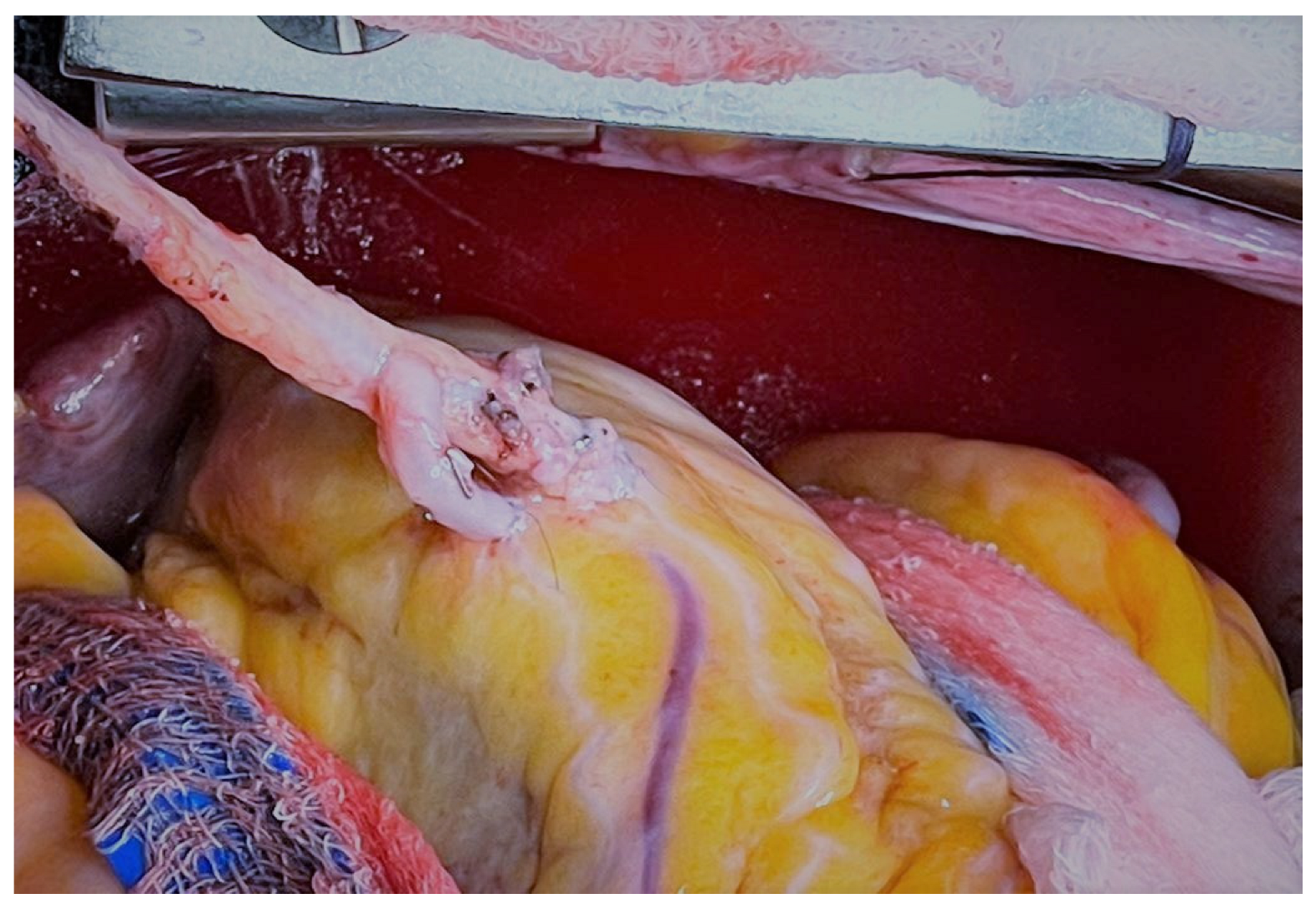
The RITA graft can be used as an in-situ graft or as a free graft coming off of the aorta, the LITA as a Y-composite conduit, or the aortic hood of the SVG or RA (Figure 1).
In a randomized control trial of 304 patients, of whom 147 were assigned to in-situ RITA and 152 to RITA Y-graft, there was no difference in patency between the two configurations at six months. The observed anastomotic patency rate was 97% in both groups [68]. No differences in clinical outcomes in terms of major adverse cerebral and cardiovascular events were reported. The extended follow-up of this cohort, including 75% of the original population, confirmed no difference in the patency rate of in-situ RITA (93%) and Y-graft (94.5%) at a mean angiographic follow-up of three years. However, the incidence of major cerebral and cardiovascular events was higher with the in-situ configuration (34% vs. 25%) at a mean clinical follow-up of seven years [69]. Similar findings were reported in a study of 1818 patients undergoing CABG with either in-situ RITA or Y-graft RITA. The patency rate was 100% for the in-situ grafts and 99.2% for the Y grafts at a mean follow-up of 17 months [70].
In a recent study of 1331 patients from the Cleveland Clinic, the patency rate of the in-situ RITA (91%) was similar to the patency rate of the RITA as a free graft from the aorta (91%) or from the hood of the SVG (77%), and as a Y-graft (89%) [71]. The authors showed that the RITA patency rate was independent of inflow configuration but was significantly influenced by the target coronary vessel. In-situ RITA to LAD had similar patency to LITA to LAD (94.4% vs. 95.4%; p = 0.50), while a lower patency rate was found when in-situ RITA was used to graft non-LAD territories [71]. Similar results were described in a study of 5776 patients undergoing BITA-CABG, showing no difference in the patency of in-situ vs. free RITA grafts (89% vs. 91%) at ten years but the patency of RITA grafts to the right coronary artery was significantly lower compared to RITA to LAD grafts [72]. The authors also identified target coronary vessel stenosis as a factor influencing RITA patency, having observed that RITA grafts anastomosed to vessels with <60% stenosis had the lowest patency rate and grafts anastomose to vessels with >80% stenosis had the highest patency rate [72].

3.3. The Impact of the Anastomotic Technique

The use of sequential anastomosis for ITA can be key to achieving effective revascularization in patients with a limited number of grafts and diffuse CAD (Figure 2).
Current evidence, albeit based on observational studies, seems to be concordant with sequential ITA having a similar patency rate as individual ITA [73,74,75,76]. A recent study including 120 propensity score matched pairs reported that the patency rate of the sequential LITA graft to the diagonal artery and LAD was similar to the patency of individual LITA-to-LAD grafts (99% and 98% vs. 98%; p > 0.05) at the angiographic follow-up of 27 months. [76]. Similar rates were found in a report of 101 patients in which patency rates for the sequential LITA to the circumflex and right coronary artery were 98% and 95%, respectively [74].
Interestingly, the design of the sequential anastomosis was reported to impact graft patency [75]. In sequential ITA grafting, the anastomosis can be performed either in a “diamond” fashion with the conduit perpendicular to the coronary target or in a parallel fashion with the conduit parallel to the coronary target. A study of 452 patients comparing LITA sequential vs. individual grafting to the circumflex showed that a “diamond” anastomosis had a numerically higher patency rate of the distal segment of the sequential graft as compared to a parallel design (98.4% vs. 90.7%; p = 0.09). In particular, the lowest patency rate (75%) was reported when the proximal anastomosis of a sequential graft to the diagonal and circumflex arteries was placed in a parallel fashion [75].
However, it is important to note that the “diamond” design is more technically demanding and requires accurate precision of the arteriotomies in order to prevent a “seagull effect”, which can lead to a flattening of the graft and impairment of graft status.
Another important factor potentially affecting sequential graft success is coronary target stenosis. Lower stenosis can result in higher competitive flow and therefore jeopardize the functionality of the graft [77]. In the Impact of Preoperative FFR on Arterial Bypass Graft Function (IMPAG) trial, which included a total of 64 patients (199 anastomoses, of whom 108 sequential), a preoperative fractional flow reserve (FFR) of at least 0.78 was associated with a better graft functionality at the six-month angiographic assessment. Lower values were associated with an anastomotic occlusion rate of 3% [78]. In a post hoc analysis of the IMPAG trial, specifically looking at sequential grafts, cut-offs of 0.80 and 0.78 were found for the first and second anastomoses of sequential grafts to the anterior circulation compared to 0.74 for the individual grafts. The FFR cut-off was higher for grafts to the postero-lateral territory being 0.81 and 0.78 for the first and second anastomoses of sequential grafts and 0.79 for individual grafts [79]. These findings suggest that attention should be paid to the severity of the target coronary vessel when sequential anastomosis is planned.

4. Radial Artery

The radial artery (RA) was first used as a conduit in 1972 by Carpentier [80]. However, due to the high tendency to vasospasm, which led to poor outcomes, the early experience with this conduit was far from promising and its use was shortly abandoned. At two years after CABG, over one-third of the RA grafts demonstrated angiographic narrowing or occlusion [81].
In the last three decades, the use of the RA has been revived due to the interest in multi-arterial coronary grafting. This was paralleled by the report of excellent 18-year angiographic outcomes [82] and by the understanding that injury to the graft during the harvesting of the conduit, as opposed to the graft itself, was the cause of the previously reported early unsatisfying outcomes. Moreover, the use of vasodilators intraoperatively and postoperatively further improved outcomes by addressing the tendency of RA grafts to spasm.to spams of the graft [83,84].
The RA is an accessible and easily procured conduit that can provide enough length to reach most coronary targets (Figure 3).
Its diameter is proximally optimal for anastomosis to the aorta or to the LITA and distally excellent for coronary artery targets [85]. On the other hand, the RA is more muscular than the other arterial conduits and thus carries an increased risk of vasospasm during harvesting.

4.1. The Impact of Target Coronary Artery and Competitive Flow

The target coronary artery is one of the most important determinants of graft patency. Target vessel considerations that influence the graft selection process are myocardial territory to be grafted, degree of proximal coronary artery occlusion, and diameter of the target vessel. The RA, as discussed, is prone to spasm and competitive flow when grafted to coronary target vessels with mild-to-moderate stenosis [86]. A study including 123 patients undergoing CABG with RA and a total of 382 distal anastomoses reported an overall patency rate of 92% at a mean angiographic follow-up of 32 months [87]. Interestingly, authors found important modifiers of RA patency. When the RA was used to graft the right coronary system, the patency rate was significantly lower when the RA was grafted to the left coronary system (79% vs. 94%; p < 0.05). Also, RA distal anastomoses to coronary vessels with >90% stenosis showed a higher patency rate compared to RA distal anastomoses to coronary vessels with 50–90% stenosis (98% vs. 83%; p < 0.05) [87]. The pivotal role of target vessel stenosis was also confirmed in an analysis of 100 CABG patients receiving RA grafts; RA grafted to vessels with ≥90% stenosis had a failure rate of 13%, which was similar to the failure rate of the LITA. The failure rate was as high as 80% in grafts anastomosed to vessels with <90% stenosis [88].
Based on this evidence, current guidelines recommend the use of the RA as the second conduit to graft significantly stenosed coronary targets and the postoperative administration of calcium-channel blockers for the following year to prevent graft spasm [1,2].

4.2. The Impact of Sequential Grafting

Sequential grafting is often used to increase the number of territories revascularized by a single conduit. However, data on the patency of single versus sequential anastomosis is limited and controversial. A study of 410 CABG patients receiving RA grafting showed that sequential anastomoses of the RA were associated with a higher likelihood of patency as compared to a single anastomosis at mean angiographic follow-up of five years (HR: 2.53, 95% CI: 1.29–5.28; p = 0.006) [89]. Conversely, a recent study including 208 patients (293 anastomosis total) undergoing CABG with skeletonized RA found a similar patency rate for sequential grafts (88.7%) and single grafts (87.4%; p = 0.88) at ten years [90]. There is a potential impact of target vessel stenosis on RA sequential graft patency. A study including 432 patients and 1221 distal RA sequential anastomoses showed that patency rates were higher for sequential anastomoses to target vessels with 76–100% stenosis compared to sequential anastomoses to target vessel with a stenosis of 51–75% (88.6% vs. 59.1%; p < 0.001) [91].

4.3. The Impact of Graft Configuration

The RA can be used as a graft directly anastomosed to the proximal aorta or as a composite conduit with other grafts, such as the ITA (Figure 4).
The available evidence seems to suggest that inflow configuration does not affect the long-term patency rate of the RA. At a mean follow-up of 6.5 years, the patency of aorta-anastomosed RA was 92%, compared with a patency rate of 86.3% for ITA-anastomosed graft (p = 0.81) [92]. However, when the RA was used to bypass target vessels with <90% stenosis, ITA-anastomosed RA grafts were associated with a higher risk of failure (p = 0.02). The impact of trans-radial catheterization.
RA grafts previously used for heart catheterization may have lower patency rates compared to those that have not been catheterized grafts [93]. Injury to the RA by the introduction and passing of catheters and wires likely produces endothelial damage to the RA lumen, especially in the proximal RA [94,95]. The patency rate can be up to 20% lower in instrumented RA compared to never-catheterized arteries [93]. This observation was also associated with the findings that the incidence of intimal hyperplasia was 30% greater in previously catheterized RA [93]. As a consequence, previously instrumented RAs should be used with caution in coronary surgery [96].

5. Gastroepiploic Artery

The gastroepiploic artery (GEA) was first used by Bailey in 1967 [97] to revascularize the posterior ischemic myocardium: however, its use as a conduit was never widely adopted and remained a grafting option only in a minority of cases. The GEA is well-suited for coronary targets on the posteroinferior aspect of the heart, such as the posterior descending artery, where it can be anastomosed as an in-situ graft. Harvesting of the GEA requires an extension of the median sternotomy beyond the xiphoid process and the opening of the peritoneum.
In a study of 1352 patients undergoing CABG with GEA grafting, the GEA patency rates were 97% at one month, 86% at five years and 67% at ten years [98]. Better long-term patency rates were reported when GEA was used as a skeletonized graft and after appropriate selection of target coronary vessel (>90% stenosis). In these settings, the patency rates were 98% at one month, 95% at five years and 90% at eight years [99]. Regarding target selection, a recent study identified the minimal coronary lumen diameter as a factor impacting GEA patency: when the lumen diameter was ≤1 mm, the patency rate was 90% at ten years compared to a patency rate of 40% for conduits anastomosed to vessels with diameter >1 mm [100].

6. Comparisons of Conduit Patency

There are few randomized trials that have directly compared the angiographic outcomes of bypass conduits, in particular RA vs. RITA. The Radial Artery Patency and Clinical Outcomes (RAPCO) system encompassed two randomized clinical trials comparing clinical and patency outcomes of the RA vs. free RITA (394 patients) and of the RA vs. SVG (225 patients), respectively [54]. With excellent completeness of follow-up (99%), the ten-year follow-up of the RAPCO demonstrated that the patency of RA was significantly higher than that of free RITA (89% vs. 80%; HR 0.45 95% CI 0.23–0.88), while the patency of RA was non-significantly better than that of the SVG (85% vs. 71%; HR 0.40 95% CI 0.15–1.00). Compared to free RITA, patients receiving RA also showed better long-term survival (91% vs. 84%, log-rank p = 0.03), while it was similar between RA and SVG (73% vs. 65%; log-rank p = 0.18).
The Radial Artery Database International Alliance (RADIAL) project compared the clinical and angiographic outcomes of the RA vs. SVG by pooling individual patient data from six trials [101]. In a pooled cohort of 1036 patients, the RADIAL investigators found that the use of RA was associated with a lower incidence of the composite endpoint of death, MI, and repeat revascularization (13% vs. 19%; HR 0.67, 95% CI 0.49–0.90) at a mean follow-up of 60 months, and with a lower incidence of graft occlusion (8% vs. 20%; HR 0.44, 95% CI 0.28–0.70) at a mean angiographic follow-up of 50 months.
In a recent network meta-analysis of 18 studies for a total of 8272 grafts analyzed, only the RA and the NT-SVG were found to be associated with a significantly lower graft occlusion rate when compared to conventionally harvested SVG at a mean weighted angiographic follow-up of five years [102]. The pooled patency rate was 94.1% (95% CI 90.0–97.6 for the RA, 91.4% (95% CI 87.3–94.3) for the NT-SVG, 89.2% (95% CI 71.2–96.5) for the RITA,86.3% (95% CI 81.2–90.2) for the conventionally harvested SVG and 61.2% (95% CI 52.2–69.4) for the GEA (Table 1).

7. Surgical Revascularization Technique and Graft Patency

7.1. On- and Off-Pump CABG

Off-pump CABG (OPCABG) is a key revascularization strategy that prevents patients from being exposed to the detrimental effects of cardio-pulmonary bypass and is currently recommended for patients with severe atherosclerotic disease of the aorta [2]. In the last decade, considerable randomized evidence has been published on both the clinical and angiographic outcomes of OPCABG compared to on-pump CABG (ONCABG).
In the Danish On-pump Versus Off-pump Randomization Study (DOORS), a total of 900 patients aged more than 70 years were assigned to either OPCABG or ONCABG and received the same heparinization protocol [103]. At six months, 481 patients underwent coronary angiography, and the proportion of occluded grafts was statistically higher in the OPCABG group vs. the ONCABG group (76% vs. 89%; p = 0.01).
Similarly, the Department of Veterans Affairs Randomized On/Off Bypass (ROOBY) trial, which randomized 2203 patients to OPCABG or ONCABG, found a statistically significant increased patency rate in ONCABG for both the arterial (91.4% vs. 85.8%; p = 0.003) and vein grafts (80.4% vs. 72.7%; p < 0.001) at 1 year [104].
The CABG Off or On Pump Revascularization Study (CORONARY) trial randomized 4752 patients to OPCABG or ONCABG [105]. In the smaller angiographic cohort, a total of 157 patients undergoing computed tomography angiography at one year were included. The investigators found that the patency index, that is, the proportion of nonoccluded grafts, was 89% in the OPCABG and 95% in the ONCABG (p = 0.09). Moreover, no difference in patency rates was found between arterial and venous grafts and target territories.
Recently, a meta-analysis of 16 RCTs totaling 6227 patients and 11,641 grafts found that OPCABG was associated with a 31% higher risk of graft failure after OPCABG (RR 1.31, 95% CI 1.17–1.46; p < 0.001) [106]. Notably, the higher graft occlusion was driven by studies in which the crossover rate, assumed as a proxy of surgeon expertise, was >10% (RR 1.31, 95% CI 1.16–1.49, p < 0.001), and there was no statistical difference in patency in studies with crossover rate <10%. Moreover, the occlusion rate in OPCABG was higher compared to ONCABG within the first year of follow-up (RR 1.34, 95% CI 1.18–1.52; p < 0.001) but was similar at longer follow-up (RR 1.15, 95% CI 0.87–1.52; p = 0.32), suggesting an effect from technical issues during the surgery. When focusing on the type of graft, SVGs were found to be more likely to fail in the OPCABG group (RR 1.40, 95% CI 1.23–1.59; p < 0.001), while no difference was found in the patency rates of arterial grafts between the two strategies.
Due to concerns related to graft patency, OPCABG is currently recommended in selected patients with severe aortic atherosclerosis and should be performed by experienced off-pump teams [1,2,107].

7.2. Minimally Invasive and Robotic CABG

In the last decade, more and more attention has been focused on reducing the invasiveness of cardiac surgical procedures paving the way to minimally invasive CABG and robotic CABG.
Minimally invasive direct CABG (MIDCAB) involves the combination of OPCABG and a minimally invasive approach through smaller surgical incisions, such as left anterolateral thoracotomy, reducing the risk of complications related to cardio-pulmonary bypass use and full sternotomy. Data on the patency of grafts after MIDCAB are scarce, and available results are mainly focused on the early patency rate of grafts. The patency rates for the LITA-LAD graft in the immediate postoperative period ranges from 96.2% to 99% [108,109,110], while at six months, it is reported to be between 95–100% for the LITA-LAD and 85% for the SVGs [109,111]. In one of the biggest series on MIDCAB, 1060 patients were follow-up for a median time of 11 years, and the patency rate of LITA-LAD was 96.8% [112]. Favorable results were also reported in a series of 140 MIDCAB patients in which the patency rate of the LITA-LAD at three years was 96.4% [113].
Robotic-assisted CABG (RCABG) represents a novel adjunct to reduce the invasiveness of cardiac surgery and achieve earlier functional recovery and lower morbidity. The reported patency rates after RCABG are similar to the rates of grafts used in conventional CABG. In a systematic review of the literature, which included 33 articles and a total of 4000 patients, the patency rates after RCABG were 97.7% at one month, 96.1% at one to five years and 93.2% after five years [114].
The optimal patency rates of minimally invasive techniques facilitate the role of these strategies in the current practice for selected patients assuring clinical and surgical benefits without compromising graft patency. An important caveat to these benefits is the expertise of the centers and surgeons performing such operations.

8. Angiographic and Clinical Outcomes

Excellent patency rates are key to achieve the clinical benefits of CABG. Despite the intuitive and biologically plausible relationship between graft status and clinical outcomes not being cleared yet, patent grafts are known to exert protective effects on the myocardium. Functioning grafts are able to not only allow flow distally to the coronary stenosis but also able to protect the proximal segment by preventing CAD progression [115].
The association between angiographic and clinical outcomes does not seem to be linear, and the consequences of graft failure are variable. For instance, failure of grafts to the LAD was reported to be more closely associated with worse clinical events than the failure of grafts to other territories. In an analysis of 1296 patients, clinical outcomes were similar between patients with and without SVGs stenosis at five years. However, significant stenosis of the SVGs grafted to the LAD was associated with a higher risk of death and cardiovascular events [116].
In the PREVENT-IV trial, SVG failure was associated with a higher risk of coronary revascularization but not death. However, failure of the LITA-LAD was strongly associated with a higher incidence of an acute clinical event [117].
In a provincial registry of 5276 patients, arterial graft failure, mainly LITA-LAD, was associated with lower survival compared to patients with vein graft failure [118].
Investigating the interplay between graft status and clinical outcomes is complex and influenced by several factors [119]. The lack of systematic angiographic graft assessment in most studies, which instead report on graft status following clinically driven angiographies, can bias the estimation of the actual impact of graft failure on clinical outcomes. Also, the temporal relationship between the two events is difficult to establish, provided that graft patency studies only determine if the graft failed and not when it occurred. It is essential to establish that graft failure occurred before the clinical event. The functionality of the failed graft should also be considered. If a graft is anastomosed to a coronary artery with a non-flow limiting lesion, competitive flow from the native coronary circulation will likely determine graft failure. However, this graft failure is likely less clinically relevant as no reduction in distal perfusion will occur, considering that native circulation will keep supplying the territory. Indeed, in the IMPAG (Impact of Preoperative FFR on Arterial Bypass Graft Function) [78] and SYNTAX-LE MANS (Synergy between PCI with TAXUS express and cardiac surgery left main angiographic substudy) [120] in which grafts either failed for competitive flow or were grafted in a situation of high risk of competitive flow (left main disease), no correlation was found between graft failure and clinical events.
Therefore, the impact of graft failure on patients’ prognosis is complex and highly variable and depends on the type and location of the failed graft and on the mechanism of failure.

9. Tailoring Surgical Revascularization to Patients
To achieve the best outcome for every patient, CABG and, in particular, the selection of conduits should be tailored to each patient.
The debate around the best second arterial graft is still ongoing in the surgical community. The current North American guidelines, however, indicate the RA as the second arterial conduit of choice to bypass highly stenosed coronary arteries [1].
With the available evidence, it is not possible to precisely define the CABG strategy for each patient. A complex interplay between patient characteristics (e.g., age, coronary anatomy, conduit availability), preoperative risk factors, patient expectations and surgeon expertise should be taken into consideration.
Life expectancy is supposed to play a major role in the decision-making process about using arterial or venous conduits. In the early follow-up period (<4 years), there is no difference in patency rates between arterial and venous grafts, but the patency rates start to be superior in arterial grafts four years after CABG [101,121]. This evidence suggests that patients with low life-expectancy would not benefit from the use of a second arterial graft. The lack of benefits for elderly patients is supported by some reports. In a post-hoc analysis of the ART, the use of BITA vs. SITA was associated with a lower risk of adverse cardiovascular events in younger patients but not in older patients [122]. A study including 26,124 patients identified an age cut-off of 70 years, after which the survival benefit of multiple arterial grafting is lost [123]. Consistently with this, the current European and North American guidelines recommend considering the use of multiple arterial grafts in patients with reasonable life expectancy [1,2].
Coronary anatomy plays a key role in the choice of conduits. As mentioned in the dedicated paragraphs, RA provides the highest benefit when used to bypass a severely stenosed coronary artery.
Patient comorbidities should guide conduit choice as well. Diabetes and obesity can represent important risk factors, particularly when concomitant, for the development of sternal wound complications in patients in which both ITAs are harvested. Therefore, these patients could benefit from other surgical strategies considering that sternal wound complications were reported to be associated with worse long-term outcomes [124].
Finally, the achievement of excellent outcomes after CABG also relies on surgeon and center expertise, as not every surgeon could be skillful in every procedure (multiple arterial grafting, off-pump, no-touch aortic technique) [125,126,127].
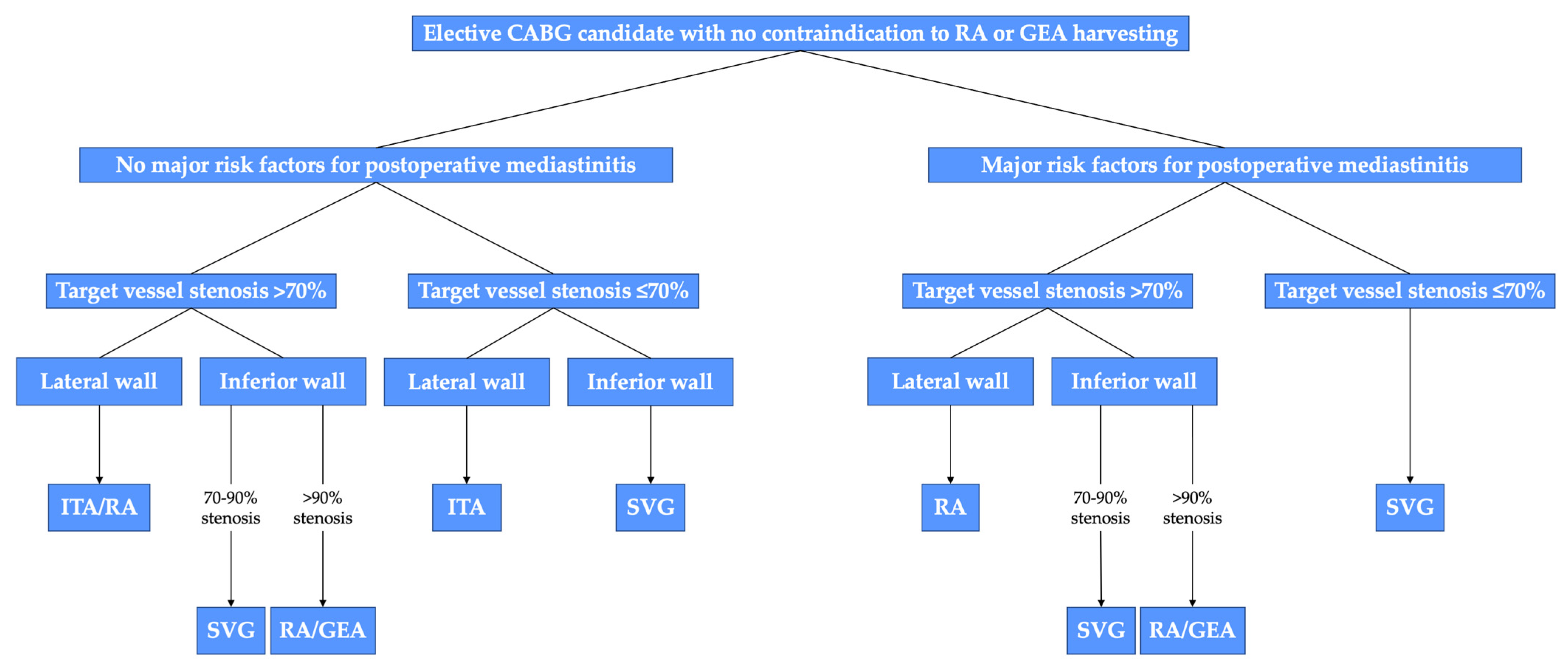
A proposed algorithm for the choice of the second conduit is shown in Figure 5.
The results from the ongoing Randomized Comparison of the Clinical Outcome of Single Versus Multiple Arterial Grafts (ROMA) trial will provide solid answers to these questions [128].
Figure 5. Algorithm for graft selection for the second target vessel in elective coronary artery bypass grafting (CABG) patients with stable coronary artery disease and without contraindications to the radial artery (RA) or gastroepiploic artery (GEA) harvesting. In case of contraindications to RA or GEA, SVG should be used. Risk factors for postoperative mediastinitis are obesity, diabetes, and severe chronic lung disease, especially in combination. ITA, internal thoracic artery; SVG, saphenous vein graft. Reproduced without changes from: “The choice of conduits in coronary artery bypass surgery” by Gaudino et al.; Journal of The American College of Cardiology. © 2015 American College of Cardiology Foundation. Published by Elsevier Inc. [129].
Jcm 12 02022 g005

10. Conclusions

The benefits of surgical revascularization are best achieved with a cautious choice of bypass conduits. The LITA to LAD is the standard of care given the proven early and late benefits. However, the choice of the second conduit is still debated. Current evidence seems to consistently points towards a clinical and angiographic benefit from the RA compared to other conduits. Selecting the conduit with the best long-term patency is most likely to ensure optimal clinical outcomes in each patient if long-term patency is considered together with a tailored treatment strategy that accounts for patient characteristics (e.g., coronary anatomy, conduit availability), preoperative risks, patient expectations, and surgeon expertise.

Author Contributions

Conceptualization, A.D., G.S.J. and M.G.; methodology, A.D., G.S.J. and M.G.; validation, A.D., G.S.J., L.H., R.P.O., G.C., K.R.A., T.A., C.M. and M.G.; writing—original draft preparation, A.D., G.S.J. and L.H.; writing—review and editing, A.D., G.S.J., L.H., R.P.O., G.C., K.R.A., T.A., C.M. and M.G.; supervision, M.G. All authors have read and agreed to the published version of the manuscript.

Funding

This research received no external funding.

Institutional Review Board Statement

Not applicable.

Informed Consent Statement

Not applicable.

Data Availability Statement

Not applicable.

Conflicts of Interest

The authors declare no conflict of interest.

References

  1. Lawton, J.S.; Tamis-Holland, J.E.; Bangalore, S.; Bates, E.R.; Beckie, T.M.; Bischoff, J.M.; Bittl, J.A.; Cohen, M.G.; DiMaio, J.M.; Don, C.W.; et al. 2021 ACC/AHA/SCAI Guideline for Coronary Artery Revascularization: A Report of the American College of Cardiology/American Heart Association Joint Committee on Clinical Practice Guidelines. Circulation 2022, 145, e18–e114. [Google Scholar] [CrossRef] [PubMed]
  2. Neumann, F.J.; Sousa-Uva, M.; Ahlsson, A.; Alfonso, F.; Banning, A.P.; Benedetto, U.; Byrne, R.A.; Collet, J.P.; Falk, V.; Head, S.J.; et al. 2018 ESC/EACTS Guidelines on myocardial revascularization. Eur. Heart J. 2019, 40, 87–165. [Google Scholar] [CrossRef] [Green Version]
  3. Gharibeh, L.; Ferrari, G.; Ouimet, M.; Grau, J.B. Conduits’ Biology Regulates the Outcomes of Coronary Artery Bypass Grafting. JACC Basic Transl. Sci. 2021, 6, 388–396. [Google Scholar] [CrossRef] [PubMed]
  4. Schwann, T.A.; Habib, R.H.; Wallace, A.; Shahian, D.M.; O’Brien, S.; Jacobs, J.P.; Puskas, J.D.; Kurlansky, P.A.; Engoren, M.C.; Tranbaugh, R.F.; et al. Operative Outcomes of Multiple-Arterial Versus Single-Arterial Coronary Bypass Grafting. Ann. Thorac. Surg. 2018, 105, 1109–1119. [Google Scholar] [CrossRef] [PubMed] [Green Version]
  5. Chan, J.; Dimagli, A.; Dong, T.; Fudulu, D.P.; Sinha, S.; Angelini, G.D. Trend and factors associated with multiple arterial revascularisation in coronary artery bypass grafting in the United Kingdom. Eur. J. Cardio-Thorac. Surg. 2022, 62, ezac284. [Google Scholar] [CrossRef]
  6. Schwann, T.A.; Tatoulis, J.; Puskas, J.; Bonnell, M.; Taggart, D.; Kurlansky, P.; Jacobs, J.P.; Thourani, V.H.; O’Brien, S.; Wallace, A.; et al. Worldwide Trends in Multi-arterial Coronary Artery Bypass Grafting Surgery 2004-2014: A Tale of 2 Continents. Semin. Thorac. Cardiovasc. Surg. 2017, 29, 273–280. [Google Scholar] [CrossRef]
  7. Favaloro, R.G. Saphenous vein autograft replacement of severe segmental coronary artery occlusion: Operative technique. Ann. Thorac. Surg. 1968, 5, 334–339. [Google Scholar] [CrossRef]
  8. Sabik, J.F. Understanding Saphenous Vein Graft Patency. Circulation 2011, 124, 273–275. [Google Scholar] [CrossRef] [Green Version]
  9. Bourassa, M.G.; Fisher, L.D.; Campeau, L.; Gillespie, M.J.; McConney, M.; Lespérance, J. Long-term fate of bypass grafts: The Coronary Artery Surgery Study (CASS) and Montreal Heart Institute experiences. Circulation 1985, 72, V71–V78. [Google Scholar]
  10. Alexander, J.H.; Hafley, G.; Harrington, R.A.; Peterson, E.D.; Ferguson, T.B.J.; Lorenz, T.J.; Goyal, A.; Gibson, M.; Mack, M.J.; Gennevois, D.; et al. Efficacy and safety of edifoligide, an E2F transcription factor decoy, for prevention of vein graft failure following coronary artery bypass graft surgery: PREVENT IV: A randomized controlled trial. JAMA 2005, 294, 2446–2454. [Google Scholar]
  11. Fitzgibbon, G.M.; Kafka, H.P.; Leach, A.J.; Keon, W.J.; Hooper, G.D.; Burton, J.R. Coronary bypass graft fate and patient outcome: Angiographic follow-up of 5065 grafts related to survival and reoperation in 1388 patients during 25 years. J. Am. Coll. Cardiol. 1996, 28, 616–626. [Google Scholar] [CrossRef] [Green Version]
  12. Souza, D. A new no-touch preparation technique. Technical notes. Scand. J. Thorac. Cardiovasc. Surg. 1996, 30, 41–44. [Google Scholar] [CrossRef] [PubMed]
  13. Dreifaldt, M.; Souza, D.S.R.; Loesch, A.; Muddle, J.R.; Karlsson, M.G.; Filbey, D.; Bodin, L.; Norgren, L.; Dashwood, M.R. The “no-touch” harvesting technique for vein grafts in coronary artery bypass surgery preserves an intact vasa vasorum. J. Thorac. Cardiovasc. Surg. 2011, 141, 145–150. [Google Scholar] [CrossRef] [PubMed] [Green Version]
  14. Dashwood, M.R.; Savage, K.; Tsui, J.C.S.; Dooley, A.; Shaw, S.G.; Fernández Alfonso, M.S.; Bodin, L.; Souza, D.S.R. Retaining perivascular tissue of human saphenous vein grafts protects against surgical and distension-induced damage and preserves endothelial nitric oxide synthase and nitric oxide synthase activity. J. Thorac. Cardiovasc. Surg. 2009, 138, 334–340. [Google Scholar] [CrossRef] [Green Version]
  15. Souza, D.S.R.; Dashwood, M.R.; Tsui, J.C.S.; Filbey, D.; Bodin, L.; Johansson, B.; Borowiec, J. Improved patency in vein grafts harvested with surrounding tissue: Results of a randomized study using three harvesting techniques. Ann. Thorac. Surg. 2002, 73, 1189–1195. [Google Scholar] [CrossRef] [PubMed]
  16. Souza, D.S.R.; Johansson, B.; Bojö, L.; Karlsson, R.; Geijer, H.; Filbey, D.; Bodin, L.; Arbeus, M.; Dashwood, M.R. Harvesting the saphenous vein with surrounding tissue for CABG provides long-term graft patency comparable to the left internal thoracic artery: Results of a randomized longitudinal trial. J. Thorac. Cardiovasc. Surg. 2006, 132, 373–378. [Google Scholar] [CrossRef] [Green Version]
  17. Samano, N.; Geijer, H.; Liden, M.; Fremes, S.; Bodin, L.; Souza, D. The no-touch saphenous vein for coronary artery bypass grafting maintains a patency, after 16 years, comparable to the left internal thoracic artery: A randomized trial. J. Thorac. Cardiovasc. Surg. 2015, 150, 880–888. [Google Scholar] [CrossRef] [PubMed] [Green Version]
  18. Pettersen, Ø.; Wiseth, R.; Hegbom, K.; Nordhaug, D.O. Pedicled Vein Grafts in Coronary Surgery Exhibit Reduced Intimal Hyperplasia at 6 Months. J. Am. Coll. Cardiol. 2016, 68, 427–429. [Google Scholar] [CrossRef] [PubMed]
  19. Deb, S.; Singh, S.K.; de Souza, D.; Chu, M.W.A.; Whitlock, R.; Meyer, S.R.; Verma, S.; Jeppsson, A.; Al-Saleh, A.; Brady, K.; et al. SUPERIOR SVG: No touch saphenous harvesting to improve patency following coronary bypass grafting (a multi-Centre randomized control trial, NCT01047449). J. Cardiothorac. Surg. 2019, 14, 85. [Google Scholar] [CrossRef] [Green Version]
  20. Tian, M.; Wang, X.; Sun, H.; Feng, W.; Song, Y.; Lu, F.; Wang, L.; Wang, Y.; Xu, B.; Wang, H.; et al. No-Touch Versus Conventional Vein Harvesting Techniques at 12 Months After Coronary Artery Bypass Grafting Surgery: Multicenter Randomized, Controlled Trial. Circulation 2021, 144, 1120–1129. [Google Scholar] [CrossRef]
  21. Ragnarsson, S.; Janiec, M.; Modrau, I.S.; Dreifaldt, M.; Ericsson, A.; Holmgren, A.; Hultkvist, H.; Jeppsson, A.; Sartipy, U.; Ternström, L.; et al. No-touch saphenous vein grafts in coronary artery surgery (SWEDEGRAFT): Rationale and design of a multicenter, prospective, registry-based randomized clinical trial. Am. Heart J. 2020, 224, 17–24. [Google Scholar] [CrossRef] [PubMed]
  22. Li, G.; Zhang, Y.; Wu, Z.; Liu, Z.; Zheng, J. Mid-term and long-term outcomes of endoscopic versus open vein harvesting for coronary artery bypass: A systematic review and meta-analysis. Int. J. Surg. 2019, 72, 167–173. [Google Scholar] [CrossRef] [PubMed]
  23. Chakos, A.; Jbara, D.; Singh, K.; Yan, T.D.; Tian, D.H. Network meta-analysis of antiplatelet therapy following coronary artery bypass grafting (CABG): None versus one versus two antiplatelet agents. Ann. Cardiothorac. Surg. 2018, 7, 577–585. [Google Scholar] [CrossRef] [PubMed] [Green Version]
  24. Saw, J.; Wong, G.C.; Mayo, J.; Bernstein, V.; Mancini, G.B.J.; Ye, J.; Skarsgard, P.; Starovoytov, A.; Cairns, J. Ticagrelor and aspirin for the prevention of cardiovascular events after coronary artery bypass graft surgery. Heart 2016, 102, 763–769. [Google Scholar] [CrossRef]
  25. Zhao, Q.; Zhu, Y.; Xu, Z.; Cheng, Z.; Mei, J.; Chen, X.; Wang, X. Effect of Ticagrelor Plus Aspirin, Ticagrelor Alone, or Aspirin Alone on Saphenous Vein Graft Patency 1 Year After Coronary Artery Bypass Grafting: A Randomized Clinical Trial. JAMA 2018, 319, 1677–1686. [Google Scholar] [CrossRef] [Green Version]
  26. Willemsen, L.M.; Janssen, P.W.A.; Peper, J.; Soliman-Hamad, M.A.; van Straten, A.H.M.; Klein, P.; Hackeng, C.M.; Sonker, U.; Bekker, M.W.A.; von Birgelen, C.; et al. Effect of Adding Ticagrelor to Standard Aspirin on Saphenous Vein Graft Patency in Patients Undergoing Coronary Artery Bypass Grafting (POPular CABG): A Randomized, Double-Blind, Placebo-Controlled Trial. Circulation 2020, 142, 1799–1807. [Google Scholar] [CrossRef]
  27. Kulik, A.; Abreu, A.M.; Boronat, V.; Kouchoukos, N.T.; Ruel, M. Ticagrelor versus aspirin 2 years after coronary bypass: Observational analysis from the TARGET trial. J. Card. Surg. 2022, 37, 1969–1977. [Google Scholar] [CrossRef]
  28. Sandner, S.; Redfors, B.; Angiolillo, D.J.; Audisio, K.; Fremes, S.E.; Janssen, P.W.A.; Kulik, A.; Mehran, R.; Peper, J.; Ruel, M.; et al. Association of Dual Antiplatelet Therapy With Ticagrelor With Vein Graft Failure After Coronary Artery Bypass Graft Surgery: A Systematic Review and Meta-analysis. JAMA 2022, 328, 554–562. [Google Scholar] [CrossRef]
  29. Post Coronary Artery Bypass Graft Trial Investigators. The effect of aggressive lowering of low-density lipoprotein cholesterol levels and low-dose anticoagulation on obstructive changes in saphenous-vein coronary-artery bypass grafts. N. Engl. J. Med. 1997, 336, 153–162. [Google Scholar] [CrossRef] [Green Version]
  30. Sacks, F.M.; Pfeffer, M.A.; Moye, L.A.; Rouleau, J.L.; Rutherford, J.D.; Cole, T.G.; Brown, L.; Warnica, J.W.; Arnold, J.M.; Wun, C.C.; et al. The effect of pravastatin on coronary events after myocardial infarction in patients with average cholesterol levels. Cholesterol and Recurrent Events Trial investigators. N. Engl. J. Med. 1996, 335, 1001–1009. [Google Scholar] [CrossRef]
  31. Christenson, J.T. Preoperative lipid control with simvastatin reduces the risk for graft failure already 1 year after myocardial revascularization. Cardiovasc. Surg. 2001, 9, 33–43. [Google Scholar] [CrossRef] [PubMed]
  32. Kulik, A.; Brookhart, M.A.; Levin, R.; Ruel, M.; Solomon, D.H.; Choudhry, N.K. Impact of statin use on outcomes after coronary artery bypass graft surgery. Circulation 2008, 118, 1785–1792. [Google Scholar] [CrossRef] [PubMed] [Green Version]
  33. Pan, E.; Nielsen, S.J.; Mennander, A.; Björklund, E.; Martinsson, A.; Lindgren, M.; Hansson, E.C.; Pivodic, A.; Jeppsson, A. Statins for secondary prevention and major adverse events after coronary artery bypass grafting. J. Thorac. Cardiovasc. Surg. 2022, 164, 1875–1886.e4. [Google Scholar] [CrossRef] [PubMed]
  34. Kulik, A.; Abreu, A.M.; Boronat, V.; Ruel, M. Intensive versus moderate statin therapy and early graft occlusion after coronary bypass surgery: The Aggressive Cholesterol Therapy to Inhibit Vein Graft Events randomized clinical trial. J. Thorac. Cardiovasc. Surg. 2019, 157, 151–161.e1. [Google Scholar] [CrossRef]
  35. Angelini, G.D.; Lloyd, C.; Bush, R.; Johnson, J.; Newby, A.C. An external, oversized, porous polyester stent reduces vein graft neointima formation, cholesterol concentration, and vascular cell adhesion molecule 1 expression in cholesterol-fed pigs. J. Thorac. Cardiovasc. Surg. 2002, 124, 950–956. [Google Scholar] [CrossRef] [Green Version]
  36. Ben-Gal, Y.; Taggart, D.P.; Williams, M.R.; Orion, E.; Uretzky, G.; Shofti, R.; Banai, S.; Yosef, L.; Bolotin, G. Expandable external support device to improve Saphenous Vein Graft Patency after CABG. J. Cardiothorac. Surg. 2013, 8, 122. [Google Scholar] [CrossRef] [PubMed] [Green Version]
  37. Murphy, G.J.; Newby, A.C.; Jeremy, J.Y.; Baumbach, A.; Angelini, G.D. A randomized trial of an external Dacron sheath for the prevention of vein graft disease: The Extent study. J. Thorac. Cardiovasc. Surg. 2007, 134, 504–505. [Google Scholar] [CrossRef] [PubMed] [Green Version]
  38. Schoettler, J.; Jussli-Melchers, J.; Grothusen, C.; Stracke, L.; Schoeneich, F.; Stohn, S.; Hoffmann, G.; Cremer, J. Highly flexible nitinol mesh to encase aortocoronary saphenous vein grafts: First clinical experiences and angiographic results nine months postoperatively. Interact Cardiovasc. Thorac. Surg. 2011, 13, 396–400. [Google Scholar] [CrossRef]
  39. Rescigno, G.; Aratari, C.; Matteucci, S.M.; Parisi, R.; Gironi, G.; Schicchi, N.; D’Alfonso, A.; Cola, V.; Torracca, L. Saphenous Vein Graft Wrapping by Nitinol Mesh: A Word of Caution. Thorac. Cardiovasc. Surg. 2015, 63, 292–297. [Google Scholar]
  40. Wan, S.; Arifi, A.A.; Chan, M.C.W.; Yip, J.H.Y.; Ng, C.S.H.; Chow, L.T.C.; Yim, A.P.C.; Jeremy, J.Y. Differential, time-dependent effects of perivenous application of fibrin glue on medial thickening in porcine saphenous vein grafts. Eur. J. Cardiothorac. Surg. 2006, 29, 742–746; discussion 747. [Google Scholar] [CrossRef] [PubMed] [Green Version]
  41. Taggart, D.P.; Ben Gal, Y.; Lees, B.; Patel, N.; Webb, C.; Rehman, S.M.; Desouza, A.; Yadav, R.; De Robertis, F.; Dalby, M.; et al. A Randomized Trial of External Stenting for Saphenous Vein Grafts in Coronary Artery Bypass Grafting. Ann. Thorac. Surg. 2015, 99, 2039–2045. [Google Scholar] [CrossRef] [Green Version]
  42. Taggart, D.P.; Webb, C.M.; Desouza, A.; Yadav, R.; Channon, K.M.; De Robertis, F.; Di Mario, C. Long-term performance of an external stent for saphenous vein grafts: The VEST IV trial. J. Cardiothorac. Surg. 2018, 13, 117. [Google Scholar] [CrossRef] [Green Version]
  43. Taggart, D.P.; Amin, S.; Djordjevic, J.; Oikonomou, E.K.; Thomas, S.; Kampoli, A.-M.; Sabharwal, N.; Antoniades, C.; Krasopoulos, G. A prospective study of external stenting of saphenous vein grafts to the right coronary artery: The VEST II study. Eur. J. Cardio-Thorac. Surg. 2017, 51, 952–958. [Google Scholar] [CrossRef] [PubMed]
  44. Taggart, D.P.; Gavrilov, Y.; Krasopoulos, G.; Rajakaruna, C.; Zacharias, J.; De Silva, R.; Channon, K.M.; Gehrig, T.; Donovan, T.J.; Friedrich, I.; et al. External stenting and disease progression in saphenous vein grafts two years after coronary artery bypass grafting: A multicenter randomized trial. J. Thorac. Cardiovasc. Surg. 2022, 164, 1532–1541.e2. [Google Scholar] [CrossRef] [PubMed]
  45. Park, S.J.; Kim, H.J.; Kim, J.B.; Jung, S.-H.; Choo, S.J.; Lee, J.W.; Chung, C.H. Sequential Versus Individual Saphenous Vein Grafting During Coronary Arterial Bypass Surgery. Ann. Thorac. Surg. 2020, 109, 1165–1173. [Google Scholar] [CrossRef]
  46. Mehta, R.H.; Ferguson, T.B.; Lopes, R.D.; Hafley, G.E.; Mack, M.J.; Kouchoukos, N.T.; Gibson, C.M.; Harrington, R.A.; Califf, R.M.; Peterson, E.D.; et al. Saphenous vein grafts with multiple versus single distal targets in patients undergoing coronary artery bypass surgery: One-year graft failure and five-year outcomes from the Project of Ex-Vivo Vein Graft Engineering via Transfection (PREVENT) IV trial. Circulation 2011, 124, 280–288. [Google Scholar] [CrossRef] [Green Version]
  47. Li, Y.; Liu, B.; Li, C.; Yu, Y.; Liu, X.; Li, L.; Li, Z.; Duan, C.; Luo, S.; Hou, W. The patency of graft and anastomoses in sequential and individual coronary artery bypass grafting: A meta-analysis. Anatol. J. Cardiol. 2020, 24, 235. [Google Scholar]
  48. Loop, F.D.; Lytle, B.W.; Cosgrove, D.M.; Stewart, R.W.; Goormastic, M.; Williams, G.W.; Golding, L.A.; Gill, C.C.; Taylor, P.C.; Sheldon, W.C. Influence of the internal-mammary-artery graft on 10-year survival and other cardiac events. N. Engl. J. Med. 1986, 314, 1–6. [Google Scholar] [CrossRef] [PubMed]
  49. Voutilainen, S.M.; Järvinen, A.A.; Verkkala, K.A.; Keto, P.E.; Heikkinen, L.O.; Voutilainen, P.E.; Harjola, P.T. Angiographic 20-year follow-up of 61 consecutive patients with internal thoracic artery grafts. Ann. Surg. 1999, 229, 154–158. [Google Scholar] [CrossRef]
  50. Barner, H.B.; Barnett, M.G. Fifteen- to twenty-one-year angiographic assessment of internal thoracic artery as a bypass conduit. Ann. Thorac. Surg. 1994, 57, 1526–1528. [Google Scholar] [CrossRef]
  51. Goldman, S.; Zadina, K.; Moritz, T.; Ovitt, T.; Sethi, G.; Copeland, J.G.; Thottapurathu, L.; Krasnicka, B.; Ellis, N.; Anderson, R.J.; et al. Long-term patency of saphenous vein and left internal mammary artery grafts after coronary artery bypass surgery: Results from a Department of Veterans Affairs Cooperative Study. J. Am. Coll. Cardiol. 2004, 44, 2149–2156. [Google Scholar] [CrossRef] [PubMed]
  52. Tatoulis, J.; Buxton, B.F.; Fuller, J.A. Patencies of 2127 arterial to coronary conduits over 15 years. Ann. Thorac. Surg. 2004, 77, 93–101. [Google Scholar] [CrossRef]
  53. Gaudino, M.; Bakaeen, F.G.; Benedetto, U.; Di Franco, A.; Fremes, S.; Glineur, D.; Girardi, L.N.; Grau, J.; Puskas, J.D.; Ruel, M.; et al. Arterial Grafts for Coronary Bypass. Circulation 2019, 140, 1273–1284. [Google Scholar] [CrossRef] [PubMed]
  54. Buxton, B.F.; Hayward, P.A.; Raman, J.; Moten, S.C.; Rosalion, A.; Gordon, I.; Seevanayagam, S.; Matalanis, G.; Benedetto, U.; Gaudino, M.; et al. Long-Term Results of the RAPCO Trials. Circulation 2020, 142, 1330–1338. [Google Scholar] [CrossRef]
  55. Gaudino, M.; Di Franco, A.; Rahouma, M.; Tam, D.Y.; Iannaccone, M.; Deb, S.; D’Ascenzo, F.; Abouarab, A.A.; Girardi, L.N.; Taggart, D.P.; et al. Unmeasured Confounders in Observational Studies Comparing Bilateral Versus Single Internal Thoracic Artery for Coronary Artery Bypass Grafting: A Meta-Analysis. J. Am. Heart Assoc. 2018, 7, e008010. [Google Scholar] [CrossRef] [Green Version]
  56. Tatoulis, J.; Wynne, R.; Skillington, P.D.; Buxton, B.F. Total Arterial Revascularization: Achievable and Prognostically Effective-A Multicenter Analysis. Ann. Thorac. Surg. 2015, 100, 1268–1275; discussion 1275. [Google Scholar] [CrossRef] [Green Version]
  57. Gaudino, M.; Bakaeen, F.; Benedetto, U.; Rahouma, M.; Di Franco, A.; Tam, D.Y.; Iannaccone, M.; Schwann, T.A.; Habib, R.; Ruel, M.; et al. Use Rate and Outcome in Bilateral Internal Thoracic Artery Grafting: Insights From a Systematic Review and Meta-Analysis. J. Am. Heart Assoc. 2018, 7, e009361. [Google Scholar] [CrossRef] [PubMed] [Green Version]
  58. Kappetein, A.P.; Dawkins, K.D.; Mohr, F.W.; Morice, M.C.; Mack, M.J.; Russell, M.E.; Pomar, J.; Serruys, P.W.J.C. Current percutaneous coronary intervention and coronary artery bypass grafting practices for three-vessel and left main coronary artery disease. Insights from the SYNTAX run-in phase. Eur. J. Cardiothorac. Surg. 2006, 29, 486–491. [Google Scholar] [CrossRef] [Green Version]
  59. Behranwala, A.A.; Raja, S.G.; Dunning, J. Is skeletonised internal mammary harvest better than pedicled internal mammary harvest for patients undergoing coronary artery bypass grafting? Interact. CardioVascular Thorac. Surg. 2005, 4, 577–582. [Google Scholar] [CrossRef] [Green Version]
  60. Kieser, T.M.; Aluthman, U.; Narine, K.; Rose, M.S. Quicker yet safe: Skeletonization of 1640 internal mammary arteries with harmonic technology in 965 patients. Eur. J. Cardio-Thorac. Surg. 2014, 45, e142–e150. [Google Scholar] [CrossRef] [Green Version]
  61. Eikelboom, J.W.; Connolly, S.J.; Bosch, J.; Dagenais, G.R.; Hart, R.G.; Shestakovska, O.; Diaz, R.; Alings, M.; Lonn, E.M.; Anand, S.S.; et al. Rivaroxaban with or without Aspirin in Stable Cardiovascular Disease. N. Engl. J. Med. 2017, 377, 1319–1330. [Google Scholar] [CrossRef]
  62. Lamy, A.; Browne, A.; Sheth, T.; Zheng, Z.; Dagenais, F.; Noiseux, N.; Chen, X.; Bakeen, F.G.; Brtko, M.; Stevens, L.-M.; et al. Skeletonized vs Pedicled Internal Mammary Artery Graft Harvesting in Coronary Artery Bypass Surgery: A Post Hoc Analysis From the COMPASS Trial. JAMA Cardiol. 2021, 6, 1042–1049. [Google Scholar] [CrossRef]
  63. Taggart, D.P.; Benedetto, U.; Gerry, S.; Altman, D.G.; Gray, A.M.; Lees, B.; Gaudino, M.; Zamvar, V.; Bochenek, A.; Buxton, B.; et al. Bilateral versus Single Internal-Thoracic-Artery Grafts at 10 Years. N. Engl. J. Med. 2019, 380, 437–446. [Google Scholar] [CrossRef]
  64. Gaudino, M.; Audisio, K.; Rahouma, M.; Chadow, D.; Cancelli, G.; Soletti, G.J.; Gray, A.; Lees, B.; Gerry, S.; Benedetto, U.; et al. Comparison of Long-term Clinical Outcomes of Skeletonized vs Pedicled Internal Thoracic Artery Harvesting Techniques in the Arterial Revascularization Trial. JAMA Cardiol. 2021, 6, 1380–1386. [Google Scholar] [CrossRef]
  65. Gaudino, M.; Toesca, A.; Nori, S.L.; Glieca, F.; Possati, G. Effect of skeletonization of the internal thoracic artery on vessel wall integrity. Ann. Thorac. Surg. 1999, 68, 1623–1627. [Google Scholar] [CrossRef]
  66. Puslecki, M.; Buczkowski, P.; Nowicki, M.; Sujka-Kordowska, P.; Ligowski, M.; Misterski, M.; Stefaniak, S.; Dąbrowski, M.; Dabrowska, A.; Bocianski, M.; et al. An innovative panel to assess endothelial integrity of pedicled and skeletonized internal thoracic artery used as aortocoronary bypass graft: A randomized comparative histologic and immunohistochemical study. J. Thorac. Dis. 2018, 10, 4865–4873. [Google Scholar] [CrossRef]
  67. Dreifaldt, M.; Samano, N.; Geijer, H.; Lidén, M.; Bodin, L.; Souza, D. Pedicled versus skeletonized internal thoracic artery grafts: A randomized trial. Asian Cardiovasc. Thorac. Ann. 2021, 29, 490–497. [Google Scholar] [CrossRef]
  68. Glineur, D.; Hanet, C.; Poncelet, A.; D’hoore, W.; Funken, J.-C.; Rubay, J.; Kefer, J.; Astarci, P.; Lacroix, V.; Verhelst, R.; et al. Comparison of bilateral internal thoracic artery revascularization using in situ or Y graft configurations: A prospective randomized clinical, functional, and angiographic midterm evaluation. Circulation 2008, 118, S216–S221. [Google Scholar] [CrossRef] [Green Version]
  69. Glineur, D.; Boodhwani, M.; Hanet, C.; de Kerchove, L.; Navarra, E.; Astarci, P.; Noirhomme, P.; El Khoury, G. Bilateral Internal Thoracic Artery Configuration for Coronary Artery Bypass Surgery. Circ. Cardiovasc. Interv. 2016, 9, e003518. [Google Scholar] [CrossRef]
  70. Calafiore, A.M.; Contini, M.; Vitolla, G.; Di Mauro, M.; Mazzei, V.; Teodori, G.; Di Giammarco, G. Bilateral internal thoracic artery grafting: Long-term clinical and angiographic results of in situ versus Y grafts. J. Thorac. Cardiovasc. Surg. 2000, 120, 990–996. [Google Scholar] [CrossRef] [Green Version]
  71. Bakaeen, F.G.; Ghandour, H.; Ravichandren, K.; Zhen-Yu Tong, M.; Soltesz, E.G.; Johnston, D.R.; Roselli, E.E.; Houghtaling, P.L.; Pettersson, G.B.; Smedira, N.G.; et al. Right Internal Thoracic Artery Patency Is Affected More by Target Choice Than Conduit Configuration. Ann. Thorac. Surg. 2022, 114, 458–466. [Google Scholar] [CrossRef]
  72. Tatoulis, J.; Buxton, B.F.; Fuller, J.A. The right internal thoracic artery: The forgotten conduit--5,766 patients and 991 angiograms. Ann. Thorac. Surg. 2011, 92, 9–15; discussion 15–17. [Google Scholar] [CrossRef]
  73. Robinson, B.M.; Paterson, H.S.; Naidoo, R.; Dhurandhar, V.; Denniss, A.R. Bilateral Internal Thoracic Artery Composite Y Grafts: Analysis of 464 Angiograms in 296 Patients. Ann. Thorac. Surg. 2016, 101, 974–980. [Google Scholar] [CrossRef]
  74. Bakay, C.; Onan, B.; Korkmaz, A.A.; Onan, I.S.; Özkara, A. Sequential in situ left internal thoracic artery grafting to the circumflex and right coronary artery areas. Ann. Thorac. Surg. 2013, 95, 63–70. [Google Scholar] [CrossRef]
  75. Ohira, S.; Doi, K.; Okawa, K.; Dohi, M.; Yamamoto, T.; Kawajiri, H.; Yaku, H. Safety and Efficacy of Sequential Left Internal Thoracic Artery Grafting to Left Circumflex Area. Ann. Thorac. Surg. 2016, 102, 766–773. [Google Scholar] [CrossRef] [Green Version]
  76. Ji, Q.; Shi, Y.; Xia, L.; Ma, R.; Shen, J.; Lai, H.; Ding, W.; Wang, C. Revascularization of Left Coronary System Using a Skeletonized Left Internal Mammary Artery—Sequential vs. Separate Grafting. Circ. J. 2017, 82, 102–109. [Google Scholar] [CrossRef] [Green Version]
  77. Nakajima, H.; Kobayashi, J.; Toda, K.; Fujita, T.; Shimahara, Y.; Kasahara, Y.; Kitamura, S. A 10-year angiographic follow-up of competitive flow in sequential and composite arterial grafts. Eur. J. Cardiothorac. Surg. 2011, 40, 399–404. [Google Scholar] [CrossRef]
  78. Glineur, D.; Grau, J.B.; Etienne, P.-Y.; Benedetto, U.; Fortier, J.H.; Papadatos, S.; Laruelle, C.; Pieters, D.; El Khoury, E.; Blouard, P.; et al. Impact of preoperative fractional flow reserve on arterial bypass graft anastomotic function: The IMPAG trial. Eur. Heart J. 2019, 40, 2421–2428. [Google Scholar] [CrossRef]
  79. Glineur, D.; Rahouma, M.; Grau, J.B.; Etienne, P.-Y.; Fortier, J.H.; Papadatos, S.; Laruelle, C.; Pieters, D.; El, K.E.; Gaudino, M. FFR Cutoff by Arterial Graft Configuration and Location. JACC Cardiovasc. Interv. 2020, 13, 143–144. [Google Scholar] [CrossRef]
  80. Carpentier, A.; Guermonprez, J.L.; Deloche, A.; Frechette, C.; DuBost, C. The aorta-to-coronary radial artery bypass graft. A technique avoiding pathological changes in grafts. Ann. Thorac. Surg. 1973, 16, 111–121. [Google Scholar] [CrossRef]
  81. Geha, A.S.; Krone, R.J.; McCormick, J.R.; Baue, A.E. Selection of coronary bypass: Anatomic, physiological, and angiographic considerations of vein and mammary artery grafts. J. Thorac. Cardiovasc. Surg. 1975, 70, 414–431. [Google Scholar] [CrossRef]
  82. Acar, C.; Ramsheyi, A.; Pagny, J.Y.; Jebara, V.; Barrier, P.; Fabiani, N.; Deloche, A.; Guermonprez, J.L.; Carpentier, A.; Barner, H.B.; et al. The radial artery for coronary artery bypass grafting: Clinical and angiographic results at five years. J. Thorac. Cardiovasc. Surg. 1998, 116, 981–989. [Google Scholar] [CrossRef] [Green Version]
  83. Aldea, G.S.; Bakaeen, F.G.; Pal, J.; Fremes, S.; Head, S.J.; Sabik, J.; Rosengart, T.; Kappetein, A.P.; Thourani, V.H.; Firestone, S.; et al. The Society of Thoracic Surgeons Clinical Practice Guidelines on Arterial Conduits for Coronary Artery Bypass Grafting. Ann. Thorac. Surg. 2016, 101, 801–809. [Google Scholar] [CrossRef] [PubMed] [Green Version]
  84. Tatoulis, J. The radial artery: An important component of multiarterial coronary surgery and considerations for its optimal harvest. JTCVS Tech. 2021, 5, 46–55. [Google Scholar] [CrossRef] [PubMed]
  85. Tatoulis, J.; Buxton, B.F.; Fuller, J.A.; Meswani, M.; Theodore, S.; Powar, N.; Wynne, R. Long-term patency of 1108 radial arterial-coronary angiograms over 10 years. Ann. Thorac. Surg. 2009, 88, 23–30. [Google Scholar] [CrossRef]
  86. van Son, J.A.; Smedts, F.; Vincent, J.G.; van Lier, H.J.; Kubat, K. Comparative anatomic studies of various arterial conduits for myocardial revascularization. J. Thorac. Cardiovasc. Surg. 1990, 99, 703–707. [Google Scholar] [CrossRef]
  87. Yie, K.; Na, C.-Y.; Oh, S.S.; Kim, J.-H.; Shinn, S.-H.; Seo, H.-J. Angiographic results of the radial artery graft patency according to the degree of native coronary stenosis. Eur. J. Cardiothorac. Surg. 2008, 33, 341–348. [Google Scholar] [CrossRef] [PubMed] [Green Version]
  88. Gaudino, M.; Tondi, P.; Benedetto, U.; Milazzo, V.; Flore, R.; Glieca, F.; Ponziani, F.R.; Luciani, N.; Girardi, L.N.; Crea, F.; et al. Radial Artery as a Coronary Artery Bypass Conduit: 20-Year Results. J. Am. Coll. Cardiol. 2016, 68, 603–610. [Google Scholar] [CrossRef]
  89. Hosono, M.; Murakami, T.; Hirai, H.; Sasaki, Y.; Suehiro, S.; Shibata, T. The Risk Factor Analysis for the Late Graft Failure of Radial Artery Graft in Coronary Artery Bypass Grafting. Ann. Thorac. Cardiovasc. Surg. 2019, 25, 32–38. [Google Scholar] [CrossRef] [Green Version]
  90. Kasahara, H.; Shin, H.; Takahashi, T.; Murata, S.; Mori, M. Comparison of patency of single and sequential radial artery grafting in coronary artery bypass. Interact. CardioVascular Thorac. Surg. 2022, 34, 515–522. [Google Scholar] [CrossRef] [PubMed]
  91. Nakajima, H.; Kobayashi, J.; Toda, K.; Fujita, T.; Shimahara, Y.; Kasahara, Y.; Kitamura, S. Determinants for successful sequential radial artery grafting to the left circumflex and right coronary arteries. Interact. CardioVascular Thorac. Surg. 2011, 12, 125–129. [Google Scholar] [CrossRef] [Green Version]
  92. Gaudino, M.; Alessandrini, F.; Pragliola, C.; Cellini, C.; Glieca, F.; Luciani, N.; Girola, F.; Possati, G. Effect of target artery location and severity of stenosis on mid-term patency of aorta-anastomosed vs. internal thoracic artery-anastomosed radial artery grafts. Eur. J. Cardiothorac. Surg. 2004, 25, 424–428. [Google Scholar] [CrossRef]
  93. Kamiya, H.; Ushijima, T.; Kanamori, T.; Ikeda, C.; Nakagaki, C.; Ueyama, K.; Watanabe, G. Use of the radial artery graft after transradial catheterization: Is it suitable as a bypass conduit? Ann. Thorac. Surg. 2003, 76, 1505–1509. [Google Scholar] [CrossRef]
  94. Antonopoulos, A.S.; Odutayo, A.; Oikonomou, E.K.; Trivella, M.; Petrou, M.; Collins, G.S.; Antoniades, C.; SAFINOUS-CABG (Saphenous Vein Graft Failure—An Outcomes Study in Coronary Artery Bypass Grafting) Group. Development of a risk score for early saphenous vein graft failure: An individual patient data meta-analysis. J. Thorac. Cardiovasc. Surg. 2020, 160, 116–127.e4. [Google Scholar] [CrossRef] [Green Version]
  95. Staniloae, C.S.; Mody, K.P.; Sanghvi, K.; Mindrescu, C.; Coppola, J.T.; Antonescu, C.R.; Shah, S.; Patel, T. Histopathologic changes of the radial artery wall secondary to transradial catheterization. Vasc. Health Risk Manag. 2009, 5, 527–532. [Google Scholar]
  96. Chadow, D.; Soletti, G.J.; Gaudino, M. Never Again. Once Used for Cardiac Catherization the Radial Artery Cannot be Used for CABG. J. Card. Surg. 2021, 36, 4799–4800. Available online: https://onlinelibrary.wiley.com/doi/10.1111/jocs.16045 (accessed on 11 January 2023). [CrossRef]
  97. Bailey, C.P.; Hirose, T.; Aventura, A.; Yamamoto, N.; Brancato, R.; Vera, C.; O’Connor, R. Revascularization of the ischemic posterior myocardium. Dis. Chest 1967, 52, 273–285. [Google Scholar] [CrossRef] [Green Version]
  98. Suma, H.; Tanabe, H.; Takahashi, A.; Horii, T.; Isomura, T.; Hirose, H.; Amano, A. Twenty Years Experience With the Gastroepiploic Artery Graft for CABG. Circulation 2007, 116 (Suppl. 11), I–188. [Google Scholar] [CrossRef] [Green Version]
  99. Suzuki, T.; Asai, T.; Nota, H.; Kuroyanagi, S.; Kinoshita, T.; Takashima, N.; Hayakawa, M. Early and long-term patency of in situ skeletonized gastroepiploic artery after off-pump coronary artery bypass graft surgery. Ann. Thorac. Surg. 2013, 96, 90–95. [Google Scholar] [CrossRef]
  100. Akita, S.; Tajima, K.; Kato, W.; Tanaka, K.; Goto, Y.; Yamamoto, R.; Yazawa, T.; Kozakai, M.; Usui, A. The long-term patency of a gastroepiploic artery bypass graft deployed in a semiskeletonized fashion: Predictors of patency. Interact Cardiovasc. Thorac. Surg. 2019, 28, 868–875. [Google Scholar] [CrossRef] [Green Version]
  101. Gaudino, M.; Benedetto, U.; Fremes, S.; Biondi-Zoccai, G.; Sedrakyan, A.; Puskas, J.D.; Angelini, G.D.; Buxton, B.; Frati, G.; Hare, D.L.; et al. Radial-Artery or Saphenous-Vein Grafts in Coronary-Artery Bypass Surgery. N. Engl. J. Med. 2018, 378, 2069–2077. [Google Scholar] [CrossRef] [Green Version]
  102. Deng, M.X.; Lia, H.; Lee, G.; Rahouma, M.; Franco, A.D.; Demetres, M.; Angelini, G.D.; Gaudino, M.; Fremes, S.E. Angiographic Patency of Coronary Artery Bypass Conduits: An Updated Network Meta-Analysis of Randomized Trials. Braz. J. Cardiovasc. Surg. 2022, 37, 7–31. [Google Scholar] [CrossRef]
  103. Houlind, K.; Fenger-Grøn, M.; Holme, S.J.; Kjeldsen, B.J.; Madsen, S.N.; Rasmussen, B.S.; Jepsen, M.H.; Ravkilde, J.; Aaroe, J.; Hansen, P.R.; et al. Graft patency after off-pump coronary artery bypass surgery is inferior even with identical heparinization protocols: Results from the Danish On-pump Versus Off-pump Randomization Study (DOORS). J. Thorac. Cardiovasc. Surg. 2014, 148, 1812–1819.e2. [Google Scholar] [CrossRef] [Green Version]
  104. Hattler, B.; Messenger, J.C.; Shroyer, A.L.; Collins, J.F.; Haugen, S.J.; Garcia, J.A.; Baltz, J.H.; Cleveland, J.C.; Novitzky, D.; Grover, F.L.; et al. Off-Pump coronary artery bypass surgery is associated with worse arterial and saphenous vein graft patency and less effective revascularization: Results from the Veterans Affairs Randomized On/Off Bypass (ROOBY) trial. Circulation 2012, 125, 2827–2835. [Google Scholar] [CrossRef] [Green Version]
  105. Noiseux, N.; Stevens, L.-M.; Chartrand-Lefebvre, C.; Soulez, G.; Prieto, I.; Basile, F.; Mansour, S.; Dyub, A.M.; Kieser, T.M.; Lamy, A.; et al. Off-pump Versus On-pump Coronary Artery Bypass Surgery: Graft Patency Assessment With Coronary Computed Tomographic Angiography: A Prospective Multicenter Randomized Controlled Pilot Study. J. Thorac. Imaging 2017, 32, 370–377. [Google Scholar] [CrossRef]
  106. Zhou, Z.; Fu, G.; Feng, K.; Huang, S.; Chen, G.; Liang, M.; Wu, Z. Randomized evidence on graft patency after off-pump versus on-pump coronary artery bypass grafting: An updated meta-analysis. Int. J. Surg. 2022, 98, 106212. [Google Scholar] [CrossRef]
  107. Dimagli, A.; Weiss, A.J.; Bakaeen, F.G. Off-Pump Coronary Artery Bypass Grafting—Not for Every Patient, Not for Every Surgeon. JAMA Surg. 2022, 157, 310–311. [Google Scholar] [CrossRef] [PubMed]
  108. Detter, C.; Reichenspurner, H.; Boehm, D.H.; Thalhammer, M.; Raptis, P.; Schütz, A.; Reichart, B. Minimally invasive direct coronary artery bypass grafting (MIDCAB) and off-pump coronary artery bypass grafting (OPCAB): Two techniques for beating heart surgery. Heart Surg. Forum 2002, 5, 157–162. [Google Scholar]
  109. Diegeler, A.; Matin, M.; Kayser, S.; Binner, C.; Autschbach, R.; Battellini, R.; Krankenberg, H.; Mohr, F.W. Angiographic results after minimally invasive coronary bypass grafting using the minimally invasive direct coronary bypass grafting (MIDCAB) approach. Eur. J. Cardiothorac. Surg. 1999, 15, 680–684. [Google Scholar] [CrossRef] [Green Version]
  110. Mack, M.J.; Magovern, J.A.; Acuff, T.A.; Landreneau, R.J.; Tennison, D.M.; Tinnerman, E.J.; Osborne, J.A. Results of graft patency by immediate angiography in minimally invasive coronary artery surgery. Ann. Thorac. Surg. 1999, 68, 383–389; discussion 389–390. [Google Scholar] [CrossRef]
  111. Ruel, M.; Shariff, M.A.; Lapierre, H.; Goyal, N.; Dennie, C.; Sadel, S.M.; Sohmer, B.; McGinn, J.T. Results of the minimally invasive coronary artery bypass grafting angiographic patency study. J. Thorac. Cardiovasc. Surg. 2014, 147, 203–209. [Google Scholar] [CrossRef] [PubMed] [Green Version]
  112. Repossini, A.; Bacco, L.D.; Nicoli, F.; Passaretti, B.; Stara, A.; Jonida, B.; Muneretto, C. Minimally invasive coronary artery bypass: Twenty-year experience. J. Thorac. Cardiovasc. Surg. 2019, 158, 127–138.e1. [Google Scholar] [CrossRef] [PubMed]
  113. Liu, G.; Yang, M.; Wu, Y.; Wang, G.; Wang, J.; Yan, J.; Gao, C. A 7-year, single-center research and long term follow-up of graft patency of robotic total arterial off-pump coronary artery bypass grafting. Med. J. Chin. People’s Lib. Army 2016, 41, 512–517. [Google Scholar]
  114. Kitahara, H.; Nisivaco, S.; Balkhy, H.H. Graft Patency after Robotically Assisted Coronary Artery Bypass Surgery. Innovations 2019, 14, 117–123. [Google Scholar] [CrossRef]
  115. Doenst, T.; Haverich, A.; Serruys, P.; Bonow, R.O.; Kappetein, P.; Falk, V.; Velazquez, E.; Diegeler, A.; Sigusch, H. PCI and CABG for Treating Stable Coronary Artery Disease: JACC Review Topic of the Week. J. Am. Coll. Cardiol. 2019, 73, 964–976. [Google Scholar] [CrossRef]
  116. Lytle, B.W.; Loop, F.D.; Taylor, P.C.; Simpfendorfer, C.; Kramer, J.R.; Ratliff, N.B.; Goormastic, M.; Cosgrove, D.M. Vein graft disease: The clinical impact of stenoses in saphenous vein bypass grafts to coronary arteries. J. Thorac. Cardiovasc. Surg. 1992, 103, 831–840. [Google Scholar] [CrossRef]
  117. Lopes, R.D.; Mehta, R.H.; Hafley, G.E.; Williams, J.B.; Mack, M.J.; Peterson, E.D.; Allen, K.B.; Harrington, R.A.; Gibson, C.M.; Califf, R.M.; et al. Relationship between vein graft failure and subsequent clinical outcomes after coronary artery bypass surgery. Circulation 2012, 125, 749–756. [Google Scholar] [CrossRef] [Green Version]
  118. Shavadia, J.; Norris, C.M.; Graham, M.M.; Verma, S.; Ali, I.; Bainey, K.R. Symptomatic graft failure and impact on clinical outcome after coronary artery bypass grafting surgery: Results from the Alberta Provincial Project for Outcome Assessment in Coronary Heart Disease registry. Am. Heart J. 2015, 169, 833–840. [Google Scholar] [CrossRef] [PubMed]
  119. Gaudino, M.; Di Franco, A.; Bhatt, D.L.; Alexander, J.H.; Abbate, A.; Azzalini, L.; Sandner, S.; Sharma, G.; Rao, S.V.; Crea, F.; et al. The association between coronary graft patency and clinical status in patients with coronary artery disease. Eur. Heart J. 2021, 42, 1433–1441. [Google Scholar] [CrossRef]
  120. Morice, M.-C.; Feldman, T.E.E.; Mack, M.J.; Ståhle, E.; Holmes, D.R.; Colombo, A.; Morel, M.-A.; van den Brand, M.; Serruys, P.W.; Mohr, F.; et al. Angiographic outcomes following stenting or coronary artery bypass surgery of the left main coronary artery: Fifteen-month outcomes from the synergy between PCI with TAXUS express and cardiac surgery left main angiographic substudy (SYNTAX-LE MANS). EuroIntervention 2011, 7, 670–679. [Google Scholar] [CrossRef]
  121. Benedetto, U.; Raja, S.G.; Albanese, A.; Amrani, M.; Biondi-Zoccai, G.; Frati, G. Searching for the second best graft for coronary artery bypass surgery: A network meta-analysis of randomized controlled trials†. Eur. J. Cardiothorac. Surg. 2015, 47, 59–65; discussion 65. [Google Scholar] [CrossRef] [PubMed]
  122. Gaudino, M.; Di Franco, A.; Flather, M.; Gerry, S.; Bagiella, E.; Gray, A.; Pearcey, L.; Saw, T.-H.; Lees, B.; Benedetto, U.; et al. Association of Age With 10-Year Outcomes After Coronary Surgery in the Arterial Revascularization Trial. J. Am. Coll. Cardiol. 2021, 77, 18–26. [Google Scholar] [CrossRef] [PubMed]
  123. Chikwe, J.; Sun, E.; Hannan, E.L.; Itagaki, S.; Lee, T.; Adams, D.H.; Egorova, N.N. Outcomes of Second Arterial Conduits in Patients Undergoing Multivessel Coronary Artery Bypass Graft Surgery. J. Am. Coll. Cardiol. 2019, 74, 2238–2248. [Google Scholar] [CrossRef] [PubMed]
  124. Perezgrovas-Olaria, R.; Audisio, K.; Cancelli, G.; Rahouma, M.; Ibrahim, M.; Soletti, G.J.; Chadow, D.; Demetres, M.; Girardi, L.N.; Gaudino, M. Deep Sternal Wound Infection and Mortality in Cardiac Surgery: A Meta-analysis. Ann. Thorac. Surg. 2023, 115, 272–280. [Google Scholar] [CrossRef]
  125. Naito, S.; Demal, T.J.; Sill, B.; Reichenspurner, H.; Onorati, F.; Gatti, G.; Mariscalco, G.; Faggian, G.; Salsano, A.; Santini, F.; et al. Impact of Surgeon Experience and Centre Volume on Outcome After Off-Pump Coronary Artery Bypass Surgery: Results from the European Multicenter Study on Coronary Artery Bypass Grafting (E-CABG) Registry. Heart Lung Circ. 2022. [Google Scholar] [CrossRef]
  126. Ueki, C.; Yamamoto, H.; Motomura, N.; Miyata, H.; Sakata, R.; Tsuneyoshi, H. Effect of Hospital and Surgeon Procedure Volumes on the Incidence of Intraoperative Conversion During Off-Pump Coronary Artery Bypass Grafting. Semin. Thorac. Cardiovasc. Surg. 2021, 33, 49–58. [Google Scholar] [CrossRef]
  127. Taggart, D.P.; Gaudino, M.F.; Gerry, S.; Gray, A.; Lees, B.; Sajja, L.R.; Zamvar, V.; Flather, M.; Benedetto, U. Arterial Revascularization Trial Investigators Ten-year outcomes after off-pump versus on-pump coronary artery bypass grafting: Insights from the Arterial Revascularization Trial. J. Thorac. Cardiovasc. Surg. 2021, 162, 591–599.e8. [Google Scholar] [CrossRef]
  128. Gaudino, M.; Alexander, J.H.; Bakaeen, F.G.; Ballman, K.; Barili, F.; Calafiore, A.M.; Davierwala, P.; Goldman, S.; Kappetein, P.; Lorusso, R.; et al. Randomized comparison of the clinical outcome of single versus multiple arterial grafts: The ROMA trial-rationale and study protocol. Eur. J. Cardiothorac. Surg. 2017, 52, 1031–1040. [Google Scholar] [CrossRef]
  129. Gaudino, M.; Taggart, D.; Suma, H.; Puskas, J.D.; Crea, F.; Massetti, M. The Choice of Conduits in Coronary Artery Bypass Surgery. J. Am. Coll. Cardiol. 2015, 66, 1729–1737. [Google Scholar] [CrossRef]
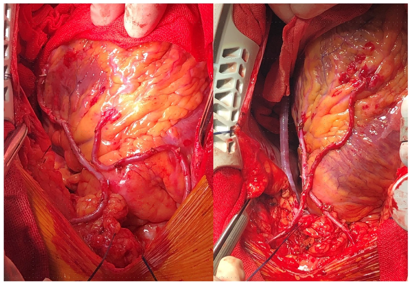
Figure 1. (Left) panel: In-situ right internal thoracic artery to left anterior descending artery with in-situ left internal thoracic artery to the diagonal branch. (Right) panel: In-situ right internal thoracic artery to left circumflex branch with in-situ left internal thoracic artery to left anterior descending. (Reproduced without changes from: “Application of bilateral internal mammary artery with different configurations in coronary artery bypass grafting” by Han et al.; Journal of Cardiothoracic Surgery, 2021; License: CC BY 4.0, https://creativecommons.org/licenses/by/4.0/, accessed on 15 January 2023).
Jcm 12 02022 g001
Figure 2. Left internal thoracic artery sequential grafting to the left anterior descending and diagonal arteries and right internal thoracic artery to the obtuse marginal artery. Reproduced from: “Hemodynamic changes during heart displacement in aorta no-touch off-pump coronary artery bypass surgery: a pilot study” by Carvalho et al.; Brazilian Journal of Cardio-Vascular Surgery, 2018; License: CC BY 4.0, https://creativecommons.org/licenses/by/4.0/, accessed on 20 February 2023).
Jcm 12 02022 g002
Figure 3. Baby-Y graft of radial artery and saphenous vein to obtuse marginal branches of left circumflex artery.
Jcm 12 02022 g003
Figure 4. Left internal thoracic artery to the left anterior descending coronary artery and radial artery Y graft to diagonal artery. Reproduced without changes from: “Revascularization strategies in OPCABG” by Vettath et al.; Cardiac Surgery Procedures, 2019; License: CC BY 3.0, https://creativecommons.org/licenses/by/3.0/, accessed on 20 February 2023).
Jcm 12 02022 g004
Table 1. Angiographic outcomes of currently used coronary artery bypass grafts.
ConduitPatency Rate
RA94.1% (95% CI 90.0–97.6)
No touch-SVG91.4% (95% CI 87.3–94.3)
RITA89.2% (95% CI 71.2–96.5)
Conventionally harvested SVG86.3% (95% CI 81.2–90.2)
GEA61.2% (95% CI 52.2–69.4)
Based on pooled estimates from a meta-analysis of 18 randomized clinical trials (8272 grafts) [102]. CI, confidence interval; LITA, left internal thoracic artery; GEA, gastroepiploic artery; RA, radial artery; RITA, right internal thoracic artery; SVG, saphenous vein graft.
Disclaimer/Publisher’s Note: The statements, opinions and data contained in all publications are solely those of the individual author(s) and contributor(s) and not of MDPI and/or the editor(s). MDPI and/or the editor(s) disclaim responsibility for any injury to people or property resulting from any ideas, methods, instructions or products referred to in the content.