Change in Descriptive Kinematic Parameters of Patients with Patellofemoral Instability When Compared to Individuals with Healthy Knees—A 3D MRI In Vivo Analysis
Abstract
1. Introduction
2. Material/Methods
2.1. Participants
2.2. MRI Setup and Protocol
2.3. Parameter
2.4. Statistics
3. Results
3.1. CCA
3.2. Patella Shift
3.3. Patella Rotation Angle
4. Discussion
Limitations
5. Conclusions
Author Contributions
Funding
Institutional Review Board Statement
Informed Consent Statement
Data Availability Statement
Acknowledgments
Conflicts of Interest
References
- Neal, B.S.; Lack, S.D.; Lankhorst, N.E.; Raye, A.; Morrissey, D.; van Middelkoop, M. Risk Factors for Patellofemoral Pain: A Systematic Review and Meta-Analysis. Br. J. Sports Med. 2019, 53, 270–281. [Google Scholar] [CrossRef] [PubMed]
- Fithian, D.C.; Paxton, E.W.; Stone, M.L.; Silva, P.; Davis, D.K.; Elias, D.A.; White, L.M. Epidemiology and Natural History of Acute Patellar Dislocation. Am. J. Sports Med. 2004, 32, 1114–1121. [Google Scholar] [CrossRef] [PubMed]
- Hawkins, R.J.; Bell, R.H.; Anisette, G. Acute Patellar Dislocations. Am. J. Sports Med. 1986, 14, 117–120. [Google Scholar] [CrossRef] [PubMed]
- Sillanpää, P.; Mattila, V.M.; Iivonen, T.; Visuri, T.; Pihlajamäki, H. Incidence and Risk Factors of Acute Traumatic Primary Patellar Dislocation. Med. Sci. Sport. Exerc. 2008, 40, 606–611. [Google Scholar] [CrossRef]
- Golant, A.; Quach, T.; Rose, J. Patellofemoral Instability: Diagnosis and Management. In Current Issues in Sports and Exercise Medicine; Hamlin, M., Ed.; InTech: Rang-Du-Fliers, France, 2013; ISBN 978-953-51-1031-6. [Google Scholar]
- Connolly, K.D.; Ronsky, J.L.; Westover, L.M.; Küpper, J.C.; Frayne, R. Differences in Patellofemoral Contact Mechanics Associated with Patellofemoral Pain Syndrome. J. Biomech. 2009, 42, 2802–2807. [Google Scholar] [CrossRef]
- Imhoff, F.B.; Funke, V.; Muench, L.N.; Sauter, A.; Englmaier, M.; Woertler, K.; Imhoff, A.B.; Feucht, M.J. The Complexity of Bony Malalignment in Patellofemoral Disorders: Femoral and Tibial Torsion, Trochlear Dysplasia, TT–TG Distance, and Frontal Mechanical Axis Correlate with Each Other. Knee Surg. Sports Traumatol. Arthrosc. 2020, 28, 897–904. [Google Scholar] [CrossRef]
- Senavongse, W.; Amis, A.A. The Effects of Articular, Retinacular, or Muscular Deficiencies on Patellofemoral Joint Stability: A biomechanical study in vitro. J. Bone Jt. Surg. 2005, 87-B, 577–582. [Google Scholar] [CrossRef]
- Feller, J.A.; Amis, A.A.; Andrish, J.T.; Arendt, E.A.; Erasmus, P.J.; Powers, C.M. Surgical Biomechanics of the Patellofemoral Joint. Arthrosc. J. Arthrosc. Relat. Surg. 2007, 23, 542–553. [Google Scholar] [CrossRef]
- Amis, A.A. Current Concepts on Anatomy and Biomechanics of Patellar Stability. Sport. Med. Arthrosc. Rev. 2007, 15, 48–56. [Google Scholar] [CrossRef]
- Clark, D.; Stevens, J.M.; Tortonese, D.; Whitehouse, M.R.; Simpson, D.; Eldridge, J. Mapping the Contact Area of the Patellofemoral Joint: The Relationship between Stability and Joint Congruence. Bone Jt. J. 2019, 101-B, 552–558. [Google Scholar] [CrossRef]
- Westphal, C.J.; Schmitz, A.; Reeder, S.B.; Thelen, D.G. Load-Dependent Variations in Knee Kinematics Measured with Dynamic MRI. J. Biomech. 2013, 46, 2045–2052. [Google Scholar] [CrossRef]
- Fick, C.N.; Jiménez-Silva, R.; Sheehan, F.T.; Grant, C. Patellofemoral Kinematics in Patellofemoral Pain Syndrome: The Influence of Demographic Factors. J. Biomech. 2022, 130, 110819. [Google Scholar] [CrossRef] [PubMed]
- Frings, J.; Dust, T.; Krause, M.; Frosch, K.-H.; Adam, G.; Warncke, M.; Welsch, G.; Henes, F.O.; Maas, K.-J. Dynamic Mediolateral Patellar Translation Is a Sex- and Size-Independent Parameter of Adult Proximal Patellar Tracking Using Dynamic 3 Tesla Magnetic Resonance Imaging. Arthrosc. J. Arthrosc. Relat. Surg. 2021, 38, 1571–1580. [Google Scholar] [CrossRef] [PubMed]
- Yao, J.; Yang, B.; Niu, W.; Zhou, J.; Wang, Y.; Gong, H.; Ma, H.; Tan, R.; Fan, Y. In Vivo Measurements of Patellar Tracking and Finite Helical Axis Using a Static Magnetic Resonance Based Methodology. Med. Eng. Phys. 2014, 36, 1611–1617. [Google Scholar] [CrossRef]
- Lange, T.; Taghizadeh, E.; Knowles, B.R.; Südkamp, N.P.; Zaitsev, M.; Meine, H.; Izadpanah, K. Quantification of Patellofemoral Cartilage Deformation and Contact Area Changes in Response to Static Loading via High-resolution MRI with Prospective Motion Correction. J. Magn. Reson. Imaging 2019, 50, 1561–1570. [Google Scholar] [CrossRef]
- Suzuki, T.; Hosseini, A.; Li, J.-S.; Gill, T.J.; Li, G. In Vivo Patellar Tracking and Patellofemoral Cartilage Contacts during Dynamic Stair Ascending. J. Biomech. 2012, 45, 2432–2437. [Google Scholar] [CrossRef]
- Yu, Z.; Yao, J.; Wang, X.; Xin, X.; Zhang, K.; Cai, H.; Fan, Y.; Yang, B. Research Methods and Progress of Patellofemoral Joint Kinematics: A Review. J. Healthc. Eng. 2019, 2019, 9159267. [Google Scholar] [CrossRef] [PubMed]
- Merican, A.M.; Amis, A.A. Iliotibial Band Tension Affects Patellofemoral and Tibiofemoral Kinematics. J. Biomech. 2009, 42, 1539–1546. [Google Scholar] [CrossRef]
- Melegari, T.M.; Parks, B.G.; Matthews, L.S. Patellofemoral Contact Area and Pressure after Medial Patellofemoral Ligament Reconstruction. Am. J. Sports Med. 2008, 36, 747–752. [Google Scholar] [CrossRef]
- Besier, T.F.; Draper, C.E.; Gold, G.E.; Beaupré, G.S.; Delp, S.L. Patellofemoral Joint Contact Area Increases with Knee Flexion and Weight-Bearing. J. Orthop. Res. 2005, 23, 345–350. [Google Scholar] [CrossRef]
- Stevens, J.M.; Eldridge, J.D.; Tortonese, D.; Whitehouse, M.R.; Krishnan, H.; Elsiwy, Y.; Clark, D. The Influence of Patellofemoral Stabilisation Surgery on Joint Congruity: An MRI Surface Mapping Study. Eur. J. Orthop. Surg. Traumatol. 2022, 32, 419–425. [Google Scholar] [CrossRef] [PubMed]
- Katchburian, M.V.; Bull, A.M.J.; Shih, Y.-F.; Heatley, F.W.; Amis, A.A. Measurement of Patellar Tracking: Assessment and Analysis of the Literature. Clin. Orthop. Relat. Res. 2003, 412, 241–259. [Google Scholar] [CrossRef] [PubMed]
- Post, W.R.; Fithian, D.C. Patellofemoral Instability: A Consensus Statement From the AOSSM/PFF Patellofemoral Instability Workshop. Orthop. J. Sport. Med. 2018, 6, 232596711775035. [Google Scholar] [CrossRef] [PubMed]
- Philippot, R.; Boyer, B.; Testa, R.; Farizon, F.; Moyen, B. Study of Patellar Kinematics after Reconstruction of the Medial Patellofemoral Ligament. Clin. Biomech. 2012, 27, 22–26. [Google Scholar] [CrossRef]
- Laprade, J.; Lee, R. Real-Time Measurement of Patellofemoral Kinematics in Asymptomatic Subjects. Knee 2005, 12, 63–72. [Google Scholar] [CrossRef]
- Nha, K.W.; Papannagari, R.; Gill, T.J.; Van de Velde, S.K.; Freiberg, A.A.; Rubash, H.E.; Li, G. In Vivo Patellar Tracking: Clinical Motions and Patellofemoral Indices. J. Orthop. Res. 2008, 26, 1067–1074. [Google Scholar] [CrossRef]
- Koh, J.L.; Stewart, C. Patellar Instability. Orthop. Clin. N. Am. 2015, 46, 147–157. [Google Scholar] [CrossRef]
- Sanders, T.L.; Pareek, A.; Johnson, N.R.; Stuart, M.J.; Dahm, D.L.; Krych, A.J. Patellofemoral Arthritis After Lateral Patellar Dislocation: A Matched Population-Based Analysis. Am. J. Sports Med. 2017, 45, 1012–1017. [Google Scholar] [CrossRef]
- Maclaren, J.; Armstrong, B.S.R.; Barrows, R.T.; Danishad, K.A.; Ernst, T.; Foster, C.L.; Gumus, K.; Herbst, M.; Kadashevich, I.Y.; Kusik, T.P.; et al. Measurement and Correction of Microscopic Head Motion during Magnetic Resonance Imaging of the Brain. PLoS ONE 2012, 7, e48088. [Google Scholar] [CrossRef]
- Fellows, R.A.; Hill, N.A.; Gill, H.S.; MacIntyre, N.J.; Harrison, M.M.; Ellis, R.E.; Wilson, D.R. Magnetic Resonance Imaging for in Vivo Assessment of Three-Dimensional Patellar Tracking. J. Biomech. 2005, 38, 1643–1652. [Google Scholar] [CrossRef]


| Patients | ±SD | Volunteers | ±SD | ||
|---|---|---|---|---|---|
| Number of subjects | 17 | 17 | |||
| Mean age (years) | 26.47 | ±7.67 | 30.00 | ±5.81 | |
| Mean height (cm) | 174.59 | ±8.73 | 176.24 | ±9.50 | |
| Mean weight (kg) | 71.35 | ±10.88 | 75.47 | ±18.27 | |
| Sex (female/male) | 10/7 | - | 10/7 | - | |
| Mean TEA distance (mm) | 77.13 | ±6.05 | 79.63 | ±7.58 | Matching reference (p = 0.245) |
| PFI Patients W/O Load Mean ± SD | Volunteers W/O Load Mean ± SD | PFI Patients 50 N Load Mean ± SD | Volunteers 50 N Load Mean ± SD | ||||||
|---|---|---|---|---|---|---|---|---|---|
| Flexion | n= | p= | n= | p= | |||||
| CCA | |||||||||
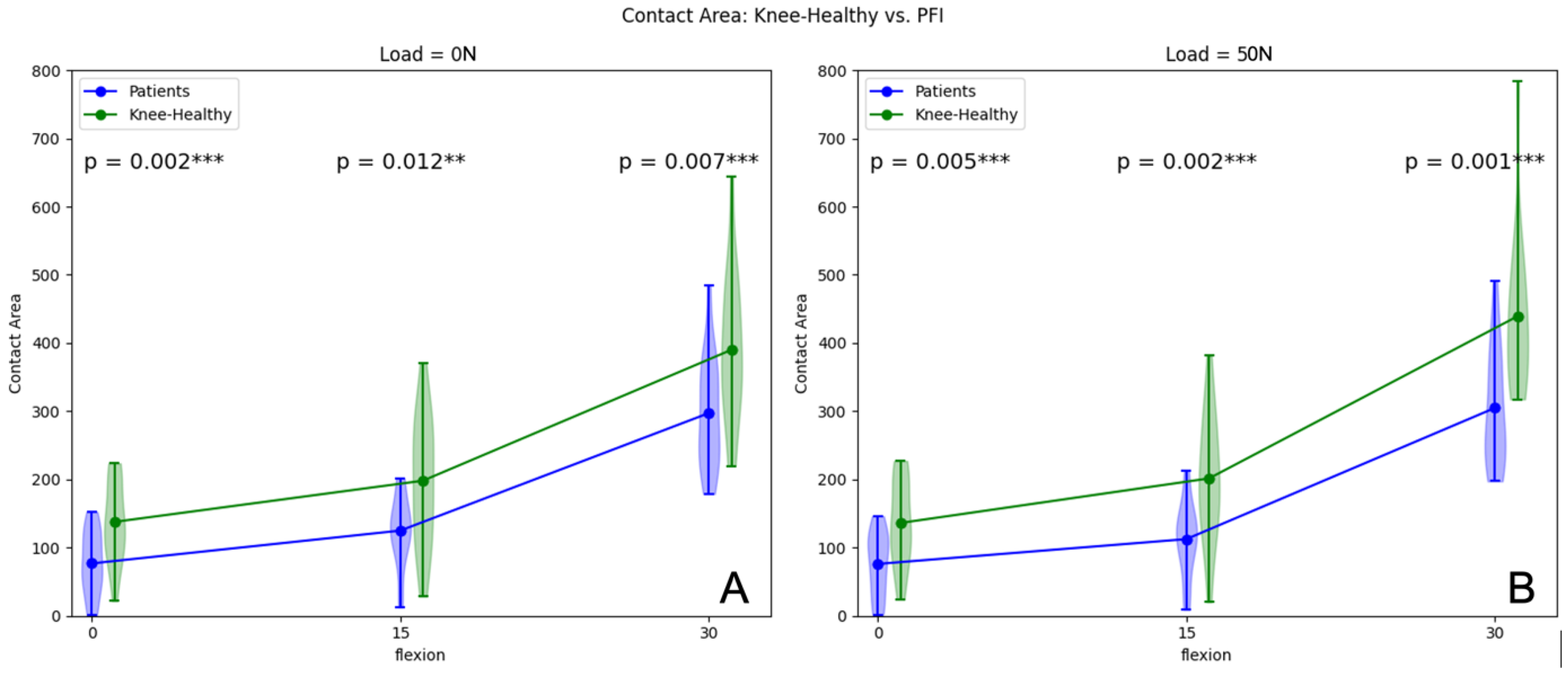
| 0° | 17/17 | 76.65 ± 47.43 | 137.52 ± 59.58 | 0.002 | 17/17 | 75.91 ± 47.70 | 135.97 ± 63.09 | 0.005 | |
| 15° | 17/17 | 124.77 ± 52.71 | 197.97 ± 93.15 | 0.012 | 17/17 | 112.27 ± 55.07 | 201.31 ± 91.41 | 0.002 | |
| 30° | 17/17 | 297.11 ± 80.74 | 389.94 ± 108.05 | 0.007 | 17/17 | 304.73 ± 86.85 | 439.17 ± 115.29 | 0.001 | |
| Shift | |||||||||
| 0° | 17/17 | 3.01 ± 1.81 | 1.73 ± 0.89 | 0.021 | 17/17 | 3.31 ± 1.93 | 1.90 ± 0.91 | 0.019 | |
| 15° | 17/17 | 2.57 ± 1.60 | 1.33 ± 0.75 | 0.015 | 17/17 | 2.73 ± 1.65 | 1.38 ± 0.76 | 0.010 | |
| 30° | 17/17 | 1.67 ± 1.41 | 0.84 ± 0.56 | 0.057 | 17/17 | 1.71 ± 1.30 | 0.76 ± 0.51 | 0.024 | |
| Rotation | |||||||||
| 0° | 17/17 | - | - | - | 17/17 | 2.07 ± 1.89 | 1.02 ± 1.94 | 0.005 | |
| 15° | 17/17 | 3.80 ± 3.40 | 2.43 ± 3.14 | 0.151 | 17/17 | 3.96 ± 3.24 | 2.56 ± 3.19 | 0.074 | |
| 30° | 17/17 | 6.32 ± 5.88 | 3.36 ± 3.17 | 0.084 | 17/17 | 6.61 ± 6.55 | 4.09 ± 3.31 | 0.267 |
| PFI Patients w/o Load Mean ± SD | PFI Patients 50 N Load Mean ± SD | Δ | Volunteers w/o Load Mean ± SD | Volunteers 50 n Load Mean ± SD | Δ | ||||||
|---|---|---|---|---|---|---|---|---|---|---|---|
| FLEX. | n= | p= | n= | p= | |||||||
| CCA | |||||||||||
| 0° | 17 | 76.65 ± 47.43 | 75.91 ± 47.70 | 0.73 ± 0.27 | 0.979 | 17 | 137.52 ± 59.58 | 135.97 ± 63.09 | −1.55 ± 3.51 | 0.758 | |
| 15° | 17 | 124.77 ± 52.71 | 112.27 ± 55.07 | 12.50± 2.36 | 0.309 | 17 | 197.97 ± 93.15 | 201.31 ± 91.41 | 3.35 ± −1.74 | 0.084 | |
| 30° | 17 | 297.11 ± 80.74 | 304.73 ± 86.85 | 7.62 ± 6.11 | 0.408 | 17 | 389.94 ± 108.05 | 439.17 ± 115.29 | 49.23 ± 7.24 | 0.042 | |
| Shift | |||||||||||
| 0° | 17 | 3.01 ± 1.81 | 3.31 ± 1.93 | 0.30 ± 0.01 | 0.001 | 17 | 1.73 ± 0.89 | 1.90 ± 0.91 | 0.17 ± 0.00 | 0.030 | |
| 15° | 17 | 2.57 ± 1.60 | 2.73 ± 1.65 | 0.16 ± 0.00 | 0.010 | 17 | 1.33 ± 0.75 | 1.38 ± 0.76 | 0.04 ± 0.00 | 0.156 | |
| 30° | 17 | 1.67 ± 1.41 | 1.71 ± 1.30 | 0.04 ±- 0.01 | 0.532 | 17 | 0.84 ± 0.56 | 0.76 ± 0.51 | 12 ± −0.01 | 0.352 | |
| Rot. | |||||||||||
| 0° | 17 | - | 2.07 ± 1.89 | 2.07 ± 1.89 | - | 17 | - | 1.02 ± 1.94 | 1.02 ± 1.94 | - | |
| 15° | 17 | 3.80 ± 3.40 | 3.96 ± 3.24 | 0.18 ± −0.18 | 0.379 | 17 | 2.43 ± 3.14 | 2.56 ± 3.19 | 1.16 ±2.04 | 0.246 | |
| 30° | 17 | 6.32 ± 5.88 | 6.61 ± 6.55 | 0.28 ± 0.67 | 0.717 | 17 | 3.36 ± 3.17 | 4.09 ± 3.31 | 2.07 ± 1.61 | 0.079 |
Disclaimer/Publisher’s Note: The statements, opinions and data contained in all publications are solely those of the individual author(s) and contributor(s) and not of MDPI and/or the editor(s). MDPI and/or the editor(s) disclaim responsibility for any injury to people or property resulting from any ideas, methods, instructions or products referred to in the content. |
© 2023 by the authors. Licensee MDPI, Basel, Switzerland. This article is an open access article distributed under the terms and conditions of the Creative Commons Attribution (CC BY) license (https://creativecommons.org/licenses/by/4.0/).
Share and Cite
Siegel, M.; Maier, P.; Taghizadeh, E.; Fuchs, A.; Yilmaz, T.; Meine, H.; Schmal, H.; Lange, T.; Izadpanah, K. Change in Descriptive Kinematic Parameters of Patients with Patellofemoral Instability When Compared to Individuals with Healthy Knees—A 3D MRI In Vivo Analysis. J. Clin. Med. 2023, 12, 1917. https://doi.org/10.3390/jcm12051917
Siegel M, Maier P, Taghizadeh E, Fuchs A, Yilmaz T, Meine H, Schmal H, Lange T, Izadpanah K. Change in Descriptive Kinematic Parameters of Patients with Patellofemoral Instability When Compared to Individuals with Healthy Knees—A 3D MRI In Vivo Analysis. Journal of Clinical Medicine. 2023; 12(5):1917. https://doi.org/10.3390/jcm12051917
Chicago/Turabian StyleSiegel, Markus, Philipp Maier, Elham Taghizadeh, Andreas Fuchs, Tayfun Yilmaz, Hans Meine, Hagen Schmal, Thomas Lange, and Kaywan Izadpanah. 2023. "Change in Descriptive Kinematic Parameters of Patients with Patellofemoral Instability When Compared to Individuals with Healthy Knees—A 3D MRI In Vivo Analysis" Journal of Clinical Medicine 12, no. 5: 1917. https://doi.org/10.3390/jcm12051917
APA StyleSiegel, M., Maier, P., Taghizadeh, E., Fuchs, A., Yilmaz, T., Meine, H., Schmal, H., Lange, T., & Izadpanah, K. (2023). Change in Descriptive Kinematic Parameters of Patients with Patellofemoral Instability When Compared to Individuals with Healthy Knees—A 3D MRI In Vivo Analysis. Journal of Clinical Medicine, 12(5), 1917. https://doi.org/10.3390/jcm12051917


