
Urethral Injuries: Diagnostic and Management Strategies for Critical Care and Trauma Clinicians
 ,
,
Abstract
:1. Background
2. Methods
3. Discussion
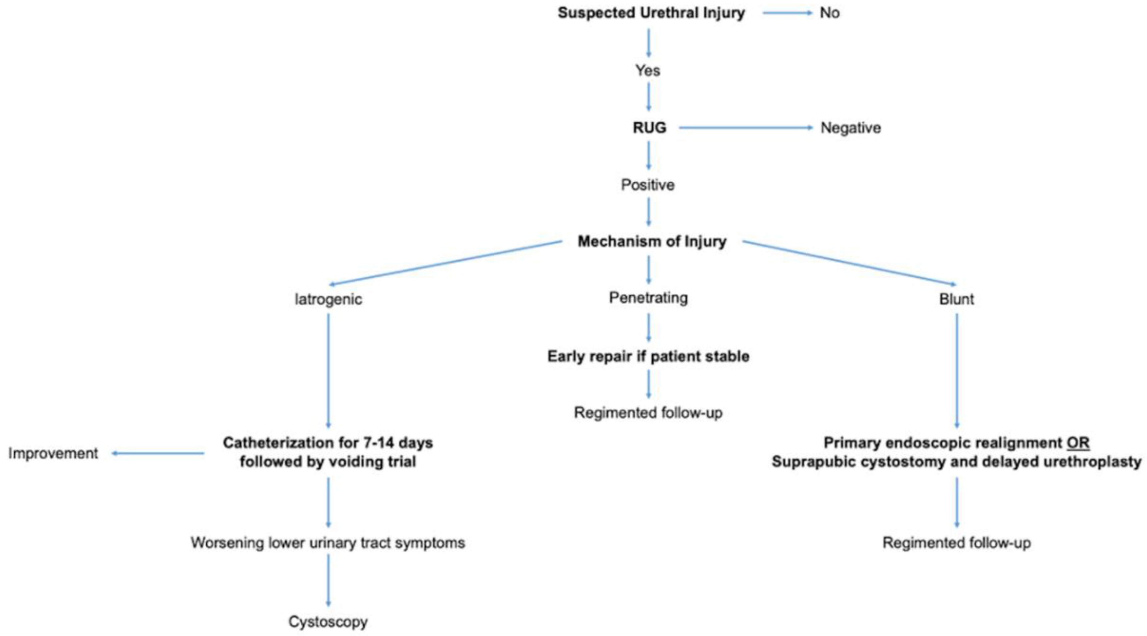
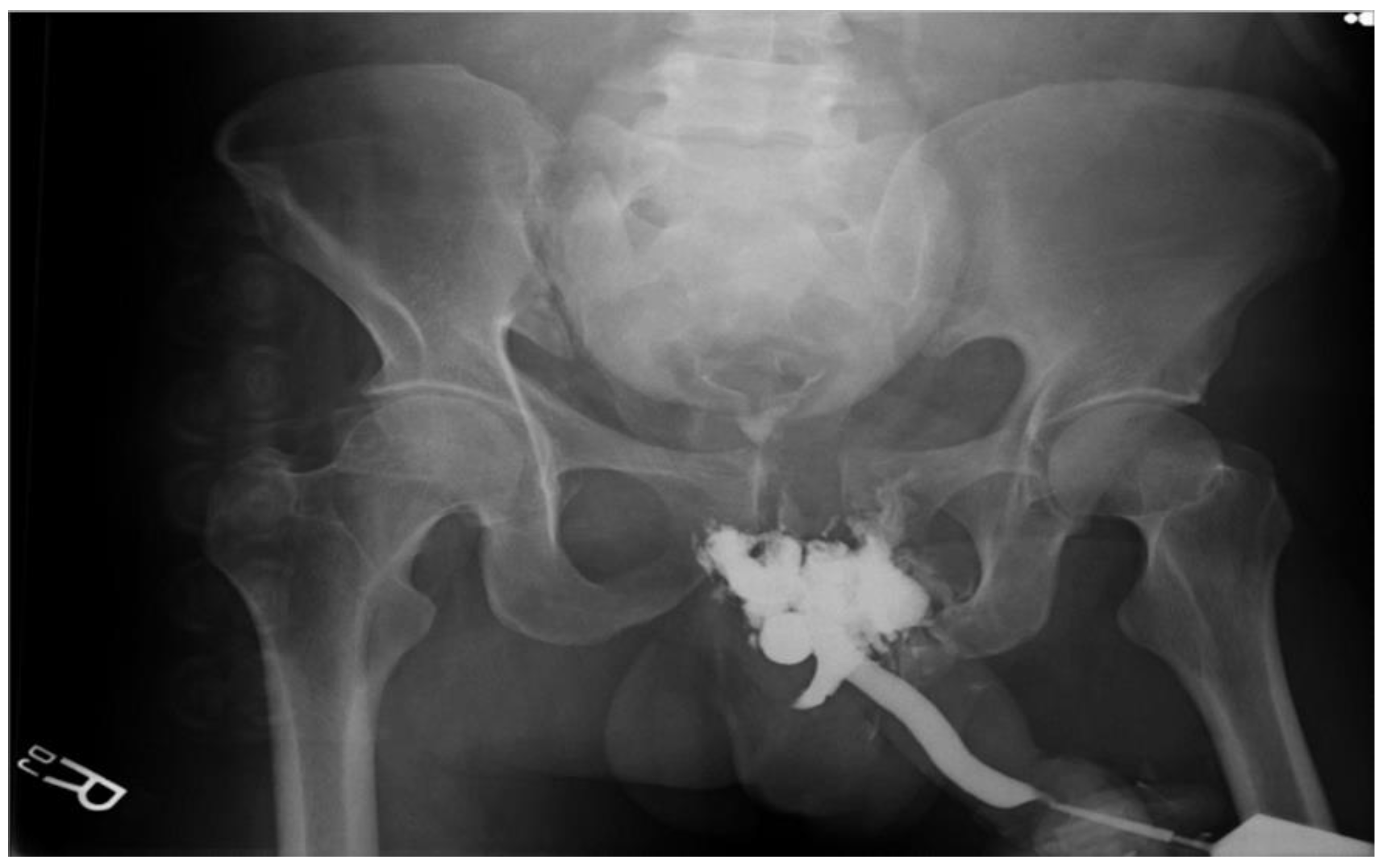
3.1. Overall Consensus on Diagnosis
3.2. Iatrogenic Urethral Injuries
3.2.1. Incidence
3.2.2. Mechanism
3.2.3. Management
3.3. Penetrating Urethral Injuries
3.3.1. Incidence
3.3.2. Mechanism
3.3.3. Management
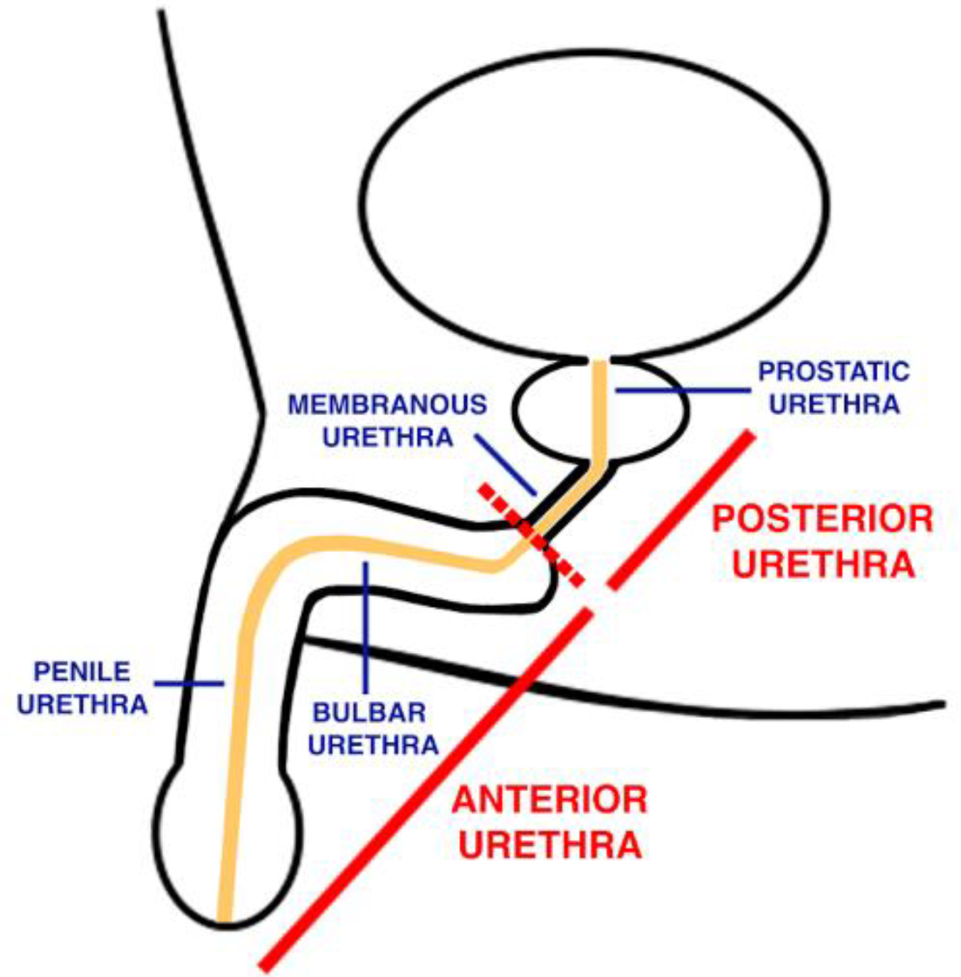
Anterior Urethral Injury
Posterior Urethral Injury
3.4. Blunt Urethral Injuries
3.4.1. Incidence
3.4.2. Mechanism
3.4.3. Management
Anterior Urethral Injury

| Author | Year | Level of Study | Number of Patients in Study | Diagnostic Procedures | Management | Complications |
|---|---|---|---|---|---|---|
| Londergan TA, Gundersen LH, van Every MJ [55] | 1997 | 4 | 6 | RUG | Fluoroscopic realignment | Procedure: Requiring interval urethrotomy; prostatic abscess. ⅖ (40%) patients had impotence—consistent with other studies. |
| Jordan GH, Virasoro R, Eltahaway EA [9] | 2006 | 3 | NA | RUG, endoscopy | Delayed excision with primary anastomosis | Procedure: Abscess, erectile dysfunction, infection |
| Dobrowolski ZF, Weglarz W, Jakubik P, et al. [44] | 2002 | 3 | 48 | RUG, voiding cystourethrogram, endoscopy | 25% surgery which included drainage, 75% conservative management | Stricture |
| Zhang Y, Zhang K, Fu Q [60] | 2018 | 3 | 1606 | RUG | Urinary diversion, primary repair (endoscopic vs. open realignment) | Infection, stricture |
| Park S, McAninch JW [56] | 2004 | 3 | 78 | RUG | Suprapubic urinary diversion, delayed presentation led to urethroplasty | Obstructive symptoms in patients with prior urethral manipulation |
Posterior Urethral Injury
| Author | Year | Level of Study | Number of Patients in Study | Diagnostic Procedures | Management | Complications |
|---|---|---|---|---|---|---|
| Dobrowolski ZF, Weglarz W, Jakubik P, et al. [44] | 2002 | 4 | 268 | RUG, voiding cystourethrogram, urethroscopy, spiral CT | Cystostomy + drainage, urethral reconstruction | Procedure: Urethral stricture, urinary tract infection, bleeding, stones, fistula |
| Koraitim MM, Reda IS [13] | 2007 | 4 | 21 | Pre-operative MRI of pelvis to aid in selection of appropriate surgical approach | End-to-end urethral anastomosis | Injury: Retropubic space scarring and fibrosis, fistula, impotence |
| El Darawany HM [37] | 2018 | 4 | 27 | CT Urography, RUG | Initial Management: Suprapubic catheterization to drain bladder Endoscopic urethral realignment | Procedure: Minimal urethral stricture |
| Koraitim MM [66] | 1996 | 4 | 100 | Clinical examination Excretory urography, retrograde urethrography | Suprapubic cystostomy indicated for incomplete urethral rupture, slight urethral distraction, and critically unstable patients, and for inadequate facilities or inexperienced surgeons Primary realignment for wide separation of urethral ends or associated injury to bladder neck or rectum | Procedure: Urethral stricture after suprabpubic cystostomy Impotence and incontinence after primary realignment Impotence and incontinence is greatest after primary suturing |
| Velarde-Ramos L, Gomez-Illanes R, Campos-Juanatey F, et al. [3] | 2016 | 3 | NA | RUG | Primary urethral realignment, delayed urethroplasty, SPC + endoscopic realignment | Procedure: Sepsis, abscess, urinoma |
| Cavalcanti AG, Krambeck R, Araujo A, et al. [67] | 2006 | 3 | 77 | RUG, surgical exploration | Primary urethral realignment/repair | Procedure: Penile curving, corporal body injury |
| Goldman SM, Sandler CM, Corriere JN Jr., et al. [12] | 1997 | 3 | NA | RUG | Delayed urethroplasty | NA |
| Elgammal MA [68] | 2009 | 3 | 53 | RUG | Suprapubic cystostomy, primary urethral realignment | Procedure: Urethral stricture |
| Jordan GH, Virasoro R, Eltahaway EA [9] | 2006 | 3 | NA | Clinical exam: direct examination RUG, cystogram | SPC + delayed repair, primary urethral realignment, endoscopic realignment | Procedure: Compartment syndrome, rhabdomyolysis |
| Protzel C, Hakenberg OW [57] | 2010 | 3 | NA | Clinical exam: direct examination, digital rectal exam RUG | Suprapubic urinary diversion + antibiotics, endoscopic transurethral splinting | NA |
| Tezval H, Tezval M, von Klot C, et al. [18] | 2007 | 3 | NA | Clinical exam: Digital rectal exam Suprapubic cystography, RUG, CT scan | Cystostomy + antibiotics, primary urethral realignment, delayed urethroplasty | Procedure: Stricture, erectile dysfunction, distraction defects, impotence, recurrent stenosis |
| Rosenstein DI, Alsikafi NF [6] | 2006 | 3 | NA | Clinical exam: Digital rectal exam Suprapubic cystography, RUG | SPC, delayed urethroplasty | Procedure: Bladder rupture, vaginal laceration, rectal tears |
| Luckhoff C, Mitra B, Cameron PA, et al. [8] | 2011 | 3 | 998 | RUG, pelvic X-ray, sonography | Primary urethral realignment | Procedure: Urethral transection |
| Cooperberg M, McAninch JW, Alsikafi NF, et al. [69] | 2007 | 3 | 134 | RUG/ voiding cystourethrogram Pelvic MRI if length of stricture is in question Doppler ultrasound for patients with erectile dysfunction to determine baseline neurological or vascular compromise | Delayed Anastomotic Posterior Urethroplasty | Procedure: Scrotal hematoma, compartment syndrome (lithotomy position), decubitus ulcer, recurrent stricture |
Complications of Urethral Injuries
Follow-Up for Urethral Injuries
Areas of Future Discovery and Improvement
4. Conclusions
Author Contributions
Funding
Institutional Review Board Statement
Informed Consent Statement
Data Availability Statement
Conflicts of Interest
References
- Martinez-Pineiro, L.; Djakovic, N.; Plas, E.; Mor, Y.; Santucci, R.A.; Serafetinidis, E.; Turkeri, L.N.; Hohenfellner, M. EAU Guidelines on Urethral Trauma. Eur. Urol. 2010, 57, 791–803. [Google Scholar] [CrossRef] [PubMed]
- Morey, A.F.; Brandes, S.; Dugi, D.D., 3rd; Armstrong, J.H.; Breyer, B.N.; Broghammer, J.A.; Erickson, B.A.; Holzbeierlein, J.; Hudak, S.J.; Pruitt, J.H.; et al. Urotrauma: AUA guideline. J. Urol. 2014, 192, 327–335. [Google Scholar] [CrossRef]
- Velarde-Ramos, L.; Gomez-Illanes, R.; Campos-Juanatey, F.; Portillo-Martin, J.A. Traumatic lesions of the posterior urethra. Actas Urol. Esp. 2016, 40, 539–548. [Google Scholar] [CrossRef] [PubMed]
- Lim, P.H.; Chng, H.C. Initial management of acute urethral injuries. Br. J. Urol. 1989, 64, 165–168. [Google Scholar] [CrossRef]
- Koraitim, M.M. Pelvic fracture urethral injuries: The unresolved controversy. J. Urol. 1999, 161, 1433–1441. [Google Scholar] [CrossRef]
- Rosenstein, D.I.; Alsikafi, N.F. Diagnosis and classification of urethral injuries. Urol. Clin. N. Am. 2006, 33, 73–85. [Google Scholar] [CrossRef]
- Balzano, F.L.; Hudak, S.J. Military genitourinary injuries: Past, present, and future. Transl. Androl. Urol. 2018, 7, 646–652. [Google Scholar] [CrossRef]
- Luckhoff, C.; Mitra, B.; Cameron, P.A.; Fitzgerald, M.; Royce, P. The diagnosis of acute urethral trauma. Injury 2011, 42, 913–916. [Google Scholar] [CrossRef]
- Jordan, G.H.; Virasoro, R.; Eltahawy, E.A. Reconstruction and management of posterior urethral and straddle injuries of the urethra. Urol. Clin. N. Am. 2006, 33, 97–109. [Google Scholar] [CrossRef]
- Maciejewski, C.; Rourke, K. Imaging of urethral stricture disease. Transl. Androl. Urol. 2015, 4, 2–9. [Google Scholar] [CrossRef]
- Bach, P.; Rourke, K. Independently interpreted retrograde urethrography does not accurately diagnose and stage anterior urethral stricture: The importance of urologist-performed urethrography. Urology 2014, 83, 1190–1193. [Google Scholar] [CrossRef] [PubMed]
- Goldman, S.M.; Sandler, C.M.; Corriere, J.N., Jr.; McGuire, E.J. Blunt urethral trauma: A unified, anatomical mechanical classification. J Urol 1997, 157, 85–89. [Google Scholar] [CrossRef] [PubMed]
- Koraitim, M.M.; Reda, I.S. Role of magnetic resonance imaging in assessment of posterior urethral distraction defects. Urology 2007, 70, 403–406. [Google Scholar] [CrossRef] [PubMed]
- Bryk, D.J.; Zhao, L.C. Guideline of guidelines: A review of urological trauma guidelines. BJU Int. 2016, 117, 226–234. [Google Scholar] [CrossRef]
- Venn, S.N.; Greenwell, T.J.; Mundy, A.R. Pelvic fracture injuries of the female urethra. BJU Int. 1999, 83, 626–630. [Google Scholar] [CrossRef] [PubMed]
- Kommu, S.S.; Illahi, I.; Mumtaz, F. Patterns of urethral injury and immediate management. Curr. Opin. Urol. 2007, 17, 383–389. [Google Scholar] [CrossRef]
- Basta, A.M.; Blackmore, C.C.; Wessells, H. Predicting urethral injury from pelvic fracture patterns in male patients with blunt trauma. J. Urol. 2007, 177, 571–575. [Google Scholar] [CrossRef] [PubMed]
- Tezval, H.; Tezval, M.; von Klot, C.; Herrmann, T.R.; Dresing, K.; Jonas, U.; Burchardt, M. Urinary tract injuries in patients with multiple trauma. World J. Urol. 2007, 25, 177–184. [Google Scholar] [CrossRef]
- Ali, M.; Safriel, Y.; Sclafani, S.J.; Schulze, R. CT signs of urethral injury. Radiogr. Rev. Publ. Radiol. Soc. N. Am. Inc. 2003, 23, 951–963. [Google Scholar] [CrossRef]
- Morey, A.F.; McAninch, J.W. Ultrasound evaluation of the male urethra for assessment of urethral stricture. J. Clin. Ultrasound JCU 1996, 24, 473–479. [Google Scholar] [CrossRef]
- Gupta, N.; Dubey, D.; Mandhani, A.; Srivastava, A.; Kapoor, R.; Kumar, A. Urethral stricture assessment: A prospective study evaluating urethral ultrasonography and conventional radiological studies. BJU Int. 2006, 98, 149–153. [Google Scholar] [CrossRef] [PubMed]
- Kashefi, C.; Messer, K.; Barden, R.; Sexton, C.; Parsons, J.K. Incidence and prevention of iatrogenic urethral injuries. J. Urol. 2008, 179, 2254–2257. [Google Scholar] [CrossRef] [PubMed]
- Santucci, R.A.; McAninch, J.W. Urethral reconstruction of strictures resulting from treatment of benign prostatic hypertrophy and prostate cancer. Urol. Clin. N. Am. 2002, 29, 417–427. [Google Scholar] [CrossRef] [PubMed]
- Elliott, S.P.; McAninch, J.W.; Chi, T.; Doyle, S.M.; Master, V.A. Management of severe urethral complications of prostate cancer therapy. J. Urol. 2006, 176, 2508–2513. [Google Scholar] [CrossRef]
- McConnell, J.D. Benign prostatic hyperplasia: Treatment guidelines and patient classification. Br. J. Urol. 1995, 76 (Suppl. S1), 29–46. [Google Scholar]
- Shaw, M.B.; Payne, S.R. A simple technique for accurate diathermy destruction of urethral meatal warts. Urology 2007, 69, 975–976. [Google Scholar] [CrossRef]
- Abdel-Hakim, A.; Bernstein, J.; Teijeira, J.; Elhilali, M.M. Urethral stricture after cardiovascular surgery, a retrospective and a prospective study. J. Urol. 1983, 130, 1100–1102. [Google Scholar] [CrossRef]
- Baktavatsalam, R.; Little, D.M.; Connolly, E.M.; Farrell, J.G.; Hickey, D.P. Complications relating to the urinary tract associated with bladder-drained pancreatic transplantation. Br. J. Urol. 1998, 81, 219–223. [Google Scholar] [CrossRef]
- Siegel, A.L. Urethral necrosis and proximal urethro-vaginal fistula resulting from tension-free vaginal tape. Int. Urogynecol. J. Pelvic Floor Dysfunct. 2006, 17, 661–664. [Google Scholar] [CrossRef]
- Summerton, D.J.; Kitrey, N.D.; Lumen, N.; Serafetinidis, E.; Djakovic, N. EAU guidelines on iatrogenic trauma. Eur. Urol. 2012, 62, 628–639. [Google Scholar] [CrossRef]
- Davis, N.F.; Quinlan, M.R.; Bhatt, N.R.; Browne, C.; MacCraith, E.; Manecksha, R.; Walsh, M.T.; Thornhill, J.A.; Mulvin, D. Incidence, Cost, Complications and Clinical Outcomes of Iatrogenic Urethral Catheterization Injuries: A Prospective Multi-Institutional Study. J. Urol. 2016, 196, 1473–1477. [Google Scholar] [CrossRef] [PubMed]
- Wagner, K.R.; Bird, E.T.; Coffield, K.S. Urinary Catheterization: A Paradigm Shift in Difficult Urinary Catheterization. Curr. Urol. Rep. 2016, 17, 82. [Google Scholar] [CrossRef] [PubMed]
- Singha, P.; Locklin, J.; Handa, H. A review of the recent advances in antimicrobial coatings for urinary catheters. Acta Biomater 2017, 50, 20–40. [Google Scholar] [CrossRef] [PubMed]
- Willette, P.A.; Coffield, S. Current trends in the management of difficult urinary catheterizations. West. J. Emerg. Med. 2012, 13, 472–478. [Google Scholar] [CrossRef] [PubMed]
- Villanueva, C.; Hemstreet, G.P., 3rd. Difficult male urethral catheterization: A review of different approaches. Int. Braz. J. Urol. Off. J. Braz. Soc. Urol. 2008, 34, 401–411. [Google Scholar] [CrossRef] [PubMed]
- Bjurlin, M.A.; Kim, D.Y.; Zhao, L.C.; Palmer, C.J.; Cohn, M.R.; Vidal, P.P.; Bokhari, F.; Hollowell, C.M. Clinical characteristics and surgical outcomes of penetrating external genital injuries. J. Trauma Acute Care Surg. 2013, 74, 839–844. [Google Scholar] [CrossRef] [PubMed]
- El Darawany, H.M. Endoscopic urethral realignment of traumatic urethral disruption: A monocentric experience. Urol. Ann. 2018, 10, 47–51. [Google Scholar] [CrossRef]
- Chrouser, K.L.; Leibovich, B.C.; Sweat, S.D.; Larson, D.W.; Davis, B.J.; Tran, N.V.; Zincke, H.; Blute, M.L. Urinary fistulas following external radiation or permanent brachytherapy for the treatment of prostate cancer. J. Urol. 2005, 173, 1953–1957. [Google Scholar] [CrossRef]
- Marguet, C.; Raj, G.V.; Brashears, J.H.; Anscher, M.S.; Ludwig, K.; Mouraviev, V.; Robertson, C.N.; Polascik, T.J. Rectourethral fistula after combination radiotherapy for prostate cancer. Urology 2007, 69, 898–901. [Google Scholar] [CrossRef]
- Maheshwari, P.N.; Shah, H.N. Immediate endoscopic management of complete iatrogenic anterior urethral injuries: A case series with long-term results. BMC Urol. 2005, 5, 13. [Google Scholar] [CrossRef]
- Mohr, A.M.; Pham, A.M.; Lavery, R.F.; Sifri, Z.; Bargman, V.; Livingston, D.H. Management of trauma to the male external genitalia: The usefulness of American Association for the Surgery of Trauma organ injury scales. J. Urol. 2003, 170, 2311–2315. [Google Scholar] [CrossRef] [PubMed]
- Cinman, N.M.; McAninch, J.W.; Porten, S.P.; Myers, J.B.; Blaschko, S.D.; Bagga, H.S.; Breyer, B.N. Gunshot wounds to the lower urinary tract: A single-institution experience. J. Trauma Acute Care Surg. 2013, 74, 725–730. [Google Scholar] [CrossRef] [PubMed]
- Tausch, T.J.; Cavalcanti, A.G.; Soderdahl, D.W.; Favorito, L.; Rabelo, P.; Morey, A.F. Gunshot wound injuries of the prostate and posterior urethra: Reconstructive armamentarium. J. Urol. 2007, 178, 1346–1348. [Google Scholar] [CrossRef] [PubMed]
- Dobrowolski, Z.F.; Weglarz, W.; Jakubik, P.; Lipczynski, W.; Dobrowolska, B. Treatment of posterior and anterior urethral trauma. BJU Int. 2002, 89, 752–754. [Google Scholar] [CrossRef]
- Brandes, S. Initial management of anterior and posterior urethral injuries. Urol. Clin. N. Am. 2006, 33, 87–95. [Google Scholar] [CrossRef]
- Mumtaz, F.; Hellawell, G.; Ralph, D. Penile Trauma. In Management of Urological Emergencies; Taylor & Francis: Abingdon-on-Thames, UK, 2004; pp. 165–180. [Google Scholar]
- Johnsen, N.V.; Dmochowski, R.R.; Mock, S.; Reynolds, W.S.; Milam, D.F.; Kaufman, M.R. Primary Endoscopic Realignment of Urethral Disruption Injuries—A Double-Edged Sword? J. Urol. 2015, 194, 1022–1026. [Google Scholar] [CrossRef]
- Chapple, C.; Barbagli, G.; Jordan, G.; Mundy, A.R.; Rodrigues-Netto, N.; Pansadoro, V.; McAninch, J.W. Consensus statement on urethral trauma. BJU Int. 2004, 93, 1195–1202. [Google Scholar] [CrossRef]
- Zaid, U.B.; Bayne, D.B.; Harris, C.R.; Alwaal, A.; McAninch, J.W.; Breyer, B.N. Penetrating Trauma to the Ureter, Bladder, and Urethra. Curr. Trauma Rep. 2015, 1, 119–124. [Google Scholar] [CrossRef]
- Mundy, A.R.; Andrich, D.E. Urethral trauma. Part I: Introduction, history, anatomy, pathology, assessment and emergency management. BJU Int. 2011, 108, 310–327. [Google Scholar] [CrossRef]
- Mundy, A.R.; Andrich, D.E. Urethral trauma. Part II: Types of injury and their management. BJU Int. 2011, 108, 630–650. [Google Scholar] [CrossRef]
- McGeady, J.B.; Breyer, B.N. Current epidemiology of genitourinary trauma. Urol. Clin. N. Am. 2013, 40, 323–334. [Google Scholar] [CrossRef] [PubMed]
- Koraitim, M.M.; Marzouk, M.E.; Atta, M.A.; Orabi, S.S. Risk factors and mechanism of urethral injury in pelvic fractures. Br. J. Urol. 1996, 77, 876–880. [Google Scholar] [CrossRef] [PubMed]
- Kong, J.P.; Bultitude, M.F.; Royce, P.; Gruen, R.L.; Cato, A.; Corcoran, N.M. Lower urinary tract injuries following blunt trauma: A review of contemporary management. Rev. Urol. 2011, 13, 119–130. [Google Scholar] [PubMed]
- Londergan, T.A.; Gundersen, L.H.; van Every, M.J. Early fluoroscopic realignment for traumatic urethral injuries. Urology 1997, 49, 101–103. [Google Scholar] [CrossRef] [PubMed]
- Park, S.; McAninch, J.W. Straddle injuries to the bulbar urethra: Management and outcomes in 78 patients. J. Urol. 2004, 171, 722–725. [Google Scholar] [CrossRef] [PubMed]
- Protzel, C.; Hakenberg, O.W. Diagnosis and treatment of lower urinary tract trauma. Der Unf. 2010, 113, 313–324. [Google Scholar] [CrossRef]
- Netto Junior, N.R.; Ikari, O.; Zuppo, V.P. Traumatic rupture of female urethra. Urology 1983, 22, 601–603. [Google Scholar] [CrossRef]
- Lowe, M.A.; Mason, J.T.; Luna, G.K.; Maier, R.V.; Copass, M.K.; Berger, R.E. Risk factors for urethral injuries in men with traumatic pelvic fractures. J. Urol. 1988, 140, 506–507. [Google Scholar] [CrossRef] [PubMed]
- Zhang, Y.; Zhang, K.; Fu, Q. Emergency treatment of male blunt urethral trauma in China: Outcome of different methods in comparison with other countries. Asian J. Urol. 2018, 5, 78–87. [Google Scholar] [CrossRef]
- Pinggera, G.M.; Rehder, P.; Bartsch, G.; Gozzi, C. Urethral trauma. Der Urologe. Ausg. A 2005, 44, 883–897. [Google Scholar] [CrossRef]
- Conort, P.; Chartier-Kastler, E.; Richard, F.; Chatelain, C. Secondary management of urethral injuries. Chir. Mem. De L’Academie De Chir. 1996, 121, 372–376. [Google Scholar]
- Lieberman, S.F.; Barry, J.M. Retreat from transpubic urethroplasty for obliterated membranous urethral strictures. J. Urol. 1982, 128, 379–381. [Google Scholar] [CrossRef] [PubMed]
- Chiou, R.K.; Gonzalez, R.; Ortlip, S.; Fraley, E.E. Endoscopic treatment of posterior urethral obliteration: Long-term followup and comparison with transpubic urethroplasty. J. Urol. 1988, 140, 508–511. [Google Scholar] [CrossRef] [PubMed]
- Moses, R.A.; Selph, J.P.; Voelzke, B.B.; Piotrowski, J.; Eswara, J.R.; Erickson, B.A.; Gupta, S.; Dmochowski, R.R.; Johnsen, N.V.; Shridharani, A.; et al. An American Association for the Surgery of Trauma (AAST) prospective multi-center research protocol: Outcomes of urethral realignment versus suprapubic cystostomy after pelvic fracture urethral injury. Transl. Androl. Urol. 2018, 7, 512–520. [Google Scholar] [CrossRef] [PubMed]
- Koraitim, M.M. Pelvic fracture urethral injuries: Evaluation of various methods of management. J. Urol. 1996, 156, 1288–1291. [Google Scholar] [CrossRef] [PubMed]
- Cavalcanti, A.G.; Krambeck, R.; Araujo, A.; Rabelo, P.H.; Carvalho, J.P.; Favorito, L.A. Management of urethral lesions in penile blunt trauma. Int. J. Urol. 2006, 13, 1218–1220. [Google Scholar] [CrossRef] [PubMed]
- Elgammal, M.A. Straddle injuries to the bulbar urethra: Management and outcome in 53 patients. Int. Braz. J. Urol. 2009, 35, 450–458. [Google Scholar] [CrossRef]
- Cooperberg, M.R.; McAninch, J.W.; Alsikafi, N.F.; Elliott, S.P. Urethral reconstruction for traumatic posterior urethral disruption: Outcomes of a 25-year experience. J. Urol. 2007, 178, 2006–2010. [Google Scholar] [CrossRef]
- Anger, J.T.; Sherman, N.D.; Dielubanza, E.; Webster, G.D.; Hegarty, P.K. Erectile function after posterior urethroplasty for pelvic fracture-urethral distraction defect injuries. BJU Int. 2009, 104, 1126–1129. [Google Scholar] [CrossRef]
- Xambre, L. Sexual (Dys)function after Urethroplasty. Adv. Urol. 2016, 2016, 9671297. [Google Scholar] [CrossRef]
- Tritschler, S.; Roosen, A.; Fullhase, C.; Stief, C.G.; Rubben, H. Urethral stricture: Etiology, investigation and treatments. Dtsch. Arztebl. Int. 2013, 110, 220–226. [Google Scholar] [CrossRef] [PubMed]
- Hampson, L.A.; McAninch, J.W.; Breyer, B.N. Male urethral strictures and their management. Nat. Rev. Urol. 2014, 11, 43–50. [Google Scholar] [CrossRef] [PubMed]
- Mundy, A.R. Management of urethral strictures. Postgrad. Med. J. 2006, 82, 489–493. [Google Scholar] [CrossRef] [PubMed]
- Wessells, H.; Angermeier, K.W.; Elliott, S.; Gonzalez, C.M.; Kodama, R.; Peterson, A.C.; Reston, J.; Rourke, K.; Stoffel, J.T.; Vanni, A.J.; et al. Male Urethral Stricture: American Urological Association Guideline. J. Urol. 2017, 197, 182–190. [Google Scholar] [CrossRef]

| Author | Year | Level of Study | Number of Patients in Study | Diagnostic Procedures | Management | Complications |
|---|---|---|---|---|---|---|
| Kommu SS, Illahi I, Mumtaz F [16] | 2007 | 4 | NA | Endoscopy at the time of urological and gynecological procedure | NA | NA |
| Rosenstein DI, Alsikafi NF [6] | 2006 | 4 | NA | RUG | Non-surgical: Using catheters made of less irritative material, intermittent catheterization, transurethral microwave therapy Surgical: Urethrotomies and repeated dilations | Procedure: Urine extravasation and scarring |
| Maheshwari PN, Shah HN [40] | 2005 | 4 | 7 | RUG, Urethroscopy | Retrograde or suprapubic immediate endoscopic realignment | Procedure: Recurrence of stricture |
| Kashefi C, Messer K, Barden R, et al. [22] | 2008 | 3 | 14 | Clinical exam: urethral and/or perineal pain, blood at the meatus, non-draining catheter with ineffective irrigation Cystoscopy, RUG | Nursing education program | Injury: urethral scarring and strictures |
| Davis NF, Quinlan MR, Bhatt NR, et al. [31] | 2016 | 3 | 37 | Antegrade/RUG | One gentle catheter attempt for junior health professionals, Education, Percutaneous Insertion of SPC, Flexible Cystoscopy + guidewire, Transurethral catheter, Catheter manipulation, Rigid cystoscopy + guidewire, Open cystostomy + SPC | Procedural: Acute urinary retention, urosepsis, bleeding, Acute kidney injury, urethral stricture disease |
| Elliot S, McAninch JW, Chi T, et al. [24] | 2006 | 3 | 48 | NA | Anastomotic urethroplasty, flap urethroplasty, perineal urethrostomy, urethral stent | Injury: Urethral stenosis, rectourinary fistula |
| Author | Year | Level of Study | Number of Patients in Study | Diagnostic Procedures | Management | Complications |
|---|---|---|---|---|---|---|
| Dobrowolski ZF, Weglarz W, Jakubik P, et al. [44] | 2002 | 4 | 255 | RUG voiding cystourethrogram, endoscopy | Urethral repair | NA |
| Bjurlin MA, Kim DY, Zhao LC, et al. [36] | 2013 | 4 | 162 | RUG | Immediate primary urethral repair, urinary diversion with delayed reconstruction | Procedure: Urethral stricture |
| Bryk DJ, Zhao LC [14] | 2016 | 4 | NA | Clinical exam: rectal exam, blood at meatus, perineal or penile hematoma, voiding dysfunction RUG | Immediate open surgical repair, except when there are other life-threatening surgeries | NA |
| Kommu SS, Illahi I, Mumtaz F [16] | 2007 | 4 | NA | RUG under fluoroscopic guidance, anteroposterior radiograph | Mild contusions: conservative management SPC with delayed primary repair for severe contusions Partial or complete disruption: suprapubic diversion, primary realignment Exploration and debridement of devitalized tissue | Procedure: Urethral stricture Injury: Infection and necrotizing fasciitis |
| Cinman NM, McAninch JW, Porten SP, et al. [42] | 2013 | 3 | 50 | Clinical exam: gross hematuria | Initial SPC with delayed reconstruction, primary urethral repair with concomitant bladder neck or rectal injury | Procedure: Urethral stricture associated with urinary diversion |
| Author | Year | Level of Study | Number of Patients in Study | Diagnostic Procedures | Management | Complications |
|---|---|---|---|---|---|---|
| Koraitim MM, Reda IS [13] | 2007 | 4 | 21 | Pre-operative MRI of pelvis to aid in selection of appropriate surgical approach | End-to-end urethral anastomosis | Injury: Retropubic space scarring and fibrosis, fistula, impotence |
| Tausch TJ, Cavalcanti AG, Soderdahl DW, et al. [43] | 2007 | 3 | 19 | RUG | Primary urethral repair with concomitant bladder neck or rectal injury, delayed reconstruction, endoscopic realignment | Erectile dysfunction, pelvic abscess, incontinence, stricture |
| Cinman NM, McAninch JW, Porten SP, et al. [42] | 2013 | 3 | 2 | Clinical exam: gross hematuria | Initial SPC with delayed reconstruction, primary endoscopic realignment | Procedure: Urethral stricture associated with urinary diversion |
Disclaimer/Publisher’s Note: The statements, opinions and data contained in all publications are solely those of the individual author(s) and contributor(s) and not of MDPI and/or the editor(s). MDPI and/or the editor(s) disclaim responsibility for any injury to people or property resulting from any ideas, methods, instructions or products referred to in the content. |
© 2023 by the authors. Licensee MDPI, Basel, Switzerland. This article is an open access article distributed under the terms and conditions of the Creative Commons Attribution (CC BY) license (https://creativecommons.org/licenses/by/4.0/).
Share and Cite
Patel, A.B.; Osterberg, E.C.; Satarasinghe, P.N.; Wenzel, J.L.; Akbani, S.T.; Sahi, S.L.; Emigh, B.J.; Wolf, J.S., Jr.; Brown, C.V.R. Urethral Injuries: Diagnostic and Management Strategies for Critical Care and Trauma Clinicians. J. Clin. Med. 2023, 12, 1495. https://doi.org/10.3390/jcm12041495
Patel AB, Osterberg EC, Satarasinghe PN, Wenzel JL, Akbani ST, Sahi SL, Emigh BJ, Wolf JS Jr., Brown CVR. Urethral Injuries: Diagnostic and Management Strategies for Critical Care and Trauma Clinicians. Journal of Clinical Medicine. 2023; 12(4):1495. https://doi.org/10.3390/jcm12041495
Chicago/Turabian StylePatel, Anish B., E. Charles Osterberg, Praveen N. Satarasinghe, Jessica L. Wenzel, Sabah T. Akbani, Saad L. Sahi, Brent J. Emigh, J. Stuart Wolf, Jr., and Carlos V. R. Brown. 2023. "Urethral Injuries: Diagnostic and Management Strategies for Critical Care and Trauma Clinicians" Journal of Clinical Medicine 12, no. 4: 1495. https://doi.org/10.3390/jcm12041495
APA StylePatel, A. B., Osterberg, E. C., Satarasinghe, P. N., Wenzel, J. L., Akbani, S. T., Sahi, S. L., Emigh, B. J., Wolf, J. S., Jr., & Brown, C. V. R. (2023). Urethral Injuries: Diagnostic and Management Strategies for Critical Care and Trauma Clinicians. Journal of Clinical Medicine, 12(4), 1495. https://doi.org/10.3390/jcm12041495