


Prognostic Role of Sonographic Decongestion in Patients with Acute Heart Failure with Reduced and Preserved Ejection Fraction: A Multicentre Study
 , , ,
, , ,  and
and
Abstract
1. Introduction
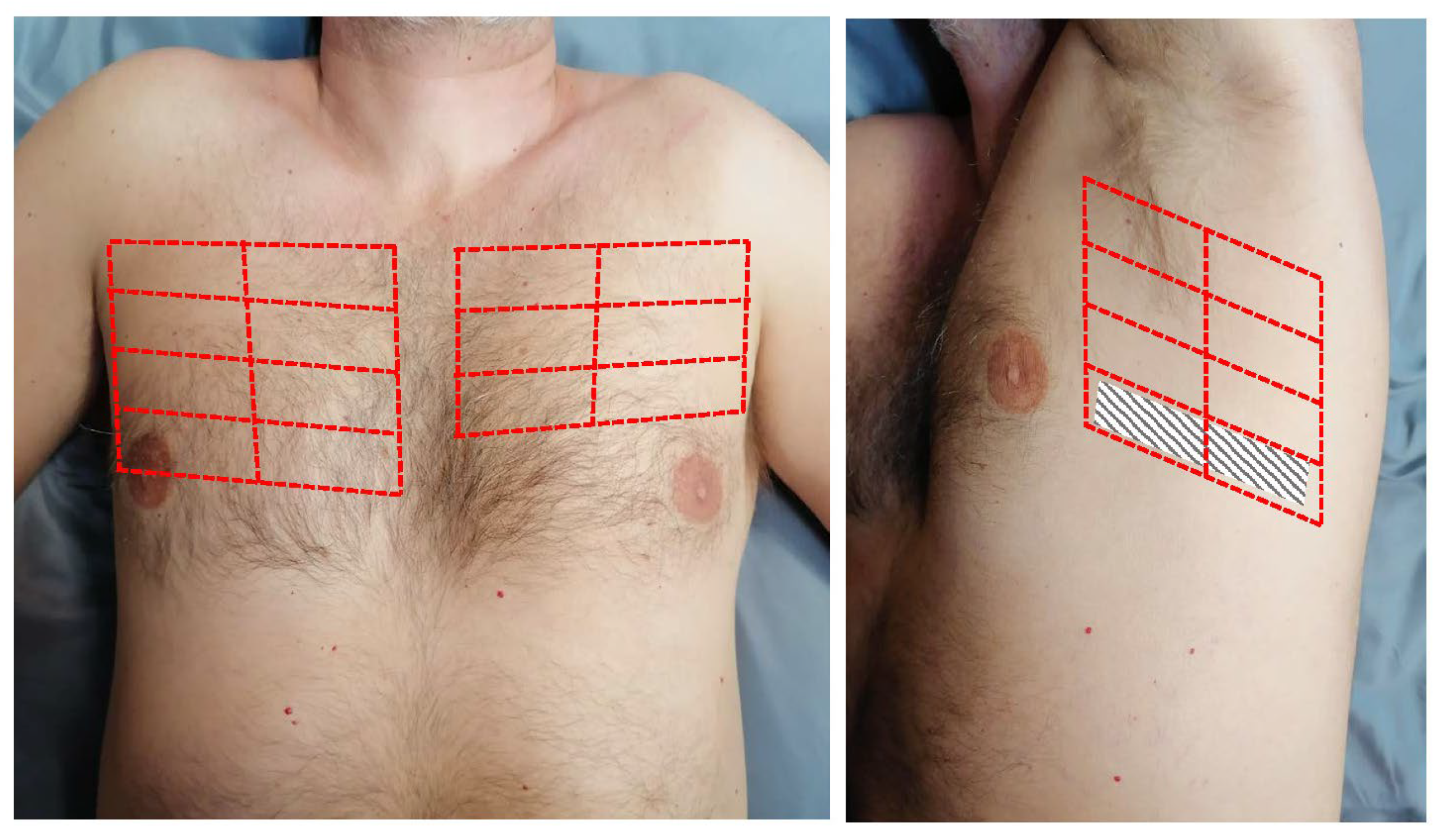
2. Materials and Methods
3. Results
3.1. Baseline Characteristics
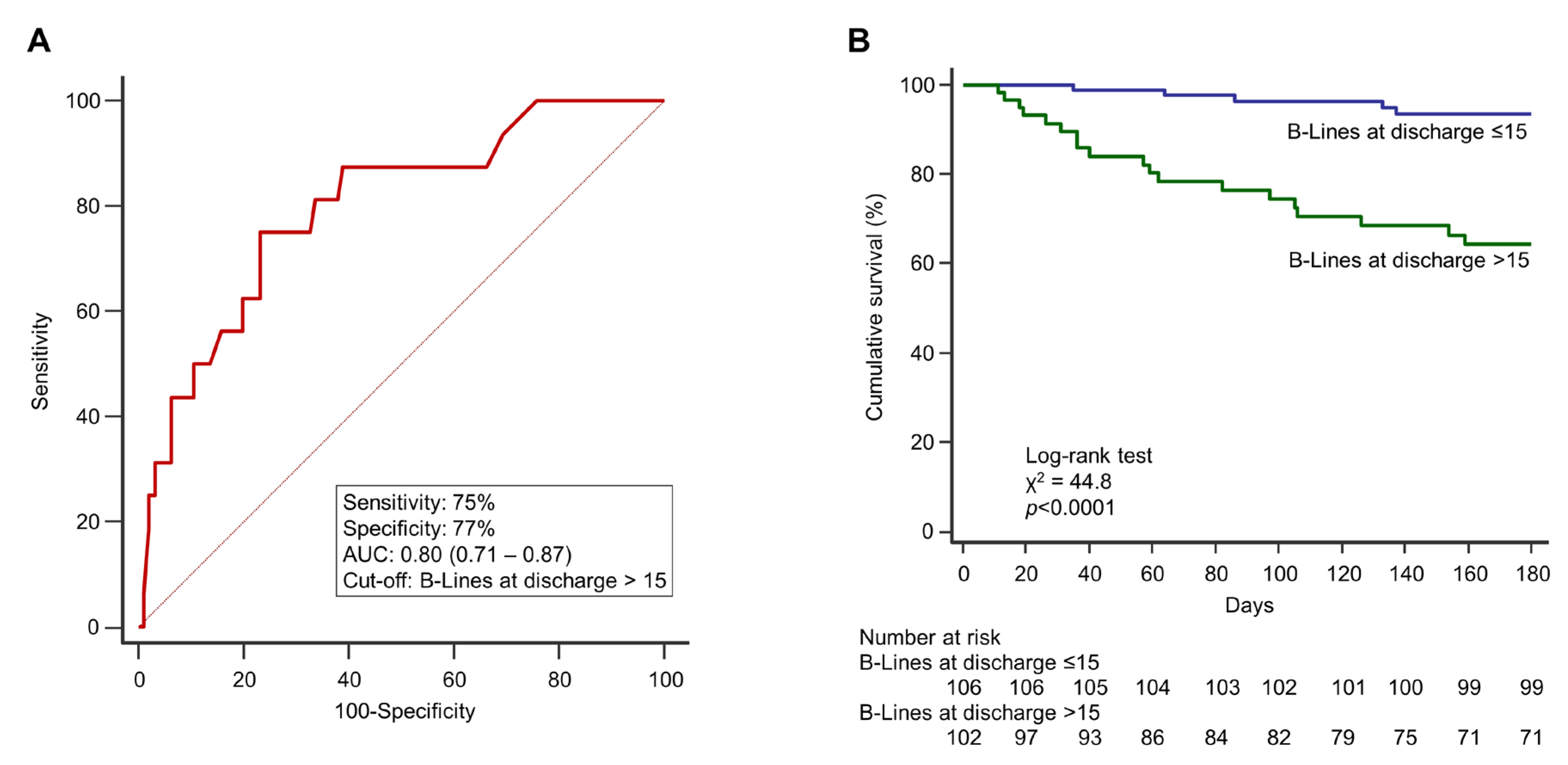
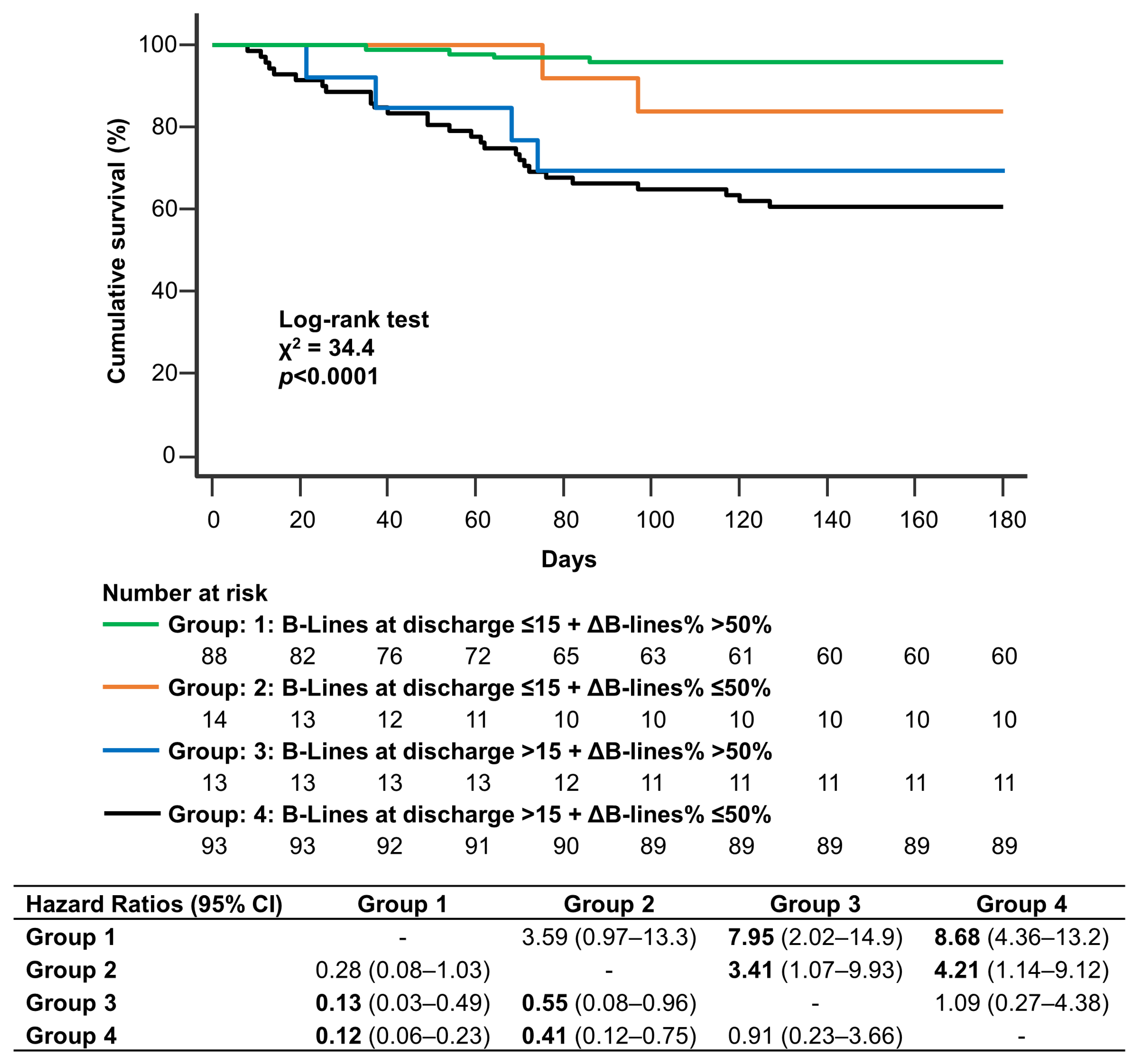
3.2. Clinical Outocomes
4. Discussion
5. Conclusions
Supplementary Materials
Author Contributions
Funding
Institutional Review Board Statement
Informed Consent Statement
Data Availability Statement
Conflicts of Interest
References
- Gheorghiade, M.; Follath, F.; Ponikowski, P.; Barsuk, J.H.; Blair, J.E.A.; Cleland, J.G.; Dickstein, K.; Drazner, M.H.; Fonarow, G.C.; Jaarsma, T.; et al. Assessing and grading congestion in acute heart failure: A scientific statement from the acute heart failure committee of the heart failure association of the European society of cardiology and endorsed by the European society of intensive care medicine. Eur. J. Heart Fail. 2010, 12, 423–433. [Google Scholar] [CrossRef] [PubMed]
- Mullens, W.; Damman, K.; Harjola, V.P.; Mebazaa, A.; Brunner-La Rocca, H.P.; Martens, P.; Testani, J.M.; Tang, W.W.; Orso, F.; Rossignol, P.; et al. The use of diuretics in heart failure with congestion—A position statement from the Heart Failure Association of the European Society of Cardiology. Eur. J. Heart Fail. 2019, 21, 137–155. [Google Scholar] [CrossRef] [PubMed]
- Zile, M.R.; Bennett, T.D.; St John Sutton, M.; Cho, Y.K.; Adamson, P.B.; Aaron, M.F.; Aranda, J.M.; Abraham, W.T.; Smart, F.W.; Stevenson, L.W.; et al. Transition from chronic compensated to acute d compensated heart failure: Pathophysiological insights obtained from continuous monitoring of intracardiac pressures. Circulation 2008, 118, 1433–1441. [Google Scholar] [CrossRef] [PubMed]
- Ponikowski, P.; Voors, A.A.; Anker, S.D.; Bueno, H.; Cleland, J.G.F.; Coats, A.J.S.; Falk, V.; González-Juanatey, J.R.; Harjola, V.-P.; Jankowska, E.A.; et al. 2016 ESC Guidelines for the diagnosis and treatment of acute and chronic heart failure. Eur. Heart J. 2016, 37, 2129–2200. [Google Scholar] [CrossRef] [PubMed]
- Agricola, E.; Bove, T.; Oppizzi, M.; Marino, G.; Zangrillo, A.; Margonato, A.; Picano, E. “Ultrasound comet-tail images”: A marker of pulmonary edema: A comparative study with wedge pressure and extravascular lung water. Chest 2005, 127, 1690–1695. [Google Scholar] [CrossRef]
- Gargani, L.; Lionetti, V.; Di Cristofano, C.; Bevilacqua, G.; Recchia, F.A.; Picano, E. Early detection of acute lung injury uncoupled to hypoxemia in pigs using ultrasound lung comets. Crit. Care Med. 2007, 35, 2769–2774. [Google Scholar]
- Baldi, G.; Gargani, L.; Abramo, A.; D’Errico, L.; Caramella, D.; Picano, E.; Giunta, F.; Forfori, F. Lung water assessment by lung ultrasonography in intensive care: A pilot study. Intensiv. Care Med. 2013, 39, 74–84. [Google Scholar] [CrossRef]
- Gargani, L.; Pang, P.S.; Frassi, F.; Miglioranza, M.; Dini, F.L.; Landi, P.; Picano, E. Persistent pulmonary congestion before discharge predicts rehospitalization in heart failure: A lung ultrasound study. Cardiovasc. Ultrasound 2015, 13, 40. [Google Scholar] [CrossRef]
- Coiro, S.; Rossignol, P.; Ambrosio, G.; Carluccio, E.; Alunni, G.; Murrone, A.; Tritto, I.; Zannad, F.; Girerd, N. Prognostic value of residual pulmonary congestion at discharge assessed by lung ultrasound imaging in heart failure. Eur. J. Heart Fail. 2015, 17, 1172–1181. [Google Scholar] [CrossRef]
- Platz, E.; Campbell, R.T.; Claggett, B.; Lewis, E.F.; Groarke, J.D.; Docherty, K.F.; Lee, M.M.Y.; Merz, A.A.; Silverman, M.; Silverman, M.; et al. Lung Ultrasound in Acute Heart Failure: Prevalence of Pulmonary Congestion and Short- and Long-Term Outcomes. JACC Heart Fail. 2019, 7, 849–858. [Google Scholar] [CrossRef]
- Öhman, J.; Harjola, V.P.; Karjalainen, P.; Lassus, J. Assessment of early treatment response by rapid cardiothoracic ultrasound in acute heart failure: Cardiac filling pressures, pulmonary congestion and mortality. Eur. Heart J. Acute Cardiovasc. Care 2018, 7, 311–320. [Google Scholar] [CrossRef]
- Mozzini, C.; Perna, M.D.D.; Pesce, G.; Garbin, U.; Pasini, A.M.F.; Ticinesi, A.; Nouvenne, A.; Meschi, T.; Casadei, A.; Soresi, M.; et al. Lung ultrasound in internal medicine efficiently drives the management of patients with heart failure and speeds up the discharge time. Intern. Emerg. Med. 2018, 13, 27–33. [Google Scholar] [CrossRef]
- Platz, E.; Merz, A.A.; Jhund, P.S.; Vazir, A.; Campbell, R.; McMurray, J.J. Dynamic changes and prognostic value of pulmonary congestion by lung ultrasound in acute and chronic heart failure: A systematic review. Eur. J. Heart Fail. 2017, 19, 1154–1163. [Google Scholar] [CrossRef]
- Januzzi, J.L. Natriuretic peptide testing: A window into the diagnosis and prognosis of heart failure. Clevel. Clin. J. Med. 2006, 73, 149–157. [Google Scholar] [CrossRef]
- Pellicori, P.; Platz, E.; Dauw, J.; Ter Maaten, J.M.; Martens, P.; Pivetta, E.; Cleland, J.G.F.; McMurray, J.J.V.; Mullens, W.; Solomon, S.D.; et al. Ultrasound imaging of congestion in heart failure: Examinations beyond the heart. Eur. J. Heart Fail. 2021, 23, 703–712. [Google Scholar] [CrossRef]
- Volpicelli, G.; Elbarbary, M.; Blaivas, M.; Lichtenstein, D.A.; Mathis, G.; Kirkpatrick, A.W.; Melniker, L.; Gargani, L.; Noble, V.E.; Via, G.; et al. International evidence-based recommendations for point-of-care lung ultrasound. In Intensive Care Medicine; Springer: Berlin/Heidelberg, Germany, 2012; pp. 577–591. [Google Scholar]
- Frassi, F.; Gargani, L.; Tesorio, P.; Raciti, M.; Mottola, G.; Picano, E. Prognostic Value of Extravascular Lung Water Assessed with Ultrasound Lung Comets by Chest Sonography in Patients with Dyspnea and/or Chest Pain. J. Card. Fail. 2007, 13, 830–835. [Google Scholar] [CrossRef]
- Gargani, L.; Sicari, R.; Raciti, M.; Serasini, L.; Passera, M.; Torino, C.; Letachowicz, K.; Ekart, R.; Fliser, D.; Covic, A.; et al. Efficacy of a remote web-based lung ultrasound training for nephrologists and cardiologists: A lust trial sub-project. Nephrol. Dial. Transplant. 2016, 31, 1982–1988. [Google Scholar] [CrossRef]
- Pivetta, E.; Goffi, A.; Nazerian, P.; Castagno, D.; Tozzetti, C.; Tizzani, P.; Tizzani, M.; Porrino, G.; Ferreri, E.; Busso, V.; et al. Lung ultrasound integrated with clinical assessment for the diagnosis of acute decompensated heart failure in the emergency department: A randomized controlled trial. Eur. J. Heart Fail. 2019, 21, 754–766. [Google Scholar] [CrossRef]
- Pugliese, N.R.; Fabiani, I.; Santini, C.; Rovai, I.; Pedrinelli, R.; Natali, A.; Dini, F.L. Value of combined cardiopulmonary and echocardiography stress test to characterize the haemodynamic and metabolic responses of patients with heart failure and mid-range ejection fraction. Eur. Heart J. Cardiovasc. Imaging 2019, 20, 828–836. [Google Scholar] [CrossRef]
- Pugliese, N.R.; Mazzola, M.; Fabiani, I.; Gargani, L.; De Biase, N.; Pedrinelli, R.; Natali, A.; Dini, F.L. Haemodynamic and metabolic phenotyping of hypertensive patients with and without heart failure by combining cardiopulmonary and echocardiographic stress test. Eur. J. Heart Fail. 2020, 22, 458–468. [Google Scholar] [CrossRef]
- Girerd, N.; Seronde, M.F.; Coiro, S.; Chouihed, T.; Bilbault, P.; Braun, F.; Kenizou, D.; Maillier, B.; Nazeyrollas, P.; Roul, G.; et al. Integrative Assessment of Congestion in Heart Failure Throughout the Patient Journey. JACC Heart Fail. 2018, 6, 273–285. [Google Scholar] [CrossRef] [PubMed]
- Ambrosy, A.P.; Pang, P.S.; Khan, S.; Konstam, M.A.; Fonarow, G.C.; Traver, B.; Maggioni, A.P.; Cook, T.; Swedberg, K.; Burnett, J.C.; et al. Clinical course and predictive value of congestion during hospitalization in patients admitted for worsening signs and symptoms of heart failure with reduced ejection fraction: Findings from the EVEREST trial. Eur. Heart J. 2013, 34, 835–843. [Google Scholar] [CrossRef] [PubMed]
- Platz, E.; Lewis, E.F.; Uno, H.; Peck, J.; Pivetta, E.; Merz, A.A.; Hempel, D.; Wilson, C.; Frasure, S.E.; Jhund, P.S.; et al. Detection and prognostic value of pulmonary congestion by lung ultrasound in ambulatory heart failure patients. Eur. Heart J. 2016, 37, 1244–1251. [Google Scholar] [CrossRef] [PubMed]
- Miglioranza, M.H.; Picano, E.; Badano, L.P.; Sant’Anna, R.; Rover, M.; Zaffaroni, F.; Sicari, R.; Kalil, R.K.; Leiria, T.L.; Gargani, L. Pulmonary congestion evaluated by lung ultrasound predicts decompensation in heart failure outpatients. Int. J. Cardiol. 2017, 240, 271–278. [Google Scholar] [CrossRef] [PubMed]
- Pellicori, P.; Shah, P.; Cuthbert, J.; Urbinati, A.; Zhang, J.; Kallvikbacka-Bennett, A.; Clark, A.L.; Cleland, J.G. Prevalence, pattern and clinical relevance of ultrasound indices of congestion in outpatients with heart failure. Eur. J. Hear Fail. 2019, 21, 904–916. [Google Scholar] [CrossRef]
- Chioncel, O.; Mebazaa, A.; Maggioni, A.P.; Harjola, V.P.; Rosano, G.; Laroche, C.; Piepoli, M.F.; Crespo-Leiro, M.G.; Lainscak, M.; Ponikowski, P.; et al. Acute heart failure congestion and perfusion status—Impact of the clinical classification on in-hospital and long-term outcomes; insights from the ESC-EORP-HFA Heart Failure Long-Term Registry. Eur. J. Heart Fail. 2019, 21, 1338–1352. [Google Scholar] [CrossRef]
- Lang, R.M.; Badano, L.P.; Mor-Avi, V.; Afilalo, J.; Armstrong, A.; Ernande, L.; Flachskampf, F.A.; Foster, E.; Goldstein, S.A.; Kuznetsova, T.; et al. Recommendations for Cardiac Chamber Quantification by Echocardiography in Adults: An Update from the American Society of Echocardiography and the European Association of Cardiovascular Imaging. J. Am. Soc. Echocardiogr. 2015, 28, 1–39e14. [Google Scholar] [CrossRef]
- Guazzi, M.; Naeije, R.; Arena, R.; Corrà, U.; Ghio, S.; Forfia, P.; Rossi, A.; Cahalin, L.P.; Bandera, F.; Temporelli, P. Echocardiography of right ventriculoarterial coupling combined with cardiopulmonary exercise testing to predict outcome in heart failure. Chest 2015, 148, 226–234. [Google Scholar] [CrossRef]
- Rivas-Lasarte, M.; Alvarez-Garcia, J.; Fernández-Martínez, J.; Maestro, A.; López-López, L.; Solé-González, E.; Pirla, M.J.; Mesado, N.; Mirabet, S.; Fluvià, P.; et al. Lung ultrasound-guided treatment in ambulatory patients with heart failure: A randomized controlled clinical trial (LUS-HF study). Eur. J. Heart Fail. 2019, 21, 1605–1613. [Google Scholar] [CrossRef]
- Marini, C.; Fragasso, G.; Italia, L.; Sisakian, H.; Tufaro, V.; Ingallina, G.; Stella, S.; Ancona, F.; Loiacono, F.; Innelli, P.; et al. Lung ultrasound-guided therapy reduces acute decompensation events in chronic heart failure. Heart 2020, 106, 1934–1939. [Google Scholar] [CrossRef]
- Felker, G.M.; Anstrom, K.J.; Adams, K.F.; Ezekowitz, J.A.; Fiuzat, M.; Houston-Miller, N.; Januzzi, J.L.; Mark, D.B.; Piña, I.L.; Passmore, G.; et al. Effect of natriuretic peptide–guided therapy on hospitalization or cardiovascular mortality in high-risk patients with heart failure and reduced ejection fraction: A randomized clinical trial. JAMA-J. Am. Med. Assoc. 2017, 318, 713–720. [Google Scholar] [CrossRef]
- Palazzuoli, A.; Ruocco, G.; Beltrami, M.; Nuti, R.; Cleland, J.G. Combined use of lung ultrasound, B-type natriuretic peptide, and echocardiography for outcome prediction in patients with acute HFrEF and HFpEF. Clin. Res. Cardiol. 2018, 107, 586–596. [Google Scholar] [CrossRef]
- Buessler, A.; Chouihed, T.; Duarte., K.; Bassand, A.; Huot-Marchand, M.; Gottwalles, Y.; Pénine, A.; André, E.; Nace, L.; Jaeger, D.; et al. Accuracy of Several Lung Ultrasound Methods for the Diagnosis of Acute Heart Failure in the ED. Chest 2020, 157, 99–110. [Google Scholar] [CrossRef]


| Variable | Total Population (n = 208) | HFrEF (n = 125) | HFpEF (n = 83) | p-Value |
|---|---|---|---|---|
| Demographics | ||||
| Age, years | 75.9 (69.6–83.5) | 74 (68.2–80) | 79.6 (71.9–86.1) | 0.005 |
| Female gender | 75 (36) | 39 (31) | 36 (43) | 0.1 |
| BSA (m2) | 1.91 (1.87–1.96) | 1.91 (1.84–2.01) | 1.87 (1.76–2.02) | 0.3 |
| BMI (kg/m2) | 27.1 (24.7–31.2) | 26.2 (26.6–29.5) | 26.8 (23.2–31.9) | 0.8 |
| Family history of CVD | 40 (19) | 21 (17) | 19 (23) | 0.1 |
| Diabetes mellitus | 74 (36) | 41 (33) | 33 (40) | 0.1 |
| Arterial hypertension | 171 (82) | 100 (80) | 71 (86) | 0.3 |
| Dyslipidaemia ^ | 80 (37) | 50 (40) | 30 (36) | 0.1 |
| CAD | 80 (37) | 51 (41) | 29 (35) | 0.1 |
| Previous MI | 73 (35) | 53 (42) | 20 (24) | 0.01 |
| Previous coronary revascularization | 75 (36) | 51 (41) | 24 (29) | 0.1 |
| Atrial fibrillation | 66 (32) | 36 (29) | 30 (36) | 0.4 |
| In-hospital evaluation | ||||
| NYHA class II at admission | 79 (38) | 40 (32) | 39 (42) | 0.2 |
| NYHA class III at admission | 69 (33) | 45 (36) | 24 (29) | 0.3 |
| NYHA class IV at admission | 60 (29) | 40 (32) | 20 (24) | 0.2 |
| Creatinine (mg/dL) at admission | 1.27 (0.99–1.55) | 1.29 (1.08–1.55) | 1.24 (0.88–1-40) | 0.1 |
| eGFR (mL/min/1.73 m2) at admission | 57 (43.8–74.1) | 55.9 (43.7–69.1) | 62.1 (49.6–81.5) | 0.1 |
| NT-proBNP (pg/mL) at admission | 4325 (2021–365) | 4601 (2099–9108) | 3004 (1322–5644) | 0.03 |
| NT-proBNP (pg/mL) at discharge * | 2742 (1140–6167) | 3109 (1228–6111) | 2254 (922–6949) | 0.3 |
| Admission chest X-ray | ||||
| Vascular congestion | 183 (88) | 111 (89) | 72 (87) | 0.7 |
| Interstitial edema | 154 (74) | 95 (76) | 59 (71) | 0.5 |
| Alveolar edema | 25 (12) | 17 (14) | 8 (10) | 0.5 |
| Unilateral pleural effusion | 48 (23) | 31 (25) | 17 (20) | 0.5 |
| Bilateral pleural effusion | 17 (8) | 11 (9) | 6 (7) | 0.8 |
| Overall in-hospital i.v.diuresis (L) | 10.5 (7.4–14.6) | 11.3 (8.1–14.6) | 9.5 (7.3–14.5) | 0.1 |
| Overall in-hospital i.v. furosemide (mg) | 340 (190–535) | 370 (220–625) | 220 (165–480) | 0.5 |
| Patients receiving i.v. inotropes | 17 (8) | 17 (14) | 0 | 0.001 |
| Hospital length of stay (days) | 7 (5–13) | 8 (6–13) | 6 (5–13) | 0.1 |
| Home medications at discharge | ||||
| Beta-blockers | 148 (71) | 94 (75) | 54 (65) | 0.2 |
| ACE inhibitor/ARB | 165 (79) | 105 (84) | 60 (72) | 0.1 |
| MRA | 139 (67) | 94 (75) | 45 (54) | 0.003 |
| Furosemide | 200 (96) | 123 (98) | 77 (93) | 0.1 |
| Furosemide dose (mg/day) | 50 (25–125) | 50 (25–75) | 75 (50–125) | 0.1 |
| Thiazide/thiazide-like diuretics | 25 (12) | 13 (10) | 12 (15) | 0.4 |
| Digoxin | 46 (22) | 36 (29) | 10 (12) | 0.007 |
| Calcium-channel blockers | 35 (17) | 11 (9) | 24 (29) | 0.0004 |
| Amiodarone | 13 (6) | 11 (9) | 2 (2) | 0.1 |
| Statins | 50 (24) | 31 (25) | 19 (23) | 0.4 |
| Oral anticoagulants | 63 (30) | 35 (28) | 28 (34) | 0.4 |
| Antiplatelet drugs | 33 (16) | 22 (18) | 11 (13) | 0.4 |
| Outcomes at 180 days | ||||
| Cardiovascular death | 5 (2) | 3 (2) | 2 (2) | 0.8 |
| Re-hospitalization for HF | 36 (17) | 22 (18) | 14 (17) | 0.7 |
| Composite end-point | 38 (18) | 23 (18) | 15 (18) | 0.8 |
| Variable | Total Population (n = 208) | HFrEF (n = 125) | HFpEF (n = 83) | p-Value |
|---|---|---|---|---|
| Echocardiography at admission | ||||
| EDV (mL/m2) | 167 (92–210) | 175 (155–210) | 160 (92–189) | <0.0001 |
| ESV (mL/m2) | 103 (75–139) | 117 (90–150) | 95 (71–122) | <0.0001 |
| LV ejection fraction (%) | 38.5 (28–55) | 32.2 (30.5–33.9) | 56.9 (55.7–58.2) | <0.0001 |
| LVMi (g/m2) | 147 (111–165) | 146 (132–161) | 135 (113–157) | 0.2 |
| Relative wall thickness | 0.34 (0.30–0.40) | 0.31 (0.30–0.33) | 0.47 (0.40–0.53) | <0.0001 |
| LAVi (mL/m2) | 43.4 (34.2–55.9) | 42.4 (32.1–52.4) | 45.3 (37.1–58.8) | 0.1 |
| Mitral regurgitation * | 71 (34) | 43 (34) | 28 (34) | 0.9 |
| Mitral stenosis * | 12 (6) | 6 (5) | 6 (7) | 0.8 |
| Aortic regurgitation * | 10 (5) | 8 (6) | 2 (2) | 0.3 |
| Aortic stenosis * | 17 (8) | 9 (7) | 8 (9) | 0.3 |
| E-wave (cm/s) | 95 (86 -120) | 96 (89–104) | 119 (99–139) | 0.01 |
| A-wave (cm/s) # | 56 (44–86) | 59 (52–67) | 66 (45–82) | 0.1 |
| E/A ratio # | 1.62 (1.07–2.51) | 1.85 (1.38–2.08) | 1.94 (1.53–2.49) | 0.01 |
| Restrictive pattern §,# | 54 (26) | 30 (24) | 24 (29) | 0.4 |
| RA minor axis (cm/m2) | 2.3 (2.1–2.6) | 2.4 (2.2–2.6) | 2.3 (2.1–2.6) | 0.5 |
| RVOT PLAX diameter (mm) | 27 (25–32) | 28 (25–30) | 29 (26–31) | 0.3 |
| TAPSE (mm) | 17.7 (16.7–18.9) | 17 (16–18) | 19 (17–21) | 0.2 |
| PASP (mmHg) | 44.2 (38.8–49.7) | 47.4 (37.7–61.2) | 44.2 (28.1–54.1) | 0.2 |
| TAPSE/PASP (mm/mmHg) | 0.45 (0.38–0.65) | 0.38 (0.25–0.58) | 0.45 (0.31–0.65) | 0.1 |
| IVC expiratory diameter (mm) | 19.3 (17.8–21.1) | 19.6 (18.2–21.3) | 19.4 (18.3–21.1) | 0.3 |
| Dilated IVC without collapse ** | 133 (64) | 80 (64) | 53 (64) | 0.9 |
| Lung ultrasound | ||||
| B-lines at admission | 39 (21–63) | 40 (21–63) | 39 (22–62) | 0.8 |
| B-lines at discharge | 15 (5–38) | 14 (5–36) | 16 (5–39) | 0.7 |
| ΔB-lines | 18 (4–37) | 19 (13–23) | 17 (9–24) | 0.7 |
| ΔB-lines% (%) | 51 (7–83) | 50 (18–82) | 53 (6–85) | 0.6 |
| Decongestion rate (B-lines/day) | 3 (0–5) | 2 (1–5) | 3 (0–6) | 0.7 |
| Parameter | B-Lines at Discharge ≤ 15 (n = 106) | B-Lines at Discharge > 15 (n = 102) | p-Value |
|---|---|---|---|
| Demographics | |||
| Age, years | 75.9 (69.6–82.5) | 75.9 (68.2–84.3) | 0.7 |
| Female gender | 37 (35) | 38 (37) | 0.6 |
| BSA (m2) | 1.95 (1.88–2.01) | 1.88 (1.81–1.94) | 0.1 |
| BMI (kg/m2) | 28.6 (24.4–32.7) | 26.6 (25.5–29.4) | 0.3 |
| Family history of CVD | 18 (17) | 22 (21) | 0.1 |
| Diabetes mellitus | 38 (37) | 36 (36) | 0.9 |
| Arterial hypertension | 90 (85) | 81 (79) | 0.4 |
| Dyslipidaemia | 40 (38) | 40 (39) | 0.1 |
| CAD | 38 (36) | 42 (41) | 0.7 |
| Prior MI | 34 (32) | 39 (38) | 0.5 |
| Prior coronary revascularization | 37 (35) | 38 (37) | 0.7 |
| Atrial fibrillation | 32 (30) | 34 (33) | 0.7 |
| In-hospital evaluation | |||
| NYHA class II at admission | 40 (38) | 35 (34) | 0.4 |
| NYHA class III at admission | 35 (33) | 34 (33) | 0.9 |
| NYHA class IV at admission | 31 (29) | 34 (33) | 0.5 |
| Creatinine (mg/dL) | 1.20 (0.90–1.41) | 1.30 (1.08–1.69) | 0.06 |
| eGFR (mL/min/1.73 m2) at admission | 57.9 (47.9–80.1) | 55.1 (40.9–70.1) | 0.1 |
| NT-proBNP (pg/mL) at admission | 3434 (1618–7127) | 5989 (2997–9470) | 0.005 |
| NT-proBNP (pg/mL) at discharge * | 1680 (1267–2999) | 3166 (2585–6724) | 0.0007 |
| Admission chest X-ray | |||
| Vascular congestion | 92 (87) | 91 (89) | 0.7 |
| Interstitial edema | 71 (67) | 83 (81) | 0.02 |
| Alveolar edema | 6 (6) | 19 (19) | 0.01 |
| Unilateral pleural effusion | 13 (12) | 36 (35) | 0.001 |
| Bilateral pleural effusion | 8 (7) | 9 (9) | 0.9 |
| In-hospital diuresis (L) | 10.5 (7.3–14.7) | 9.5 (7.5–14.5) | 0.7 |
| Intravenous furosemide (mg) | 340 (190–555) | 335 (160–500) | 0.6 |
| Intravenous inotropes | 8 (7) | 9 (9) | 0.7 |
| Hospital length of stay (days) | 7 (5–11) | 8 (5–15) | 0.3 |
| Home medications | |||
| Beta-blockers | 71 (67) | 73 (71) | 0.4 |
| ACE inhibitor/ARB | 76 (72) | 74 (73) | 0.8 |
| MRA | 68 (64) | 71 (71) | 0.4 |
| Furosemide | 101 (95) | 99 (97) | 0.9 |
| Furosemide dose (mg/die) | 50 (25–75) | 75 (50–125) | 0.1 |
| Thiazide/thiazide-like diuretics | 12 (11) | 13 (13) | 0.7 |
| Digoxin | 22 (21) | 24 (24) | 0.6 |
| Calcium-channel blockers | 21 (20) | 14 (14) | 0.2 |
| Amiodarone | 5 (5) | 8 (7) | 0.6 |
| Statins | 24 (23) | 26 (26) | 0.8 |
| Oral anticoagulants | 30 (28) | 33 (32) | 0.7 |
| Antiplatelet drugs | 16 (15) | 17 (17) | 0.6 |
| Echocardiography at admission | |||
| EDV (mL/m2) | 163 (97–201) | 170 (113–216) | 0.2 |
| ESV (mL/m2) | 97 (71–124) | 111 (82–145) | 0.1 |
| LV ejection fraction (%) | 40 (30–50) | 34.5 (25–55) | 0.6 |
| LVMi (g/m2) | 146 (104–157) | 148 (113–170) | 0.5 |
| Relative wall thickness | 0.34 (0.31–0.40) | 0.33 (0.30–0.42) | 0.8 |
| LAVi (mL/m2) | 41.4 (32.6–53.9) | 44.5 (35.2–57.6) | 0.002 |
| Mitral regurgitation | 28 (27) | 43 (42) | 0.03 |
| Mitral stenosis | 4 (4) | 8 (8) | 0.2 |
| Aortic regurgitation | 5 (5) | 5 (5) | 0.9 |
| Aortic stenosis | 10 (9) | 7 (7) | 0.4 |
| E-wave (cm/s) | 100 (89–130) | 93 (83–119) | 0.4 |
| A-wave (cm/s) | 50 (42–88) | 66 (49–81) | 0.4 |
| E/A ratio | 1.61 (1.07–2.16) | 1.90 (1.12–2.77) | 0.4 |
| Restrictive pattern § | 29 (28) | 25 (24) | 0.8 |
| RA minor axis (cm/m2) | 2.3 (2.1–2.5) | 2.4 (2.1–2.7) | 0.3 |
| RVOT PLAX diameter (mm) | 28 (25–31) | 27 (24–32) | 0.9 |
| TAPSE (mm) | 18 (16–22) | 16 (14–19) | 0.02 |
| PASP (mmHg) | 34.9 (24.5–42.2) | 50.4 (40.2–61.6) | 0.04 |
| TAPSE/PASP (mm/mmHg) | 0.57 (0.40–0.69) | 0.38 (0.27–0.45) | 0.03 |
| IVC expiratory diameter (mm) | 17.6 (16–19.2) | 22.1 (19.1–25.2) | 0.009 |
| Dilated IVC without collapse | 56 (53) | 77 (75) | 0.001 |
| Lung ultrasound | |||
| B-lines at admission | 28 (15–44) | 43 (22–63) | 0.01 |
| B-lines at discharge | 6 (3–8) | 28 (19–45) | <0.0001 |
| ΔB-lines | −22 (−36–−8) | −7 (−23–−6) | <0.0001 |
| ΔB-lines% (%) | −81 (−90–−60) | −26 (−42–−26) | <0.0001 |
| Decongestion rate (ΔB-lines/day) | −4 (−7–−2) | −2 (−4–0) | <0.0001 |
| Outcomes at 180 days | |||
| Cardiovascular death | 0 | 5 (5) | 0.005 |
| Re-hospitalization for HF | 7 (7) | 29 (28) | <0.0001 |
| Composite end-point | 7 (7) | 31 (30) | <0.0001 |
| Parameter | Overall Population (n = 208) | HFpEF (n = 83) | HFrEF (n = 125) | |||
|---|---|---|---|---|---|---|
| HR (95% CI) | p-Value | HR (95% CI) | p-Value | HR (95% CI) | p-Value | |
| NT-proBNP (pg/mL) at admission | 1.00 (1.00–1.01) | 0.017 | 1.00 (0.99–1.01) | 0.132 | 1.00 (0.99–1.01) | 0.588 |
| Mitral regurgitation at admission | 3.47 (1.02–11.7) | 0.042 | 4.10 (0.50–3.35) | 0.188 | 4.63 (0.73–2.95) | 0.105 |
| IVC expiratory diameter (mm) at admission | 1.15 (1.03–1.29) | 0.012 | 1.11 (0.76–1.62) | 0.602 | 1.15 (1.06–1.66) | 0.015 |
| B-lines at discharge | 1.02 (1.01–1.05) | 0.023 | 1.08 (1.01–1.16) | 0.024 | 1.07 (1.02–1.12) | 0.007 |
| ΔB-lines% | 0.99 (0.99–1.01) | 0.281 | 0.99 (0.99–1.01) | 0.398 | 0.96 (0.94–0.99) | 0.009 |
Disclaimer/Publisher’s Note: The statements, opinions and data contained in all publications are solely those of the individual author(s) and contributor(s) and not of MDPI and/or the editor(s). MDPI and/or the editor(s) disclaim responsibility for any injury to people or property resulting from any ideas, methods, instructions or products referred to in the content. |
© 2023 by the authors. Licensee MDPI, Basel, Switzerland. This article is an open access article distributed under the terms and conditions of the Creative Commons Attribution (CC BY) license (https://creativecommons.org/licenses/by/4.0/).
Share and Cite
Pugliese, N.R.; Mazzola, M.; Bandini, G.; Barbieri, G.; Spinelli, S.; De Biase, N.; Masi, S.; Moggi-Pignone, A.; Ghiadoni, L.; Taddei, S.; et al. Prognostic Role of Sonographic Decongestion in Patients with Acute Heart Failure with Reduced and Preserved Ejection Fraction: A Multicentre Study. J. Clin. Med. 2023, 12, 773. https://doi.org/10.3390/jcm12030773
Pugliese NR, Mazzola M, Bandini G, Barbieri G, Spinelli S, De Biase N, Masi S, Moggi-Pignone A, Ghiadoni L, Taddei S, et al. Prognostic Role of Sonographic Decongestion in Patients with Acute Heart Failure with Reduced and Preserved Ejection Fraction: A Multicentre Study. Journal of Clinical Medicine. 2023; 12(3):773. https://doi.org/10.3390/jcm12030773
Chicago/Turabian StylePugliese, Nicola R., Matteo Mazzola, Giulia Bandini, Greta Barbieri, Stefano Spinelli, Nicolò De Biase, Stefano Masi, Alberto Moggi-Pignone, Lorenzo Ghiadoni, Stefano Taddei, and et al. 2023. "Prognostic Role of Sonographic Decongestion in Patients with Acute Heart Failure with Reduced and Preserved Ejection Fraction: A Multicentre Study" Journal of Clinical Medicine 12, no. 3: 773. https://doi.org/10.3390/jcm12030773
APA StylePugliese, N. R., Mazzola, M., Bandini, G., Barbieri, G., Spinelli, S., De Biase, N., Masi, S., Moggi-Pignone, A., Ghiadoni, L., Taddei, S., Sicari, R., Pang, P. S., De Carlo, M., & Gargani, L. (2023). Prognostic Role of Sonographic Decongestion in Patients with Acute Heart Failure with Reduced and Preserved Ejection Fraction: A Multicentre Study. Journal of Clinical Medicine, 12(3), 773. https://doi.org/10.3390/jcm12030773