Article

Comparative Study of Short-Term Efficacy and Safety of Radical Surgery with or without Hyperthermic Intraperitoneal Chemotherapy in Colorectal Cancer with T4 Stage: A Propensity Score Matching Analysis

Department of Gastrointestinal Surgery, Union Hospital, Tongji Medical College, Huazhong University of Science and Technology, Wuhan 430022, China
*
Authors to whom correspondence should be addressed.
These authors contributed equally to this work.
J. Clin. Med. 2023, 12(3), 1145; https://doi.org/10.3390/jcm12031145
Submission received: 16 December 2022 / Revised: 28 January 2023 / Accepted: 30 January 2023 / Published: 1 February 2023

Abstract

:
Background: Hyperthermic intraperitoneal chemotherapy (HIPEC) in T4 colorectal cancer (CRC) remains controversial. The study aimed to explore the safety and efficacy of radical surgery (RS) with HIPEC in T4 CRC. Methods: Adverse events after HIPEC were estimated by Common Terminology Criteria for Adverse Events version 5.0. The efficacy was evaluated using recurrence-free survival (RFS) and overall survival (OS). Propensity score matching (PSM) was used to reduce the effects of confounders between groups. Results: Of the 417 patients (263 men and 154 women), 165 patients were treated with RS + HIPEC and 252 patients with RS alone. There was no significant difference in the incidence of all adverse events after PSM. Overall RFS and OS were not significantly different at 24 months (p = 0.580 and p = 0.072, respectively). However, in patients with T4b stage CRC (92.1% vs. 77.3%, p = 0.048) and tumor size ≥ 5 cm (93.0% vs. 80.9%, p = 0.029), RFS in the two groups showed a significant difference at 24 months. Conclusions: In summary, the safety of HIPEC in T4 CRC was confirmed. Compared with RS, though RS + HIPEC did not benefit the overall cohort at 24 months, RS + HIPEC could benefit patients with T4b stage CRC and tumor size ≥ 5 cm in RFS.

1. Introduction

Colorectal cancer (CRC) is one of the most common malignant tumors of the digestive system. The latest data show that in the United States, CRC had the third highest incidence in both males and females [1]. In Asian populations, a 2018 Chinese cancer statistics report showed that CRC incidence and mortality ranked third and fifth among all malignancies [2]. The common distant metastases of CRC include liver, lung, and peritoneal carcinomatosis (PC), among which PC has the worst prognosis and shortest survival [3,4]. The prognosis of CRC with PC is poor, and the median overall survival (OS) is only 5–7 months [5]. Studies have shown that the 3-year peritoneal metastasis rate of pT4 colorectal cancer is as high as 20–36.7% [6]. So, the T4 stage is a high-risk factor for PC in patients with CRC.
For T4 CRC, many reports have described positive intraperitoneal treatments, including radical surgery (RS) with hyperthermic intraperitoneal chemotherapy (HIPEC), to improve the quality of life and survival of patients with PC [7]. RS combined with HIPEC is currently the recommended treatment method for CRC with PC, according to the consensus in many countries [8,9,10]. However, whether HIPEC improves peritoneal metastasis-free survival in patients with T4 CRC remains controversial. Klaver et al. reported that the 18-month peritoneal metastasis-free survival of patients with T4 or perforated colon cancer did not improve with the adjuvant HIPEC plus oxaliplatin [11], while A. Arjona-Sanchez et al. reported that RS + HIEPC with mitomycin C for locally advanced colon cancer could improve the loco-regional control rate (97% vs. 87%, p = 0.025) [12].
There is a lack of convincing results elucidating the safety and efficacy of RS combined with HIPEC in patients with CRC. Therefore, this large-scale retrospective study aimed to explore the safety and efficacy of tumor cytoreductive surgery combined with HIPEC in patients with T4 CRC.

2. Patients and Methods
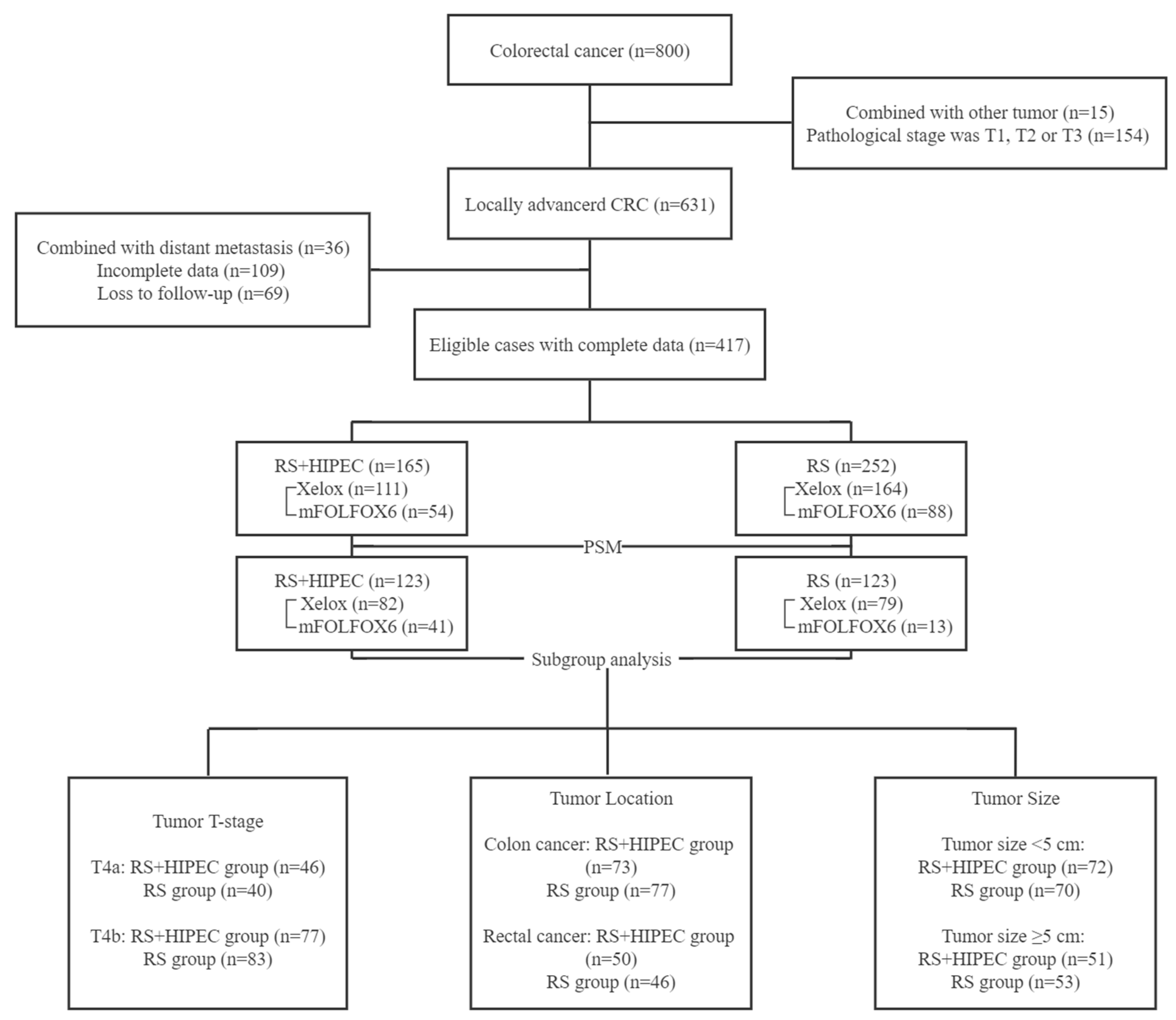
2.1. Study Cohort

We reviewed the hospital records of patients diagnosed with T4 CRC and treated with RS with or without HIPEC, from January 2019 to April 2020, at Wuhan Union Hospital, Tongji Medical College, Huazhong University of Science and Technology. Participants were included with the following criteria: (1) confirmation of colorectal adenocarcinoma by postoperative pathology (pT ≥ 4), (2) no distant metastasis or PC detected before the operation through computed tomography (CT) or magnetic resonance imaging (MRI), (3) adjuvant chemotherapy taken after discharge, and (4) a tumor location in patients with rectal cancer above the peritoneal reflection. Preoperative MRI and colonoscopy showed at least 7 cm from the lower border of the tumor to the anal verge (above the peritoneal reflection). The exclusion criteria included: (1) incomplete clinical and pathological data preoperatively, (2) detectable tumor metastasis found during the operation, (3) patients with a history of other malignancies within 5 years, and (4) emergency surgery due to perforation, ileus, etc. Institutional ethical approval [2021] LUNSHENGZI (0601) was received for the study.
The patients were classified into (a) the experimental group: RS + HIPEC, depending on whether they received HIPEC treatment, and (b) the control group: RS alone. This retrospective study was approved by the institutional review board, and patient information and privacy were closely protected.

2.2. Data Collection

Patient data were collected through the hospital’s database and included sex, age, height, weight, history, American Society of Anesthesiologists (ASA) classification, operation method, tumor location, maximum tumor diameter, tumor histological type, surgical margin, nerve invasion, vascular invasion, lymphatic metastasis, depth of invasion, length of postoperative hospital stay, and HIPEC-related information. Body mass index (BMI) and the Charlson comorbidity index (CCI) were used to evaluate height and weight and basic preoperative medical history, respectively. Additionally, lymph node metastasis and depth of invasion were evaluated using the American Joint Committee on Cancer TNM staging system.
The Common Terminology Criteria for Adverse Events version 5.0 was used to record adverse events of grade 2 and above that occurred postoperatively with or without HIPEC. The adverse events included anemia, hypoalbuminemia, myelosuppression, wound complications, abdominal infection, pulmonary infection, postoperative bleeding, gastroparesis, anastomotic leakage, ileus, electrolyte disorder (Supplementary Table S1).

2.3. Follow-Up

The latest time of follow-up was in June 2022. Patients were followed up every 3 months during the first 2 years after surgery. Follow-up data were collected from outpatient visits or re-examination results that met the follow-up requirements sent by patients through the network. Based on individual patient necessity, CT of lungs, abdomen, and pelvis, enhanced CT, MRI, or colonoscopy were performed during outpatient follow-up.
The treatment effectiveness was evaluated using: (1) treatment efficacy indicators, including tumor recurrence, metastasis, and death; (2) recurrence-free survival (RFS), which stood for the time from RS until the patient’s recurrence (local recurrence and peritoneal metastasis) or last contact, and the time point of progression was the date of the first detectable new lesions; (3) OS, which was the time from the initial RS to death due to any cause or the end of follow-up.

2.4. Treatment

None of the patients included in this study received neoadjuvant therapy or radiotherapy. All patients had been informed about the operation procedure and HIPEC therapy due to their cT4 preoperative staging and voluntarily signed an informed consent form for the procedure. HIPEC was conducted according to the formal standards and specifications of the clinical application of HIPEC in China, constituted by the Peritoneal Surface Oncology Committee of the China Anti-Cancer Association [13]. Clinicians performed radical resection of CRC (resection of the corresponding colon and rectum plus regional lymph node dissection), following the principles of mesorectal excision and tumor-free operation, and the surgical procedure was referred to the Chinese protocol of diagnosis and treatment of CRC (2020 edition) [2].
The HIPEC was implemented immediately after RS. For HIPEC, four catheters were placed in the abdominal cavity. Extracorporeal circulation pipes were connected to a HIPEC perfusion machine (BRTRG-II, Bright Medical Tech, Guangzhou, China) (Supplementary Figure S1). The perfusion flow rate was 400 mL/min, the perfusion bag was filled with 2 L/m2 normal saline, the perfusion time was 60 min, and the perfusion temperature was 43 ± 0.5 ℃. HIPEC antitumor drugs chosen were mitomycin, lobaplatin, raltitrexed, or oxaliplatin, and the dosage was based on the patient’s body surface area (refer to guidelines for systemic chemotherapy [2,10]) (Supplementary Table S2).
Postoperative adjuvant chemotherapy started 3–4 weeks after HIPEC. Patients with poor physical condition could extend it appropriately, but it should not exceed 8 weeks after operation at the latest. The chemotherapy scheme was determined by clinicians according to the pathological stage, molecular classification, and risk factors, referring to NCCN Guidelines (2018) [10]. The XELOX or mFOLFOX6 scheme was preferred.

2.5. Statistical Analysis

The reporting standard of CTCAE 5.0 was used to define the HIPEC-related adverse events. OS and RFS were compared between RS and RS + HIEPC groups. A separate analysis was performed for tumor T-stage, tumor location, and tumor size. IBM SPSS Statistics version 24.0 (IBM Corp., Armonk, NY, USA) was used for statistical processing, and GraphPad Prism version 8.0 (GraphPad Software, San Diego, CA, USA) was used for mapping. Categorical data are expressed as percentages, continuous data as mean or median, normally distributed data as mean ± standard deviation (SD), and survival data as a Kaplan–Meier curve. Categorical variables were analyzed by means of the Pearson chi-square test or Fisher exact test, and continuous variables were analyzed by means of Student’s t-test or Mann–Whitney U test. The survival data were reported using Kaplan–Meier estimates, and differences were determined by the log-rank test. Propensity score matching (PSM) was used to reduce the influence of biases and confounding variables to make a more reasonable comparison between the experimental and control groups (Supplementary Figure S2). A 1:1 PSM was conducted for 16 clinically relevant variables in Table 1. The matching quality was assessed using absolute standard differences, with a value < 5% considered insignificant. Statistical significance was set at two-sided p-values < 0.05.

3. Results

3.1. Baseline Clinicopathological Characteristics of Patients

The baseline clinicopathological characteristics of the patients are listed in Table 1. Using the inclusion and exclusion criteria, we identified 417 patients from 800, 165 managed with RS + HIPEC and 252 with RS alone (Figure 1). There were 263 men and 154 women, with a median age of 61 (24–86) years. After PSM, there were 246 patients in total, 150 men and 96 women, with a median age of 60 (24–86) years. Before PSM, there was no significant difference between the two groups with respect to sex (p = 0.208, χ2 = 1.584), CCI (p = 0.462, χ2 = 0.737), ASA (p = 0.494, χ2 = 0.685), tumor location (p = 0.247, χ2 = 2.800), tumor size (p = 0.177, Z = 1.353), tumor differentiation (p = 0.335, χ2 = 2.187), pN (p = 0.115, χ2 = 7.432), number of resected lymph nodes (p = 0.059, Z = 1.895), nerve invasion (p = 0.817, χ2 = 0.054), vascular invasion (p = 0.153, χ2 = 2.043), mismatch repair mutation (p = 0.897, χ2 = 0.017), and adjuvant chemotherapy (p = 0.256, χ2 = 2.722). However, age (p = 0.004, Z = −2.873), BMI (p = 0.007, Z = 2.725), surgical procedure (p < 0.001, χ2 = 20.609), pT (p < 0.001, χ2 = 20.531), and length of postoperative stay (p = 0.048, Z = −1.987) were significantly different between the RS + HIPEC and RS groups.
After PSM, there was no statistically significant difference in any of the baseline clinicopathological characteristics between the RS + HIPEC and RS groups. The baseline clinicopathological characteristics of the patients after PSM are listed in Table 2.

3.2. Adverse Events

The details and rates of adverse events globally or in the groups before and after PSM are listed in Table 3.
Before PSM, there was no significant difference between the RS + HIPEC and RS groups in the incidence of moderate or severe anemia (p = 0.968, χ2 = 0.002), hypoalbuminemia (p = 0.050, χ2 = 3.853), myelosuppression (p = 0.216, χ2 = 1.531), wound complications (p = 0.611, χ2 = 0.259), abdominal infection (p = 0.825, χ2 = 0.049), pulmonary infection (p = 0.764, χ2 = 0.090), postoperative bleeding (p = 0.668, χ2 = 0.184), anastomotic leakage (p = 0.753, χ2 = 0.009), ileus (p = 0.858, χ2 = 0.032), electrolyte disorder (p = 0.295, χ2 = 1.097), or abdominal discomfort (p = 0.321, χ2 = 0.983).
After PSM, there was no significant difference in the occurrence of any adverse events between the RS + HIPEC and RS groups.

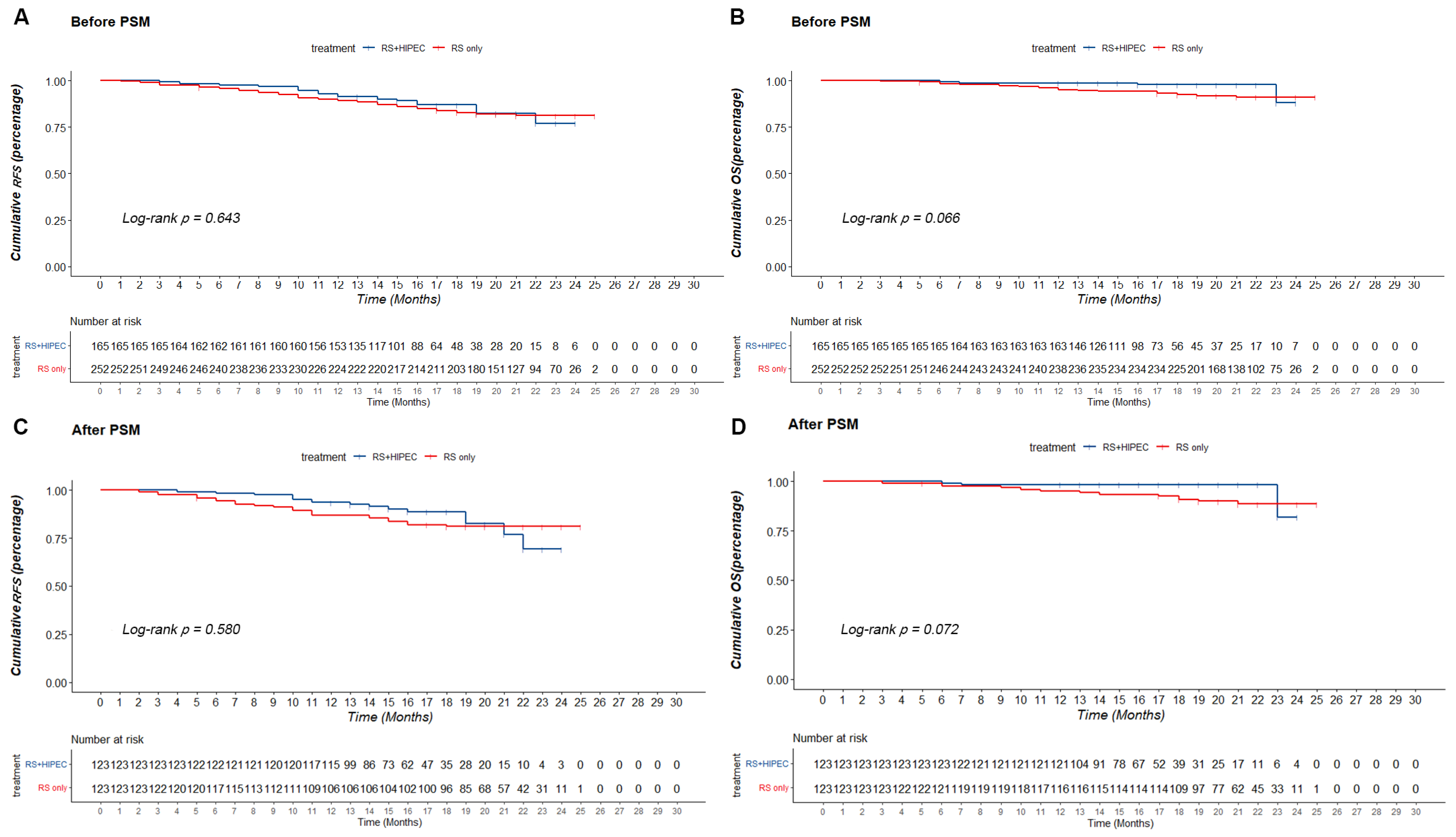
3.3. Comparison of Prognosis

Follow-up data from 417 patients were included in the analysis. Before PSM, the RS + HIPEC group had a lower median follow-up time than the RS group, with 77.0 vs. 81.2% RFS and 91.9 vs. 92.3% OS at 24 months, in the RS + HIPEC and RS groups, respectively. The corresponding cumulative RFS and OS on the Kaplan–Meier survival curve were not significantly different (p = 0.643, χ2 = 0.215 and p = 0.066, χ2 = 3.381, respectively) (Figure 2).
After PSM, the RS + HIPEC group had a lower median follow-up time than the RS group, with 16 (12–27) and 21 (3–25) months, respectively. On the Kaplan–Meier survival curve, the RFS of both groups at 24 months (73.4 vs. 81.1%) showed no significant difference (p = 0.580, χ2 = 0.442). However, the OS of both groups at 24 months (82.0 vs. 88.5%) was not significantly different (p = 0.072, χ2 = 3.242) (Figure 2).

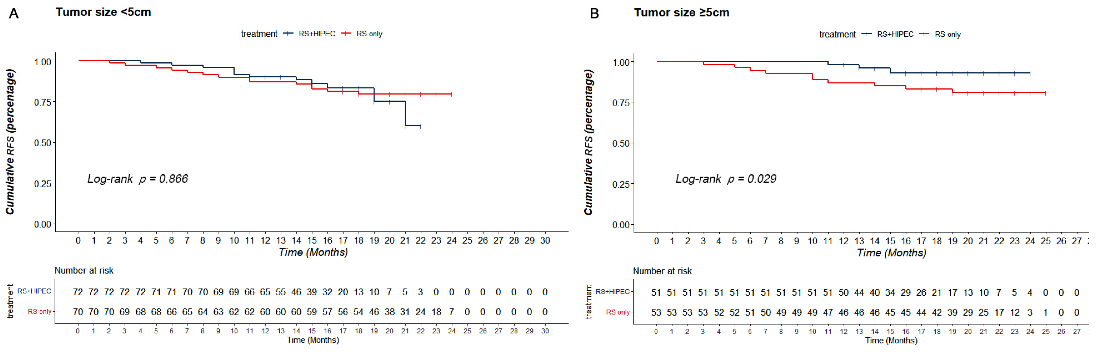
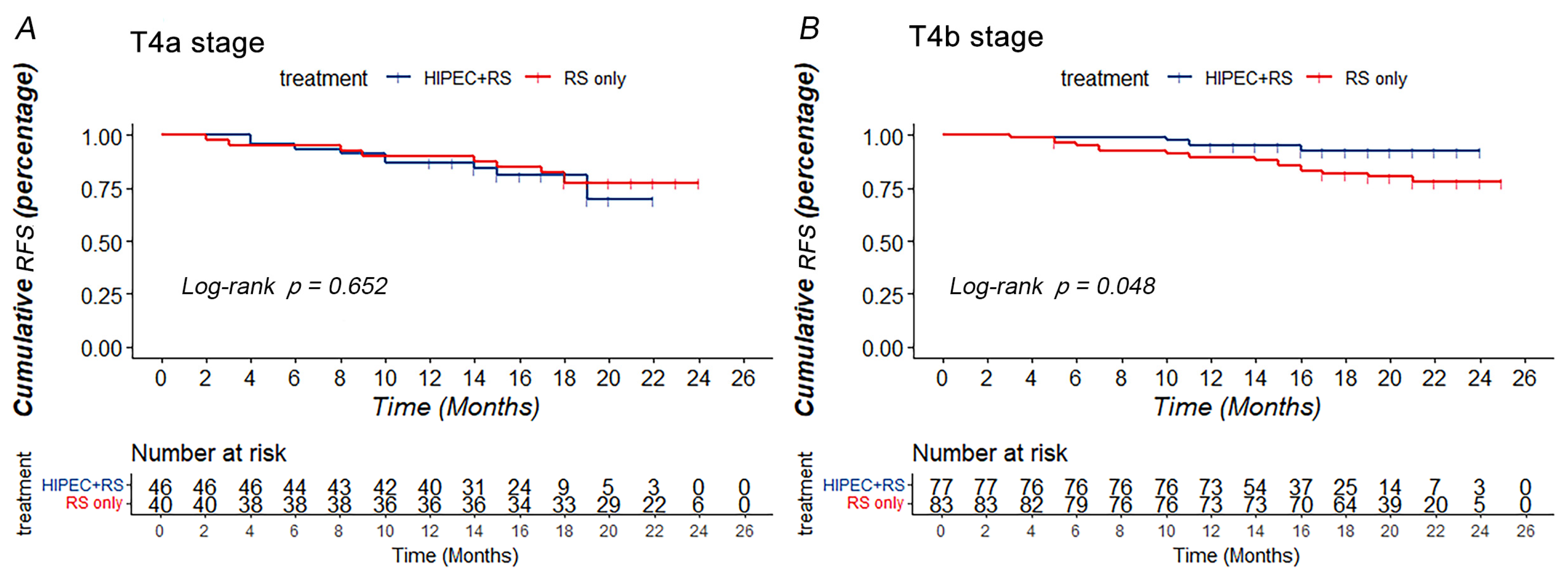
3.4. Stratified Analysis of Prognosis

Stratified analysis was used to further explore the factors influencing RFS and OS in patients with CRC. The patients were divided into T stage, tumor location, and size groups to determine the influence of these factors on effectiveness.
After PSM, when the patients had stage T4a tumors (n = 86), the RFS and OS of both groups (nRS + HIPEC = 46; nRS = 40) showed no significant difference (p = 0.652, χ2 = 0.203 and p = 0.282, χ2 = 1.156, respectively). For patients with stage T4b tumors (n = 160), the RS + HIPEC group (n = 77) had better cumulative RFS in 24 months (92.1 vs. 77.3%) than those in the RS group (n = 83) (p = 0.048, χ2 = 3.902) (Figure 3). However, the OS of both groups was not significantly different (p = 0.161, χ2 = 1.964) (Supplementary Figure S3).
After PSM, for patients with colon cancer (n = 150), the RFS and OS of the two groups (nRS + HIPEC = 73; nRS = 77) showed no significant differences (p = 1.000, χ2 < 0.001 and p = 0.177, χ2 = 1.820, respectively). In addition, for patients with rectal cancer (n = 96), the RFS and OS of both groups (nRS + HIPEC = 50; nRS = 46) were not significantly different (p = 0.419, χ2 = 0.654 and p = 0.223, χ2 = 1.482, respectively) (Supplementary Figure S4).
After PSM, with respect to the patients with tumor size < 5 cm (n = 142), the RFS and OS of both groups (nRS + HIPEC = 72; nRS = 70) showed no significant difference (p = 0.866, χ2 = 0.029 and p = 0.062, χ2 = 3.486, respectively). For patients with tumor size ≥ 5 cm (n = 104), the RS + HIPEC group (n = 51) had better cumulative RFS in 24 months (93.0 vs. 80.9%) than those in the RS group (n = 53) (p = 0.029, χ2 = 4.744) (Figure 4). However, the OS of both groups was not significantly different (p = 0.470, χ2 = 0.522) (Supplementary Figure S5).

4. Discussion

Several reports have confirmed that HIPEC can clear free cancer cell in the peritoneal cavity and can be used as an effective adjuvant treatment for preventing PC after surgery for advanced peritoneal tumors [14,15,16,17,18]. Several clinical studies have reported that cytoreductive surgery combined with HIPEC could significantly improve OS or RFS in CRC with PC [19,20,21]. Since there is high-level clinical evidence supporting the therapeutic role of HIPEC in peritoneal metastasis of CRC, HIPEC has become the recommended method for CRC with PC in many countries [8,9,10]. Nevertheless, the use of HIPEC in T4 CRC is controversial. Our study confirmed the safety of RS combined with HIPEC in treating T4 CRC and its short-term efficacy in patients with T4b stage CRC and tumor size ≥ 5 cm.
Regarding the adverse events occurring in RS + HIPEC in patients with PC, Verwaal et al. reported grade III–IV adverse reactions, including leukopenia (17%), gastrointestinal fistula (15%), hemorrhage (14%), and heart failure (12%) [20]. Goere et al. reported that grade III–IV postoperative complications occurred in 41% of patients who underwent second-look surgery and HIPEC [22]. Quenet et al. reported that at 60 days postoperatively, the grade III–V morbidity rate in the comparator arm was significantly higher than that in the HIPEC arm (24.1 vs. 13.6%, p = 0.030) [19]. Klaver et al. reported that HIPEC-related complications occurred in 14% of patients who underwent HIPEC [11]. HIPEC increased the risk of complications in patients with CRC, which was different from that in the control group, but it was within the acceptable range. In our study, the two most common complications were hypoalbuminemia (19.5%) and anemia (17.9%) in the RS + HIPEC group. After PSM, there was no significant difference in the statistics for all adverse events between two groups. Among these adverse events, the most concerning to clinicians is the occurrence of gastrointestinal anastomotic leakage after HIPEC treatment. Similarly, recent research has indicated that the occurrence of gastrointestinal anastomotic leakage has no clear relationship with HIPEC treatment. Additionally, HIPEC does not increase the risk of anastomotic leakage compared to conventional gastrointestinal surgery [7,11,23,24]. It is feasible and stable to carry out HIPEC by experienced medical and nursing staff following technical standards.
Since HIPEC was applied in patients with CRC, much research has focused on the effectiveness of HIPEC in patients with CRC. Our study showed that the RFS (73.4 vs. 81.1%, p = 0.580) and OS (82.0 vs. 88.5%, p = 0.072) between RS + HIPEC and RS groups on overall analysis did not show significant differences before and after PSM. The COLOPEC trial showed no difference in disease-free survival (DFS) at 18 months (69.0 vs. 69.3%; p = 0.99), OS at 18 months (93.0 vs. 94.1%; p = 0.82), or peritoneal metastases-free survival at 18 months (80.9 vs. 76.2%, p = 0.28) between the experimental and control groups [11]. The PROPHYLOCHIP trial showed no difference in the 5-year DFS (42 vs. 49%, p = 0.82) and 5-year OS (68 vs. 72%, p-value not reported) between the HIPEC and control groups after a median follow-up of 50.8 months [22,25]. Both RCTs investigated the preventive effect of HIPEC in patients with CRC with high risk factors for PC. The effectiveness of HIPEC in preventing PC in CRC has not been presented in terms of currently reported results. Nonetheless, results of HIPECT4 showed that RS + HIEPC for locally advanced colon cancer could improve the loco-regional control rate (35.3 ± 0.4 vs. 33.2 ± 0.8 months), with 3-year loco-regional control rates of 97% and 87% (p = 0.025) [12]. Meanwhile, our research showed that HIPEC could improve the RFS of T4 CRC patients with T4b stage CRC (92.1 vs. 77.3%, p = 0.048) and tumor size ≥ 5 cm at 24 months (93.0 vs. 80.9%, p = 0.029).
Patient selection is an important factor that influences the efficacy of HIPEC in preventing PC. For patients with T4 stage CRC, D. Hompes et al. showed that the incidence of PC was 13.2% at 3 years after RS in stage II-III CRC, including 20–36.7% in pT4 stage [6]. Even after RS and regular adjuvant chemotherapy, the 3-year DFS of patients with pT4 CRC is only 58–61%, which is far lower than that of patients with T1, T2, or T3 CRC [26]. A retrospective control study showed that among patients with T4 CRC with high risk factors of peritoneal metastasis (T3/4, mucinous adenocarcinoma/signet ring cell carcinoma), the risk of peritoneal metastasis in the treatment group receiving RS combined with HIPEC was 4%, while the risk in the control group without HIPEC was 28% [27]. It suggested that the use of HIPEC in selective population may help to reduce the risk of PC. Previous studies have shown that the T stage is an important factor that affects the prognosis of patients with locally advanced gastric cancer after surgery with prophylactic HIPEC [28]. However, few studies have reported the effect of T staging and tumor size on the application of HIPEC in T4 CRC. After a comprehensive follow-up, we confirmed the role of HIPEC in patients with T4b stage CRC and tumor size ≥ 5 cm. These patient characteristics can be a predictor of HIPEC effectiveness and define the possible window of opportunity for HIPEC to prevent colorectal PC.
This study had some limitations. First, it was retrospective, and its conclusions need to be verified by prospective cohort studies. Second, the follow-up period was not long enough to obtain a long-term prognosis. Finally, the shorter follow-up period may overstate the short-term prognosis, requiring further long-term follow-up and more subjects with specific stratification in the later stage.

5. Conclusions

Thus, the safety of HIPEC in patients with T4 CRC can be guaranteed. Furthermore, regarding prognosis, RS + HIPEC did not show the expected advantages in the overall cohort at 24 months, and a longer-term follow-up is required. However, patients with T4 CRC who had T4b stage CRC and tumor size ≥ 5 cm and underwent RS combined with HIPEC had better RFS than patients who underwent simple RS at 24 months.

Supplementary Materials

The following supporting information can be downloaded at: https://www.mdpi.com/article/10.3390/jcm12031145/s1, Table S1: Definition of CTCAE V5.0 adverse events; Table S2: HIPEC drug utilization; Figure S1: Perfusion equipment and pipe connection. (A) The BRTRG-I hyperthermic perfusion intraperitoneal treatment system (Bright Medical Tech, Guangzhou, China). (B) Pipe connection. The red tube is the inlet tube, and the blue tube is the outlet tube. The perfusion fluid enters the abdominal cavity from the red tube below and flows out through the blue tube above. (C) Four tubes were used for circulatory perfusion; two each were placed in the upper and lower abdomen; Figure S2: Propensity score matching (PSM). (A) Histogram of propensity score. (B) Distribution of propensity score; Figure S3: Subgroup analysis based on T stage. (A) OS of RS + HIPEC group and RS group after PSM when the tumor was at T4a stage. (B) OS of RS + HIPEC group and RS group after PSM when the tumor was at T4b stage; Figure S4: Subgroup analysis based on tumor location. (A) RFS of RS + HIPEC group and RS group after PSM when the tumor was at colon. (B) OS of RS + HIPEC group and RS group after PSM when the tumor was at colon. (C) R FS of RS + HIPEC group and RS group after PSM when the tumor was at rectum. (D) OS of RS + HIPEC group and RS group after PSM when the tumor was at rectum; Figure S5: Subgroup analysis based on tumor size. (A) OS of RS + HIPEC group and RS group after PSM when the tumor was <5cm. (B) OS of RS + HIPEC group and RS group after PSM when the tumor was ≥5cm.

Author Contributions

X.G.: Conceptualization, Validation, Formal analysis, Investigation, Data curation, Writing—Original Draft, Writing—Review and Editing. Y.L. (Yao Lin): Methodology, Formal analysis, Data curation, Writing—Review and Editing. C.S.: Formal analysis, Investigation. Y.L. (Yuan Li): Investigation, Resources. X.Z.: Investigation. J.L.: Investigation. F.X.: Investigation, Data curation. T.R.: Investigation, Data curation. C.W.: Conceptualization, Resources, Supervision, Project administration, Funding acquisition. K.T.: Resources, Supervision, Project administration, Funding acquisition. All authors have read and agreed to the published version of the manuscript.

Funding

This research was funded by the National Nature Science Foundation of China, grant number No.82073324.

Institutional Review Board Statement

The Ethics Committee of Tongji Medical College, Huazhong University of Science and Technology gave a final approval (No: UHCT-IEC-SOP-016-03-01) for this study.

Informed Consent Statement

The data were collected anonymized and centrally, and the requirement for written informed consent was therefore waived. The study was performed in accordance with the Declaration of Helsinki.

Data Availability Statement

The original anonymous dataset is available on request from the corresponding author.

Acknowledgments

All authors’ sincere appreciation also go to the teachers and students from the Department of Gastrointestinal Surgery, Union Hospital, Tongji Medical College, Huazhong University of Science and Technology, who participated in this study with great cooperation. Moreover, Xikai Guo would like to thank his girlfriend, QianYun Li, for her encouragement and support.

Conflicts of Interest

The authors declare no conflict of interest.

References

  1. Siegel, R.L.; Miller, K.D.; Fuchs, H.E.; Jemal, A. Cancer Statistics, 2021. CA Cancer J. Clin. 2021, 71, 7–33. [Google Scholar] [CrossRef]
  2. Hospital Authority of National Health Commission of the People’s Repuhlic of China; Chinese Society of Oncology CMA. Chinese protocol of diagnosis and treatment of colorectal cancer (2020 edition). Chin. J. Pract. Surg. 2020, 6, 601–625. [Google Scholar]
  3. Jayne, D.G.; Fook, S.; Loi, C.; Seow-Choen, F. Peritoneal carcinomatosis from colorectal cancer. Br. J. Surg. 2002, 89, 1545–1550. [Google Scholar] [CrossRef] [PubMed]
  4. Koppe, M.J.; Boerman, O.C.; Oyen, W.J.G.; Bleichrodt, R.P. Peritoneal Carcinomatosis of Colorectal Origin. Ann. Surg. 2006, 243, 212–222. [Google Scholar] [CrossRef]
  5. Kerscher, A.G.; Chua, T.C.; Gasser, M.; Maeder, U.; Kunzmann, V.; Isbert, C.; Germer, C.T.; Pelz, J.O.W. Impact of peritoneal carcinomatosis in the disease history of colorectal cancer management: A longitudinal experience of 2406 patients over two decades. Br. J. Cancer 2013, 108, 1432–1439. [Google Scholar] [CrossRef] [PubMed]
  6. Hompes, D.; Tiek, J.; Wolthuis, A.; Fieuws, S.; Penninckx, F.; Van Cutsem, E.; D’Hoore, A. HIPEC in T4a colon cancer: A defendable treatment to improve oncologic outcome? Ann. Oncol. 2012, 23, 3123–3129. [Google Scholar] [CrossRef]
  7. Auer, R.C.; Sivajohanathan, D.; Biagi, J.; Conner, J.; Kennedy, E.; May, T. Indications for hyperthermic intraperitoneal chemotherapy with cytoreductive surgery: A systematic review. Eur. J. Cancer 2020, 127, 76–95. [Google Scholar] [CrossRef]
  8. Esquivel, J.; Piso, P.; Verwaal, V.; Bachleitner-Hofmann, T.; Glehen, O.; González-Moreno, S.; Deraco, M.; Pelz, J.; Alexander, R.; Glockzin, G. American Society of peritoneal surface malignancies opinion statement on defining expectations from cytoreductive surgery and hyperthermic intraperitoneal chemotherapy in patients with colorectal cancer. J. Surg. Oncol. 2014, 110, 777–778. [Google Scholar] [CrossRef]
  9. O’Dwyer, S.; Verwaal, V.J.; Sugarbaker, P.H. Evolution of Treatments for Peritoneal Metastases from Colorectal Cancer. J. Clin. Oncol. 2015, 33, 2122–2123. [Google Scholar] [CrossRef]
  10. Benson, A.B.; Venook, A.P.; Al-Hawary, M.M.; Cederquist, L.; Chen, Y.J.; Ciombor, K.K.; Cohen, S.; Cooper, H.S.; Deming, D.; Engstrom, P.F.; et al. NCCN Guidelines Insights: Colon Cancer, Version 2.2018. J. Natl. Compr. Cancer Netw. 2018, 16, 359–369. [Google Scholar] [CrossRef]
  11. Klaver, C.E.; Wisselink, D.D.; Punt, C.J.; Snaebjornsson, P.; Crezee, J.; Aalbers, A.G.; Brandt, A.; Bremers, A.J.; Burger, J.W.; Fabry, H.F.; et al. Adjuvant hyperthermic intraperitoneal chemotherapy in patients with locally advanced colon cancer (COLOPEC): A multicentre, open-label, randomised trial. Lancet Gastroenterol. Hepatol. 2019, 4, 761–770. [Google Scholar] [CrossRef] [PubMed]
  12. Arjona-Sanchez, A.; Cano-Osuna, M.T.; Gutierrez, A.; Segura, J.J.; Perez, E.; Concepcion, V.; Sanchez, S.; Garcia, A.; Prieto, I.; Sanchez, P.B.; et al. Adjuvant hyperthermic intraperitoneal chemotherapy in locally advanced colon cancer (HIPECT4): A randomized phase III study. Ann. Oncol. 2022, 33 (Suppl. S7), S680. [Google Scholar] [CrossRef]
  13. Peritoneal Surface Oncology Committee of China Anti-Cancer Association. Chinese Expert Cnsensus on the Clinical Application of China Hyperthermic Intraperitoneal Chemotherapy. Natl. Med. J. China 2020, 100, 89–96. [Google Scholar]
  14. Klaver, C.E.; Musters, G.D.; Bemelman, W.A.; Punt, C.J.; Verwaal, V.J.; Dijkgraaf, M.G.; Aalbers, A.G.; van der Bilt, J.D.; Boerma, D.; Bremers, A.J.; et al. Adjuvant hyperthermic intraperitoneal chemotherapy (HIPEC) in patients with colon cancer at high risk of peritoneal carcinomatosis; the COLOPEC randomized multicentre trial. BMC Cancer 2015, 15, 1–9. [Google Scholar] [CrossRef] [PubMed]
  15. Baratti, D.; Kusamura, S.; Iusco, D.; Gimondi, S.; Pietrantonio, F.; Milione, M.; Guaglio, M.; Bonomi, S.; Grassi, A.; Virzì, S.; et al. Hyperthermic Intraperitoneal Chemotherapy (HIPEC) at the Time of Primary Curative Surgery in Patients with Colorectal Cancer at High Risk for Metachronous Peritoneal Metastases. Ann. Surg. Oncol. 2016, 24, 167–175. [Google Scholar] [CrossRef] [PubMed]
  16. Narasimhan, V.; Britto, M.; Pham, T.; Warrier, S.; Naik, A.; Lynch, A.C.; Michael, M.; Tie, J.; Ramsay, R.; Heriot, A. Evolution of Cytoreductive Surgery and Hyperthermic Intraperitoneal Chemotherapy for Colorectal Peritoneal Metastases: 8-Year Single-Institutional Experience. Dis. Colon Rectum 2019, 62, 1195–1203. [Google Scholar] [CrossRef]
  17. Lei, Z.Y.; Guan, T.P.; Luo, J.L.; Tang, H.S.; Cui, S.Z. Rationality of performing hyperthermic intraperitoneal chemotherapy 5-8 weeks after primary tumor resection for patients with locally advanced colorectal cancer—Based on COLOPEC. Chin. J. Gastrointest. Surg. 2019, 22, 1115–1117. [Google Scholar]
  18. Birgisson, H.; Enblad, M.; Artursson, S.; Ghanipour, L.; Cashin, P.; Graf, W. Patients with colorectal peritoneal metastases and high peritoneal cancer index may benefit from cytoreductive surgery and hyperthermic intraperitoneal chemotherapy. Eur. J. Surg. Oncol. 2020, 46, 2283–2291. [Google Scholar] [CrossRef]
  19. Quenet, F.; Elias, D.; Roca, L.; Goere, D.; Ghouti, L.; Pocard, M.; Facy, O.; Arvieux, C.; Lorimier, G.; Pezet, D.; et al. A UNICANCER phase III trial of hyperthermic intra-peritoneal chemotherapy (HIPEC) for colorectal peritoneal carcinomatosis (PC): PRODIGE 7. J. Clin. Oncol. 2018, 36, 3503. [Google Scholar] [CrossRef]
  20. Verwaal, V.J.; van Ruth, S.; de Bree, E.; van Slooten, G.W.; van Tinteren, H.; Boot, H.; Zoetmulder, F.A. Randomized Trial of Cytoreduction and Hyperthermic Intraperitoneal Chemotherapy Versus Systemic Chemotherapy and Palliative Surgery in Patients with Peritoneal Carcinomatosis of Colorectal Cancer. J. Clin. Oncol. 2003, 21, 3737–3743. [Google Scholar] [CrossRef]
  21. Verwaal, V.J.; Bruin, S.; Boot, H.; van Slooten, G.; van Tinteren, H. 8-Year Follow-up of Randomized Trial: Cytoreduction and Hyperthermic Intraperitoneal Chemotherapy Versus Systemic Chemotherapy in Patients with Peritoneal Carcinomatosis of Colorectal Cancer. Ann. Surg. Oncol. 2008, 15, 2426–2432. [Google Scholar] [CrossRef] [PubMed]
  22. Goere, D.; Glehen, O.; Quenet, F.; Ducreux, M.; Guilloit, J.M.; Texier, M.; Benhamou, E.; Elias, D.; BIG-RENAPE and PRODIGE. Results of a randomized phase 3 study evaluating the potential benefit of a secondlook surgery plus HIPEC in patients at high risk of developing colorectal peritoneal metastases (PROPHYLOCHIP- NTC01226394). J. Clin. Oncol. Conf. 2018, 36 (Suppl. S1), 3531. [Google Scholar] [CrossRef]
  23. Bonnot, P.E.; Piessen, G.; Kepenekian, V.; Decullier, E.; Pocard, M.; Meunier, B.; Bereder, J.M.; Abboud, K.; Marchal, F.; Quenet, F.; et al. Cytoreductive Surgery with or Without Hyperthermic Intraperitoneal Chemotherapy for Gastric Cancer With Peritoneal Metastases (CYTO-CHIP study): A Propensity Score Analysis. J. Clin. Oncol. 2019, 37, 2028–2040. [Google Scholar] [CrossRef] [PubMed]
  24. Baratti, D.; Kusamura, S.; Azmi, N.; Guaglio, M.; Montenovo, M.; Deraco, M. Colorectal Peritoneal Metastases Treated by Perioperative Systemic Chemotherapy and Cytoreductive Surgery with or Without Mitomycin C-Based HIPEC: A Comparative Study Using the Peritoneal Surface Disease Severity Score (PSDSS). Ann. Surg. Oncol. 2019, 27, 98–106. [Google Scholar] [CrossRef]
  25. Goéré, D.; Glehen, O.; Quenet, F.; Guilloit, J.M.; Bereder, J.M.; Lorimier, G.; Thibaudeau, E.; Ghouti, L.; Pinto, A.; Tuech, J.J.; et al. Second-look surgery plus hyperthermic intraperitoneal chemotherapy versus surveillance in patients at high risk of developing colorectal peritoneal metastases (PROPHYLOCHIP–PRODIGE 15): A randomised, phase 3 study. Lancet Oncol. 2020, 21, 1147–1154. [Google Scholar] [CrossRef]
  26. Grothey, A.; Sobrero, A.F.; Shields, A.F.; Yoshino, T.; Paul, J.; Taieb, J.; Souglakos, J.; Shi, Q.; Kerr, R.; Labianca, R.; et al. Duration of Adjuvant Chemotherapy for Stage III Colon Cancer. New Engl. J. Med. 2018, 378, 1177–1188. [Google Scholar] [CrossRef]
  27. Sammartino, P.; Sibio, S.; Biacchi, D.; Cardi, M.; Mingazzini, P.; Rosati, M.S.; Cornali, T.; Sollazzo, B.; Atta, J.M.; Di Giorgio, A. Long-term results after proactive management for locoregional control in patients with colonic cancer at high risk of peritoneal metastases. Int. J. Color. Dis. 2014, 29, 1081–1089. [Google Scholar] [CrossRef]
  28. Fan, B.; Bu, Z.; Zhang, J.; Zong, X.; Ji, X.; Fu, T.; Jia, Z.; Zhang, Y.; Wu, X. Phase II trial of prophylactic hyperthermic intraperitoneal chemotherapy in patients with locally advanced gastric cancer after curative surgery. BMC Cancer 2021, 21, 216. [Google Scholar] [CrossRef]
Figure 1. Flow chart. A total of 417 out of 800 T4 CRC patients were included in the study. Inclusion criteria: (1) confirmation of colorectal adenocarcinoma by postoperative pathology (pT ≥ 4), (2) no distant metastasis or PC detected before the operation through computed tomography (CT) or magnetic resonance imaging (MRI), (3) adjuvant chemotherapy taken after discharge, and (4) a tumor location in patients with rectal cancer above the peritoneal reflection. Preoperative MRI or colonoscopy showed at least 7 cm from the lower border of the tumor to the anal verge (above the peritoneal reflection). The exclusion criteria included: (1) incomplete clinical and pathological data preoperatively, (2) tumor metastasis found during the operation, (3) patients with a history of other malignancies within 5 years, and (4) emergency surgery due to perforation, ileus, etc.
Jcm 12 01145 g001
Figure 2. Kaplan–Meier method was used to compare recurrence-free survival (RFS) and overall survival (OS) between RS + HIPEC group and RS group. (A) RFS of RS + HIPEC group and RS group before PSM. (B) OS of RS + HIPEC group and RS group before PSM. (C) RFS of RS + HIPEC group and RS group after PSM. (D) OS of RS + HIPEC group and RS group after PSM.
Jcm 12 01145 g002
Figure 3. Subgroup analysis based on T stage. (A) RFS of RS + HIPEC group and RS group after PSM when the tumor was at T4a stage. (B) RFS of RS + HIPEC group and RS group after PSM when the tumor was at T4b stage.
Jcm 12 01145 g003
Figure 4. Subgroup analysis based on tumor size. (A) RFS of RS + HIPEC group and RS group after PSM when the tumor was <5 cm. (B) RFS of RS + HIPEC group and RS group after PSM when the tumor was ≥5 cm.
Jcm 12 01145 g004
Table 1. Baseline clinicopathological characteristics of patients with T4 colorectal cancer before PSM.
CharacteristicNo (%)χ2/Zp-Value
Overall
(n = 417)
RS + HIPEC
(n = 165)
RS Alone
(n = 252)
Age, years
Mean ± SD59.78 ± 12.31957.66 ± 12.30161.17 ± 12.156−2.8730.004
≤60204 (48.9)86 (52.1)118 (46.8)
>60213 (51.1)79 (47.9)134 (53.2)
Sex 1.5840.208
Female154 (36.9)67 (40.6)87 (34.5)
Male263 (63.1)98 (59.4)165 (65.5)
BMI
Mean ± SD22.74 ± 3.49223.31 ± 3.31822.37 ± 3.5592.7250.007
Charlson Comorbidity Index 0.7370.462
0307 (73.6)119 (72.1)188 (74.6)
177 (18.5)32 (19.4)45 (17.9)
221 (5.0)8 (4.8)13 (5.2)
≥312 (2.9)6 (3.6)6 (2.4)
ASA Score 0.6850.494
189 (21.3)29 (17.6)60 (23.8)
2303 (72.7)128 (77.6)175 (69.4)
≥325 (6.0)8 (4.8)17 (6.8)
Surgical Procedures 20.609<0.001
laparoscopy371 (89.0)161 (97.6)210 (83.3)
laparotomy46 (11.0)4 (2.4)42 (16.7)
Tumor Location 2.8000.247
right semicolon105 (25.2)45 (27.3)60 (23.8)
left semicolon137 (32.9)59 (35.8)78 (31.0)
rectum175 (42.0)61 (37.0)114 (45.2)
Tumor Size, cm
Mean ± SD4.764 ± 2.2194.945 ± 2.5104.645 ± 2.0021.3530.177
<5248 (59.5)93 (56.4)155 (61.5)
≥5169 (40.5)72 (43.6)97 (38.5)
Tumor differentiation 2.1870.335
poor or undifferentiation29 (7.0)11 (6.7)18 (7.1)
Well or moderately388 (93.0)154 (93.3)234 (92.9)
pT status 17.490<0.001
pT4a217 (52.0)65 (39.4)152 (60.3)
pT4b200 (48.0)100 (60.6)100 (39.7)
No. of resected lymph nodes 1.8950.059
Mean ± SD20.32 ± 8.85321.33 ± 7.49319.66 ± 9.597
pN status 7.4320.115
pN0234 (56.1)89 (53.9)145 (57.5)
pN1a50 (12.0)19 (11.5)31 (12.3)
pN1b58 (13.9)32 (19.4)26 (10.3)
pN2a38 (9.1)13 (7.9)25 (9.9)
pN2b37 (8.9)12 (7.3)25 (9.9)
nerve invasion 0.0540.817
No260 (62.4)104 (63.0)156 (61.9)
Yes157 (37.6)61 (37.0)96 (38.1)
vascular invasion 2.0430.153
No292 (70.0)109 (66.1)183 (72.6)
Yes125 (30.0)56 (33.9)69 (27.4)
MMR positive 0.0170.897
No390 (93.5)154 (93.3)236 (93.7)
Yes27 (6.5)11 (6.7)16 (6.3)
Post-surgery stay time
Mean ± SD12.20 ± 6.76911.39 ± 3.85212.73 ± 8.095−1.9870.048
Adjuvant chemotherapy 2.7220.256
XELOX275 (65.9)111 (67.3)164 (65.1)
mFOLFOX6142 (34.1)54 (32.7)88 (34.9)
Follow-up time, months
Mean ± SD18.95 ± 3.85716.66 ± 3.44920.45 ± 3.345
Median (range)20 (3–27)16 (12–27)21 (3–25)
PSM: propensity score matching; SD: standard deviation; BMI: body mass index; ASA: American Society of Anesthesiologists; MMR: mismatch repair mutation.
Table 2. Baseline clinicopathological characteristics of patients with T4 colorectal cancer after PSM.
CharacteristicNo (%)χ2/Zp-Value
Overall
(n = 246)
RS + HIPEC
(n = 123)
RS Alone
(n = 123)
Age, years
Mean ± SD59.03 ± 12.23858.49 ± 11.55459.58 ± 12.910−0.6970.486
≤60125 (50.8)60 (48.8)65 (52.8)
>60121 (49.2)63 (51.2)58 (47.2)
Sex 0.2730.601
Female96 (39.0)50 (40.7)46 (37.4)
Male150 (61.0)73 (59.3)77 (62.6)
BMI
Mean ± SD22.875 ± 3.43422.839 ± 3.15822.912 ± 3.702−0.1680.867
Charlson Comorbidity Index 1.2870.864
0182 (74.0)91 (74.0)91 (74.0)
146 (18.7)22 (17.9)24 (19.5)
212 (4.9)6 (4.9)6 (4.9)
≥36 (2.4)4 (3.2)2 (1.6)
ASA Score 1.0920.779
146 (18.7)24 (19.5)22 (17.9)
2185 (75.2)92 (74.8)93 (75.6)
315 (6.1)7 (5.7)8 (6.5)
Surgical Procedures 0.1470.701
laparoscopy239 (97.2)119 (96.7)120 (97.6)
laparotomy7 (2.8)4 (3.3)3 (2.4)
Tumor Location 0.3670.836
right semicolon66 (26.8)33 (26.8)33 (26.8)
left semicolon84 (34.1)40 (32.5)44 (35.8)
rectum96 (39.0)50 (40.7)46 (37.4)
Tumor Size, cm
Mean ± SD4.797 ± 2.3494.74 ± 2.3934.85 ± 2.313−0.3660.715
<5142 (57.7)72 (58.5)70 (56.9)
≥5104 (42.3)51 (41.5)53 (43.1)
Tumor differentiation 0.1190.942
poor or undifferentiation19 (7.7)10 (8.1)9 (7.3)
Well or moderately227 (92.3)113 (91.9)114 (92.7)
pT status 0.6440.422
pT4a86 (35.0)46 (37.4)40 (32.5)
pT4b160 (65.0)77 (62.6)83 (67.5)
No. of resected lymph nodes −0.1720.863
Mean ± SD21.08 ± 9.96920.97 ± 7.14821.19 ± 12.185
pN status 1.9760.740
pN0138 (56.1)66 (53.7)72 (58.5)
pN1a31 (12.6)14 (11.4)17 (13.8)
pN1b37 (15.0)22 (17.9)15 (12.2)
pN2a21 (8.5)11 (8.9)10 (8.1)
pN2b19 (7.7)10 (8.1)9 (7.3)
nerve invasion 1.8290.176
No164 (66.7)77 (62.6)87 (70.7)
Yes82 (33.3)46 (37.4)36 (29.3)
vascular invasion 0.0720.788
No162 (65.9)80 (65.0)82 (66.7)
Yes84 (34.1)43 (35.0)41 (33.3)
MMR positive 0.0001.000
No230 (93.5)115 (93.5)115 (93.5)
Yes16 (6.5)8 (6.5)8 (6.5)
Post-surgery stay time
Mean ± SD11.75 ± 7.84210.84 ± 3.37912.66 ± 10.507−1.8300.068
Adjuvant chemotherapy 2.8840.236
XELOX161 (65.4)82 (66.7)79 (64.2)
mFOLFOX685 (34.6)41 (33.3)44 (35.8)
Follow-up time, months
Mean ± SD18.32 ± 3.92516.37 ± 3.43120.28 ± 3.383
Median (range)19 (3–27)16 (12–27)21 (3–25)
PSM: propensity score matching; SD: standard deviation; BMI: body mass index; ASA: American Society of Anesthesiologists; MMR: mismatch repair mutation.
Table 3. Adverse events of patients with T4 colorectal cancer before and after PSM.
Adverse EventBefore PSMAfter PSM
No (%)χ2p-ValueNo (%)χ2p-Value
Overall
(n = 417)
RS + HIPEC
(n = 165)
RS Alone
(n = 232)
Overall
(n = 246)
RS + HIPEC
(n = 123)
RS Alone
(n = 123)
Anemia83 (19.9)33 (20.0)50 (19.8)0.0020.96846 (18.7)22 (17.9)24 (19.5)0.1070.744
Hypoalbuminemia113 (27.1)36 (21.8)77 (30.6)3.8530.05059 (24.0)24 (19.5)35 (28.5)2.6980.100
Myelosuppression1 (0.2)1 (0.6)0 (0.0)1.5310.2161 (0.4)1 (0.8)0 (0.0)1.0040.316
Wound complications20 (4.8)9 (5.5)11 (4.4)0.2590.6119 (3.7)4 (3.3)5 (4.1)0.1150.734
Abdomen infection3 (0.7)1 (0.6)2 (0.8)0.0490.8251 (0.4)0 (0.0)1 (0.8)1.0040.316
Pulmonary infection14 (3.4)5 (3.0)9 (3.6)0.0900.76411 (4.5)5 (4.1)6 (4.9)0.0950.758
Postoperative bleeding4 (1.0)2 (1.2)2 (0.8)0.1840.6682 (0.8)1 (0.8)1 (0.8)0.0001.000
Anastomotic leakage6 (1.4)2 (1.2)4 (1.6)0.0090.7534 (1.6)1 (0.8)3 (2.4)1.0170.313
Ileus7 (1.7)3 (1.8)4 (1.6)0.0320.8587 (2.8)3 (2.4)4 (3.3)0.1470.701
Electrolyte disturbance12 (2.9)3 (1.8)9 (3.6)1.0970.2956 (2.4)2 (1.6)4 (3.3)0.6830.408
Abdomen discomfort9 (2.2)5 (3.0)4 (1.6)0.9830.3215 (2.0)2 (1.6)3 (2.4)0.2040.651
PSM: propensity score matching.
Disclaimer/Publisher’s Note: The statements, opinions and data contained in all publications are solely those of the individual author(s) and contributor(s) and not of MDPI and/or the editor(s). MDPI and/or the editor(s) disclaim responsibility for any injury to people or property resulting from any ideas, methods, instructions or products referred to in the content.

