Article

Podocyte Infolding Glomerulopathy: A Case Series Report and Literature Review

1
Department of Nephrology, Sichuan Provincial People’s Hospital, University of Electronic Science and Technology of China, Chengdu 610072, China
2
Chinese Academy of Sciences Sichuan Translational Medicine Research Hospital, Chengdu 610072, China
3
The Renal and Metabolic Division, George Institute for Global Health, University of New South Wales, Sydney 2042, Australia
4
Nephrology Department, Chengdu First People’s Hospital, Chengdu 610021, China
5
Concord Clinical School, University of Sydney, Sydney 2006, Australia
6
The Faculty of Medicine and Health Sciences, Macquarie University, Sydney 2109, Australia
*
Author to whom correspondence should be addressed.
These authors contributed equally to this work.
J. Clin. Med. 2023, 12(3), 1088; https://doi.org/10.3390/jcm12031088
Submission received: 3 January 2023 / Revised: 19 January 2023 / Accepted: 28 January 2023 / Published: 30 January 2023
(This article belongs to the Special Issue Clinical Advances in Metabolic Syndrome and Kidney Disease)

Abstract

:
Background: Podocyte infolding glomerulopathy (PIG) is a peculiar and very rare manifestation in renal pathology. Its underlying pathogenesis mechanism and clinical characteristics remain unclear due to sparse reports. Objective: To further elucidate the clinical profile of PIG by carefully reporting our four cases and a comprehensive review of cases in the literature. Methods: This study retrospectively reviewed four cases of PIG from 2010 to 2022 in our centre. Clinical and pathological profiles were reported. PIG cases in the literature were searched in the MEDLINE database and analysed together with our cases. Results: Four cases of PIG identified from our centre and 40 cases from the current literature were reported. The pooled analysis of these 44 cases indicated 79.5% (35/44) were females, 93.2% (41/44) were East Asians, and 63.6% (28/44) were reported in Japan. The average age was 42.0 ± 12.5 years old. The average amount of proteinuria at the time of renal biopsy was 3.06 ± 3.2 g/day. The most reported comorbidities were connective tissue diseases, mainly systemic lupus erythematosus, and 20.5% (9/44) of the cases did not have any contaminant disease. Most of the cases (81.8%, 36/44) had been treated with immunosuppressants, of which a combination of corticosteroids and one other type of immunosuppressant was most commonly reported. In addition, 45.4% (20/44) and 34.1% (15/44) of the cases had achieved complete response and partial response, respectively, after treatment. Whole exosome sequencing indicated mutations in the INF2 gene. Conclusions: PIG is a rare condition and seen in relatively younger populations, often associated with connective tissue diseases clinically and one or two other glomerulopathies histologically. The outcomes following immunosuppressive treatment are relatively good. Mutations in INF2 might be involved in the development of PIG; however, the implications of these results need to be investigated.

1. Introduction

Podocyte infolding glomerulopathy (PIG) is a very rare phenomenon that was first reported as a unique pathological manifestation in 1992 [1] in Japan and officially proposed as a new disease entity in 2008 by the Japanese Society of Nephrology in 2008 [2]. PIG is characterized by peculiar microstructures of microspheres, microtubules, or both within the thickened glomerular basement membrane (GBM), accompanied by podocyte infolding [2]. It is not included as a separate category in the current classification of glomerular diseases. Thus far, most reported cases have been in East Asians.
The underlying pathogenesis mechanism of PIG remains unclear. Successive pathology sections analysis indicated that PIG might be a continuous process of podocyte processes from primary infolding to bubbling [2]. The initiation factor of the primary infolding is not clear. The only gene reported to be involved in PIG so far is SMARCAL1, detected by whole exosome sequencing in a child with Schimke immune-osseous dysplasia (SIOD) [3]. However, the contributions of these mutations to the development of PIG were unclear.
The clinical features are highly diversified in PIG. It has been reported that PIG is associated with other diseases, mostly connective tissue diseases, including systemic lupus erythematosus (SLE) and Sjogren Syndrome (SS) [4,5,6,7]. However, there are also cases of PIG that were not associated with any disease entity [2,8,9]. Whether PIG is a new disease entity or just a specific renal pathological finding of coexisting diagnoses remain controversial.
To improve our understating of PIG, we reported here the largest PIG case series study from a single center thus far and comprehensively reviewed the reported cases in the literature alongside our case series.

2. Case Presentation
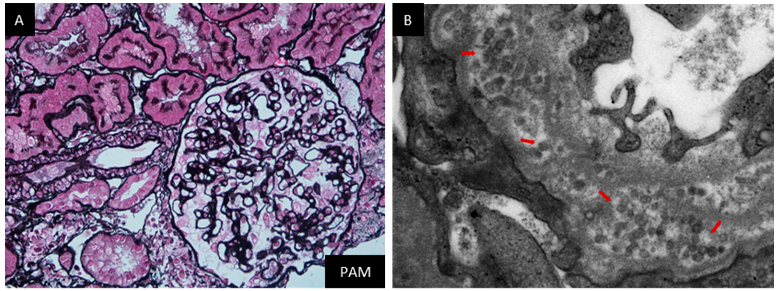
2.1. Case 1

This patient was a 26-year-old lady, without a known history, who presented with intermittent edema in both lower extremities for one month and denied any accompanying symptoms. Physical examination on admission indicated no remarkable findings except for mild pitting edema in both legs. Remarkable laboratory investigations at this admission were as follows: WBC 12.60 × 10⁹/L, HGB 123 g/L, PLT 161 × 109/L, proteinuria 2.74 g/day, hematuria (−), serum creatinine (Cr) 56 μmol/L, complement C3 0.96 g/L, and total cholesterol (TC) 5.14 mmol/L. All autoimmune antibodies tested negative. A renal biopsy was done on the third day after admission. Immunofluorescence (IF) staining was positive for IgG and IgM in the capillary walls and mesangial regions (Figure 1A,B) and negative for subtypes of IgG and PLA2R antibodies. Light microscopy found global glomerulosclerosis in 2/15 glomeruli, segmental sclerosis with synechial attachment to Bowman’s capsule in 7/15 glomeruli, mild mesangial matrix expansion, and diffuse thickening of glomerular basement membrane (GBM) (Figure 1C). Marked vacuolar degeneration and granular degeneration in renal tubular epithelial cells and mild interstitial fibrosis and tubular atrophy (<5%) were also noticed. Electron microscopy found thickening of the GBM, diffuse podocyte foot process effacement, and prominent microspheres forming clusters within the GBM (Figure 1D). She was diagnosed with focal segmental glomerular sclerosis (FSGS), membranous nephropathy, and PIG. Corticosteroid therapy was prescribed, starting with 50 mg of prednisone daily. Her proteinuria began to decrease after 2 months and remained around 1 g/day. The prednisone was tapered gradually from the third month and stopped at the sixth month. Eighty milligrams of Valsartan daily was prescribed afterward. Her proteinuria remained in the range of 0.7–1.2 g/day, and her serum Cr remained normal.

2.2. Case 2

This patient was a 47-year-old gentleman, without a known history, who presented with edema in both lower extremities for more than 10 days. He complained of progressive edema starting from both ankles to his knees. Foamy urine was also noticed. At admission, physical examination only found pitting edema in both legs. Remarkable lab investigations at this admission were as follows: WBC 9.25 × 10⁹/L, HGB 163 g/L, PLT 280 × 109/L, proteinuria 9.96 g/day, hematuria (−), urine albumin-to-creatinine 5277.5 μg/mg, Cr 94.4 μmol/L, C3 0.95 g/L, and TC 6.01 mmol/L. All serum autoimmune antibodies tested negative. The result of the anti-PLA2R antibody was also negative. A renal biopsy was done on the third day after admission. IF staining was positive for IgG in the capillary walls (Figure 2A) and negative for IgM, IgA, subtypes of IgG, and PLA2R. Light microscopy found segmental sclerosis with podocyte dysplasia in 3/14 glomeruli, marked hyperplasia of overlying epithelial cells in 2/14 glomeruli, and diffuse thickening of GBM (Figure 2B). Mild interstitial fibrosis and tubular atrophy (<5%) was also noticed. Electron microscopy found diffuse podocyte foot process effacement, multiple podocyte infolding, and prominent microspheres forming clusters within the GBM (Figure 2C). The diagnosis was FSGS and PIG. His treatment included 30 mg of prednisone daily and 1 mg of tacrolimus twice daily. The proteinuria began to decrease after two months and then remained in the range of 1.2–1.9 g/day. The prednisone was tapered gradually from the third month and stopped at the end of the seventh month. Tacrolimus still continued at the time of this report. His serum Cr was normal throughout the follow-up period.

2.3. Case 3

This patient was a 48-year-old lady with a history of SLE for 20 years who presented with edema in both lower extremities for one week. After the diagnosis of SLE, she had been initially treated with prednisone and mycophenolate mofetil (MMF) for five years, which was then changed to prednisone and tacrolimus. She claimed having a renal biopsy 10 years ago, but the detailed results were unavailable. At this admission, she was receiving 10 mg of prednisone daily, 1.0 mg of tacrolimus twice daily, 100 mg of hydroxychloroquine, and 80 mg of valsartan daily. Physical examination indicated mild edema in both lower extremities and old-standing butterfly erythematosus on both cheeks. Remarkable laboratory investigations at this admission were as follows: WBC 2.31 × 10⁹/L, HGB 105 g/L, PLT 119 × 109/L, proteinuria 4.37 g/day, hematuria (−), urine albumin-to-creatinine 5967.6 μg/mg, Cr 59.3 μmol/L, C3 0.727 g/L, ANA 1:100 (+), anti-SSA-Ab (+++), and anti-Ro52-Ab (+++). A repeat renal biopsy was done on the fourth day after admission. IF staining was positive for IgA and IgM in the mesangial regions (Figure 3A,B) and PLA2R in the capillary walls and negative for IgG and subclass IgG. Light microscopy found global glomerulosclerosis in 6/20 glomeruli, segmental sclerosis with synechial attachment to Bowman’s capsule in 2/20 glomeruli, mild mesangial matrix expansion, and diffuse thickening of GBM (Figure 3C). Focal interstitial fibrosis and tubular atrophy (about 15%) was also noticed. Electron microscopy found diffuse podocyte foot process effacement and prominent microspheres within the GBM (Figure 3D). The diagnosis was membranous nephropathy and PIG. The patient was then treated with 360 mg of belimumab once a month based on her existing combination therapy of prednisone and tacrolimus. Her proteinuria began to decrease from 5.43 g/day at discharge to 0.91 g/day after one month and remained in the range of 0.9–1.4 g/day for eight months, but it was gradually increased to 2.0–3.8 g/day afterwards. The triple regimen continued at this report. Her serum Cr was normal throughout the follow-up period.

2.4. Case 4

This patient was a 57-year-old lady, without a known history, who presented with intermittent edema in both lower extremities for more than two months. She denied any other symptoms. Physical examination at admission indicated mild edema in both lower extremities. Remarkable lab investigations at this admission were as follows: WBC 5.09 × 10⁹/L, HGB 137 g/L, PLT 164 × 109/L, proteinuria 3.54 g/day, hematuria (−), serum Cr 33.3 μmol/L, serum ALB 26.8 g/L, C3 0.91 g/L, ANA 1:100 (+), anti-SSA-Ab (+), and anti-AHA-Ab (+). A renal biopsy was done on the third day after the admission. IF staining was nonspecific for IgG, IgA, IgM, C3, C1q, and all subtypes of IgG and PLA2R. Light microscopy found mild mesangial matrix expansion and mild thickening of GBM, supporting mild glomerular abnormality (Figure 4A). Scattered interstitial fibrosis and tubular atrophy (about 5%) was also noticed. Electron microscopy showed diffuse podocyte foot process effacement, mild podocyte infolding, and prominent microspheres within the GBM (Figure 4B). The diagnosis was mild mesangial proliferative glomerulopathy and PIG. The patient was initially treated with 45 mg of prednisone daily. Her proteinuria began to decrease from 4.2 g/day at discharge to 0.48 g/day after two months. The prednisone was tapered gradually from the third month and stopped at the eighth month. Her proteinuria increased again to 4 g/day after one year. The patient was then treated with 10 mg of prednisone daily and 1.0 mg of tacrolimus twice daily to date. Her proteinuria was maintained in the range of 2.2–3.2 g/day afterwards. Her serum Cr was normal throughout the follow-up period.

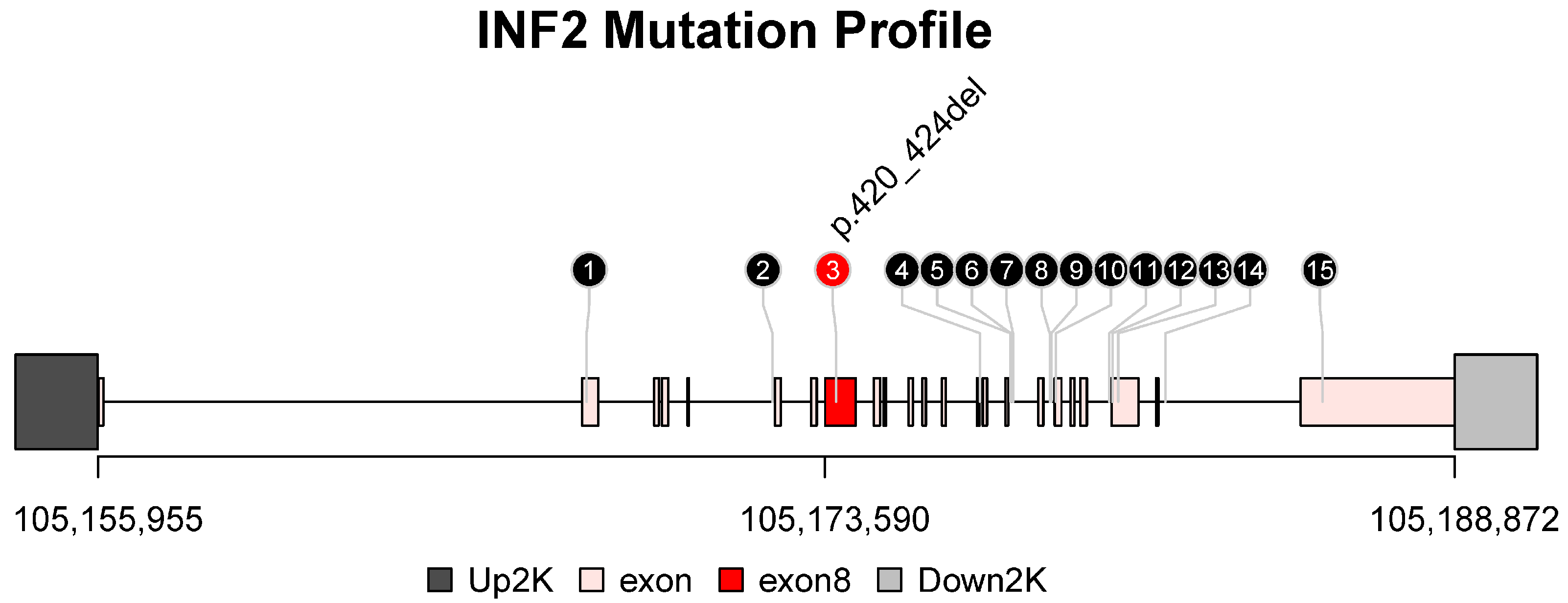
2.5. Whole Exosome Sequencing Results

Whole exosome sequencing of three of these four patients indicated mutations in the INF2 gene. All single nucleotide polymorphisms (SNPs) and insertion–deletion mutations (INDELs) were filtered to retain only variants classified as “PASS”. The retained variants were loaded into the R environment (v4.2.0; R Foundation for Statistical Computing, Vienna, Austria) and then visualized using the lolliplot function, described previously [10], using a Bioconductor package trackViewer (version 1.32.1; Bioconductor, Boston, MA, USA) (Figure 5). Among all SNPs and INDELs, the third mutation p.420_424del, a non-frameshift deletion located in exon8, was a harmful mutation and labelled with a red circle. The underlying meaning of these findings were to be investigated due to the small sample size of this study.

2.6. Literature Review

A comprehensive literature review on PIG identified 40 cases (see Section S1 in Supplementary Materials). All of them were included in the pool analysis alongside the four cases being reported from our center. The detailed characteristics of clinical and histological profiles of these 44 cases are shown in Table 1 and Table 2, respectively.
Among these 44 cases, 79.5% (35/44) were females, 93.2% (41/44) were East Asians, and 63.6% (28/44) were reported in Japan (see Figure S1). The average age was 42.0 ± 12.5 years old. At the time of renal biopsy, the median (interquartile range) serum creatinine level was 61.9 (52.3) μmol/L, and the average amount of proteinuria was 3.06 ± 3.2 g/day.
An analysis of clinical profiles of these cases indicated the most reported comorbidities were connective tissue diseases, mainly systemic lupus erythematosus (see Figure S2), and 20.5% (9/44) of the cases did not have any contaminant disease. Hematuria was not common. Most of the cases (81.8%, 36/44) were treated with immunosuppressants, of which a combination of corticosteroids and one other type of immunosuppressant was most reported. Furthermore, 45.4% (20/44) and 34.1% (15/44) of the cases have achieved complete response and partial response, respectively, after treatment.
An analysis of the histological profiles of renal biopsies indicated 31.8% (14/44) of the cases were all negative for immunofluorescence (IF) staining (see Figure S3). The most seen deposit on IF staining was IgG. Dense deposits on electron microscopy were not common and only seen in 27.2% (12/44) of the cases.

3. Discussion

Here we reported the largest case series of PIG from a single center so far. The pooled analysis of our cases and the cases in the literature indicated PIG was seen in relatively young populations, often associated with connective tissue diseases clinically and one or two other glomerulopathies histologically. The outcomes following immunosuppressive treatment were relatively good. We also reported for the first time that mutations in INF2 might be involved in the development of PIG; however, the significance of these results were yet unclear.
PIG is a peculiar and very rare pathological manifestation which lacks commonality both clinically and histologically. Although it was proposed as a disease entity in 2008 [2], PIG shows great heterogeneities in multiple aspects. It has been reported in both primary and secondary glomerular diseases and has been observed without any marked glomerular nephropathy. As seen in our literature review, SLE is the most reported comorbidity (17/44, 38.6%) [2,4]. Other connective tissue diseases, such as primary biliary cirrhosis, rheumatoid arthritis, mixed connective tissue disease, and Sjogren syndrome, were also reported [5,6,8,11]. Viral infection was reported in a few cases [2,4]. Membranous nephropathy (MN) and focal segmental glomerular sclerosis (FSGS) were the most common histopathological findings associated with PIG, with 12 and 8 cases reported in the literature and this case series, respectively. There were also a significant proportion of cases that showed only mild glomerular changes. In case three, electron-dense deposits were observed in the thickened GBM, which could be explained by her SLE. This should be differentiated from thickened GBM seen in diabetic nephropathy, smoking and obesity, and when uniform thickening of GBM can also be seen but could be distinguished based on the lack of electron-dense deposits and organized ultrastructure. Thickening of GBM in PIG might be caused by a loss of balance between degradation and biosynthesis of the podocyte matrix [2].
The role of PIG in the pathogenesis of glomerulopathies is still unclear. There are a few hypotheses. For example, it was reported that the formation of PIG might be related to the role of special types of complement activation in situ on the microstructures [18]. Since this phenomenon is a dynamic process from primary infolding to bubbling of podocyte foot processes, which has been observed in serial section electronic microscopy, we also suspected that deficits of cytoskeletal structures might be responsible for this change. The whole exosome sequencing results indicated three of the four cases in our case series carried mutations in INF2, which is a known causative gene of genetic FSGS [19]. INF2 is extensively expressed in renal podocytes, of which the mutations cause the mislocalization of INF2 in podocytes and impaired actin dynamics, subsequently damaging the integrity of the glomerular filtration barrier [20,21,22]. Although our whole exosome sequencing results indicated all four patients carried mutations of INF2, these findings did not support the specifically causative relationship between INF2 mutations and PIG. Future work on potential changes in cytoskeletal structures of podocytes in PIG might help to shed light on this link and will be our next step.
Clinical outcomes of PIG were relatively good. In this review, most cases (34/44, 77.3%) received immunosuppressive treatment consisting of corticosteroids only or with a combination of other immunosuppressants, and nearly 80% (35/44) of the patients had achieved at least partial response. Only less than 5% (2/44) of the patients had shown progression. The coexistence of connective tissue diseases and PIG were the main indications for immunosuppressive treatment. Some patients also received only supportive treatment and had achieved complete or partial response. In our study population, all four patients achieved at least partial response. From this point of view, PIG is relatively benign, even if it is classified as a new disease entity.
To our acknowledge, this is the largest single center case series so far on PIG. PIG is quite heterogenous, both clinically and pathologically. Whether this should be considered as a distinctive disease or merely a pathological representation is inconclusive, according to current understanding. We reported for the first time that mutations in INF2 might be involved in the development of PIG. However, our study also had some limitations. Our genetic sequencing analysis did not include pedigree studies due to travel restrictions caused by COVID-19. In addition, the mutations in INF2 lacked specificity for PIG, preventing us from performing any causative analysis. The clinical significance and underlying mechanisms of PIG need further investigations.

4. Conclusions

PIG is a peculiar and very rare manifestation in renal histopathology. The pathogenesis and clinical significance of PIG are still unclear. This pooled analysis indicated PIG was seen in relatively younger populations, often associated with connective tissue diseases clinically and one or two other glomerulopathies histologically. The outcomes following immunosuppressive treatment were relatively good. Mutations in INF2 might be involved in the development of PIG; however, the implications of these mutations need to be investigated.

Supplementary Materials

The following supporting information can be downloaded at: https://www.mdpi.com/article/10.3390/jcm12031088/s1, Section S1. Literature review methodology; Figure S1: Demographic characteristics of podocyte infolding cases in literature and this case series report; Figure S2: Clinical characteristics of podocyte infolding cases in literature and this case series report; Figure S3: Histological characteristics of podocyte infolding cases in literature and this case series report.

Author Contributions

Conceptualization, Y.F. and P.Z.; data curation, Y.F., W.W. (Wei Wang 1), Y.Z., T.C. and P.Z.; formal analysis, Y.F. and P.Z.; funding acquisition, Y.F.; methodology, Y.F. and W.W. (Wei Wang 2); supervision, G.L.; visualization, Y.Z.; writing—original draft, Y.F. and P.Z.; writing—review and editing, W.W. (Wei Wang 1), T.C., W.W. (Wei Wang 2), G.L. and A.Y.W. All authors have read and agreed to the published version of the manuscript.

Funding

This research was partly funded by a clinical research grant from Sichuan Provincial People’s Hospital (2020LY02). Y.F. is supported in part by an Australian Government Research Training Program Scholarship (RTP) for study towards a Ph.D. in the Faculty of Medicine, UNSW, Australia. A/Prof Amanda Y Wang is supported by the National Heart Foundation Vanguard Grant, Australia.

Institutional Review Board Statement

This study was approved by the ethics committee of Sichuan Provincial People’s Hospital (No. 2017.124). Written informed consent was obtained from the patients for publication of this case series report and any accompanying images. Copies of the written consents to participate in this study from the patients are available for review from the editor of this journal.

Informed Consent Statement

Copies of the written consents to publish this study from the patients are available for review from the editor of this journal.

Data Availability Statement

The data and materials reported in this work are available on reasonable request from the correspondence.

Acknowledgments

We sincerely thank the patients and their families as well as the healthcare professionals involved in the patients’ treatment.

Conflicts of Interest

The authors declare no conflict of interest.

References

  1. Sato, H.; Saito, T.; Yoshinaga, K. Intramembranous fine deposit disease associated with collagen disorders: A new morphological entity? Virchows Arch A 1992, 420, 447–451. [Google Scholar] [CrossRef] [PubMed]
  2. Joh, K.; Taguchi, T.; Shigematsu, H.; Kobayashi, Y.; Sato, H.; Nishi, S.; Katafuchi, R.; Nomura, S.; Fujigaki, Y.; Utsunomiya, Y.; et al. Proposal of podocytic infolding glomerulopathy as a new disease entity: A review of 25 cases from nationwide research in Japan. Clin. Exp. Nephrol. 2008, 12, 421–431. [Google Scholar] [CrossRef] [PubMed]
  3. Xiong, S.; Shuai, L.; Li, X.; Dang, X.; Wu, X.; He, Q. Podocytic infolding in Schimke immuno-osseous dysplasia with novel SMARCAL1 mutations: A case report. BMC Nephrol. 2020, 21, 170. [Google Scholar] [CrossRef] [PubMed]
  4. Liu, X.; Huang, J.; Zhang, K.; Niu, Y.; Liu, Y.; Cui, C.; Yu, C. A case of Podocytic Infolding Glomerulopathy with SLE and literature review. BMC Nephrol. 2021, 22, 410. [Google Scholar] [CrossRef]
  5. Matthai, S.M.; Mohapatra, A.; Mathew, A.J.; Roy, S.; Varughese, S.; Danda, D.; Tamilarasi, V. Podocyte Infolding Glomerulopathy (PIG) in a Patient with Undifferentiated Connective Tissue Disease: A Case Report. Am. J. Kidney Dis. 2018, 72, 149–153. [Google Scholar] [CrossRef]
  6. Shi, J.; Zheng, R.; Gao, H.; Zhao, Z.; Wu, H.; Zhang, Z. Podocyte infolding glomerulopathy with undifferentiated connective tissue disease: A case report. Ultrastruct. Pathol. 2020, 44, 245–248. [Google Scholar] [CrossRef]
  7. Zhang, T.; Sun, W.; Xue, J.; Chen, J.; Jiang, Q.; Mou, L.; Du, H. Podocytic infolding glomerulopathy: Two new cases with connective tissue disease and literature review. Clin. Rheumatol. 2019, 38, 1521–1528. [Google Scholar] [CrossRef]
  8. Iguchi, A.; Sohma, A.; Yamazaki, H.; Ito, T.; Saeki, T.; Ito, Y.; Imai, N.; Osawa, Y.; Narita, I. A case of podocytic infolding glomerulopathy with focal segmental glomerulosclerosis. Case Rep. Nephrol. Urol. 2013, 3, 110–116. [Google Scholar] [CrossRef]
  9. Kwon, K.W.; Jeong, H.J.; Lee, J.H. Podocytic infolding glomerulopathy: A case report. Ultrastruct. Pathol. 2016, 40, 374–377. [Google Scholar] [CrossRef]
  10. Ou, J.; Zhu, L.J. trackViewer: A Bioconductor package for interactive and integrative visualization of multi-omics data. Nat. Methods 2019, 16, 453–454. [Google Scholar] [CrossRef]
  11. Ting, J.A.; Hung, W.; McRae, S.A.; Barbour, S.J.; Copland, M.; Riazy, M. Podocyte Infolding Glomerulopathy, First Case Report from North America. Can J. Kidney Health Dis. 2021, 8, 20543581211048357. [Google Scholar] [CrossRef] [PubMed]
  12. Fang, J.Y.; Song, A.H.; Shen, B.; Liu, Y.L. A Case of Podocytic Infolding Glomerulopathy with Primary Sjogren’s Syndrome and Hashimoto’s Thyroiditis. Chin. Med. J. 2018, 131, 2747–2748. [Google Scholar] [CrossRef] [PubMed]
  13. Harada, M.; Kamijo, Y.; Ehara, T.; Shimojo, H.; Shigematsu, H.; Higuchi, M. A case of podocytic infolding glomerulopathy with multiple myeloma. BMC Nephrol. 2014, 15, 32. [Google Scholar] [CrossRef] [PubMed] [Green Version]
  14. Malvar, A.; Davila, P.; Ferrari, M.; Delgado, P.; Iscoff, P.; Lococo, B.; Alberton, V. Podocyte infolding glomerulopathy; report of the first case in Latin America and review of the literature. Nefrologia 2020, 40, 469–473. [Google Scholar] [CrossRef] [PubMed]
  15. Manabe, S.; Sato, M.; Kataoka, H.; Taneda, S.; Mochizuki, T.; Nitta, K. Cell invasion in glomerular basement membrane: Infolding glomerulopathy. Kidney Int. 2020, 98, 1623. [Google Scholar] [CrossRef]
  16. Pandit, A.P.; Rennke, H.G.; Denker, B.M. Podocytic Infolding Glomerulopathy in a Patient with Phospholipase A2 Receptor-Positive Membranous Nephropathy and Review of the Literature. Nephron 2021, 145, 496–502. [Google Scholar] [CrossRef]
  17. Wostmann, F.; Muller, R.U.; Gobel, H.; Benzing, T. Becker JU, Bartram MP: Case report: A peculiar glomerulopathy in a patient suffering from nephrotic syndrome. BMC Nephrol. 2019, 20, 326. [Google Scholar] [CrossRef] [Green Version]
  18. Hinglais, N.; Kazatchkine, N.D.; Bhakdi, S.; Appay, M.-D.; Mandet, C.; Grossetete, J.; Bariety, J. Immunohistochemical study of the C5d-9 complex of complement in human kidneys. Kidney Int. 1986, 30, 399–410. [Google Scholar] [CrossRef] [Green Version]
  19. Subramanian, B.; Chun, J.; Perez-Gill, C.; Yan, P.; Stillman, I.E.; Higgs, H.N.; Alper, S.L.; Schlondorff, J.S.; Pollak, M.R. FSGS-Causing INF2 Mutation Impairs Cleaved INF2 N-Fragment Functions in Podocytes. J. Am. Soc. Nephrol. 2020, 31, 374–391. [Google Scholar] [CrossRef]
  20. Zhao, Y.; Zhang, H.; Wang, H.; Ye, M.; Jin, X. Role of formin INF2 in human diseases. Mol. Biol. Rep. 2022, 49, 735–746. [Google Scholar] [CrossRef]
  21. Panzer, L.; Trube, L.; Klose, M.; Joosten, B.; Slotman, J.; Cambi, A. Linder S: The formins FHOD1 and INF2 regulate inter- and intra-structural contractility of podosomes. J. Cell Sci. 2016, 129, 298–313. [Google Scholar] [PubMed] [Green Version]
  22. Krendel, M.; Pruyne, D. New Paradigm for Cytoskeletal Organization in Podocytes: Proteolytic Fragments of INF2 Formin Function Independently of INF2 Actin Regulatory Activity. J. Am. Soc. Nephrol. 2020, 31, 235–236. [Google Scholar] [CrossRef] [PubMed]
Figure 1. Representative pathological images of renal biopsy specimen form Case 1. Note: IF staining was positive for IgM (A) and IgG (B) in capillary walls and mesangial regions. Light microscopy indicated mild mesangial matrix expansion and diffuse thickening of GBM ((C), PASM + Masson, 400×). Electron microscopy showed thickening of the GBM, diffuse podocyte foot process effacement, and prominent microspheres forming clusters (red arrow) within the GBM ((D), 20,000×).
Jcm 12 01088 g001
Figure 2. Representative pathological images of renal biopsy specimen form Case 2. Note: IF staining was positive for IgG (A) in capillary walls. Light microscopy showed segmental sclerosis with podocyte proliferation in 3/14 glomeruli and diffuse thickening of GBM ((B), PASM + Masson, 400×). Electron microscopy showed diffuse podocyte foot process effacement, multiple podocyte infolding (blue arrow), and prominent microspheres forming clusters (red arrow) within the GBM ((C), 20,000×).
Jcm 12 01088 g002
Figure 3. Representative pathological images of renal biopsy specimen form Case 3. Note: IF staining was positive for IgM and IgA in mesangial regions (A,B). Light microscopy indicated segmental sclerosis with synechial attachment to Bowman’s capsule in 2/20 glomeruli, mild mesangial matrix expansion, and diffuse thickening of GBM ((C), PASM + Masson, 400×). Electron microscopy found diffuse podocyte foot process effacement and prominent microspheres (red arrow) in the GBM ((D), 20,000×).
Jcm 12 01088 g003
Figure 4. Representative pathological images of renal biopsy specimen form Case 4. Note: Light microscopy showed mild mesangial matrix expansion and mild thickening of GBM ((A), PASM + Masson, 400×). Electron microscopy showed diffuse podocyte foot process effacement, mild podocyte infolding, and prominent microspheres (red arrow) within the GBM ((B), 50,000×).
Jcm 12 01088 g004
Figure 5. Lolliplot illustrating the mutation profile in INF2 based on the whole exosome sequencing results of the patients. Note: All SNPs and INDELs in INF2 were plotted. The numbers inside the circle represented the mutation order. The start and end positions were indicated on the horizontal axis. All exons were filled in mistyrose. The upstream and downstream 2K regions were coloured with dark grey and light grey, respectively. Harmful mutations were represented with red circles, while harmless mutations were represented with black circles.
Jcm 12 01088 g005
Table 1. Characteristics of clinical profiles of PIG cases reported in the literature and in this case series.
Case NoSexRaceCountryAgeComorbidityLab Investigations at Renal BiopsyTreatment
Cr (μmol/L)Proteinuria *HematuriaTherapyResponse
1 [3]MAsianJapan31SLE168.00.5 g/dayAbsentPSL (20 mg Qd)CR
2 [3]FAsianJapan37SLE106.11 g/dayAbsentPSL (20 mg Qd), MMFCR after 6 m
3 [3]FAsianJapan40SLE44.21.5 g/dayNRPSLNR
4 [3]FAsianJapan30SLE44.21.6 g/dayMildPSL (pulsex3)CR after 1 m
5 [3]FAsianJapan61SLE, Takayasu’s arteritis79.61.7 g/dayAbsentPSL (40 mg Qd), CsAPR after 7 y
6 [3]FAsianJapan29SLE, hydronephrosis due to lupus cystitis61.91.6 g/dayAbsentPSL (20 mg Qd)PR after 5 y
7 [2]FAsianJapan46SLE, hydronephrosis due to lupus cystitis44.20.6 g/dayAbsentPSL (15 mg Qd)PR after 2 y
8 [2]FAsianJapan27SLE35.42.7 g/dayAbsentPSL (30 mg Qd), MMFCR after 6 m
9 [2]MAsianJapan53SLE, bilateral urethral stone79.63.1 g/dayPresentPSL (30 mg Qd)CR after 8 m
10 [2]FAsianJapan23SLE44.21.8 g/dayPresentPSL (40 mg Qd)CR after 5 y
11 [2]FAsianJapan31SLE79.60.5 g/dayAbsentPSL (20 mg Qd)CR
12 [2]FAsianJapan24SLE, SS53.06.0 g/dayAbsentPSL (pulsex3)CR after 9 w
13 [2]MAsianJapan49PBC, SS, cystitis, finally SLE97.22.2 g/dayAbsentARB, PSL (40 mg Qd)CR after 9 y
14 [2]FAsianJapan20SLE?123.81.4 g/dayAbsentPSL (40 mg Qd)CR
15 [2]FAsianJapan47RA, pSS53.01.3 g/dayAbsentARB, PSL (20 mg Qd)PR
16 [2]FAsianJapan51pSS53.03.7 g/dayAbsentPSL (40 mg Qd)CR after 1 m
17 [2]FAsianJapan30MCTD79.60.3 g/dayAbsentPSL (15 mg Qd)NR
18 [2]FAsianJapan54Basedow’s disease221.06.0 g/dayAbsent-PR after 3 m
19 [2]FAsianJapan57Hypothyroidism, chronic thyroiditis53.00.3 g/dayAbsent-CR after 2 m
20 [2]MAsianJapan45Absent61.92.6 g/dayAbsentPSL (50 mg Qd)PR after 2 m
21 [2]FAsianJapan42Ovarian mature teratoma68.17.5 g/dayPresentARBPR after 1 m
22 [2]FAsianJapan69Absent79.61.6 g/dayAbsent-PR after 1 m
23 [2]MAsianJapan46HBV infection106.14.0 g/dayAbsentDiureticsPR
24 [2]MAsianJapan59Tumor lysis syndrome450.80.6 g/dayAbsentPSL (pulsex3)NR
25 [2]FAsianJapan45Absent70.71.5 g/dayAbsentPSL (30 mg Qd)CR after 1 y
26 [3]MAsianChina4SIODNormal3.7 g/dayNRP, tacrolimus, ACEINormal renal function
27 [4]FAsianChina61SLE, benign ovarian tumor, EBV infection75.12.1 g/dayPresentHCQ, ARB, PSL (40 mg Qd), CTXPR
28 [5]FAsianIndia45UCTD145.95.8 g/dayPresentHigh dose PSL, MMF, RTXPR
29 [6]FAsianChina33UCTD36.22.1 g/dayAbsentNRNR
30 [7]FAsianChina27pSS168.20.6 g/dayAbsentPSL (48 mg Qd), HCQProgression
31 [7]FAsianChina23SLE47.116.8 g/dayPresentPSL (40 mg Qd), HCQPR
32 [8]FAsianJapan14Absent48.62.35 g/dayAbsentPSL (40 mg Qd)CR after 9 m
33 [9]FAsianSouth Korea44Absent39.8PCR = 67.1/88.29 mg/mgNRPSL (10 mg Qd)CR after 3 m
34 [11]FAsianCanada52Prior HBV infection with immunity61uACR = 1600 mg/molPresentARB, P (1 mg/kg, Qd), spironolactonePR, worsened after stopping P
35 [12]FAsianChina52pSS, Hashimoto’s thyroiditisNRNRNRNRNR
36 [13]MAsianJapan79MM113.21.4 g/dayAbsentPSL (20 mg Qd)PR, but death after 2 m
37 [14]FLatin American CaucasianArgentina38SLENormalNS range proteinuriaNRCO + CTXGood response, but relapsed
38 [15]FAsianJapan35SclerodermaNRNRNRNRNR
39 [16]FNRUSA60Absent61.97.3 g/dayAbsentRTXPR
40 [17]FCaucasianGermany56RA386.3uACR = 6200 mg/gAbsentHigh hose P + RTXCR
41FAsianChina26Absent82.32.74 g/dayPresentPrednisone (50 mg Qd, tapered to stop)PR
42MAsianChina47Absent94.49.96 g/dayAbsentPrednisone (30 mg Qd) + tacrolimus (1 mg Bid)PR
43FAsianChina48SLE59.35.4 g/dayAbsentPrednisone (30 mg Qd) + tacrolimus (1 mg Bid) ± Belimumab (10 mg/kg/w)PR
44FAsianChina57Absent33.33.5 g/dayMildPrednisone (45 mg Qd)CR, but relapsed
Abbreviations: ACEI, angiotensin converting enzyme inhibitor; ARB, angiotensin receptor blocker; Bid, twice daily; CO, corticosteroid; Cr, creatinine; CR, complete response; CsA, cyclosporin; F, female; HBV, hepatitis B virus; HCQ, hydroxychloroquine; M, male; m, month; MCTD, mixed connective tissue disease; MM, multiple myeloma; MMF, mycophenolate mofetil; NR, not reported; NS, nephrotic syndrome; P, prednisone; PBC, primary biliary cirrhosis; PR, partial response; PSL, prednisolone; pSS, primary Sjogren syndrome; Qd, once daily; RA, rheumatoid arthritis; RTX, rituximab; SIOD, Schimke immune-osseous dysplasia; SLE, systemic lupus erythematosus; SS, Sjogren syndrome; UCTD, undifferentiated connective tissue disease; w, week; y, year. * The value was the amount of proteinuria per day, unless specified.
Table 2. Characteristics of pathological profiles of PIG cases reported in the literature and in this case series.
Case NoSexAgeRenal Pathology
IF StainingHypercellularityMesangial DepositGBM ThickeningFPEMicrospheresMicrotubulesDense DepositLM Manifestations
1 [2]M31All negativeAbsentMildPresentPresentPresentAbsentAbsentMGA
2 [2]F37G, A, C3, C1qAbsentMildPresentPresentPresentPresentAbsentLN Class II
3 [2]F40G, A, C3, C1qNRMildPresentPresentPresentPresentAbsentLN Class II
4 [2]F30G, A, C3, C1q, C5b-9PresentMildPresentPresentPresentAbsentMesangium, subendothelialLN Class II
5 [2]F61G, M, C1qAbsentMildPresentPresentPresentAbsentPresentLN Class II
6 [2]F29All negativeAbsentAbsentPresentPresentPresentPresentAbsentMN
7 [2]F46All negativeAbsentAbsentPresentPresentPresentPresentAbsentMN
8 [2]F27G, A, M, C3, C1q, C5b-9AbsentAbsentPresentPresentPresentPresentGBM, subendothelial, subepithelialLN Class V
9 [2]M53All negativeAbsentAbsentPresentPresentPresentPresentAbsentMN
10 [2]F23GPresentMildPresentPresentPresentPresentGBM, subendothelial, subepithelialLN Class V
11 [2]F31GAbsentAbsentPresentPresentPresentPresentGBMLN Class V
12 [2]F24G, M, C1qMildMildPresentPresentPresentPresentGBMLN Class V
13 [2]M49G, APresentPresentPresentPresentAbsentPresentAbsentMPGN (Type 3)
14 [2]F20GAbsentAbsentPresentNRPresentAbsentGBMMGA
15 [2]F47G, A, MAbsentAbsentPresentNRPresentAbsentGBMMGA
16 [2]F51All negativeAbsentAbsentPresentPresentPresentAbsentAbsentMGA
17 [2]F30GAbsentAbsentPresentPresentPresentAbsentAbsentMGA
18 [2]F54All negativePresentPresentPresentPresentPresentAbsentAbsentFSGS
19 [2]F57All negativeAbsentAbsentPresentPresentPresentAbsentAbsentFSGS
20 [2]M45G, A, C3AbsentPresentPresentPresentPresentPresentAbsentFSGS
21 [2]F42GPresentAbsentPresentPresentPresentAbsentAbsentFSGS + MN
22 [2]F69G, A, M, C3AbsentAbsentPresentPresentPresentAbsentAbsentMN
23 [2]M46GAbsentAbsentPresentPresentPresentPresentAbsentMN
24 [2]M59MAbsentAbsentPresentPresentPresentAbsentAbsentMN
25 [2]F45G, A, C3AbsentAbsentPresentPresentPresentPresentAbsentMN
26 [3]M4All negativeNRNRPresentPresentPresentAbsentAbsentFocal or global sclerosis
27 [4]F61M, C3PresentPresentPresentPresentPresentAbsentAbsentLN
28 [5]F45G, C3AbsentAbsentPresentPresentPresentAbsentAbsentMN
29 [6]F33M, G, C1q, C3MildMildPresentPresentPresentAbsentMesangium, subepithelialProliferative glomerulonephritis
30 [7]F27MPresentMildPresentPresentPresentAbsentAbsentChronic interstitial nephritis
31 [7]F23MAbsentAbsentPresentPresentPresentAbsentAbsentMGA
32 [8]F14M, C3, C1qAbsenceAbsenceAbsentPresencePresentAbsentAbsentFSGS
33 [9]F44MAbsentAbsentPresentPresentPresentPresentMesangium, intramembraneMGA
34 [11]F52All negativeAbsentAbsentAbsentPresentPresentPresentAbsentGlobal and segmental sclerosis
35 [12]F52All negativeNRNRPresentPresentPresentAbsentAbsentMGA
36 [13]M79All negativeAbsentAbsentPresentPresentPresentAbsentAbsentMN
37 [14]F38All negativeAbsentPresentPresentPresentPresentPresentAbsentFSGS
38 [15]F35Full house pattern, faint IgGPresentMildPresentPresentPresentPresentMesangiumLN Class II
39 [16]F60G, M, A, C3, PLA2rAbsentMildAbsentPresentPresentPresentSubepithelialMN
40 [17]F56All negativeAbsentAbsentPresentPresentAbsentAbsentAbsentAcute tubular injury
41F26G, MMildPresentPresentPresentPresentAbsentPresentFSGS
42M47GMildAbsentPresentPresentPresentAbsentAbsentFSGS
43F48A, MNonePresentPresentPresentPresentAbsentAbsentMN
44F57All negativeAbsentMildMildPresentPresentPresentAbsentMGA
Abbreviations: A, IgA; F, female; FPE, foot process effacement; FSGS, focal segmental glomerular sclerosis; G, IgG; GBM, glomerular basement membrane; IF, immunofluorescent; LM, light microscopy; LN, lupus nephritis; M, male; M (in IF staining column), IgM; MGA, mild glomerular abnormality; MN, membranous nephropathy; MPGN, membranous proliferative glomerulonephritis; NR, not reported.
Disclaimer/Publisher’s Note: The statements, opinions and data contained in all publications are solely those of the individual author(s) and contributor(s) and not of MDPI and/or the editor(s). MDPI and/or the editor(s) disclaim responsibility for any injury to people or property resulting from any ideas, methods, instructions or products referred to in the content.

