Article

The Somatic Mutational Landscape of Mismatch Repair Deficient Prostate Cancer

1
Department of Urology, Fudan University Shanghai Cancer Center, Shanghai 200032, China
2
Department of Oncology, Shanghai Medical College, Fudan University, Shanghai 200032, China
*
Authors to whom correspondence should be addressed.
These authors contributed equally to this work.
J. Clin. Med. 2023, 12(2), 623; https://doi.org/10.3390/jcm12020623
Submission received: 3 December 2022 / Revised: 9 January 2023 / Accepted: 10 January 2023 / Published: 12 January 2023

Abstract

:
Prostate cancers with mismatch repair deficiency (MMR-d) have aggressive clinical and histological features, and they are potentially responsive to immunotherapy. However, its rarity prevents the analysis of the underlying biology. Here, we collected the genomic data of 2664 primary prostate tumors and 1409 metastatic prostate tumors from the GENIE and TCGA databases. A total of 69 (2.59%) primary and 60 (4.26%) metastatic MMR-d tumors were identified among these tumors. Single nucleotide variant (SNV) frequencies of 34 candidate genes (including KMT2D (46.4%), ZFHX3 (33.3%), JAK1 (31.9%), and RNF43 (27.5%)) and 16 candidate genes (including KMT2D (33.3%) and JAK1 (28.3%)) were higher in MMR-d primary tumors and MMR-d metastatic tumors, respectively. The tumor mutation burden (TMB) was higher in primary MMR-d tumors. Homozygous deletions of EPCAM and EPAS1 were enriched in MMR-d primary tumors, while EPCAM deletions were enriched in metastatic MMR-d tumors. For genomic rearrangement events, TMPRSS2-ETS fusions were less frequent in primary MMR-d tumors. Our study indicates MMR-d prostate cancers have unique genomic features. These may play an important role in providing therapeutic targets for the treatment of this subset of prostate cancer patients.

1. Introduction

DNA mismatch repair (MMR) is a mechanism for repairing incorrect base matches as well as insertion/deletion errors that occur during replication. MMR-deficient (MMR-d) status usually results from pathogenic alterations in the MMR core genes (MLH1, MSH2, MSH6, and PMS2), which are also associated with high microsatellite instability and high tumor mutational burden [1,2,3].
A prospective study indicated that men with MSH2 or MSH6 germline pathogenic mutations are at a significantly higher risk of prostate cancer (PCa) than non-carriers [4]. Several studies, however, have shown that MMR-d PCa are associated with aggressive histologic characteristics such as cribriform and ductal adenocarcinomas [5,6,7,8]. Furthermore, MMR-d PCa patients also have a high prevalence of Gleason ≥ 8, often present with de novo metastases, and respond poorly to androgen deprivation therapy (ADT) and next-generation antiandrogens [9,10,11]. Despite the fact that the presence of MMR-d status guides the use of immunotherapy, nearly half of these patients do not benefit from immunotherapy [12,13].
MMR-d is relatively rare in PCa, estimated at 3–5% of cases [11,12,14]. Due to their low prevalence, the molecular features of MMR-d PCa tumors remain incompletely described. We propose that MMR-d tumors represent a distinct phenotype in PCa. Recognition of genomic alterations in MMR-d PCa tumors may enable understanding of the mechanisms of MMR-d associated PCa carcinogenesis and progression, which may have the potential to provide downstream implications for treatment. In this study, we integrated the genomic data of primary and metastatic PCa from the TCGA and GENIE databases to depict the somatic mutational profiles in MMR-d prostate tumors and compare them with MMR-p tumors.

2. Materials and Methods

2.1. Samples and Data Processing

The mutation files for SNV, CNA, and SV and clinical data from The Cancer Genome Atlas Prostate Adenocarcinoma (TCGA-PRAD) were retrieved from cBioportal (https://www.cbioportal.org/study/summary?id=prad_tcga_pan_can_atlas_2018, accessed on 12 September 2022). The SNV, CNA, and SV and clinical data from the American Association for Cancer Research (AACR) Project Genomics Evidence Neoplasia Information Exchange (GENIE) for prostate adenocarcinomas were downloaded from Sage Bionetworks Synapse platform release 12.0 (https://www.synapse.org, accessed on 12 September 2022). The GENIE data was filtered to retain only primary and metastatic samples with sequencing panels including SNV and CNA profiling of MMR genes (MLH1, MSH2, MSH6, and PMS2). The 14 retained panels were: COLU-CCCP-V1, DFCI-ONCOPANEL-1, DFCI-ONCOPANEL-2, DFCI-ONCOPANEL-3, DFCI-ONCOPANEL-3.1, MSK-IMPACT341, MSK-IMPACT410, MSK-IMPACT468, MSK-IMPACT505, UCSF-IDTV5-TO, UCSF-NIMV4-TO, VICC-01-DX1, VICC01-T5A, and VICC-01-T7. If a patient had multiple primary or metastatic samples, one sample was selected using the following hierarchy: (1) the sample was identified as a MMR-d tumor, (2) the sample had the maximal amount of available genomic data, and (3) if the first two criteria were not met, the sample was selected randomly. The sample sizes of the final analyzed population for primary and metastatic tumors by dataset and genomic data type are shown in Figure 1.

2.2. Identification of MMR-Deficient (MMR-d) Tumors

MMR-d status was determined by assessing the presence of SNV, CNA, and SV involving MMR genes in tumors. Pathogenic SNV of MMR genes were identified by the following process: (1) variants reported as oncogenic or likely oncogenic in OncoKB (http://oncokb.org, accessed on 12 September 2022) [15] were retained, and neutral variants were removed; and (2) the impact of the remaining variants with uncertain significance was further evaluated through SIFT and PolyPhen-2 [16,17]. Only variants classified as deleterious in SIFT and as probable or possibly damaging in PolyPhen-2 were retained. Tumors with pathogenic SNV were classified as MMR-d. However, tumors with homozygous deletions at MMR genes or SV affecting MMR genes were also classified as MMR-d.

2.3. Genomic Profiling Analyses

Tumor mutational burden (TMB) was calculated using SNV data from the TCGA database with whole-exome sequencing (WES) profiling. The capture size for the WES data was set to 50 Mbp. Differences in TMB by tumor MMR status were assessed. The candidate genes for analysis were identified from the literature [14,18,19], which included 14 hereditary genes in PCa, 7 gene interactors with MMR genes, and 27 genes recurrently mutated in PCa. In addition to this, we supplemented the top 20 genes with the highest SNV or CNV frequencies in primary or metastatic PCa samples from TCGA and GENIE. Since not all candidate genes were analyzed in the GENIE panels, the sample size for frequency calculations varied across genes. To ensure sufficient power, only genes profiled in at least 60% of tumors were studied further (Supplementary Table S1). The details of SNV, CNV, and SV analyses were presented in Supplementary Method.

3. Statistical Analysis

The frequencies of SNVs, CNAs, and SVs by MMR status were compared using the two-sided Fisher’s exact test. The Bonferroni’s correction was used to control the false discovery rate (FDR ≤ 1%).

4. Results

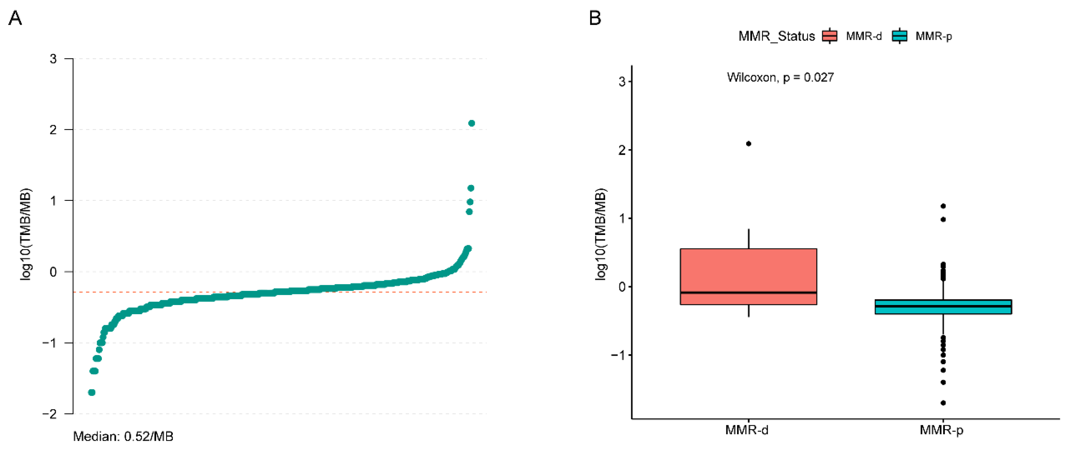
4.1. MMR-d Prostate Tumors

The genomic data of 2664 primary and 1409 metastatic prostate adenocarcinomas was collected from the American Association for Cancer Research Project Genomics Evidence Neoplasia Information Exchange (GENIE) and The Cancer Genome Atlas (TCGA) databases (Figure 1). A total of sixty-nine (2.59%) primary tumors harbored pathogenic SNVs, CNAs, or SVs leading to MMR-d, while 2595 tumors (97.41%) did not have pathogenic alterations at MMR genes and were classified as MMR-p. Seven tumors had pathogenic SNVs at MLH1, twenty-two tumors had pathogenic SNVs at MSH2, twenty tumors had pathogenic SNVs at MSH6, and fourteen tumors had pathogenic SNVs at PMS2 (Figure 2). A total of one tumor had homozygous deletion at MLH1, two tumors had homozygous deletion at PMS2, two tumors had homozygous deletion at MSH2, one tumor had homozygous deletion at MSH6, and seven tumors had homozygous deletion at both MSH2 and MSH6. A total of two tumors had SVs affecting MLH1, two tumors had SVs affecting MSH2, and three tumors had SVs affecting MSH6 (Supplementary Table S2).
A total of 60 (4.26%) metastatic tumors were identified as MMR-d, while 1349 tumors (97.41%) were MMR-p. A total of seven tumors had pathogenic SNVs at MLH1, ten tumors had pathogenic SNVs at MSH2, twenty-three tumors had pathogenic SNVs at MSH6, and eleven tumors had pathogenic SNVs at PMS2 (Figure 2). A total of two tumors had homozygous deletion at MLH1, three tumors had homozygous deletion at MSH2, two tumors had homozygous deletion at MSH6, and eight tumors had homozygous deletion at both MSH2 and MSH6. A total of one tumor had SVs affecting MLH1, five tumors had SVs affecting MSH2, and two tumors had SVs affecting MSH6 (Supplementary Table S3).

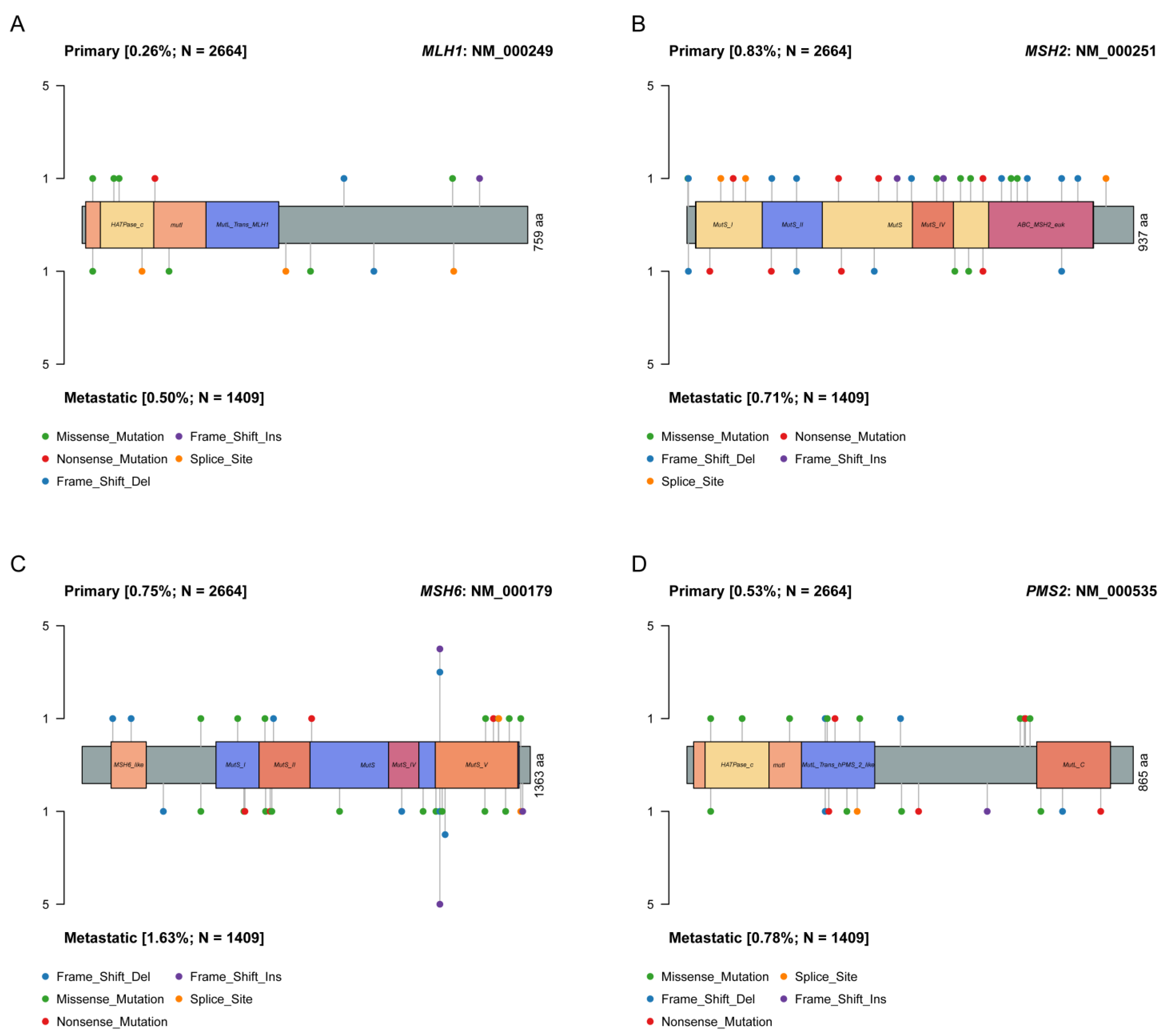
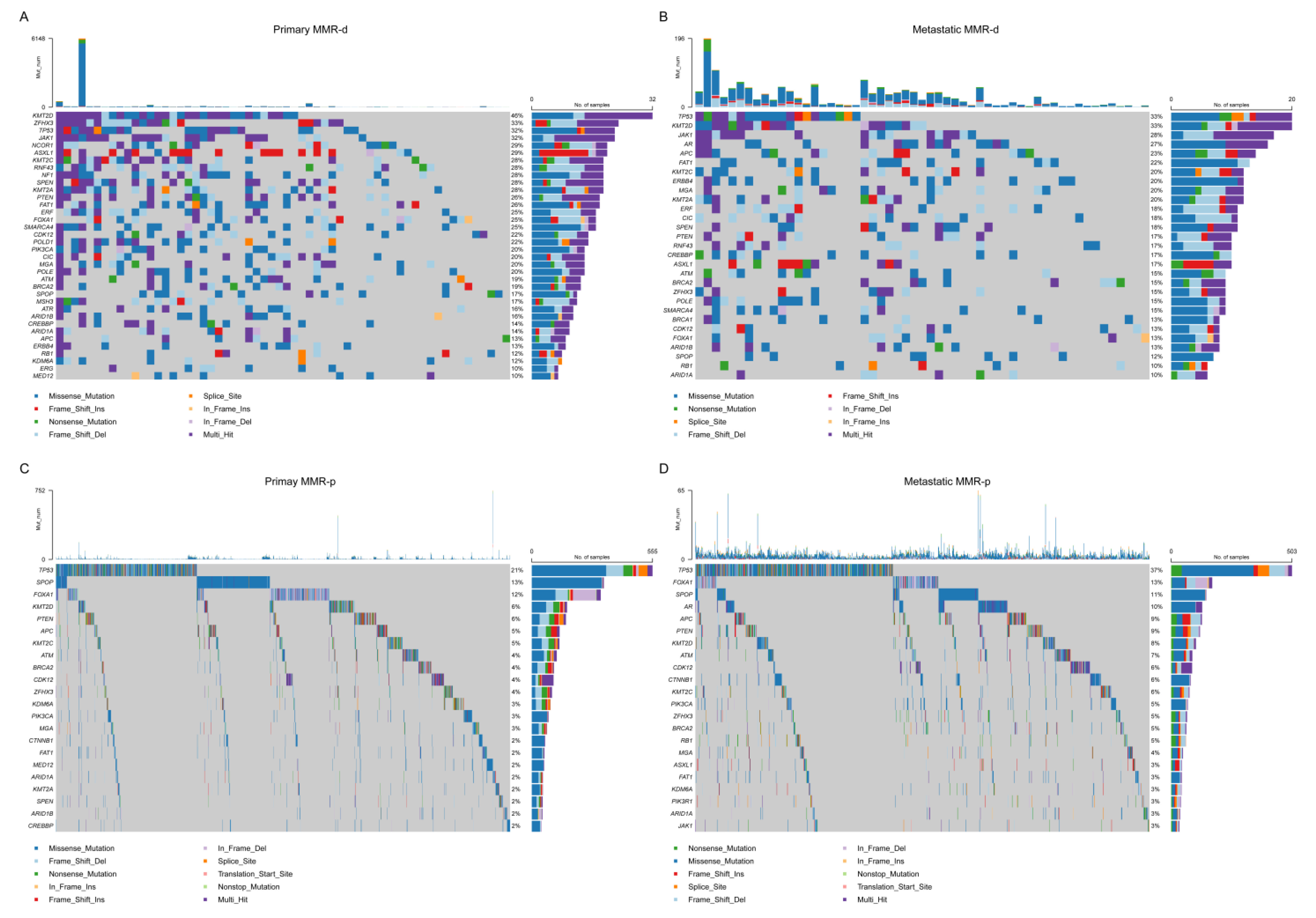
4.2. Single Nucleotide Variant (SNV) Analyses

The most frequent SNVs of candidate genes in MMR-d and MMR-p primary tumors derived by combining TCGA and GENIE data are shown in Figure 3a,c. The SNV frequency of 34 candidate genes in MMR-d primary tumors was significantly higher than in MMR-p primary tumors. A total of 19 of these genes had a SNV frequency ≥20% in MMR-d tumors. The most significant differences by MMR status were found in JAK1 (31.9% vs. 1.5%, Bonferroni-adjusted p = 2.1 × 10⁻²¹), ASXL1 (29.0% vs. 1.0%, Bonferroni-adjusted p = 5.6 × 10⁻²¹), KMT2D (46.4% vs. 6.2%, Bonferroni-adjusted p = 2.1 × 10⁻¹⁹), and RNF43 (27.5% vs. 1.08%, Bonferroni-adjusted p = 1.1 × 10⁻¹⁷) (Supplementary Table S4). For metastatic tumors from GENIE, a total of sixteen candidate genes had significantly higher SNV frequencies in MMR-d tumors, of which six genes (KMT2D (33.3%), JAK1 (28.3%), FAT1 (21.7%), ERBB4 (20.0%), KMT2A (20.0%), and MGA (20.0%)) had a SNV frequency ≥ 20% in MMR-d tumors. TP53 had the highest SNV frequency in both MMR-d and MMR-p tumors, but its frequency did not differ between MMR-d (33.3%) and MMR-p (37.3%) metastatic tumors (p = 0.56) (Figure 3b,d, Supplementary Table S5). Among 490 patients with pmary tumors from the TCGA database, the median SNV TMB was 0.52 per MB (range: 0.02–122.96) (Figure 4a). SNV TMB was significantly higher in MMR-d tumors than in MMR-p tumors (median: 0.82 vs. 0.52, p = 0.024) (Figure 4b).

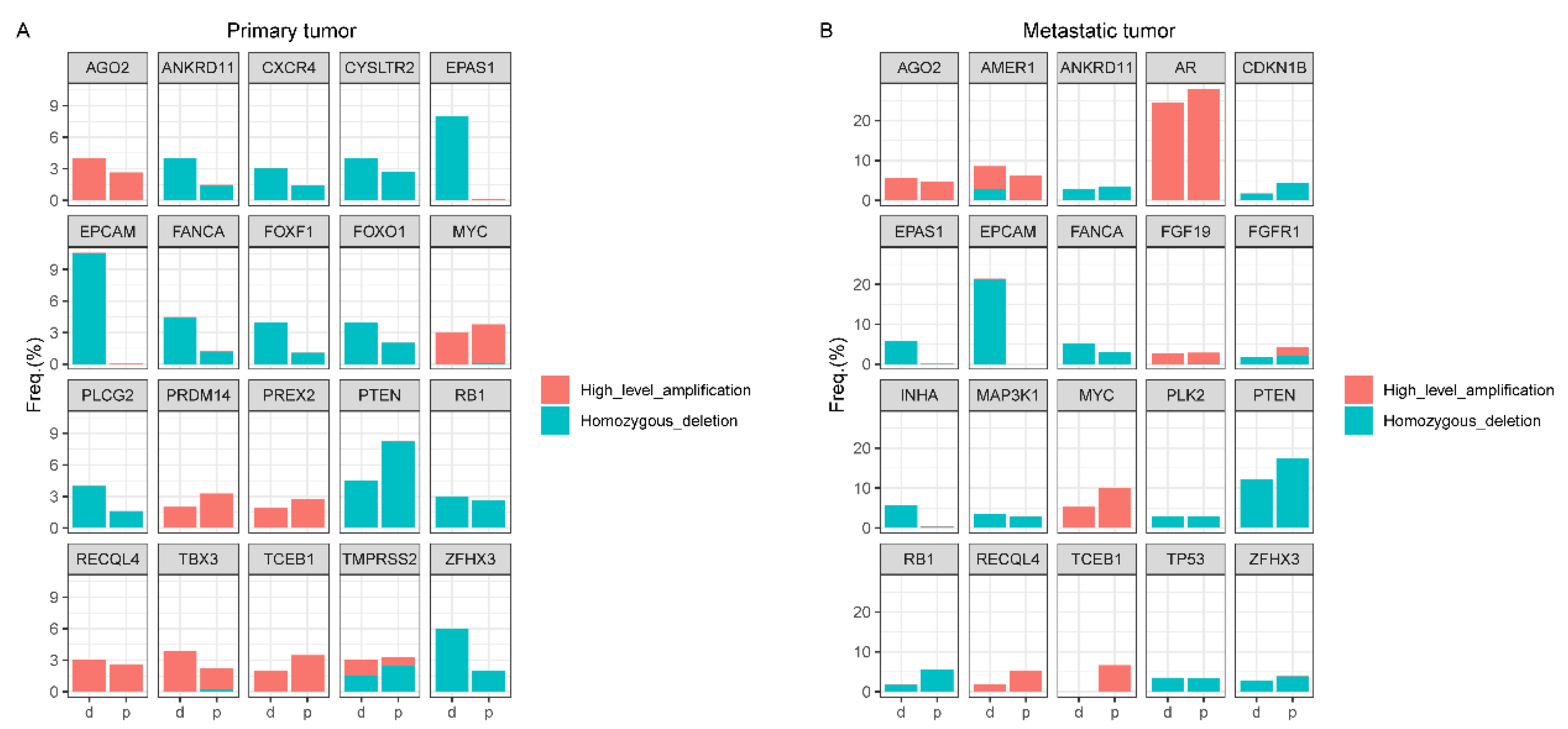
4.3. Copy Number Alteration (CNA) Analyses

For primary tumors from the TCGA and GENIE databases, homozygous deletions at EPCAM (10.6% vs. 0%, Bonferroni-adjusted p = 2.6 × 10⁻¹⁰) and EPAS1 (8.0% vs. 0.05%, Bonferroni-adjusted p = 7.0 × 10⁻⁵) were significantly more frequent in MMR-d tumors (Figure 5a, Supplementary Table S6). Among metastatic tumors from the GENIE database, the frequency of homozygous deletions at EPCAM (21.4% vs. 0%, Bonferroni-adjusted p = 4.2 × 10⁻¹⁶) was significantly higher in MMR-d tumors (Figure 5b, Supplementary Table S7). There were no other gene differences in homozygous deletion frequencies by MMR status after multiple testing correction. The homozygous deletion frequency at PTEN was non-significantly lower in MMR-d tumors than in MMR-p tumors (primary: 4.5% vs. 8.2%, metastatic: 12.3% vs. 17.6%). The frequencies of homozygous deletions at ETV1 (3.0% vs. 0.1%, p = 0.0037), KMT2C (3.9% vs. 0.19%, p = 0.0076), and FANCA (4.8% vs. 1.17%, p = 0.0499) were nominally higher in MMR-d primary tumors, as were the frequencies of homozygous deletions at EPAS1 (5.7% vs. 0.1%, p = 0.0030) and INHA (5.7% vs. 0.3%, p = 0.0094) in MMR-d metastatic tumors. Nevertheless, there was no significant difference in these frequencies after FDR correction.
No significant differences in the frequency of genes with high-level amplification were found between MMR-d and MMR-p tumors, both in primary tumors and metastatic tumors. The high-level amplification frequencies at MYC (primary: 3.0% vs. 3.7%, metastatic: 5.3% vs. 10.0%) and AR (primary: 0% vs. 1.3%, metastatic: 24.6% vs. 27.9%) were non-significantly lower in MMR-d tumors than MMR-p tumors (Supplementary Tables S8 and S9).

4.4. Structural Variant (SV) Analyses

For primary tumors, the frequencies of TMPRSS2- and ETS-related SVs were evaluated using the TCGA and GENIE databases. TMPRSS2-EST fusions were less common in MMR-d primary tumors than MMR-p primary tumors (32.0% vs. 54.2%, p = 0.041). The frequencies of TMPRSS2-ERG, TMPRSS2-intragenic, and other SVs affecting ETS genes were insignificantly less in MMR-d tumors than in MMR-p tumors. As for metastatic tumorsrom the GENIE database, the prevalence of TMPRSS2- and ETS-related SVs did not differ by MMR status (Table 1).

5. Discussion

MMR-d status was not common in PCa patients. However, they have attracted the attention of clinicians since the FDA granted approval for the PD-1 inhibitor pembrolizumab for the treatment of MMR-d tumors regardless of the cancers’ tissue of origin in May 2017 [20]. Immunotherapy has shown promising efficacy in MMR-d PCa patients, but there is growing evidence that PD-1 inhibitors are not effective in all MMR-d patients, and some MMR-p patients are also sensitive to immunotherapy [12,21]. In addition, it was suggested that MMR-d status could also lead to BRCA1/2 mutations, but these concurrent BRCA1/2 mutations do not lead to PARP-inhibitor sensitivity [22]. To improve the understanding of MMR-d in PCa, our work provides integrated analyses of the genomic landscape in primary and metastatic prostate tumors.
In our study, we found that the TMB in MMR-d tumors was significantly higher than in MMR-p tumors in the context of a low overall TMB in PCa, which is consistent with previous findings [10,11]. However, despite a significant increase in the mutation frequencies of most candidate genes in MMR-d tumors, we found no significant increase in the frequency of the most common genomic aberrations in PCa driver genes, such as TP53, FOXA1, and SPOP. In addition, other molecular events such as PTEN deletion, RB1 deletion, and MYC amplification were less common in MMR-d tumors, although they did not reach a significant difference. While these genomic aberrations usually indicate highly aggressive tumors as well as a poor prognosis, MMR-d tumors had a lower incidence of these molecular events yet still exhibited a highly malignant biology and responded poorly to ADT and next-generation antiandrogens [10,11]. These results suggest that MMR-d PCa tumors possess a unique molecular profile, and the development of this group of tumors may not be dependent on the common tumor pathways activated in PCa.
Notably, we observed that KMT2D, also known as MLL2, mutated in close to half of MMR-d primary tumors and in 1/3 of MMR-d metastatic tumors. KMT2D encodes a histone methyltransferase that methylates the Lys-4 position of histone H3, and it plays an important role in DNA damage repair. Mutations in KMT2D occur widely in numerous types of tumors and have been proven to be directly associated with genomic instability [23,24]. It is worth noting that a recent study showed that KMT2D-mutant tumors exhibit enhanced immune infiltration in the tumor microenvironment, and patients with KMT2D mutations are sensitive to immunotherapy [25]. Combined with clinical data indicating that nearly half of MMR-d prostate cancer patients are sensitive to immunotherapy [12,13], a proportion similar to the proportion of KMT2D mutations in MMR-d tumors. Therefore, we speculate that the high mutation rate in KMT2D may be partly accountable for the favorable efficacy of immunotherapy in MMR-d PCa patients, and the identification of KMT2D mutations may have important implications for further subdividing the population of PCa patients who can benefit from immunotherapy. In addition, mutations in KMT2D in PCa were also shown to be associated with aggressive biologic behaviors as well as a poor prognosis [26], which was consistent with the clinical features of MMR-d PCa patients, suggesting that KMT2D mutations may be engaged in the carcinogenesis and progression of MMR-d tumors and that it might be a potential therapeutic target for MMR-d PCa patients. In addition, we found that the SNV frequency at JAK1 was also significantly increased among MMR-d tumors. JAK1 encodes a membrane protein that is a member of a group of protein tyrosine kinases (PTKs). The encoded kinase phosphorylates STAT proteins and plays a crucial role in affecting the expression of genes that mediate inflammation, epithelial remodeling, and metastatic cancer progression. It is a pivotal constituent of the interleukin-6 (IL-6)/JAK1/STAT3 immune and inflammation response and is a therapeutic target for alleviating cytokine storms [27]. More importantly, there is evidence that loss-of-function mutations at JAK1 promote a lack of response to interferon gamma, leading to resistance to immunotherapy in tumor cells [28]. Therefore, mutations in JAK1 may contribute to primary resistance to immunotherapy in some PCa patients with MMR-d. However, it is noteworthy that Laura et al. reported that in MMR-d PCa patients, the mutations of JAK1 did not associate with diminished efficacy of immunotherapy, Hence, the JAK1 mutations may be passenger mutations occurring in MMR-d PCa patients, which, like genes containing microsatellite tracts (such as RNF43), have a higher propensity for secondary mutations in MMR-d tumors [29]. We also analyzed the four MMR genes separately and found that mutations in some genes, like KMT2D and FAT1, were mainly enriched in tumors carrying deleterious aberrations in MSH2 and MSH6, which reflect that tumors carrying different MMR gene aberrations may have different genomic features. Given the low number of tumor specimens analyzed per individual MMR gene, we plan to validate these findings in a larger cohort of samples. Furthermore, considering that we do not currently treat patients carrying different MMR gene mutations differently in clinical decision-making, we believe it is reasonable to have them grouped together for the present analysis. Combining these results, we believe that a comprehensive assessment of the genomic characteristics of PCa patients is necessary before the administration of immunotherapy.
We also found higher frequencies of homozygous deletions for EPCAM and EPAS1 in MMR-d primary tumors and for EPCAM in MMR-d metastatic tumors. EPCAM encodes a carcinoma-associated antigen, which is being used as a target for immunotherapy treatment [30]. It is located upstream of MSH2, and the germline deletions at EPCAM can cause MSH2 epigenetic inactivation, leading to Lynch syndrome in the absence of MSH2 pathogenic alterations [31,32]. Considering that EPCAM deletions have a relatively high frequency in MMR-d tumors (21.4% in MMR-d metastatic tumors), we presume that the prevalence of Lynch syndrome in MMR-d prostate cancer may be higher than previously thought [33].
Genomic alterations in ETS-related genes (mainly ERG) due to a fusion between the androgen receptor regulatory gene promoter of TMPRSS2 and the ETS transcription factors are detected in approximately half of PCa tumors [34]. Several studies have indicated that TMPRSS2-ETS fusions perform a critical role in PCa tumorigenesis [35,36]. However, in our study, we found that MMR-d tumors had a lower incidence of TMPRSS2-ETS fusions relative to MMR-p tumors, which is consistent with previous findings in a small MMR-d PCa cohort [5]. It suggests that the mechanism of tumorigenesis in MMR-d PCa tumors is likely different from that of most PCa tumors.
To the best of our knowledge, our analysis has represented the largest primary and metastatic MMR-d prostate tumors to date and investigated the characteristics of multi-class somatic mutations. Although we identified possible associations between the characteristic somatic mutations in MMR-d PCa tumors and the efficacy of immunotherapy, there are still some limitations in our study. Firstly, although more than 90% of MMR-d tumors are caused by somatic mutations, germline mutations in MMR genes and EPCAM account for 5–10% of dMMR tumors, which are also known as Lynch syndrome [37]. Thus, we may have misclassified a small portion of MMR-d tumors as MMR-p tumors. This misclassification may mitigate the differences in the pattern of alterations between MMR-d and MMR-p tumors. Second, since the GENIE database did not provide detailed clinical data on patients, we were unable to compare the clinical characteristics among patients with MMR-d PCa tumors and those with MMR-p PCa tumors. Third, due to the relatively low proportion of MMR-d status in PCa, the limited number of MMR-d tumor specimens restricts us from doing further analysis by classifying different MMR gene mutations, and the genomic alterations do not fully reflect the DNA repair capacity. In addition, the inclusion of patients with potentially non-pathogenic MMR missense mutations might have diluted our findings. However, we only retained variants reported as oncogenic or likely oncogenic in OncoKB (http://oncokb.org, accessed on 12 September 2022) and removed variants unless they were classified as deleterious in SIFT and as probably damaging or possibly damaging in PolyPhen-2 (as detailed in the Methods section). With this strategy, >70% of missense mutations involving MMR genes were filtered, and the percentage of MMR-d tumors (2.59% in primary tumors and 4.26% in metastatic tumors) is consistent with previous studies (3–5%) [11,12,14]. If all missense mutations were removed, then only 1.6% of primary tumors and 2.7% of metastatic tumors would be classified as MMR-d tumors, a significantly lower proportion than reported in previous studies.

6. Conclusions

In conclusion, our study reveals the unique somatic mutational landscape of MMR-d PCa and provides partial evidence from this perspective to explain why this population is potentially sensitive to immunotherapy. These results also provide information for the development of therapeutic targets and clinical management for this group of patients.

Supplementary Materials

The following supporting information can be downloaded at: https://www.mdpi.com/article/10.3390/jcm12020623/s1, Supplementary Method: Supplementary method for genomic profiling analyses [14,18,19,38]; Table S1: Candidate Gene Genlist for analysis; Table S2: List of MMR-d primary tumors; Table S3: List of MMR-d metastatic tumors; Table S4: SNV frequency by MMR status in primary tumors; Table S5: SNV frequency by MMR status in metastatic tumors; Table S6: CNV frequency by MMR status in primary tumors (Homozygous Deletion); Table S7: CNV frequency by MMR status in metastatic tumors (Homozygous Deletion); Table S8: CNV frequency by MMR status in primary tumors (High level of amplification); Table S9: CNV frequency by MMR status in metastatic tumors (High level of amplification).

Author Contributions

Conceptualization, Y.Z., D.Y., and B.F.; software, B.F., Y.W., and J.P.; methodology, B.F., Y.W., and T.Z.; validation, Y.Z. and D.Y.; formal analysis, B.F., Y.W., and T.Z.; writing—original draft preparation, B.F.; writing—review and editing, Y.W. and Y.Z.; visualization, B.F.; supervision, D.Y.; project administration, D.Y.; funding acquisition, Y.Z. All authors have read and agreed to the published version of the manuscript.

Funding

National Nature Science Foundation of China (82172621, YZ); National Nature Science Foundation of China (81972375, YZ); Shanghai Medical Innovation Research Special Project (21Y11904300, YZ); Chinese Anti-Cancer Association-Hengrui PARP Inhibitor Cancer Research Foundation; Oriental Scholar Professorship, Shanghai Municipal Commission of Education, China; Shanghai Shenkang Research Physician Innovation and Transformation Ability Training Project: SHDC2022CRD035; General Program of Beijing Xisike Clinical Oncology Research Foundation: Y-MSDZD2021-0230; and the General Program of the Beijing Xisike Clinical Oncology Research Foundation (Y-2019AZMS-0012, YZ).

Institutional Review Board Statement

Not applicable.

Informed Consent Statement

The GENIE and TCGA consortia were responsible for obtaining patient informed consent or waivers of consent.

Data Availability Statement

The data of this study are available from the AACR Project GENIE database (https://www.synapse.org/#!Synapse:syn7222066/wiki/410924, accessed on 12 September 2022) and The Cancer Genome Atlas (https://www.cbioportal.org/study/summary?id=prad_tcga_pan_can_atlas_2018, accessed on 12 September 2022).

Conflicts of Interest

The authors declare that they have no competing interests.

References

  1. Strand, M.; Prolla, T.A.; Liskay, R.M.; Petes, T.D. Destabilization of tracts of simple repetitive DNA in yeast by mutations affecting DNA mismatch repair. Nature 1993, 365, 274–276. [Google Scholar] [CrossRef] [PubMed]
  2. Shia, J. Evolving approach and clinical significance of detecting DNA mismatch repair deficiency in colorectal carcinoma. Semin. Diagn. Pathol. 2015, 32, 352–361. [Google Scholar] [CrossRef] [PubMed] [Green Version]
  3. Sedhom, R.; Antonarakis, E.S. Clinical implications of mismatch repair deficiency in prostate cancer. Future Oncol. 2019, 15, 2395–2411. [Google Scholar] [CrossRef] [PubMed]
  4. Bancroft, E.K.; Page, E.C.; Brook, M.N.; Thomas, S.; Taylor, N.; Pope, J.; McHugh, J.; Jones, A.B.; Karlsson, Q.; Merson, S.; et al. A prospective prostate cancer screening programme for men with pathogenic variants in mismatch repair genes (IMPACT): Initial results from an international prospective study. Lancet Oncol. 2021, 22, 1618–1631. [Google Scholar] [CrossRef]
  5. Wyvekens, N.; Tsai, H.K.; Sholl, L.M.; Tucci, J.; Giannico, G.A.; Gordetsky, J.B.; Hirsch, M.S.; Barletta, J.A.; Acosta, A.M. Histopathological and genetic features of mismatch repair-deficient high-grade prostate cancer. Histopathology 2022, 80, 1050–1060. [Google Scholar] [CrossRef]
  6. Schweizer, M.T.; Cheng, H.H.; Tretiakova, M.S.; Vakar-Lopez, F.; Klemfuss, N.; Konnick, E.Q.; Mostaghel, E.A.; Nelson, P.S.; Yu, E.Y.; Montgomery, B.; et al. Mismatch repair deficiency may be common in ductal adenocarcinoma of the prostate. Oncotarget 2016, 7, 82504–82510. [Google Scholar] [CrossRef] [Green Version]
  7. Schweizer, M.T.; Antonarakis, E.S.; Bismar, T.A.; Guedes, L.B.; Cheng, H.H.; Tretiakova, M.S.; Vakar-Lopez, F.; Klemfuss, N.; Konnick, E.Q.; Mostaghel, E.A.; et al. Genomic Characterization of Prostatic Ductal Adenocarcinoma Identifies a High Prevalence of DNA Repair Gene Mutations. JCO Precis. Oncol. 2019, 3, 1–9. [Google Scholar] [CrossRef]
  8. Antonarakis, E.S.; Shaukat, F.; Isaacsson Velho, P.; Kaur, H.; Shenderov, E.; Pardoll, D.M.; Lotan, T.L. Clinical Features and Therapeutic Outcomes in Men with Advanced Prostate Cancer and DNA Mismatch Repair Gene Mutations. Eur. Urol. 2019, 75, 378–382. [Google Scholar] [CrossRef]
  9. Graham, L.S.; Montgomery, B.; Cheng, H.H.; Yu, E.Y.; Nelson, P.S.; Pritchard, C.; Erickson, S.; Alva, A.; Schweizer, M.T. Mismatch repair deficiency in metastatic prostate cancer: Response to PD-1 blockade and standard therapies. PLoS ONE 2020, 15, e0233260. [Google Scholar] [CrossRef]
  10. Nava Rodrigues, D.; Rescigno, P.; Liu, D.; Yuan, W.; Carreira, S.; Lambros, M.B.; Seed, G.; Mateo, J.; Riisnaes, R.; Mullane, S.; et al. Immunogenomic analyses associate immunological alterations with mismatch repair defects in prostate cancer. J. Clin. Investig. 2018, 128, 4441–4453. [Google Scholar] [CrossRef]
  11. Ritch, E.; Fu, S.Y.F.; Herberts, C.; Wang, G.; Warner, E.W.; Schonlau, E.; Taavitsainen, S.; Murtha, A.J.; Vandekerkhove, G.; Beja, K.; et al. Identification of Hypermutation and Defective Mismatch Repair in ctDNA from Metastatic Prostate Cancer. Clin. Cancer Res. 2020, 26, 1114–1125. [Google Scholar] [CrossRef] [PubMed]
  12. Abida, W.; Cheng, M.L.; Armenia, J.; Middha, S.; Autio, K.A.; Vargas, H.A.; Rathkopf, D.; Morris, M.J.; Danila, D.C.; Slovin, S.F.; et al. Analysis of the Prevalence of Microsatellite Instability in Prostate Cancer and Response to Immune Checkpoint Blockade. JAMA Oncol. 2019, 5, 471–478. [Google Scholar] [CrossRef] [PubMed]
  13. Barata, P.; Agarwal, N.; Nussenzveig, R.; Gerendash, B.; Jaeger, E.; Hatton, W.; Ledet, E.; Lewis, B.; Layton, J.; Babiker, H.; et al. Clinical activity of pembrolizumab in metastatic prostate cancer with microsatellite instability high (MSI-H) detected by circulating tumor DNA. J. Immunother. Cancer 2020, 8, e001065. [Google Scholar] [CrossRef]
  14. Robinson, D.; Van Allen, E.M.; Wu, Y.M.; Schultz, N.; Lonigro, R.J.; Mosquera, J.M.; Montgomery, B.; Taplin, M.E.; Pritchard, C.C.; Attard, G.; et al. Integrative clinical genomics of advanced prostate cancer. Cell 2015, 161, 1215–1228. [Google Scholar] [CrossRef] [Green Version]
  15. Chakravarty, D.; Gao, J.; Phillips, S.M.; Kundra, R.; Zhang, H.; Wang, J.; Rudolph, J.E.; Yaeger, R.; Soumerai, T.; Nissan, M.H.; et al. OncoKB: A Precision Oncology Knowledge Base. JCO Precis. Oncol. 2017, 1, 1–16. [Google Scholar] [CrossRef] [PubMed]
  16. Ng, P.C.; Henikoff, S. SIFT: Predicting amino acid changes that affect protein function. Nucleic Acids Res. 2003, 31, 3812–3814. [Google Scholar] [CrossRef] [Green Version]
  17. Adzhubei, I.; Jordan, D.M.; Sunyaev, S.R. Predicting functional effect of human missense mutations using PolyPhen-2. Curr. Protoc. Hum. Genet. 2013, 76, 7–20. [Google Scholar] [CrossRef] [Green Version]
  18. Cancer Genome Atlas Research, N. The Molecular Taxonomy of Primary Prostate Cancer. Cell 2015, 163, 1011–1025. [Google Scholar]
  19. Mao, R.; Krautscheid, P.; Graham, R.P.; Ganguly, A.; Shankar, S.; Ferber, M.; Hegde, M.; Committee, A.L.Q.A. Genetic testing for inherited colorectal cancer and polyposis, 2021 revision: A technical standard of the American College of Medical Genetics and Genomics (ACMG). Genet. Med. 2021, 23, 1807–1817. [Google Scholar] [CrossRef]
  20. Marcus, L.; Lemery, S.J.; Keegan, P.; Pazdur, R. FDA Approval Summary: Pembrolizumab for the Treatment of Microsatellite Instability-High Solid Tumors. Clin. Cancer Res. 2019, 25, 3753–3758. [Google Scholar] [CrossRef] [Green Version]
  21. Antonarakis, E.S.; Piulats, J.M.; Gross-Goupil, M.; Goh, J.; Ojamaa, K.; Hoimes, C.J.; Vaishampayan, U.; Berger, R.; Sezer, A.; Alanko, T.; et al. Pembrolizumab for Treatment-Refractory Metastatic Castration-Resistant Prostate Cancer: Multicohort, Open-Label Phase II KEYNOTE-199 Study. J. Clin. Oncol. 2020, 38, 395–405. [Google Scholar] [CrossRef] [PubMed]
  22. Sokol, E.S.; Jin, D.X.; Fine, A.; Trabucco, S.E.; Maund, S.; Frampton, G.; Molinero, L.; Antonarakis, E.S. PARP Inhibitor Insensitivity to BRCA1/2 Monoallelic Mutations in Microsatellite Instability-High Cancers. JCO Precis. Oncol. 2022, 6, e2100531. [Google Scholar] [CrossRef] [PubMed]
  23. Safran, M.; Dalah, I.; Alexander, J.; Rosen, N.; Iny Stein, T.; Shmoish, M.; Nativ, N.; Bahir, I.; Doniger, T.; Krug, H.; et al. GeneCards Version 3: The human gene integrator. Database 2010, 2010, baq020. [Google Scholar] [CrossRef] [Green Version]
  24. Kantidakis, T.; Saponaro, M.; Mitter, R.; Horswell, S.; Kranz, A.; Boeing, S.; Aygun, O.; Kelly, G.P.; Matthews, N.; Stewart, A.; et al. Mutation of cancer driver MLL2 results in transcription stress and genome instability. Genes Dev. 2016, 30, 408–420. [Google Scholar] [CrossRef] [PubMed] [Green Version]
  25. Wang, G.; Chow, R.D.; Zhu, L.; Bai, Z.; Ye, L.; Zhang, F.; Renauer, P.A.; Dong, M.B.; Dai, X.; Zhang, X.; et al. CRISPR-GEMM Pooled Mutagenic Screening Identifies KMT2D as a Major Modulator of Immune Checkpoint Blockade. Cancer Discov. 2020, 10, 1912–1933. [Google Scholar] [CrossRef]
  26. Lv, S.; Ji, L.; Chen, B.; Liu, S.; Lei, C.; Liu, X.; Qi, X.; Wang, Y.; Lai-Han Leung, E.; Wang, H.; et al. Histone methyltransferase KMT2D sustains prostate carcinogenesis and metastasis via epigenetically activating LIFR and KLF4. Oncogene 2018, 37, 1354–1368. [Google Scholar] [CrossRef] [Green Version]
  27. Gruber, C.N.; Calis, J.J.A.; Buta, S.; Evrony, G.; Martin, J.C.; Uhl, S.A.; Caron, R.; Jarchin, L.; Dunkin, D.; Phelps, R.; et al. Complex Autoinflammatory Syndrome Unveils Fundamental Principles of JAK1 Kinase Transcriptional and Biochemical Function. Immunity 2020, 53, 672–684.e11. [Google Scholar] [CrossRef]
  28. Zaretsky, J.M.; Garcia-Diaz, A.; Shin, D.S.; Escuin-Ordinas, H.; Hugo, W.; Hu-Lieskovan, S.; Torrejon, D.Y.; Abril-Rodriguez, G.; Sandoval, S.; Barthly, L.; et al. Mutations Associated with Acquired Resistance to PD-1 Blockade in Melanoma. N. Engl. J. Med. 2016, 375, 819–829. [Google Scholar] [CrossRef]
  29. Sena, L.A.; Fountain, J.; Isaacsson Velho, P.; Lim, S.J.; Wang, H.; Nizialek, E.; Rathi, N.; Nussenzveig, R.; Maughan, B.L.; Velez, M.G.; et al. Tumor Frameshift Mutation Proportion Predicts Response to Immunotherapy in Mismatch Repair-Deficient Prostate Cancer. Oncologist 2021, 26, e270–e278. [Google Scholar] [CrossRef]
  30. Chaudry, M.A.; Sales, K.; Ruf, P.; Lindhofer, H.; Winslet, M.C. EpCAM an immunotherapeutic target for gastrointestinal malignancy: Current experience and future challenges. Br. J. Cancer 2007, 96, 1013–1019. [Google Scholar] [CrossRef]
  31. Ligtenberg, M.J.; Kuiper, R.P.; Chan, T.L.; Goossens, M.; Hebeda, K.M.; Voorendt, M.; Lee, T.Y.; Bodmer, D.; Hoenselaar, E.; Hendriks-Cornelissen, S.J.; et al. Heritable somatic methylation and inactivation of MSH2 in families with Lynch syndrome due to deletion of the 3’ exons of TACSTD1. Nat. Genet. 2009, 41, 112–117. [Google Scholar] [CrossRef] [PubMed]
  32. Kloor, M.; Voigt, A.Y.; Schackert, H.K.; Schirmacher, P.; von Knebel Doeberitz, M.; Blaker, H. Analysis of EPCAM protein expression in diagnostics of Lynch syndrome. J. Clin. Oncol. 2011, 29, 223–227. [Google Scholar] [CrossRef]
  33. Ryan, S.; Jenkins, M.A.; Win, A.K. Risk of prostate cancer in Lynch syndrome: A systematic review and meta-analysis. Cancer Epidemiol. Biomark. Prev. 2014, 23, 437–449. [Google Scholar] [CrossRef] [PubMed] [Green Version]
  34. Tomlins, S.A.; Rhodes, D.R.; Perner, S.; Dhanasekaran, S.M.; Mehra, R.; Sun, X.W.; Varambally, S.; Cao, X.; Tchinda, J.; Kuefer, R.; et al. Recurrent fusion of TMPRSS2 and ETS transcription factor genes in prostate cancer. Science 2005, 310, 644–648. [Google Scholar] [CrossRef] [PubMed]
  35. King, J.C.; Xu, J.; Wongvipat, J.; Hieronymus, H.; Carver, B.S.; Leung, D.H.; Taylor, B.S.; Sander, C.; Cardiff, R.D.; Couto, S.S.; et al. Cooperativity of TMPRSS2-ERG with PI3-kinase pathway activation in prostate oncogenesis. Nat. Genet. 2009, 41, 524–526. [Google Scholar] [CrossRef] [Green Version]
  36. Li, F.; Yuan, Q.; Di, W.; Xia, X.; Liu, Z.; Mao, N.; Li, L.; Li, C.; He, J.; Li, Y.; et al. ERG orchestrates chromatin interactions to drive prostate cell fate reprogramming. J. Clin. Investig. 2020, 130, 5924–5941. [Google Scholar] [CrossRef] [PubMed]
  37. Deshpande, M.; Romanski, P.A.; Rosenwaks, Z.; Gerhardt, J. Gynecological Cancers Caused by Deficient Mismatch Repair and Microsatellite Instability. Cancers 2020, 12, 3319. [Google Scholar] [CrossRef]
  38. Mayakonda, A.; Lin, D.C.; Assenov, Y.; Plass, C.; Koeffler, H.P. Maftools: Efficient and comprehensive analysis of somatic variants in cancer. Genome Res. 2018, 28, 1747–1756. [Google Scholar] [CrossRef]
Figure 1. The workflow chart of the study. SNV = single nucleotide variant; TMB = tumor mutational burden; CNV = copy number variation; SV = structural variant; and MMR-d = mismatch repair-deficient.
Jcm 12 00623 g001
Figure 2. Locations of pathogenic mutations in MMR genes. Lollipop plots showing (A) pathogenic mutations of MLH1, (B) pathogenic mutations of MSH2, (C) pathogenic mutations of MSH6 and (D) pathogenic mutations of PMS2. The mutations in primary tumors are displayed above the lollipop, and the mutations in metastatic tumors are displayed below the lollipop. aa = amino acid; Del = deletion; Ins = insertion.
Jcm 12 00623 g002
Figure 3. Oncoplot of single nucleotide variants in primary and metastatic MMR-d and MMR-p tumors. Plots correspond to (A) primary mismatch repair-deficient tumors, (B) metastatic mismatch repair-deficient tumors, (C) primary mismatch repair-proficient tumors, and (D) metastatic mismatch repair-proficient tumors. Only the most frequently altered candidate genes are shown in the figure. MMR-d = mismatch repair-deficient; MMR-p = mismatch repair-proficient; Del = deletion; Ins = insertion.
Jcm 12 00623 g003
Figure 4. Tumor mutational burden in primary MMR-d and MMR-p tumor samples. (A) The distribution of TMB in all tumors. (B) The distribution of TMB by MMR status. The Y-axis represents the log10-transformed tumor mutation burden per megabase. TMB = tumor mutational burden; MMR-d = mismatch repair-deficient; and MMR-p = mismatch repair-proficient.
Jcm 12 00623 g004
Figure 5. Copy number variant frequency in MMR-d and MMR-p tumors. Plots correspond to (A) primary and (B) metastatic tumors. Only the most frequently altered candidate genes are shown in the figure. MMR-d = mismatch repair-deficient; MMR-p = mismatch repair-proficient; d = mismatch repair-deficient; p = mismatch repair-proficient.
Jcm 12 00623 g005
Table 1. Structural variant (SV) frequency by MMR status for primary and metastatic prostate tumors.
SV TypePrimary MMR-dPrimary MMR-pp-ValueMetastatic MMR-dMetastatic MMR-pp-Value
TMPRSS2-ETS
Present8 (32.0%)783 (54.2%)0.04111 (35.5%)360 (48.3%)0.200
Absent17 (68.0%)661 (45.8%) 20 (64.5%)386 (51.7%)
TMPRSS2-ERG
Present8 (32.0%)755 (52.3%)0.06710 (32.3%)353 (47.3%)0.141
Absent17 (68.0%)689 (47.7%) 21 (67.7%)393 (52.7%)
ETS-others
Present0 (0%)62 (4.3%)0.6221 (3.2%)22 (2.9%)0.613
Absent25 (100%)1382 (95.7%) 30 (96.8%)724 (97.1%)
ERG-others
Present0 (0%)32 (2.2%)1.0001 (3.2%)6 (0.8%)0.249
Absent25 (100%)1412 (97.8%) 30 (96.8%)740 (99.2%)
TMPRSS2-intragenic
Present1 (5.0%)75 (7.4%)1.0001 (3.2%)54 (7.2%)0.718
Absent19 (95.0%)943 (92.6%) 30 (96.8%)692 (92.8%)
p-values were from the two-sided Fisher’s exact test. MMR = mismatch repair; MMR-d = mismatch repair-deficient; MMR-p = mismatch repair-proficient.
Disclaimer/Publisher’s Note: The statements, opinions and data contained in all publications are solely those of the individual author(s) and contributor(s) and not of MDPI and/or the editor(s). MDPI and/or the editor(s) disclaim responsibility for any injury to people or property resulting from any ideas, methods, instructions or products referred to in the content.

