Systematic Review

Short-Term Heart Rate Variability in Metabolic Syndrome: A Systematic Review and Meta-Analysis

by
Johan E. Ortiz-Guzmán
1,†,
Sara Mollà-Casanova
2,3,†,
Pilar Serra-Añó
2,3,
Óscar J. Arias-Mutis
4,5,6,
Conrado Calvo
1,7,
Alexandra Bizy
4,
Antonio Alberola
1,
Francisco J. Chorro
5,6,8 and
Manuel Zarzoso
2,6,*
1
Department of Physiology, Universitat de València, Av. Blasco Ibáñez 15, 46010 Valencia, Spain
2
Department of Physiotherapy, Universitat de València, Street Gascó Oliag 5, 46010 Valencia, Spain
3
UBIC Research Group, Department of Physiotherapy, Universitat de València, Street Gascó Oliag 5, 46010 Valencia, Spain
4
CEU Cardenal Herrera, Department of Biomedical Sciences, Street Lluís Vives 1, 46115 Valencia, Spain
5
Health Research Institute—Instituto de Investigación Sanitaria del Hospital Clínico Universitario de Valencia (INCLIVA) Valencia, Street Menéndez y Pelayo 4, 46010 Valencia, Spain
6
Centro de Investigación Biomédica en Red de Enfermedades Cardiovasculares (CIBER-CV) Madrid, Av. Monforte de Lemos, 3–5, Pavilion 11, Floor 0, 28029 Madrid, Spain
7
CSIC-UPV, Instrumentation for Molecular Imaging Technologies Research Institute (I3M), Universitat Politècnica de València, 46022 Valencia, Spain
8
Department of Medicine, Universitat de València, Av. Blasco Ibáñez 15, 46010 Valencia, Spain
*
Author to whom correspondence should be addressed.
These authors contributed equally to this work.
J. Clin. Med. 2023, 12(18), 6051; https://doi.org/10.3390/jcm12186051
Submission received: 8 August 2023 / Revised: 5 September 2023 / Accepted: 15 September 2023 / Published: 19 September 2023
(This article belongs to the Special Issue Cardiovascular Autonomic Function: From Bench to Bedside)

Abstract

:
Background: Our aim was to determine the differences in short-term heart rate variability (HRV) between patients with metabolic syndrome (MS) and healthy controls. Methods: We searched electronic databases for primary works with short-term HRV recordings (≤30 min) that made comparisons between individuals with MS versus healthy controls. This systematic review and meta-analysis (MA) was performed according to PRISMA guidelines and registered at PROSPERO (CRD42022358975). Results: Twenty-eight articles were included in the qualitative synthesis and nineteen met the criteria for the MA. Patients with MS showed decreased SDNN (−0.36 [−0.44, −0.28], p < 0.001), rMSSD (−7.59 [−9.98, −5.19], p < 0.001), HF (−0.36 [−0.51, −0.20], p < 0.00001) and LF (−0.24 [−0.38, −0.1], p = 0.001). In subsequent subanalyses, we found a decrease in SDNN (−0.99 (−1.45, −0.52], p < 0.001), rMSSD (−10.18 [−16.85, −3.52], p < 0.01) and HF (−1.04 [−1.97, −0.1] p < 0.05) in women. In men, only LF showed a significant lower value (−0.26 [−0.5, −0.02], p < 0.05). We could not perform MA for non-linear variables. Conclusions: Patients with MS showed changes in time-domain analyses, with lower values in SDNN and rMSSD. Regarding frequency-domain analyses, MS patients showed a decrease in HF and LF When sex was used as a grouping variable, the MA was only possible in one of both sexes (men or women) in rMSSD and LF/HF. Lastly, when data for both men and women were available, subanalyses showed a different behavior compared to mixed analyses for SDNN, HF and LF, which might point towards a different impact of MS in men and women.

1. Introduction

Metabolic syndrome (MS) consists of a diverse combination of cardiometabolic risk factors and some associated conditions, such as abdominal obesity, decreased HDL and increased LDL cholesterol levels, elevated triglycerides, impaired glucose tolerance, and hypertension, that predispose to high risk of both cardiovascular disease and type 2 diabetes [1,2]. It is estimated that between 20% and 25% of the adult population worldwide has MS [3], a prevalence that tends to increase due to the current lifestyle habits, especially in western societies. The exposure to artificial light, constant availability of processed and ultra-processed food, and excess of stressful stimuli in daily or work environments, among other conditions, favor the development of cardio-metabolic risk factors and related comorbidities, like cardiovascular diseases, type 2 diabetes, obesity and cancer [4,5,6].
Regarding non-communicable chronic diseases (NCDs), scientific evidence links their appearance and progression to a state of dysregulation of the autonomic nervous system (ANS), in which a predominance of sympathetic tone is observed [7,8]. For some authors, the appearance of an alteration in the regulation of the sympathetic nervous system could be a red flag and a primary risk factor for the development of MS, thus the development of therapeutic actions focused on autonomic monitoring and control could help to detect the severity of MS, as well as attenuating its adverse outcomes [9].
Therefore, the evaluation of ANS function could be a very useful tool when diagnosing or monitoring patients with MS. Indeed, the study of heartbeat fluctuations through heart rate variability (HRV) is an interesting and non-invasive measurement method which allows to observe and analyze the behavior of cardiac autonomic balance [10]. Long (24 h), short (5–15 min) and ultra-short (10–20 s) term recordings have been used to assess changes in HRV in MS patients in order to evaluate the degree of autonomic dysfunction, which predisposes to cardiac arrhythmic events and sudden cardiac death [11]. Indeed, several descriptive and randomized controlled trials that analyzed short-term HRV, have identified modifications in some of its components, but in many cases, the results are not consistent, as shown by recent studies [12]. Furthermore, despite the fact that severity and the number of components of MS seem to be associated with changes in HRV [13], to date no previous study has quantitatively synthesized (i.e., meta-analysis) the findings of published research that describe the disfunction in the ANS presented in MS, using short-term HRV.
Hence, we undertook this systematic review and meta-analysis in order to determine the differences in short-term heart rate variability (HRV) between patients with and without MS, which would enable us to characterize the cardiac autonomic dysfunction induced by this pathological condition and the most frequently reported explanatory variables.

2. Materials and Methods

2.1. Search Strategy

The International Prospective Register of Systematic Reviews (PROSPERO, https://www.crd.york.ac.uk/prospero/ (accessed on 19 October 2022)) was used to register the study (CRD42022358975). Moreover, the PRISMA guidelines (Preferred Reporting Items for Systematic Reviews and Meta-analyses) [14] was used to conduct it. The search was made in the specialized databases of the US National Library of Medicine (PubMed), the Web of Science and Scopus, and was delimited in the review topic by keywords linked to specific MESH terms. The terms used in these database were “metabolic syndrome” linked to “heart rate variability”, “cardiac autonomic control”, “cardiac autonomic function”, or “cardiac autonomic modulation”. The boolean characters AND and OR were used to construct the search equations, and the exploration was limited to the title and abstract fields (Table 1). With these search equations, we searched for original works between 25 June 2022, and 1 September 2022.

2.2. Study Inclusion/Exclusion Criteria

The inclusion criteria for this systematic review and metanalysis were: (i) original works; (ii) performed in humans; (iii) with HRV recordings shorter than 30 min (between 2 and 30 min) (iv) with comparisons data between people with diagnosed with MS (MS+) versus healthy (i.e., people without MS) as a control group (MS−); (v) that analyses time or frequency domains or non-linear variables; and (vi) written in English. A detailed description of the variables can be found in Supplementary Tables S1–S3.
Exclusion criteria included (i) systematic reviews of the literature and/or meta-analyses, (ii) bibliographic reviews, (iii) letters to the editor or (iv) conference communications.

2.3. Quality Assessment

To assess the methodological quality of the studies, the recommendations made by Law et al. [15,16] for the review of quantitative articles were used. The scale includes 16 items to assess each article, and the result is expressed as a percentage, which is calculated by adding the total number of items contained and dividing this result by 16. In accordance with this, the articles with a less or equal score than 50% were classified as “low methodological quality”, between 51 and 75% as “good methodological quality”; and a score higher than 75% was defined as an “excellent methodological quality” [16].

2.4. Data Extraction

The main information of each work was collected in a Microsoft Excel (2019) table, including general information of the study (title, authors, journal and year of publication, objective), subjects’ characteristics (sample size by groups, sex, age, weight, height, Body Mass Index-BMI-), HRV recordings procedures (recording time and hour, body position, ventilation control, previous fasting, time and frequency and non-linear variables, criteria for MS diagnostic, and key outcomes.
A total of 28 studies were included in the qualitative syntesis (i.e., systematic review). Then, an accurate qualitative synthesis was performed. Methodological and clinical diversity (i.e., heterogeneity) was assessed in order to determine if a meta-analysis (MA) was appropriate. Therefore, the I2; statistical test was used following the recommendations of Cochrane Handbook [17]: non-important heterogeneity was ranged between 0–40%; moderate heterogeneity was ranged between 30%–60%; substantial heterogeneity was ranged between 50–90%; and considerable heterogeneity was ranged between 75–100% [17]. To detect potential differences in the methodology of included studies, high heterogeneity was taken into account. Moreover, to reduce risk of bias and the heterogeneity, the studies with less than 5 min or 250 intervals of recording were excluded from the quantitative synthesis (i.e., MA) [11].
The Review Manager software 5.4 for Windows (RevMan Version 5.4, The Nordic Cochrane Center, The Cochrane Collaboration, Copenhagen, Denmark) was used to perform the MA and to analyze the differences between groups. It was conducted when 3 or more articles assessed and measured the same outcome. Before pooling the data, comparisons were grouped as MS+ versus MS−, and Cohen’s d and the 95% Confidence Interval (CI) was used to report the differences between these two groups. Standard mean difference (SMD) was used when different outcome measures were combined; while mean difference (MD) was used when different outcome measures were not combined.
Finally, subgroup analyses were carried out taking into account the factor “sex”. This analysis was only performed when three or more studies included separate data for both men and women.

3. Results

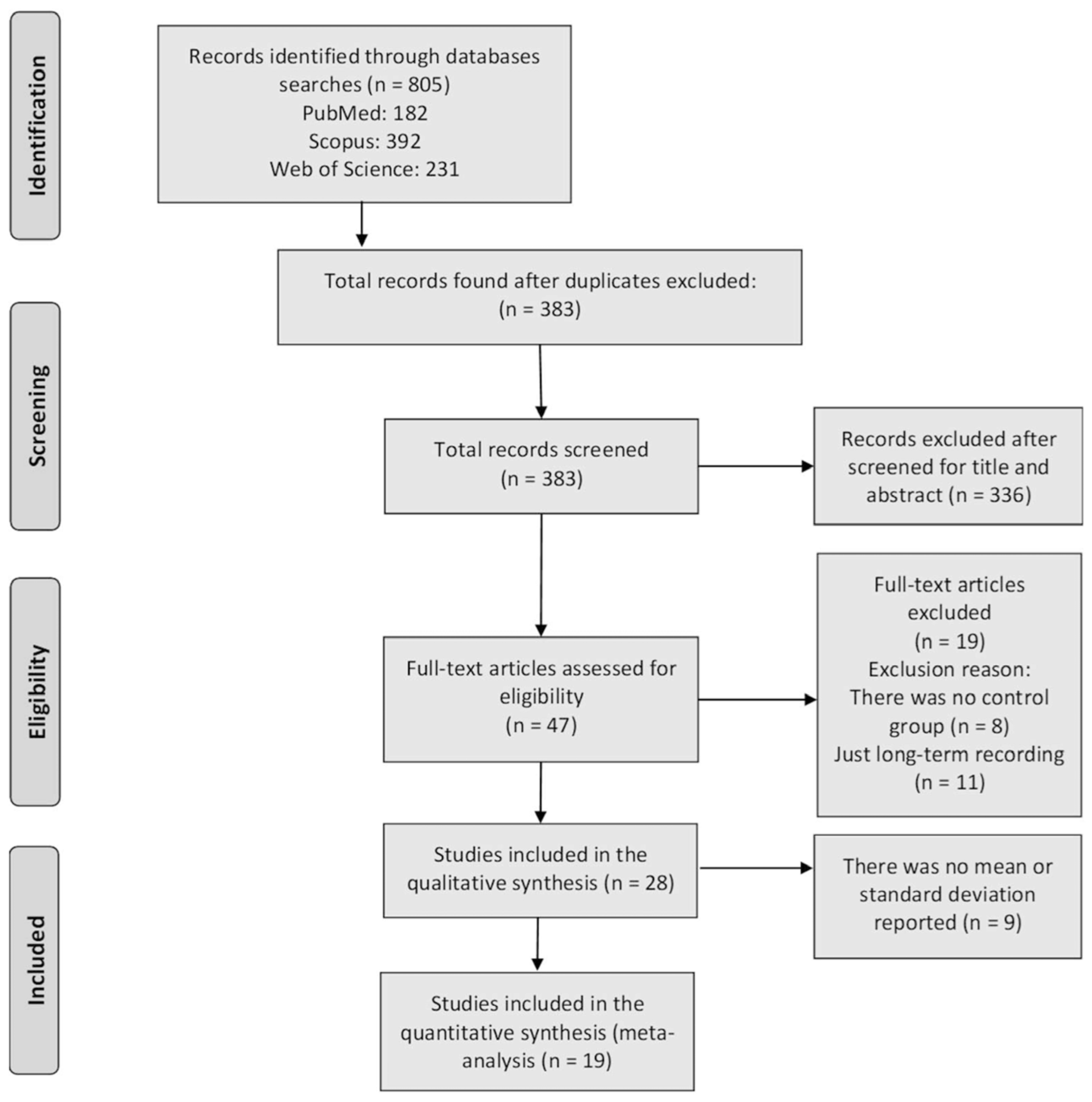
3.1. Identification of Studies

In the initial review, 805 articles were identified (182 in PUBMED, 392 in Scopus and 231 in Web of Science). The duplicated studies between selected databases were excluded (422), title and abstract of the remained 383 records were reviewed and a total of 336 additional studies were excluded (201 for not measuring HRV, 61 for analyzing only 1 component of MS, 48 for not being original articles and 26 for being studies in animal models). Subsequently, 47 full-text papers were retrieved, which were reviewed to ensure that they met the inclusion and exclusion criteria. Of these, 8 were excluded from the final analysis for not making comparisons between people with MS+ vs. MS−, and 11 for just reporting long-term recordings. No study was considered as “low methodological quality”, thus 28 articles were included in the final qualitative synthesis (Figure 1). Finally, 9 more articles were excluded from the quantitative meta-analysis (MA), as they did not report mean and/or standard deviations. Thus, the MA was carried out with 19 articles (Figure 1).

3.2. Quality Assessment

The methodological evaluation showed 82% (23) of the articles valued as “excellent methodological quality”, 18% (5) as “good methodological quality” and no reviewed study was scored as “low methodological quality”. The mean scored of the methodological quality of the reviewed article was 85% (“excellent methodological quality”).

3.3. Study and Patient Characteristics

The main studies’ characteristics reviewed are shown in Table 2. We found 1 study published before the year 2000, 9 between 2002 and 2010, and 18 between 2012 and 2020. The population under investigations originated from: North America (n = 4), including 3 from the United States [18,19,20] and 1 from Canada [21]; South America (n = 5), all of them from Brazil [9,10,22,23,24]; Europe (n = 7), finding 1 from England [25], 3 from Finland [26,27,28], 1 from France [29], 1 from Portugal [30] and 1 from Serbia [31]; and Asia (n = 12), where 4 were from Taiwan [32,33,34,35], 3 from South Korea [36,37,38], 2 from China [39,40], 2 from India [41,42], and 1 from Japan [43].
Regarding sex, 3 studies included only men [19,25,38], 3 studies included only women [9,22,23] and 22 studies were carried out in both sexes [10,18,20,21,26,27,28,29,30,31,32,33,34,35,36,37,39,40,41,42,43,44]. With respect to age, 5 papers included people between 18 and 40 years old [10,22,26,30,41], 16 papers evaluated the population over 40 years old [9,18,19,20,23,25,28,29,31,32,35,38,39,40,42,44], 6 more included populations between 18 and 79 years old [21,33,34,36,37,43] and 1 study was done in children between 6 and 8 years old [27]. The 68% (12,499) of all individuals included in this review (18,440) were people with MS.
Following the criteria described for the quantitative analysis, the variables R-R, pNN50, NN50, RRtri, TINN and all non-linear variables were not analyzed quantitatively (highlighted in bold in Table 2). The other linear variables, both in the time domain and frequency domain, were included in the quantitative analysis (i.e., SDNN, rMSSD, HF, LF, LF/HF). According to the recommendations from the Task Force of The European Society of Cardiology and The North American Society of Pacing and Electrophysiology [11], VLF and TP were not analyzed either, since their analysis is not recommended in short-term recordings given that their physiological explanation is much less defined and the existence of a specific physiological process attributable to these heart period changes might even be questioned. All the articles included in the MA analyzed the HRV from cardiac recordings.

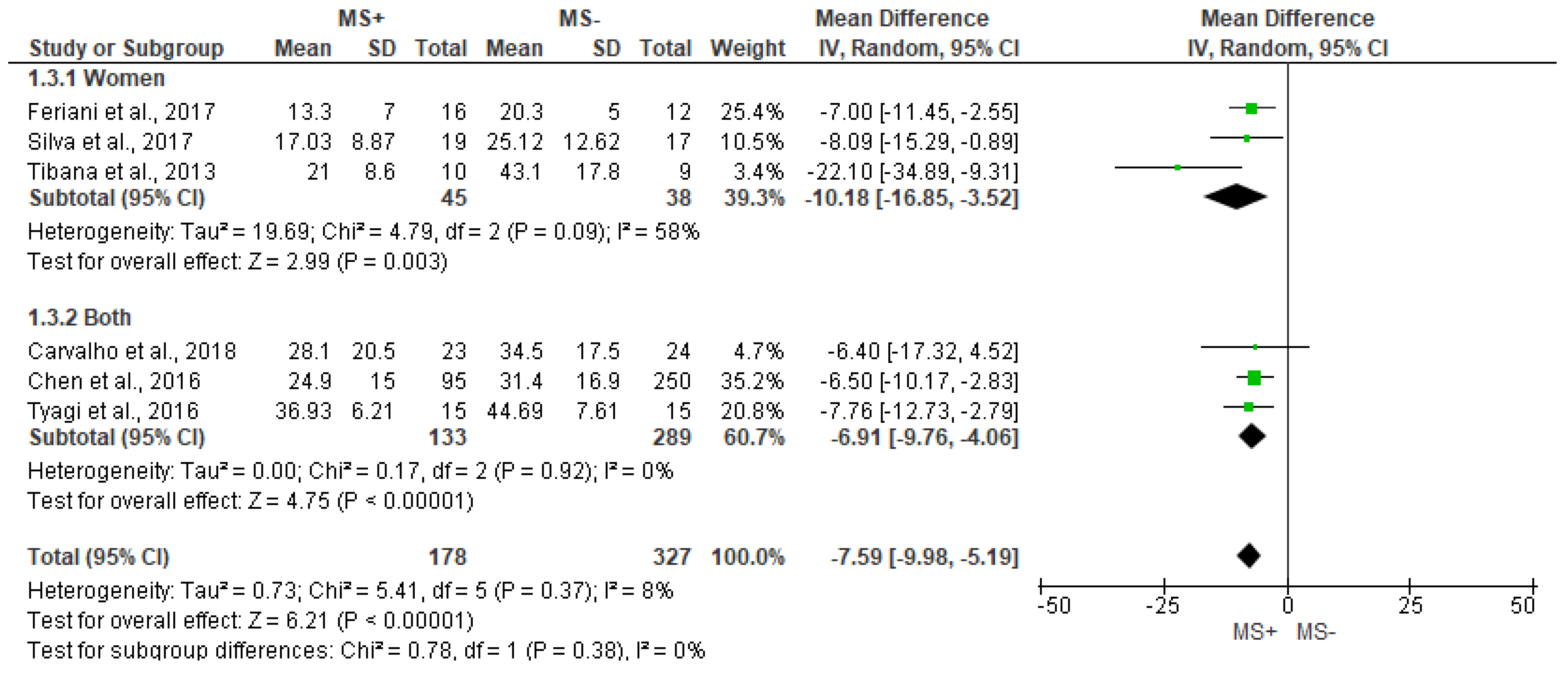
3.4. Time Domain Analysis Outcomes

In total, 17 of the 28 studies included in this review analyzed time domain variables. The main results of the qualitative analyses are provided in Table 3.
In the case of the R-R interval, lower values were reported in men older than 40 years with MS [25] and in women between 20 and 40 years with MS+ [22]. Other studies also reported significantly lower values in R-R when analyzing data from mixed groups [10,41,42,44]. Stuckey et al. reported a decrease in the R-R interval when performing the analysis only in women but not when combining men and women in the same group [21]. Regarding pNN50, two studies report lower values in men and women with MS+ [41,42], one study only in women [23] and another study did not find differences between groups [10].
The SDNN outcome is the most studied by the different works reviewed. Figure 2 shows significant differences between groups for SDNN outcome, being lower in people with MS+ (SMD = −0.36 [95%IC = −0.44, −0.28], p < 0.001), with low heterogeneity between reports (I2 = 9%). In addition, sub analyses showed significant differences between groups in women (−0.99 [−1.45, −0.52], p < 0.001), but not in men (p = 0.09).
Significant differences between groups were found for rMSSD (MD = −7.59 [95%IC = −9.98, −5.19], p < 0.001), with small heterogeneity between studies (I2 = 8%, Figure 3). In the same way, women also showed lower values of rMSSD in the MS+ group when compared with MS− (−10.18 [−16.85, −3.52], p = 0.003), while differences between groups could not be assessed in men sub analysis, due to lack of data.

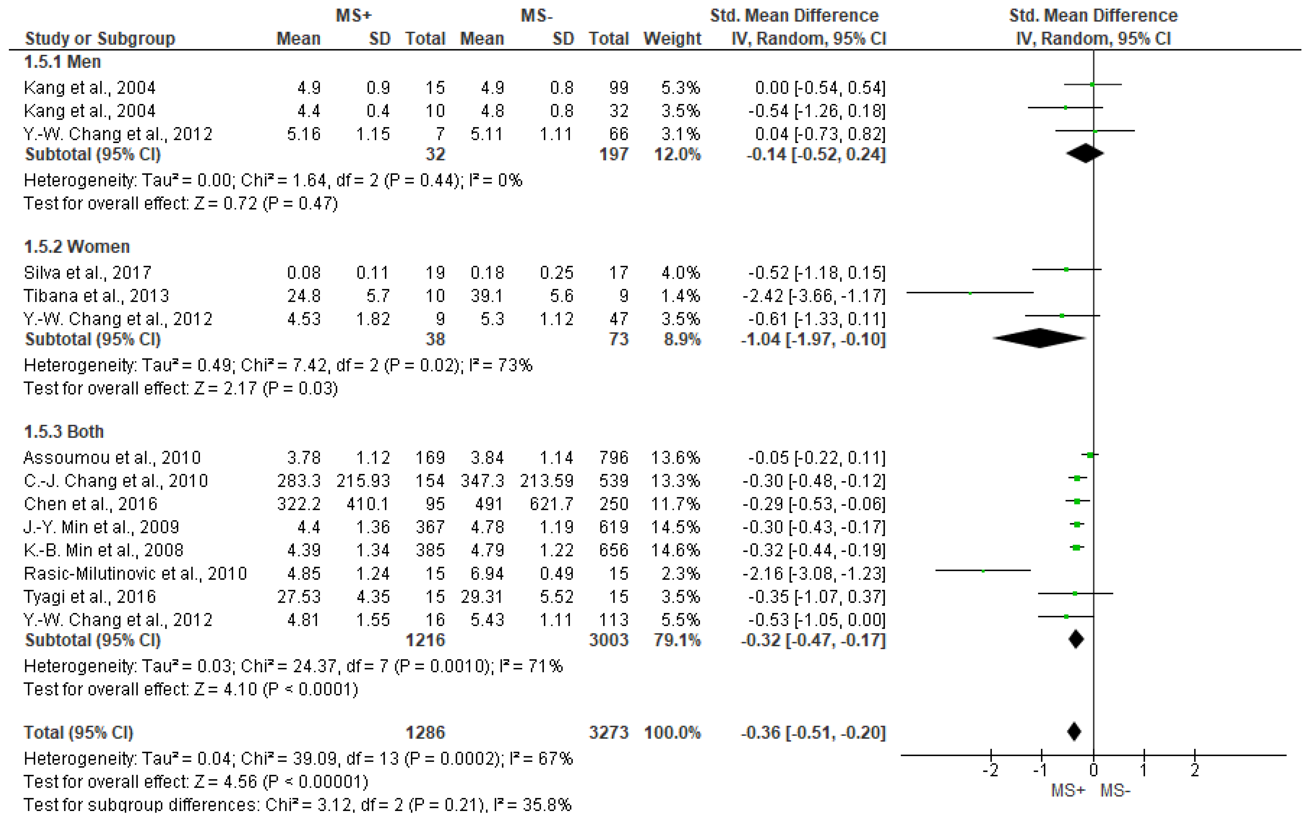
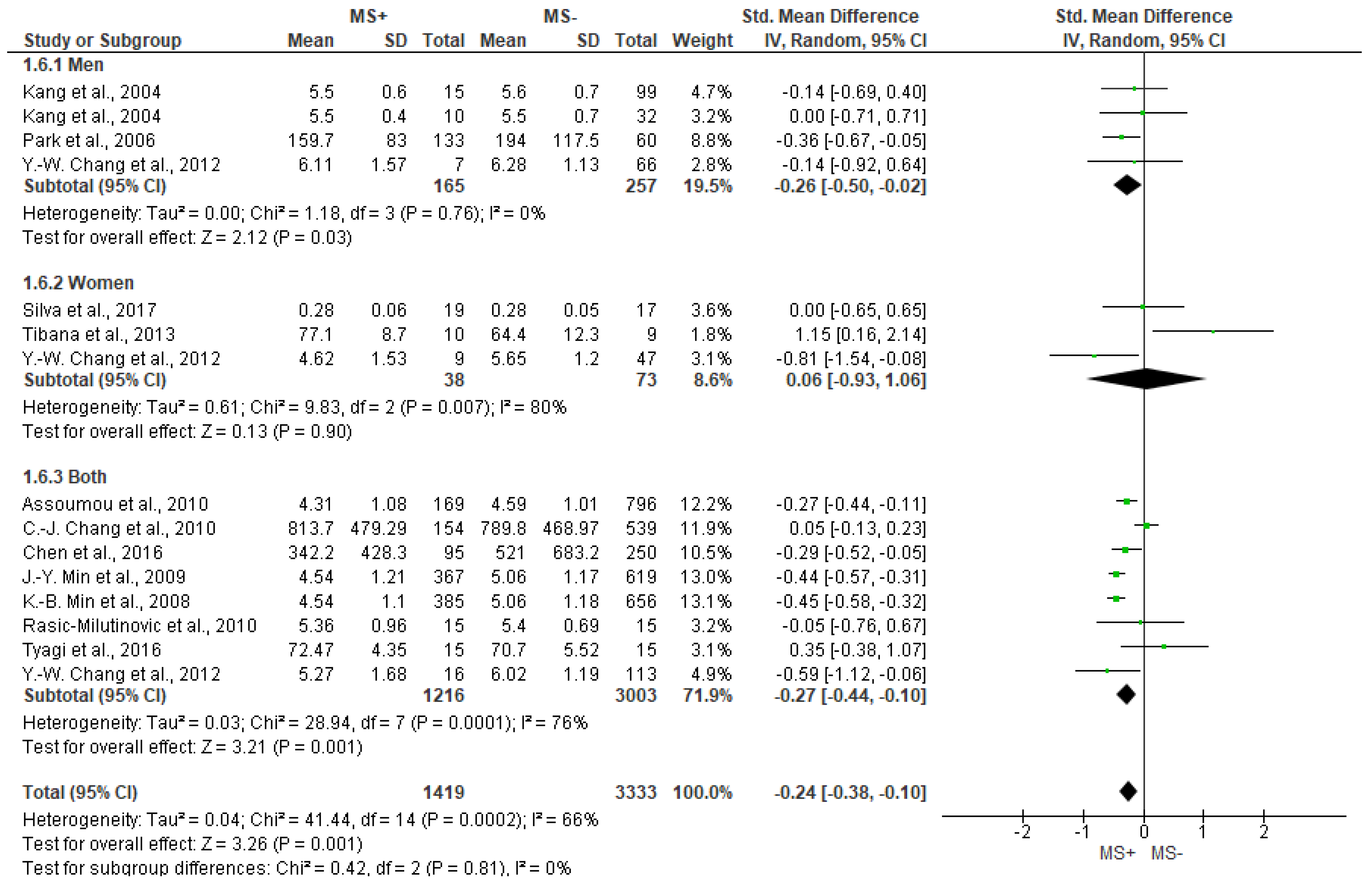
3.5. Frequency Domain Analysis Outcomes

For the spectral variables of the HRV, 25 studies carried out frequency domain analyses. A summary of the main results about its qualitative is showed in Table 4.
Regarding HF outcome, and with substantial heterogeneity, Figure 4 shows significant differences between groups (SMD = −0.36 [−0.51, −0.20], p < 0.001), showing lower values for the MS+ group (men and women together) (−0.32 [−0.47, −0.17], p < 0.001) and women only (−1.04 [−1.97, −0.10], p = 0.03), but not for men (p = 0.47).
Figure 5 depicts the different analyses of LF. Significant lower values of LF for MS+ group compared to MS− were found (SMD = −0.24 [−0.38, −0.1], p = 0.001), even though the results show substantial heterogeneity (I2 = 66%). Regarding subgroup analyses, mixed (both men and women) (−0.27 [−0.44, −0.1], p = 0.001) and men groups also showed significantly lower values of LF for MS+ group (−0.26 [−0.50, −0.02], p = 0.03) compared with MS−, while women did not show significant differences between groups (p = 0.90).
Regarding the LF/HF outcome, we found high heterogeneity (I2 = 87%) between studies and no significant differences (p = 0.76) were found between groups in both the general analysis and subanalyses (Figure 6).

3.6. Non-Linear Analysis Outcomes

Regarding the non-linear analysis, only 3 studies included these variables (Table 5). Stuckey et al. found no significant differences for SD1 of the Poincare Plot, for men or women, but they did find a significant increase for SD2 in MS+ women compared to MS− women [21]. No significant differences were found in α1 (detrended fluctuations analysis) or approximate entropy (ApEn) [21]. Silva et al. analyzed the records of 19 MS+ women and compared them to the records of 17 sex-match MS−, but they did not report differences in entropy (Shannon entropy, ShanEn) [9]. Finally, Carvalho et al. found a significant decrease for SD1 in young men and women, but not in SD2 and α1 of the Poincare analysis, or ShanEn [10].

4. Discussion

We conducted this systematic review and MA to determine the difference in short-term heart rate variability (HRV) between patients with metabolic syndrome (MS) and healthy controls. A total of 28 moderate-high quality studies were reviewed, 19 of which were included in the MA.
The main findings were: (1) patients with MS showed changes in short-term HRV in the time-domain analyses, having a decrease of all the parameters included in the MA: SDNN and rMSSD; (2) regarding the frequency-domain analyses, MS group reported a decrease in HF and LF, with the exception of the LF/HF relationship, which was not modified; (3) when sex was taken into account, the MA was only possible in one of both sexes (men or women) in rMSSD and LF/HF; (4) when data for both men and women were available, sex subanalyses showed a different behavior that the mixed analyses for SDNN, HF and LF, which might point towards a different impact of MS in men and women, (5) we could not perform the MA in non-linear parameters, RR and pNN50 due to the lack of studies and data availability.
It has been suggested that the SDNN is an indicator of the global behavior of the HRV [9,19,45] which being dependent on the analysed signal length, has shown to correlate well with the TP of the spectral analysis [11]. The MA showed that SDNN is decreased in patients with MS, which was previously described by most studies [10,18,32,34,36,37], thus clearly altering cardiac autonomic control in patients with MS. This suggests overall lower HRV, and therefore, reduced parasympathetic cardiac control in MS patients.
The other variable of the time-domain analyses reviewed in this MA is the rMSSD, directly linked to short-term HRV components [46], which is corelated to HF of the frequency-domain [11] and provides information on parasympathetic activity [9,45]. We found a decrease in rMSSD in people with MS, in both the mixed group and the subgroup of women. For Carvalho et al. [10], MS in young adults (<40 years) causes a decrease in parasympathetic modulation, which is also reported by Chen et al. [34] and Tyagi et al. [41] in those older than 40 years. It has been reported that in MS, C-reactive protein levels are increased because of a chronic condition of low-grade inflammation, which could cause the ANS dysregulation [34,47], thus increasing sympathetic activity and decreasing parasympathetic activity. For some authors, the deterioration of cardiac parasympathetic modulation is the main cause that would explain the alteration in autonomic control in MS, also suggesting that this worsening is closely related to fasting glycemia levels [9], states of insulin resistance [48] and type 2 diabetes [49].
In the frequency-domain analyses, the decrease in HF found in the MA reinforces the hypothesis that MS favors a lower parasympathetic activity [11,45]. HF presents significantly lower values in people with MS, both in the analysis of the data for men and women together (mixed group), and for the subgroup of women. On the other hand, the LF component of the spectral analysis, which has been suggested to reflect the sympathetic behavior exclusively by some studies [10,50], and as a parameter that could be modulated by both the sympathetic and parasympathetic branches of the ANS by others [9,45,51,52,53], decreased in the mixed group. LF components have been related in part to baroreflex control of HR to maintain BP homeostasis. Some studies have identified a relation between reduced neural baroreflex pathway and baroreceptor resetting with MS subjects and those at risk of high BP [54]. A weaker baroreflex response results in less efficient BP maintenance and lower HRV. The explanation of the underlying mechanisms could be difficult to interpret due to the controversy about its main modulating mechanism, likely multifactorial and non-linear, but it is important to emphasize that the LF component is decreased with MS, which indicates a lower HRV. Finally, since both LF and HF decreased, the MA showed no significant changes in the ratio LF/HF. This might be aligned with previous doubts by some authors casting that the LF/HF ratio should not be considered as a strong index to analyse the balance between the sympathetic and parasympathetic activities [55] since LF might itself be biased as a solid sympathetic indicator.
Regarding the analysis by sex, MS+ women group showed significantly lower values of SDNN compared with those in the MS− group, while men sub analysis did not show significant differences. Some authors have proposed that this behavior could be explained by physiological stress that, in the case of women, is exacerbated even at early ages, due to a greater systemic response to inflammatory processes [45,56]. In addition, LF was decreased only in men and, due to the important role that central obesity plays in the etiology of MS [26], it is important to highlight that men included in the studies reviewed in this MA showed high rates of central obesity, which favours chronic exposure to oxidative stress and increased risk of autonomic dysfunction, as has been reported [19,26]. However, LF was not modified in the group of women which could be explained, at least in part, by the fact that in premenopausal women the accumulation of abdominal fat is less common than in men at the same ages. Since central obesity is considered a key factor in the etiology of MS [26,33], the age of women who were evaluated in the studies included in the MA could explain the results. On the other hand, SDNN [19,38] and HF [33,38] did not reach statistical significance in men, even though the reviewed studies agree that these values showed a tendency to be lower, suggesting that parasympathetic modulation could be decreased also in men with MS.
HRV temporal series are non-stationary and non-linear in nature, likely due to non-linear interaction amongst different cardiovascular regulatory mechanisms. The non-linear analysis has been proposed to be a tool with high value for the analysis of complex systems and the predictability of a time series, which results from the complexity of the mechanisms that regulate HRV, analyzing the fractal behavior of the signal (detrended fluctuations analysis) and the complexity or regularity in the data series (entropy) [21]. The works published to date, however, do not allow a MA of these variables. Nevertheless, the available evidence points towards a decrease of SD1 in both men and women with MS [10], while SD2 seems to increase in women with MS [21]. SD1 has been associated directly to parasympathetic activity [57] and correlating well with SDNN and rMSSD, while SD2, has been reported to be related to both sympathetic and parasympathetic activity. SD2 has also been inversely related to sympathetic activity in sedentary and non-sedentary subjects [58]. On the other hand, DFA and entropy do not show significant changes in MS group compared with controls [9,10,21]. However, it is important to emphasize that, as indicated above, these are findings reported by a small sample of studies, so future works that analyze the non-linear behavior of HRV would be necessary to reach conclusive analyses, even though some authors question the physiological significance of using the non-linear analysis for short-term recordings [59].
Among cardiovascular complications, cardiac autonomic dysfunction is one of the most important, and it has been associated with a significant increase in morbidity and mortality independent of other risk factors [60,61]. Indeed, it has been reported that reduced HRV is a marker of autonomic dysfunction, and it is already evident in patients with MS, even before the development of type-2 diabetes [62]. Thus, early detection of autonomic dysfunction in prediabetic patients using HRV would be as important as the screening for MS, since the progression of cardiovascular denervation is partly reversible or can be delayed in the early stages of the disease [63,64].

5. Limitations

One of the main limitations of the MA is the high degree of heterogeneity presented in some of the parameters studied. Likewise, due to the low number of studies included in certain analyses, it was not possible to analyze the behavior of short-term HRV by sex in some variables, especially in the case of women. In addition, more studies are required to be able to develop quantitative analyzes about the non-linear variables of HRV, even though its clinical significance and utility in short-term recordings is questionable. On the other hand, and taking into account that the behavior of the HRV is largely conditioned by factors such as the body position in which the recordings are made, electrode position and/or recording method, the time of day and circadian rhythms, whether or not there was fasting prior to the recordings, the use of drugs and respiratory rate should be clearly reported in studies measuring HRV. Such factors may have indeed greater impact on short-term recordings than long-term recordings. Several recording methods have been used in the analysed studies, such as ECG measurements, RR recordings with digital heart rate bands, and pulse-wave recordings. However, in order to reduce variability and heterogeneity, only HRV analyses derived from ECG recordings were included in the MA. In addition, the recent development and use of wearable devices and phone applications might be useful for the study of HRV, specially for long-term recordings, as well as for obtaining self-reported assessments in the patient’s natural environment, but a meticulous process of validation is needed for their use in scientific research. In this line, efforts should be made to standardize the recording protocols, which in turn would reduce variability in the measurements and would improve data quality.

6. Conclusions

In conclusion, we found that patients with MS had differences in short-term HRV in the time-domain analyses, showing a decrease of SDNN and rMSSD. Regarding the frequency-domain analyses, patients with MS showed a decrease in HF and LF. When sex was taken into account, the MA was only possible in one of both sexes (men or women) in rMSSD and LF/HF. Lastly, when data for both men and women were available, sex subanalyses showed a different behavior than the mixed analyses for SDNN, HF and LF, which might point towards a different impact of MS in men and women. Regarding the analysis of non-linear variables (entropy, DFA, Poincare Plot), the results are not conclusive, due to the low number of datasets found for analysis.

Supplementary Materials

The following supporting information can be downloaded at: https://www.mdpi.com/article/10.3390/jcm12186051/s1, Table S1: Description of main time-domain HRV measurements reviewed; Table S2: Description of main frequency-domain HRV measurements reviewed; Table S3: Description of main non-linear HRV measurements reviewed.

Author Contributions

J.E.O.-G., S.M.-C. and M.Z. contributed to the conception of the work. J.E.O.-G., S.M.-C., Ó.J.A.-M., C.C. and A.B. provided the data necessary for the meta-analysis. All authors contributed to the analysis and interpretation of results. All authors contributed drafting and revising the manuscript. All authors have read and agreed to the published version of the manuscript.

Funding

This work was supported by Instituto de Salud Carlos III (PI21/01984 and CB16/11/00486) to F.J.C. and M.Z., and Generalitat Valenciana (CIAICO/2021/215) to PSA. SMC was supported by a predoctoral fellowship awarded by the Universitat de València (INV19-01-13-07).

Institutional Review Board Statement

The study was registered at the International Prospective Register of Systematic Re-views (PROSPERO) at https://www.crd.york.ac.uk/prospero/ (CRD42022358975 (accessed on 19 October 2022)).

Informed Consent Statement

Not applicable.

Data Availability Statement

All data generated or analyzed during this study are included in the published studies and their supplementary information files.

Conflicts of Interest

The authors declare no conflict of interest.

References

  1. Alberti, K.G.M.M.; Zimmet, P.; Shaw, J. Metabolic Syndrome—A New World-Wide Definition. A Consensus Statement from the International Diabetes Federation. Diabet. Med. 2006, 23, 469–480. [Google Scholar] [CrossRef] [PubMed]
  2. Zupkauskiene, J.; Lauceviciene, I.; Navickas, P.; Ryliskyte, L.; Puronaite, R.; Badariene, J.; Laucevicius, A. Changes in Health-Related Quality of Life, Motivation for Physical Activity, and the Levels of Anxiety and Depression after Individualized Aerobic Training in Subjects with Metabolic Syndrome. Hell. J. Cardiol. 2022, 66, 41–51. [Google Scholar] [CrossRef] [PubMed]
  3. Sperling, L.S.; Mechanick, J.I.; Neeland, I.J.; Herrick, C.J.; Després, J.P.; Ndumele, C.E.; Vijayaraghavan, K.; Handelsman, Y.; Puckrein, G.A.; Araneta, M.R.G.; et al. The CardioMetabolic Health Alliance Working Toward a New Care Model for the Metabolic Syndrome. J. Am. Coll. Cardiol. 2015, 66, 1050–1067. [Google Scholar] [CrossRef] [PubMed]
  4. Cornier, M.-A.; Dabelea, D.; Hernandez, T.L.; Lindstrom, R.C.; Steig, A.J.; Stob, N.R.; Van Pelt, R.E.; Wang, H.; Eckel, R.H. The Metabolic Syndrome. Endocr. Rev. 2008, 29, 777–822. [Google Scholar] [CrossRef]
  5. Kumanyika, S.; Afshin, A.; Arimond, M.; Lawrence, M.; McNaughton, S.A.; Nishida, C. Approaches to Defining Healthy Diets: A Background Paper for the International Expert Consultation on Sustainable Healthy Diets. Food Nutr. Bull. 2020, 41, 7S–30S. [Google Scholar] [CrossRef]
  6. Tudoran, C.; Tudoran, M.; Cut, T.G.; Lazureanu, V.E.; Bende, F.; Fofiu, R.; Enache, A.; Pescariu, S.A.; Novacescu, D. The Impact of Metabolic Syndrome and Obesity on the Evolution of Diastolic Dysfunction in Apparently Healthy Patients Suffering from Post-COVID-19 Syndrome. Biomedicines 2022, 10, 1519. [Google Scholar] [CrossRef] [PubMed]
  7. Yu, T.Y.; Lee, M.K. Autonomic Dysfunction, Diabetes and Metabolic Syndrome. J. Diabetes Investig. 2021, 12, 2108–2111. [Google Scholar] [CrossRef]
  8. Spallone, V.; Ziegler, D.; Freeman, R.; Bernardi, L.; Frontoni, S.; Pop-Busui, R.; Stevens, M.; Kempler, P.; Hilsted, J.; Tesfaye, S.; et al. Cardiovascular Autonomic Neuropathy in Diabetes: Clinical Impact, Assessment, Diagnosis, and Management. Diabetes. Metab. Res. Rev. 2011, 27, 639–653. [Google Scholar] [CrossRef]
  9. Silva, L.R.B.E.; Zamuner, A.R.; Gentil, P.; Alves, F.M.; Leal, A.G.F.; Soares, V.; Silva, M.S.; Vieira, M.F.; Simoes, K.; Pedrino, G.R.; et al. Cardiac Autonomic Modulation and the Kinetics of Heart Rate Responses in the On- and off-Transient during Exercise in Women with Metabolic Syndrome. Front. Physiol. 2017, 8, 542. [Google Scholar] [CrossRef]
  10. Carvalho, L.P.; Di Thommazo-Luporini, L.; Mendes, R.G.; Cabiddu, R.; Ricci, P.A.; Basso-Vanelli, R.P.; Oliveira-Junior, M.C.; Vieira, R.P.; Bonjorno-Junior, J.C.; Oliveira, C.R.; et al. Metabolic Syndrome Impact on Cardiac Autonomic Modulation and Exercise Capacity in Obese Adults. Auton. Neurosci. Basic Clin. 2018, 213, 43–50. [Google Scholar] [CrossRef]
  11. Malik, M.; John Camm, A.; Thomas Bigger, J.; Breithardt, G.; Cerutti, S.; Cohen, R.J.; Coumel, P.; Fallen, E.L.; Kennedy, H.L.; Kleiger, R.E.; et al. Heart Rate Variability: Standards of Measurement, Physiological Interpretation, and Clinical Use. Circulation 1996, 93, 1043–1065. [Google Scholar] [CrossRef]
  12. Stuckey, M.I.; Tulppo, M.P.; Kiviniemi, A.M.; Petrella, R.J. Heart Rate Variability and the Metabolic Syndrome: A Systematic Review of the Literature. Diabetes. Metab. Res. Rev. 2014, 30, 784–793. [Google Scholar] [CrossRef] [PubMed]
  13. Routledge, F.S.; Campbell, T.S.; McFetridge-Durdle, J.A.; Bacon, S.L. Improvements in Heart Rate Variability with Exercise Therapy. Can. J. Cardiol. 2010, 26, 303–312. [Google Scholar] [CrossRef] [PubMed]
  14. Moher, D.; Liberati, A.; Tetzlaff, J.; Altman, D.G.; Group, P. Preferred Reporting Items for Systematic Reviews and Meta-Analyses: The PRISMA Statement. Int. J. Surg. 2009, 8, 336–341. [Google Scholar] [CrossRef] [PubMed]
  15. Law, M.; Stewart, D.; Letts, L.; Pollock, N.; Bosch, J.; Westmorland, M. Critical Review Form–Quantitative Studies. McMaster Univ. Occup. Ther. Evid.-Based Pract. Res. Group. 1998. Available online: https://www.google.com/url?sa=t&rct=j&q=&esrc=s&source=web&cd=&ved=2ahUKEwiIodnPhbaBAxXGNt4KHa2QD6wQFnoECBEQAQ&url=https%3A%2F%2Fwww.mcgill.ca%2Fcpengine%2Ffiles%2Fcpengine%2Fquanreview_form1.doc&usg=AOvVaw1XWw38sWQFiLCfZ26bhqWz&opi=89978449 (accessed on 1 February 2023).
  16. Faber, I.R.; Bustin, P.M.; Oosterveld, F.G.; Elferink-Gemser, M.T.; Nijhuis-Van der Sanden, M.W. Assessing Personal Talent Determinants in Young Racquet Sport Players: A Systematic Review. J Sport. Sci 2016, 34, 395–410. [Google Scholar] [CrossRef]
  17. Shuster, J.J. Cochrane Handbook for Systematic Reviews of Interventions Version 5.1.0; Higgins, J.P.T., Green, S., Eds.; The Cochrane Collaboration: London, UK, 2011. [Google Scholar]
  18. Liao, D.P.; Sloan, R.P.; Cascio, W.E.; Folsom, A.R.; Liese, A.D.; Evans, G.W.; Cai, J.W.; Sharrett, A.R. Multiple Metabolic Syndrome Is Associated with Lower Heart Rate Variability-The Atherosclerosis Risk in Communities Study. Diabetes Care 1998, 21, 2116–2122. [Google Scholar] [CrossRef]
  19. Park, S.K.; Schwartz, J.; Weisskopf, M.; Sparrow, D.; Vokonas, P.S.; Wright, R.O.; Coull, B.; Nie, H.; Hu, H. Low-Level Lead Exposure, Metabolic Syndrome, and Heart Rate Variability: The VA Normative Aging Study. Environ. Health Perspect. 2006, 114, 1718–1724. [Google Scholar] [CrossRef]
  20. Pennathur, S.; Jaiswal, M.; Vivekanandan-Giri, A.; White, E.A.; Ang, L.; Raffel, D.M.; Rubenfire, M.; Pop-Busui, R. Structured Lifestyle Intervention in Patients with the Metabolic Syndrome Mitigates Oxidative Stress but Fails to Improve Measures of Cardiovascular Autonomic Neuropathy. J. Diabetes Complicat. 2017, 31, 1437–1443. [Google Scholar] [CrossRef]
  21. Stuckey, M.I.; Kiviniemi, A.; Gill, D.P.; Shoemaker, J.K.; Petrella, R.J. Associations between Heart Rate Variability, Metabolic Syndrome Risk Factors, and Insulin Resistance. Appl. Physiol. Nutr. Metab. 2015, 40, 734–740. [Google Scholar] [CrossRef]
  22. Tibana, R.A.; Boullosa, D.A.; Leicht, A.S.; Prestes, J. Women with Metabolic Syndrome Present Different Autonomic Modulation and Blood Pressure Response to an Acute Resistance Exercise Session Compared with Women without Metabolic Syndrome. Clin. Physiol. Funct. Imaging 2013, 33, 364–372. [Google Scholar] [CrossRef]
  23. Feriani, D.J.; Coelho-Júnior, H.J.; Scapini, K.B.; de Moraes, O.A.; Mostarda, C.; Ruberti, O.M.; Uchida, M.C.; Caperuto, É.C.; Irigoyen, M.C.; Rodrigues, B.; et al. Effects of Inspiratory Muscle Exercise in the Pulmonary Function, Autonomic Modulation, and Hemodynamic Variables in Older Women with Metabolic Syndrome. J. Exerc. Rehabil. 2017, 13, 218–226. [Google Scholar] [CrossRef] [PubMed]
  24. Maciel, A.W.S.; Pinto, L.M.; Campos, R.C.A.; Ferreira, A.C.; Dias-Filho, C.A.A.; Dias, C.J.M.; Pires, F.d.O.; Urtado, C.B.; Rodrigues, B.; Mostarda, C.T.; et al. Acute Effects of Resistance Exercise With Blood Flow Restriction in Elderly Women: A Pilot Study. J. Aging Phys. Act. 2021, 29, 361–371. [Google Scholar] [CrossRef] [PubMed]
  25. Brunner, E.J.; Hemingway, H.; Walker, B.R.; Page, M.; Clarke, P.; Juneja, M.; Shipley, M.J.; Kumari, M.; Andrew, R.; Seckl, J.R.; et al. Adrenocortical, Autonomic, and Inflammatory Causes of the Metabolic Syndrome-Nested Case-Control Study. Circulation 2002, 106, 2659–2665. [Google Scholar] [CrossRef] [PubMed]
  26. Koskinen, T.; Kähönen, M.; Jula, A.; Mattsson, N.; Laitinen, T.; Keltikangas-Järvinen, L.; Viikari, J.; Välimäki, I.; Rönnemaa, T.; Raitakari, O.T.; et al. Metabolic Syndrome and Short-Term Heart Rate Variability in Young Adults: The Cardiovascular Risk in Young Finns Study. Diabet. Med. 2009, 26, 354–361. [Google Scholar] [CrossRef] [PubMed]
  27. Leppanen, M.H.; Haapala, E.A.; Veijalainen, A.; Seppala, S.; Oliveira, R.S.; Lintu, N.; Laitinen, T.; Tarvainen, M.P.; Lakka, T.A. Associations of Cardiometabolic Risk Factors with Heart Rate Variability in 6- to 8-Year-Old Children: The PANIC Study. Pediatr. Diabetes 2020, 21, 251–258. [Google Scholar] [CrossRef] [PubMed]
  28. Kangas, P.; Tikkakoski, A.; Uitto, M.; Viik, J.; Bouquin, H.; Niemelä, O.; Mustonen, J.; Pörsti, I.; Niemela, O.; Mustonen, J.; et al. Metabolic Syndrome Is Associated with Decreased Heart Rate Variability in a Sex-Dependent Manner: A Comparison between 252 Men and 249 Women. Clin. Physiol. Funct. Imaging 2019, 39, 160–167. [Google Scholar] [CrossRef]
  29. Assoumou, H.G.N.; Pichot, V.; Barthelemy, J.C.; Dauphinot, V.; Celle, S.; Gosse, P.; Kossovsky, M.; Gaspoz, J.M.; Roche, F. Metabolic Syndrome and Short-Term and Long-Term Heart Rate Variability in Elderly Free of Clinical Cardiovascular Disease: The PROOF Study. Rejuvenation Res. 2010, 13, 653–663. [Google Scholar] [CrossRef]
  30. Soares-Miranda, L.; Sandercock, G.; Vale, S.; Santos, R.; Abreu, S.; Moreira, C.; Mota, J. Metabolic Syndrome, Physical Activity and Cardiac Autonomic Function. Diabetes-Metab. Res. Rev. 2012, 28, 363–369. [Google Scholar] [CrossRef]
  31. Rasic-Milutinovic, Z.R.; Milicevic, D.R.; Milovanovic, B.D.; Perunicic-Pekovic, G.B.; Pencic, B.D. Do Components of Metabolic Syndrome Contribute to Cardiac Autonomic Neuropathy in Non-Diabetic Patients? SAUDI Med. J. 2010, 31, 650–657. [Google Scholar]
  32. Chang, C.-J.; Yang, Y.-C.; Lu, F.-H.; Lin, T.-S.; Chen, J.-J.; Yeh, T.-L.; Wu, C.-H.; Wu, J.-S. Altered Cardiac Autonomic Function May Precede Insulin Resistance in Metabolic Syndrome. Am. J. Med. 2010, 123, 432–438. [Google Scholar] [CrossRef]
  33. Chang, Y.-W.; Lin, J.-D.; Chen, W.-L.; Yen, C.-F.; Loh, C.-H.; Fang, W.-H.; Wu, L.-W. Metabolic Syndrome and Short-Term Heart Rate Variability in Adults with Intellectual Disabilities. Res. Dev. Disabil. 2012, 33, 1701–1707. [Google Scholar] [CrossRef] [PubMed]
  34. Chen, Y.-J.; Lin, C.-L.C.-L.; Li, C.-R.C.-R.; Huang, S.-M.S.-M.; Chan, J.Y.-H.J.Y.-H.; Fang, W.-H.W.-H.; Chen, W.-L.W.-L.W.-L. Associations among Integrated Psychoneuroimmunological Factors and Metabolic Syndrome. Psychoneuroendocrinology 2016, 74, 342–349. [Google Scholar] [CrossRef] [PubMed]
  35. Chang, Y.-M.; Shiao, C.-C.; Huang, Y.-T.; Chen, I.-L.; Yang, C.-L.; Leu, S.-C.; Su, H.-L.; Kao, J.-L.; Tsai, S.-C.; Jhen, R.-N.; et al. Impact of Metabolic Syndrome and Its Components on Heart Rate Variability during Hemodialysis: A Cross-Sectional Study. Cardiovasc. Diabetol. 2016, 15, 16. [Google Scholar] [CrossRef] [PubMed]
  36. Min, K.-B.; Min, J.-Y.; Paek, D.; Cho, S.-I. The Impact of the Components of Metabolic Syndrome on Heart Rate Variability: Using the NCEP-ATP III and IDF Definitions. PACE-PACING Clin. Electrophysiol. 2008, 31, 584–591. [Google Scholar] [CrossRef]
  37. Min, J.-Y.; Paek, D.; Cho, S.-I.; Min, K.-B. Exposure to Environmental Carbon Monoxide May Have a Greater Negative Effect on Cardiac Autonomic Function in People with Metabolic Syndrome. Sci. Total Environ. 2009, 407, 4807–4811. [Google Scholar] [CrossRef]
  38. Kang, M.G.; Koh, S.B.; Cha, B.S.; Park, J.K.; Woo, J.M.; Chang, S.J. Association between Job Stress on Heart Rate Variability and Metabolic Syndrome in Shipyard Male Workers. YONSEI Med. J. 2004, 45, 838–846. [Google Scholar] [CrossRef]
  39. Li, Z.; Tang, Z.-H.; Zeng, F.; Zhou, L. Associations between the Severity of Metabolic Syndrome and Cardiovascular Autonomic Function in a Chinese Population. J. Endocrinol. Investig. 2013, 36, 993–999. [Google Scholar] [CrossRef]
  40. Guo, Y.; Cao, L.; Zhou, Y.; Xiao, L.; Zhang, X.; Yuan, J.; Chen, W. Cardiometabolic Traits Mediated the Relationship from Urinary Polycyclic Aromatic Hydrocarbons Metabolites to Heart Rate Variability Reduction: A Community-Based Study. Environ. Pollut. 2018, 243, 28–36. [Google Scholar] [CrossRef]
  41. Tyagi, A.; Cohen, M.; Reece, J.; Telles, S.; Jones, L. Heart Rate Variability, Flow, Mood and Mental Stress During Yoga Practices in Yoga Practitioners, Non-Yoga Practitioners and People with Metabolic Syndrome. Appl. Psychophysiol. Biofeedback 2016, 41, 381–393. [Google Scholar] [CrossRef]
  42. Endukuru, C.K.; Gaur, G.S.; Yerrabelli, D.; Sahoo, J.; Vairappan, B. Impaired Baroreflex Sensitivity and Cardiac Autonomic Functions Are Associated with Cardiovascular Disease Risk Factors among Patients with Metabolic Syndrome in a Tertiary Care Teaching Hospital of South-India. DIABETES Metab. Syndr. Res. Rev. 2020, 14, 2043–2051. [Google Scholar] [CrossRef]
  43. Saito, I.; Maruyama, K.; Eguchi, E.; Kato, T.; Kawamura, R.; Takata, Y.; Onuma, H.; Osawa, H.; Tanigawa, T. Low Heart Rate Variability and Sympathetic Dominance Modifies the Association between Insulin Resistance and Metabolic Syndrome:—The Toon Health Study. Circ. J. 2017, 81, 1447–1453. [Google Scholar] [CrossRef] [PubMed]
  44. MacAgnan, F.E.; Pandolfo Feoli, A.M.; Russomano, T.; Feoli, A.M.P.; Russomano, T. Acute Physical Effort Increases Sympathovagal Balance Responses to Autonomic Stimulation in Metabolic Syndrome. Metab. Syndr. Relat. Disord. 2019, 17, 67–74. [Google Scholar] [CrossRef]
  45. Supriya, R.; Li, F.-F.F.-F.; Yang, Y.-D.Y.-D.; Liang, W.; Baker, J.S. Association between Metabolic Syndrome Components and Cardiac Autonomic Modulation among Children and Adolescents: A Systematic Review and Meta-Analysis. Biology 2021, 10, 699. [Google Scholar] [CrossRef] [PubMed]
  46. Shaffer, F.; Ginsberg, J.P. An Overview of Heart Rate Variability Metrics and Norms. Front. Public Health 2017, 5, 258. [Google Scholar] [CrossRef]
  47. Yang, T.; Chu, C.H.; Hsieh, P.C.; Hsu, C.H.; Chou, Y.C.; Yang, S.H.; Bai, C.H.; You, S.L.; Hwang, L.C.; Chung, T.C.; et al. C-Reactive Protein Concentration as a Significant Correlate for Metabolic Syndrome: A Chinese Population-Based Study. Endocrine 2013, 43, 351–359. [Google Scholar] [CrossRef] [PubMed]
  48. Miller, A.W.; Jason Sims, J.; Canavan, A.; Hsu, T.; Ujhelyi, M.R. Impaired Vagal Reflex Activity in Insulin-Resistant Rats. J. Cardiovasc. Pharmacol. 1999, 33, 698–702. [Google Scholar] [CrossRef] [PubMed]
  49. Carnethon, M.R.; Jacobs, D.R.; Sidney, S.; Liu, K. Influence of Autonomic Nervous System Dysfunction on the Development of Type 2 Diabetes: The CARDIA Study. Diabetes Care 2003, 26, 3035–3041. [Google Scholar] [CrossRef] [PubMed]
  50. Yokobori, Y.; Nakane, H.; Uehara, C.; Nagasawa, T.; Mitsuyama, S.; Ohkawa, K.; Kario, K.; Ozawa, S. Temporal Relationships among Changes in the RR-Interval and the Powers of the Low- and High-Frequency Components of Heart Rate Variability in Normal Subjects. Physiol. Rep. 2023, 11, e15557. [Google Scholar] [CrossRef]
  51. Porto, A.A.; Benjamim, C.J.R.; Gonzaga, L.A.; Luciano de Almeida, M.; Bueno Júnior, C.R.; Garner, D.M.; Valenti, V.E. Caffeine Intake and Its Influences on Heart Rate Variability Recovery in Healthy Active Adults after Exercise: A Systematic Review and Meta-Analysis. Nutr. Metab. Cardiovasc. Dis. 2022, 32, 1071–1082. [Google Scholar] [CrossRef]
  52. Jarczok, M.N.; Weimer, K.; Braun, C.; Williams, D.P.; Thayer, J.F.; Gündel, H.O.; Balint, E.M. Heart Rate Variability in the Prediction of Mortality: A Systematic Review and Meta-Analysis of Healthy and Patient Populations. Neurosci. Biobehav. Rev. 2022, 143, 104907. [Google Scholar] [CrossRef]
  53. Nunan, D.; Sandercock, G.R.H.; Brodie, D.A. A Quantitative Systematic Review of Normal Values for Short-Term Heart Rate Variability in Healthy Adults. PACE-Pacing Clin. Electrophysiol. 2010, 33, 1407–1417. [Google Scholar] [CrossRef] [PubMed]
  54. Zanoli, L.; Empana, J.P.; Estrugo, N.; Escriou, G.; Ketthab, H.; Pruny, J.F.; Castellino, P.; Laude, D.; Thomas, F.; Pannier, B.; et al. The Neural Baroreflex Pathway in Subjects with Metabolic Syndrome: A Sub-Study of the Paris Prospective Study III. Medicine 2016, 95, e2472. [Google Scholar] [CrossRef] [PubMed]
  55. Billman, G.E. The LF/HF Ratio Does Not Accurately Measure Cardiac Sympatho-Vagal Balance. Front. Physiol. 2013, 4, 26. [Google Scholar] [CrossRef]
  56. Stefanaki, C.; Michos, A.; Latsios, G.; Tousoulis, D.; Peppa, M.; Zosi, P.; Boschiero, D.; Bacopoulou, F. Sexual Dimorphism of Heart Rate Variability in Adolescence: A Case-Control Study on Depression, Anxiety, Stress Levels, Body Composition, and Heart Rate Variability in Adolescents with Impaired Fasting Glucose. Int. J. Environ. Res. Public Health 2020, 17, 2688. [Google Scholar] [CrossRef] [PubMed]
  57. Hoshi, R.A.; Pastre, C.M.; Vanderlei, L.C.M.; Godoy, M.F. Poincaré Plot Indexes of Heart Rate Variability: Relationships with Other Nonlinear Variables. Auton. Neurosci. Basic Clin. 2013, 177, 271–274. [Google Scholar] [CrossRef] [PubMed]
  58. Navarro-Lomas, G.; Alejandro, D.L.O.; Jurado-Fasoli, L.; Castillo, M.J.; Femia, P.; Amaro-Gahete, F.J. Assessment of Autonomous Nerve System through Non-Linear Heart Rate Variability Outcomes in Sedentary Healthy Adults. PeerJ 2020, 8, e10178. [Google Scholar] [CrossRef] [PubMed]
  59. Maestri, R.; Pinna, G.D.; Porta, A.; Balocchi, R.; Sassi, R.; Signorini, M.G.; Dudziak, M.; Raczak, G. Assessing Nonlinear Properties of Heart Rate Variability from Short-Term Recordings: Are These Measurements Reliable? Physiol. Meas. 2007, 28, 1067–1077. [Google Scholar] [CrossRef]
  60. Young, L.H.; Wackers, F.J.T.; Chyun, D.A.; Davey, J.A.; Barrett, E.J.; Taillefer, R.; Heller, G.V.; Iskandrian, A.E.; Wittlin, S.D.; Filipchuk, N.; et al. Cardiac Outcomes after Screening for Asymptomatic Coronary Artery Disease in Patients with Type 2 Diabetes the DIAD Study: A Randomized Controlled Trial. JAMA 2009, 301, 1547–1555. [Google Scholar] [CrossRef]
  61. Pop-Busui, R.; Evans, G.W.; Gerstein, H.C.; Fonseca, V.; Fleg, J.L.; Hoogwerf, B.J.; Genuth, S.; Grimm, R.H.; Corson, M.A.; Prineas, R. Effects of Cardiac Autonomic Dysfunction on Mortality Risk in the Action to Control Cardiovascular Risk in Diabetes (ACCORD) Trial. Diabetes Care 2010, 33, 1578–1584. [Google Scholar] [CrossRef]
  62. Balcioglu, A.S.; Akinci, S.; Cicek, D.; Eldem, H.O.; Coner, A.; Bal, U.A.; Muderrisoglu, H. Which Is Responsible for Cardiac Autonomic Dysfunction in Non-Diabetic Patients with Metabolic Syndrome: Prediabetes or the Syndrome Itself? Diabetes Metab. Syndr. Clin. Res. Rev. 2016, 10, S13–S20. [Google Scholar] [CrossRef]
  63. Howorka, K.; Pumprla, J.; Haber, P.; Koller-Strametz, J.; Mondrzyk, J.; Schabmann, A. Effects of Physical Training on Heart Rate Variability in Diabetic Patients with Various Degrees of Cardiovascular Autonomic Neuropathy. Cardiovasc. Res. 1997, 34, 206–214. [Google Scholar] [CrossRef] [PubMed]
  64. Ortiz-Guzmán, J.E.; Mollà-Casanova, S.; Arias-Mutis, Ó.J.; Bizy, A.; Calvo, C.; Alberola, A.; Chorro, F.J.; Zarzoso, M. Differences in Long-Term Heart Rate Variability between Subjects with and without Metabolic Syndrome: A Systematic Review and Meta-Analysis. J. Cardiovasc. Dev. Dis. 2023, 10, 203. [Google Scholar] [CrossRef] [PubMed]
Figure 1. Flowchart of article selection.
Jcm 12 06051 g001
Figure 2. Forest plot showing the SDNN between metabolic syndrome (MS+) versus control (MS−).
Jcm 12 06051 g002
Figure 3. Forest plot showing the rMSSD between metabolic syndrome (MS+) versus control (MS−).
Jcm 12 06051 g003
Figure 4. Forest plot showing the HF between metabolic syndrome (MS+) versus control (MS−).
Jcm 12 06051 g004
Figure 5. Forest plot showing the LF between metabolic syndrome (MS+) versus control (MS−).
Jcm 12 06051 g005
Figure 6. Forest plot showing the LF/HF between metabolic syndrome (MS+) versus control (MS−).
Jcm 12 06051 g006
Table 1. Search strategy.
DatabaseSearch Equation
PubMed((“heart rate variability” [Title/Abstract] or “autonomic control” [Title/Abstract] or “HRV” [Title/Abstract] or “cardiac autonomic control” [Title/Abstract] or “cardiac autonomic function” [Title/Abstract] or “cardiac autonomic modulation” [Title/Abstract]) AND (“metabolic syndrome” [Title/Abstract]))
Web of Science(“heart rate variability” or “autonomic control” or “HRV” or “cardiac autonomic control” or “cardiac autonomic function” or “cardiac autonomic modulation”) and (“metabolic syndrome”)
Scopus(TITLE-ABS-KEY (“metabolic syndrome”) and TITLE-ABS-KEY (“heart rate variability”) or TITLE-ABS-KEY (“autonomic control”) or TITLE-ABS-KEY (“HRV”) or TITLE-ABS-KEY (“cardiac autonomic control”) or TITLE-ABS-KEY (“cardiac autonomic function”) or TITLE-ABS-KEY (“cardiac autonomic modulation”))
Table 2. Summary of the studies.
ReferenceMethodological Evaluation (%)nAge (Years)GenderMS DefinitionRecording CharacteristicsAnalyzed HRV Variables
Recording Time (min)Body PositionTimeFrequencySpectral MethodsNon-Linear
Liao et al., 1998 [18]81%235945–64BothHTA, DM-2, dislipidemia2SupineSDNNHF, LF, LF/HFFFTNo
Brunner et al., 2002 [25]63%18345–63MenNCEP-ATP III5SupineSDNNTP, LF, HFBlackman-TukeyNo
Kang et al., 2004 [38]69%15641–55Men≥3 risk factors5SittingSDNN, rMSSDHF, LF, LF/HFNRNo
Park et al., 2006 [19]88%41364–79MenNCEP-ATP III7SittingSDNNHF, LF, LF/HFFFTNo
K.-B. Min et al., 2008 [36]88%104120–87BothNCEP-ATP III, IDF5SittingSDNNLF, HFNRNo
J.-Y. Min et al., 2009 [37]69%98620–87BothNCEP-ATP III5SittingSDNNLF, HFFFTNo
Koskinen et al., 2009 [26]88%188924–39BothNCEP-ATP III, IDF, EGIR3SupineNoLH, HF, TP, LF/HFFFTNo
Assoumou et al., 2010 [29]94%101064–66BothNCEP-ATP III5SupineNoTP, HF, LF, VLF, ULF, LF/HFFFTNo
C.-J. Chang et al., 2010 [32]75%128936–48BothNCEP-ATP III5SupineSDNNHF, LF, LF/HFFFTNo
Rasic-Milutinovic et al., 2010 [31]88%4745–65BothNCEP ATP IIINRNRSDNN
rMSSD
HF, LF, LF/HF, TP, VLFFFTNo
Y.-W. Chang et al., 2012 [33]88%12919–62BothNCEP-ATP III5SupineNoLH, HF, TP, LF/HFFFTNo
Soares-Miranda et al., 2012 [30]81%16319–21BothN/A5SupinerMSSD, SDNN, NN50, pNN50HF, LF/HFFFTSD1, SD2
Tibana et al., 2013 [22]88%1930–40WomenNCEP-ATP III5NRR-R, SDNN, rMSSDHF, LF, LF/HFFFTNo
Li et al., 2013 [39]94%211950–70BothNCEP-ATP III15SupineNoLH, HF, TP, LF/HFNRNo
Stuckey et al., 2015 [21]88%22023–70BothNCEP-ATP III5SupineSDNN, rMSSDLF, HFFFTSD1, SD2, α1, Aprox. Entropy
Chen et al., 2016 [34]88%34520–65Both¤5NRSDNN, rMSSDVLF, LF, HF, TPFFTNo
Tyagi et al., 2016 [41]56%3040–50BothIDF5NRrMSSD, pNN50, R-RHF, LF, LF/HFFFTNo
Y.-M. Chang et al., 2016 [35]88%17550–80BothIDF5SupineNoLF, HF, LF/HF, TP, VLFFFTNo
Silva et al., 2017 [9]94%3640–50Women§12SittingSDNN, rMSSDHF, LF, LF/HFFFTShannon Entropy
Feriani et al., 2017 [23]94%2865–75WomenNCEP-ATP III20NRSDNN, rMSSD, pNN50HF, LF, LF/HFFFTNo
Saito et al., 2017 [43]94%201630–79Both≥3 risk factors5NRSDNN, rMSSDHF, LF, LF/HFNRNo
Pennathur et al., 2017 [20]94%5040–60BothNCEP-ATP III5SupineNoHF, LF, LF/HFWavelet transformNo
Guo et al., 2018 [40]100%247645–70BothNCEP-ATP III5SittingNoLF, HF, LF/HF, VLF, TPNRNo
Carvalho et al., 2018 [10]88%6630–40BothNCEP-ATP III300 consecutives R-R intervalsSupineR-R, rMSSD, pNN50, RRtri, TINNNoN/ASD1, SD2, α1, Shannon Entropy
MacAgnan et al., 2019 [44]88%1440–60BothNCEP ATP III250–350 consecutives R-R intervalsSupineR-RHF, LF, LF/HFAutoregressive algorithmNo
Kangas et al., 2019 [28]88%57240–60BothIC5SupineNoTP, LF, HF, LF/HFFFTNo
Leppanen et al., 2020 [27]94%4436–8BothNR5SupineR-R, rMSSDHF, LF, LF/HFNRNo
Endukuru et al., 2020 [42]94%17640–55BothNCEP ATP III5SupineR-R, SDNN, pNN50, NN50, rMSSDLF, HF, LF/HF, VLF, TPNRNo
NCEP-ATP III: National Cholesterol Education Program’s Adult Treatment Panel III. IDF: International Diabetes Federation. EGIR: European Group for the Study of Insulin Resistance. HTA: Arterial hypertension. DM-2: Type 2 Diabetes Mellitus. ¤ The country-specific definition applied by Taiwan’s Ministry of Healthand Welfare. § Waist circumference (≥80 cm) and the presence of at least two criteria. IC: Consensus definition from several national and international organizations. min: minutes. FFT: Fast Fourier transformation. NR: not reported. N/A: Not Applicable.
Table 3. Reported changes in time domain analyses (short-term HRV) in MS.
ReferenceSDNNrMSSDR-RpNN50
Liao et al., 1998 [18]
Brunner et al., 2002 [25]
Kang et al., 2004 [38]=
Park et al., 2006 [19]=
K.-B. Min et al., 2008 [36]
J.-Y. Min et al., 2009 [37]
C.-J. Chang et al., 2010 [32]
Tibana et al., 2013 [22]
Stuckey et al., 2015 [21]w=w
Chen et al., 2016 [34]
Tyagi et al., 2016 [41]
Silva et al., 2017 [9]
Feriani et al., 2017 [23]
Saito et al., 2017 [43]=
Carvalho et al., 2018 [10]==
MacAgnan et al., 2019 [44]
Endukuru et al., 2020 [42]
w: only in women; =: without change; ↓: lower values in MS group.
Table 4. Reported changes in frequency domain analyses (short-term HRV) in MS.
ReferenceHFLFLF/HF
Liao et al., 1998 [18]=
Brunner et al., 2002 [25]
Kang et al., 2004 [38]===
Park et al., 2006 [19]===
K.-B. Min et al., 2008 [36]
J.-Y. Min et al., 2009 [37]
Koskinen et al., 2009 [26]w
Assoumou et al., 2010 [29]=
C.-J. Chang et al., 2010 [32]=
Rasic-Milutinovic et al., 2010 [31]
Y.-W. Chang et al., 2012 [33]===
Tibana et al., 2013 [22]
Li et al., 2013 [39]
Stuckey et al., 2015 [21]=w=
Chen et al., 2016 [34]
Tyagi et al., 2016 [41]
Y.-M. Chang et al., 2016 [35]===
Silva et al., 2017 [9]=
Feriani et al., 2017 [23]
Saito et al., 2017 [43]=
Pennathur et al., 2017 [20]==
Guo et al., 2018 [40]=
MacAgnan et al., 2019 [44]
Kangas et al., 2019 [28]m=
Endukuru et al., 2020 [42]
m: only in men; w: only in women; =: without change; ↓: lower values in MS group; ↑: upper values in MS group.
Table 5. Reported changes in non-linear analyses (short-term HRV) in MS.
ReferenceSD1SD2α1ApEnShanEn
Stuckey et al., 2015 [21]=w==
Silva et al., 2017 [9] =
Carvalho et al., 2018 [10]== =
w: only in women; =: without change.
Disclaimer/Publisher’s Note: The statements, opinions and data contained in all publications are solely those of the individual author(s) and contributor(s) and not of MDPI and/or the editor(s). MDPI and/or the editor(s) disclaim responsibility for any injury to people or property resulting from any ideas, methods, instructions or products referred to in the content.

